Uniporter

Last updated
Comparison of transport proteins Porters.PNG
Comparison of transport proteins

Uniporters, also known as solute carriers or facilitated transporters, are a type of membrane transport protein that passively transports solutes (small molecules, ions, or other substances) across a cell membrane. [1] It uses facilitated diffusion for the movement of solutes down their concentration gradient from an area of high concentration to an area of low concentration. [2] Unlike active transport, it does not require energy in the form of ATP to function. Uniporters are specialized to carry one specific ion or molecule and can be categorized as either channels or carriers. [3] Facilitated diffusion may occur through three mechanisms: uniport, symport, or antiport. The difference between each mechanism depends on the direction of transport, in which uniport is the only transport not coupled to the transport of another solute. [4]

Contents

Uniporter carrier proteins work by binding to one molecule or substrate at a time. Uniporter channels open in response to a stimulus and allow the free flow of specific molecules. [2]

There are several ways in which the opening of uniporter channels may be regulated:

  1. Voltage – Regulated by the difference in voltage across the membrane
  2. Stress – Regulated by physical pressure on the transporter (as in the cochlea of the ear)
  3. Ligand – Regulated by the binding of a ligand to either the intracellular or extracellular side of the cell

Uniporters are found in mitochondria, plasma membranes and neurons.The uniporter in the mitochondria is responsible for calcium uptake. [1] The calcium channels are used for cell signaling and triggering apoptosis. The calcium uniporter transports calcium across the inner mitochondrial membrane and is activated when calcium rises above a certain concentration. [5] The amino acid transporters function in transporting neutral amino acids for neurotransmitter production in brain cells. [6] Voltage-gated potassium channels are also uniporters found in neurons and are essential for action potentials. [7] This channel is activated by a voltage gradient created by sodium-potassium pumps. When the membrane reaches a certain voltage, the channels open, which depolarizes the membrane, leading to an action potential being sent down the membrane. [8] Glucose transporters are found in the plasma membrane and play a role in transporting glucose. They help to bring glucose from the blood or extracellular space into cells usually to be utilized for metabolic processes in generating energy. [9]

Uniporters are essential for certain physiological processes in cells, such as nutrient uptake, waste removal, and maintenance of ionic balance.

Discovery

Facilitated diffusion using transport proteins Scheme facilitated diffusion in cell membrane-en.svg
Facilitated diffusion using transport proteins

Early research in the 19th and 20th centuries on osmosis and diffusion provided the foundation for understanding the passive movement of molecules across cell membranes. [10]

In 1855, the physiologist Adolf Fick was the first to define osmosis and simple diffusion as the tendency for solutes to move from a region of higher concentration to a lower concentration, also very well-known as Fick's Laws of Diffusion. [11] Through the work of Charles Overton in the 1890s, the concept that the biological membrane is semipermeable became important to understanding the regulation of substances in and out of the cells. [11] The discovery of facilitated diffusion by Wittenberg and Scholander suggested that proteins in the cell membrane aid in the transport of molecules. [12] In the 1960s - 1970s, studies on the transport of glucose and other nutrients highlighted the specificity and selectivity of membrane transport proteins. [13]

Technological advancements in biochemistry helped isolate and characterize these proteins from cell membranes. Genetic studies on bacteria and yeast identified genes responsible for encoding transporters. This led to the discovery of glucose transporters (GLUT proteins), with GLUT1 being the first to be characterized. [14] Identification of gene families encoding various transporters, such as solute carrier (SLC) families, also advanced knowledge on uniporters and its functions. [14]

Newer research is focusing on techniques using recombinant DNA technology, electrophysiology and advanced imaging to understand uniporter functions. These experiments are designed to clone and express transporter genes in host cells to further analyze the three-dimensional structure of uniporters, as well as directly observe the movement of ions through proteins in real-time. [14] The discovery of mutations in uniporters has been linked to diseases such as GLUT1 deficiency syndrome, cystic fibrosis, Hartnup disease, primary hyperoxaluria and hypokalemic periodic paralysis. [15]

Types

Glucose transporter (GLUTs)

The glucose transporter (GLUTs) is a type of uniporter responsible for the facilitated diffusion of glucose molecules across cell membranes. [9] Glucose is a vital energy source for most living cells, however, due to its large size, it cannot freely move through the cell membrane. [16] The glucose transporter is specialized in transporting glucose specifically across the membrane. The GLUT proteins have several types of isoforms, each distributed in different tissues and exhibiting different kinetic properties. [16]

Glucose transporter Glucose-6-phosphatase system.svg
Glucose transporter

GLUTs are integral membrane proteins composed of 12 α-helix membrane spanning regions. [16] The GLUT proteins are encoded by the SLC2 genes and categorized into three classes based on amino acid sequence similarity. [17] Humans have been found to express fourteen GLUT proteins. Class I GLUTs include GLUT1, one of the most studied isoforms, and GLUT2. [16] GLUT1 is found in various tissues like the red blood cells, brain, and blood-brain barrier and is responsible for basal glucose uptake. [16] GLUT2 is predominantly found in the liver, pancreas, and small intestines. [16] It plays an important role in insulin secretion from pancreatic beta cells. Class II includes the GLUT3 and GLUT4. [16] GLUT3, primarily found in the brain, neurons and placenta, has a high affinity for glucose in facilitating glucose uptake into neurons. [16] GLUT4 plays a role in insulin-regulated glucose uptake and is mainly found in insulin-sensitive tissues such as muscle and adipose tissue. [16] Class III includes GLUT5, found in the small intestine, kidney, testes, and skeletal muscle. [16] Unlike the other GLUTs, GLUT5 specifically transports fructose rather than glucose. [16]

Glucose transporters allow glucose molecules to move down their concentration gradient from areas of high glucose concentration to areas of low concentration. This process often involves bringing glucose from the extracellular space or blood into the cell. The concentration gradient set up by glucose concentrations fuels the process without the need for ATP. [18]

When glucose binds to the glucose transporter, the protein channels change shape and undergo a conformational change to transport the glucose across the membrane. Once the glucose unbinds, the protein returns to its original shape. The glucose transporter is essential for carrying out physiological processes that require high energy demands in the brain, muscles, and kidneys by providing an adequate amount of energy substrate for metabolism. Diabetes, an example of a condition that involves glucose metabolism, highlights the importance of the regulation of glucose uptake in disease management. [19]

Mitochondrial Ca2+ uniporter (MCU)

The mitochondrial calcium uniporter (MCU) is a protein complex located in the inner mitochondrial matrix that functions to take up calcium ions (Ca2+) into the matrix from the cytoplasm. [20] The transport of calcium ions is specifically used in cellular function for regulating energy production in the mitochondria, cytosolic calcium signaling, and cell death. The uniporter becomes activated when cytoplasmic levels of calcium rise above 1 uM. [20]

The MCU complex comprises 4 parts: the port-forming subunits, regulatory subunits MICU1 and MICU2, and an auxiliary subunit, EMRE. [21] These subunits work together to regulate the uptake of calcium in the mitochondria. Specifically, the EMRE subunit functions for the transport of calcium, and the MICU subunit functions in tightly regulating the activity of MCU to prevent the overload of calcium concentrations in the cytoplasm. [21] Calcium is fundamental for signaling pathways in cells, as well as for cell death pathways. [21] The function of the mitochondrial uniporter is critical for maintaining cellular homeostasis.

The MICU1 and MICU2 subunits are a heterodimer connected by a disulfide bridge. [20] When there are high levels of cytoplasmic calcium, the MICU1-MICU2 heterodimer undergoes a conformational change. [20] The heterodimer subunits have cooperative activation, which means Ca2+ binding to one MICU subunit in the heterodimer induces a conformational change on the other MICU subunits. The uptake of calcium is balanced by the sodium-calcium exchanger. [21]

Large neutral amino acid transporter (LAT1)

SLC3 protein coding gene for LAT1 Protein SLC3A2 PDB 2dh2.png
SLC3 protein coding gene for LAT1

The L-type amino acid transporter (LAT1) is a uniporter that mediates the transport of neutral amino acids like L-tryptophan, leucine, histidine, proline, alanine, etc. [6] LAT1 favors the transport of amino acids with large branched or aromatic side chains. The amino acid transporter functions to move essential amino acids into the intestinal epithelium, placenta, and blood-brain barrier for cellular processes such as metabolism and cell signaling. [22] The transporter is of particular significance in the central nervous system as it provides the necessary amino acids for protein synthesis and neurotransmitter production in brain cells. [22] Aromatic amino acids like phenylalanine and tryptophan are precursors for neurotransmitters like dopamine, serotonin, and norepinephrine. [22]

LAT1 is a membrane protein of the SLC7 family of transporters and works in conjunction with the SLC3 family member 4F2hc to form a heterodimeric complex known as the 4F2hc complex. [6] The heterodimer consists of a light chain and a heavy chain covalently bonded by a disulfide bond. The light chain is the one that carries out transport, while the heavy chain is needed to stabilize the dimer. [6]

There is some controversy over whether LAT1 is an uniporter or an antiporter. The transporter has uniporter characteristics of transporting amino acids into cells in a unidirectional manner down the concentration gradient. However, recently it has been found that the transporter has antiporter characteristics of exchanging neutral amino acids for abundant intracellular amino acids. [23] Over-expression of LAT1 has been found in human cancer and is associated with playing a role in cancer metabolism. [24]

Equilibrative nucleoside transporters (ENTs)

The nucleoside transporters, or equilibrative nucleoside transporters, are uniporters that transport nucleosides, nucleobases, and therapeutic drugs across the cell membrane. [25] Nucleosides serve as building blocks for nucleic acid synthesis and are key components for energy metabolism in creating ATP/ GTP. [26] They also act as ligands for purinergic receptors such as adenosine and inosine. ENTs allow the transport of nucleosides down their concentration gradient. They also have the ability to deliver nucleoside analogs to intracellular targets for the treatment of tumors and viral infections. [26]

ENTs are part of the Major Facilitator Superfamily (MFS) and are suggested to transport nucleosides using a clamp-and-switch model. [26] In this model, the substrate first binds to the transporter, which leads to a conformational change that forms an occluded state (clamp). Then, the transporter switches to face the other side of the membrane and releases the bound substrate (switching). [26]

ENTs have been found in protozoa and mammals. In humans, they have been discovered as ENT3 (hENT1-3) and ENT4 (hENT4) transporters. [25] ENTs are expressed across all tissue types, but certain ENT proteins have been found to be more abundant in specific tissues. hENT1 is found mostly in the adrenal glands, ovary, stomach and small intestines. [25] hENT2 is expressed mostly in neurological tissues and small parts of the skin, placenta, urinary bladder, heart muscle and gallbladder. [25] hENT3 is expressed highly in the cerebral cortex, lateral ventricle, ovary and adrenal gland. [25] hENT4 is more commonly known as the plasma membrane monoamine transporter (PMAT), as it facilitates the movement of organic cations and biogenic amines across the membrane. [25]

Mechanism

Mechanism of uniport transport across cell membrane Uniporter attivo.gif
Mechanism of uniport transport across cell membrane

Uniporters work to transport molecules or ions by passive transport across a cell membrane down its concentration gradient.

Upon binding and recognition of a specific substrate molecule on one side of the uniporter membrane, a conformational change is triggered in the transporter protein. [27] This causes the transporter protein to change its three-dimensional shape, which ensures the substrate molecule is captured within the transporter proteins structure. The conformational change leads to the translocation of the substrate across the membrane onto the other side. [27] On the other side of the membrane, the uniporter undergoes another conformational change in the release of the substrate molecule. The uniporter returns to its original conformation to bind another molecule for transport. [27]

Unlike symporters and antiporters, uniporters transport one molecule/ion in a single direction based on the concentration gradient. [28] The entire process depends on the substrate's concentration difference across the membrane to be the driving force for the transport by uniporters. [28] Cellular energy in the form of ATP is not required for this process. [28]

Physiological processes

Uniporters play an essential role in carrying out various cellular functions. Each uniporter is specialized to facilitate the transport of a specific molecule or ion across the cell membrane. Examples of a few of the physiological roles uniporters aid in include: [29]

  1. Nutrient Uptake: Uniporters facilitate the transport of essential nutrients into the cell. Glucose transporters (GLUTs) are uniporters that uptake glucose for energy production. [29]
  2. Ion homeostasis: Uniporters facilitate in maintaining the balance of ions (i.e., Na+ K+ , Ca2+ , Cl ) within cells [30]
  3. Metabolism: Uniporters are involved in the transport of essential ions, amino acids and molecules required for the metabolic pathway, protein synthesis and energy production [20]
  4. Cell signaling: Calcium uniporters help regulate intercellular calcium levels essential for signal transduction [1]
  5. Waste removal: Uniporters aid in removing metabolic waste products and toxins from cells
  6. pH regulation: Transport of ions by uniporters also helps to maintain the overall acid-base balance within cells [31]

Mutations

Mutations in genes encoding uniporters lead to dysfunctional transporter proteins being formed. This loss of function in uniporters causes disruption in cellular function which leads to various diseases and disorders.

Gene with mutationDiseaseResult of disease
Mutations in the SLC2A1 gene which encode glucose transporters (GLUTs) [32] GLUT1 Deficiency Syndrome [32] Impaired glucose transport across the blood-brain barriers, and neurological symptoms such as seizures, development delay, and movement disorders [33]
Mutations in the CFTR gene encoding ion channels [32] Cystic Fibrosis [32] Problems with breathing and digestion due to thick mucus forming; affects multiple organs, primarily the lungs and digestive system [33]
Mutation in KCNA1 gene encoding potassium channels [32] Hypokalemic Periodic Paralysis [32] Periodic muscle weakness; associated with low potassium levels due to altered transport activity [33]
Mutations in the SLC6A19 gene encoding amino acid transporter [32] Hartnup Disease [32] Impaired absorption of certain amino acid in the intestines and kidneys [33]
Mutations in the AGXT gene encoding peroxisomal membrane transporter [32] Primary Hyperoxaluria [32] Metabolic disease; Leads to accumulation of oxalate in causing kidney stone and damage [33]

See also

Related Research Articles

<span class="mw-page-title-main">Adenosine triphosphate</span> Energy-carrying molecule in living cells

Adenosine triphosphate (ATP) is a nucleotide that provides energy to drive and support many processes in living cells, such as muscle contraction, nerve impulse propagation, and chemical synthesis. Found in all known forms of life, it is often referred to as the "molecular unit of currency" for intracellular energy transfer.

<span class="mw-page-title-main">Facilitated diffusion</span> Biological process

Facilitated diffusion is the process of spontaneous passive transport of molecules or ions across a biological membrane via specific transmembrane integral proteins. Being passive, facilitated transport does not directly require chemical energy from ATP hydrolysis in the transport step itself; rather, molecules and ions move down their concentration gradient according to the principles of diffusion.

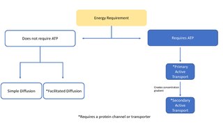
In cellular biology, active is the movement of molecules or ions across a cell membrane from a region of lower concentration to a region of higher concentration—against the concentration gradient. Active transport requires cellular energy to achieve this movement. There are two types of active transport: primary active transport that uses adenosine triphosphate (ATP), and secondary active transport that uses an electrochemical gradient. This process is in contrast to passive transport, which allows molecules or ions to move down their concentration gradient, from an area of high concentration to an area of low concentration, without energy.

<span class="mw-page-title-main">Sodium–potassium pump</span> Enzyme found in the membrane of all animal cells

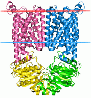
The sodium–potassium pump is an enzyme found in the membrane of all animal cells. It performs several functions in cell physiology.

<span class="mw-page-title-main">Mediated transport</span> Transportation of substances via membrane

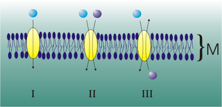
Mediated transport refers to transport mediated by a membrane transport protein. Substances in the human body may be hydrophobic, electrophilic, contain a positively or negatively charge, or have another property. As such there are times when those substances may not be able to pass over the cell membrane using protein-independent movement. The cell membrane is imbedded with many membrane transport proteins that allow such molecules to travel in and out of the cell. There are three types of mediated transporters: uniport, symport, and antiport. Things that can be transported are nutrients, ions, glucose, etc, all depending on the needs of the cell. One example of a uniport mediated transport protein is GLUT1. GLUT1 is a transmembrane protein, which means it spans the entire width of the cell membrane, connecting the extracellular and intracellular region. It is a uniport system because it specifically transports glucose in only one direction, down its concentration gradient across the cell membrane.

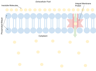
A membrane transport protein is a membrane protein involved in the movement of ions, small molecules, and macromolecules, such as another protein, across a biological membrane. Transport proteins are integral transmembrane proteins; that is they exist permanently within and span the membrane across which they transport substances. The proteins may assist in the movement of substances by facilitated diffusion, active transport, osmosis, or reverse diffusion. The two main types of proteins involved in such transport are broadly categorized as either channels or carriers. Examples of channel/carrier proteins include the GLUT 1 uniporter, sodium channels, and potassium channels. The solute carriers and atypical SLCs are secondary active or facilitative transporters in humans. Collectively membrane transporters and channels are known as the transportome. Transportomes govern cellular influx and efflux of not only ions and nutrients but drugs as well.

<span class="mw-page-title-main">Antiporter</span> Class of transmembrane transporter protein

An antiporter is an integral membrane protein that uses secondary active transport to move two or more molecules in opposite directions across a phospholipid membrane. It is a type of cotransporter, which means that uses the energetically favorable movement of one molecule down its electrochemical gradient to power the energetically unfavorable movement of another molecule up its electrochemical gradient. This is in contrast to symporters, which are another type of cotransporter that moves two or more ions in the same direction, and primary active transport, which is directly powered by ATP.

<span class="mw-page-title-main">Cotransporter</span> Type of membrane transport proteins

Cotransporters are a subcategory of membrane transport proteins (transporters) that couple the favorable movement of one molecule with its concentration gradient and unfavorable movement of another molecule against its concentration gradient. They enable coupled or cotransport and include antiporters and symporters. In general, cotransporters consist of two out of the three classes of integral membrane proteins known as transporters that move molecules and ions across biomembranes. Uniporters are also transporters but move only one type of molecule down its concentration gradient and are not classified as cotransporters.

<span class="mw-page-title-main">ABC transporter</span> Gene family

The ABC transporters, ATP synthase (ATP)-binding cassette transporters are a transport system superfamily that is one of the largest and possibly one of the oldest gene families. It is represented in all extant phyla, from prokaryotes to humans. ABC transporters belong to translocases.

<span class="mw-page-title-main">Glucose transporter</span> Family of monosaccharide transport proteins

Glucose transporters are a wide group of membrane proteins that facilitate the transport of glucose across the plasma membrane, a process known as facilitated diffusion. Because glucose is a vital source of energy for all life, these transporters are present in all phyla. The GLUT or SLC2A family are a protein family that is found in most mammalian cells. 14 GLUTS are encoded by the human genome. GLUT is a type of uniporter transporter protein.

<span class="mw-page-title-main">GLUT4</span> Protein

Glucose transporter type 4 (GLUT4), also known as solute carrier family 2, facilitated glucose transporter member 4, is a protein encoded, in humans, by the SLC2A4 gene. GLUT4 is the insulin-regulated glucose transporter found primarily in adipose tissues and striated muscle. The first evidence for this distinct glucose transport protein was provided by David James in 1988. The gene that encodes GLUT4 was cloned and mapped in 1989.

<span class="mw-page-title-main">Ion transporter</span> Transmembrane protein that moves ions across a biological membrane

In biology, an ion transporter is a transmembrane protein that moves ions across a biological membrane to accomplish many different biological functions, including cellular communication, maintaining homeostasis, energy production, etc. There are different types of transporters including pumps, uniporters, antiporters, and symporters. Active transporters or ion pumps are transporters that convert energy from various sources—including adenosine triphosphate (ATP), sunlight, and other redox reactions—to potential energy by pumping an ion up its concentration gradient. This potential energy could then be used by secondary transporters, including ion carriers and ion channels, to drive vital cellular processes, such as ATP synthesis.

<span class="mw-page-title-main">GLUT1</span> Uniporter protein

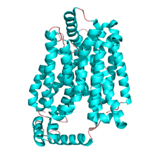
Glucose transporter 1, also known as solute carrier family 2, facilitated glucose transporter member 1 (SLC2A1), is a uniporter protein that in humans is encoded by the SLC2A1 gene. GLUT1 facilitates the transport of glucose across the plasma membranes of mammalian cells. This gene encodes a facilitative glucose transporter that is highly expressed in erythrocytes and endothelial cells, including cells of the blood–brain barrier. The encoded protein is found primarily in the cell membrane and on the cell surface, where it can also function as a receptor for human T-cell leukemia virus (HTLV) I and II. GLUT1 accounts for 2 percent of the protein in the plasma membrane of erythrocytes.

The galactose permease or GalP found in Escherichia coli is an integral membrane protein involved in the transport of monosaccharides, primarily hexoses, for utilization by E. coli in glycolysis and other metabolic and catabolic pathways (3,4). It is a member of the Major Facilitator Super Family (MFS) and is homologue of the human GLUT1 transporter (4). Below you will find descriptions of the structure, specificity, effects on homeostasis, expression, and regulation of GalP along with examples of several of its homologues.

The solute carrier (SLC) group of membrane transport proteins include over 400 members organized into 66 families. Most members of the SLC group are located in the cell membrane. The SLC gene nomenclature system was originally proposed by the HUGO Gene Nomenclature Committee (HGNC) and is the basis for the official HGNC names of the genes that encode these transporters. A more general transmembrane transporter classification can be found in TCDB database.

<span class="mw-page-title-main">Mitochondrial membrane transport protein</span>

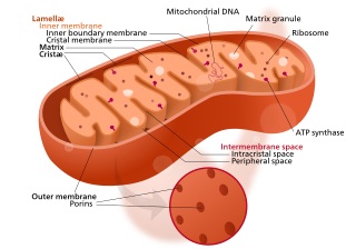
Mitochondrial membrane transport proteins, also known as mitochondrial carrier proteins, are proteins which exist in the membranes of mitochondria. They serve to transport molecules and other factors, such as ions, into or out of the organelles. Mitochondria contain both an inner and outer membrane, separated by the inter-membrane space, or inner boundary membrane. The outer membrane is porous, whereas the inner membrane restricts the movement of all molecules. The two membranes also vary in membrane potential and pH. These factors play a role in the function of mitochondrial membrane transport proteins. There are 53 discovered human mitochondrial membrane transporters, with many others that are known to still need discovered.

Translocase is a general term for a protein that assists in moving another molecule, usually across a cell membrane. These enzymes catalyze the movement of ions or molecules across membranes or their separation within membranes. The reaction is designated as a transfer from “side 1” to “side 2” because the designations “in” and “out”, which had previously been used, can be ambiguous. Translocases are the most common secretion system in Gram positive bacteria.

The insulin transduction pathway is a biochemical pathway by which insulin increases the uptake of glucose into fat and muscle cells and reduces the synthesis of glucose in the liver and hence is involved in maintaining glucose homeostasis. This pathway is also influenced by fed versus fasting states, stress levels, and a variety of other hormones.

The mitochondrial calcium uniporter (MCU) is a transmembrane protein that allows the passage of calcium ions from a cell's cytosol into mitochondria. Its activity is regulated by MICU1 and MICU2, which together with the MCU make up the mitochondrial calcium uniporter complex.

The ATP:ADP Antiporter (AAA) Family is a member of the major facilitator superfamily. Members of the AAA family have been sequenced from bacteria and plants.

References

  1. 1 2 3 Zhang XC, Han L (2016). "Uniporter substrate binding and transport: reformulating mechanistic questions". Biophys Rep. 2 (2–4): 45–54. doi:10.1007/s41048-016-0030-7. PMC   5138270 . PMID   28018963.
  2. 1 2 Alberts, Bruce (1998). Essential cell biology : an introduction to the molecular biology of the cell. Garland. ISBN   0-8153-2045-0. OCLC   36847771.
  3. Wolfersberger MG (November 1994). "Uniporters, symporters and antiporters". J Exp Biol. 196: 5–6. doi:10.1242/jeb.196.1.5. PMID   7823043.
  4. Pratt CA, Voet D, Voet JG (2002). Fundamentals of biochemistry. Wiley. pp. 264–6. ISBN   0-471-41759-9. OCLC   48137160.
  5. Hoppe, U. (2010). "Mitochondrial Calcium Channels". FEBS Letters. 584 (10): 1975–81. Bibcode:2010FEBSL.584.1975H. doi: 10.1016/j.febslet.2010.04.017 . PMID   20388514. S2CID   33664763.
  6. 1 2 3 4 Häfliger P, Charles RP (May 2019). "The L-Type Amino Acid Transporter LAT1-An Emerging Target in Cancer". Int J Mol Sci. 20 (10): 2428. doi: 10.3390/ijms20102428 . PMC   6566973 . PMID   31100853.
  7. Kim DM, Nimigean CM (May 2016). "Voltage-Gated Potassium Channels: A Structural Examination of Selectivity and Gating". Cold Spring Harb Perspect Biol. 8 (5): a029231. doi:10.1101/cshperspect.a029231. PMC   4852806 . PMID   27141052.
  8. OpenStax College (2013). Chapter 12.4 The Action Potential. OpenStax College. pp. 523–531. ISBN   978-1938168130.
  9. 1 2 Olson AL, Pessin JE (1996). "Structure, function, and regulation of the mammalian facilitative glucose transporter gene family". Annu Rev Nutr. 16: 235–56. doi:10.1146/annurev.nu.16.070196.001315. PMID   8839927.
  10. Cooper GM (2000). "12.2 Transport of Small Molecules". The Cell: A Molecular Approach (2nd ed.). Sunderland MA: Sinauer Associates. ISBN   0-87893-106-6. NBK9847.
  11. 1 2 Stillwell W (2016). "Membrane Transport". An Introduction to Biological Membranes. pp. 423–51. doi:10.1016/B978-0-444-63772-7.00019-1. ISBN   978-0-444-63772-7. PMC   7182109 .
  12. Rubinow SI, Dembo M (April 1977). "The facilitated diffusion of oxygen by hemoglobin and myoglobin". Biophys J. 18 (1): 29–42. Bibcode:1977BpJ....18...29R. doi:10.1016/S0006-3495(77)85594-X. PMC   1473276 . PMID   856316.
  13. Wright EM, Loo DD, Hirayama BA (April 2011). "Biology of human sodium glucose transporters". Physiol Rev. 91 (2): 733–94. doi:10.1152/physrev.00055.2009. PMID   21527736.
  14. 1 2 3 Thorens B, Mueckler M (February 2010). "Glucose transporters in the 21st Century". Am J Physiol Endocrinol Metab. 298 (2): E141–5. doi:10.1152/ajpendo.00712.2009. PMC   2822486 . PMID   20009031.
  15. Shamseldin HE, Alasmari A, Salih MA, Samman MM, Mian SA, Alshidi T, Ibrahim N, Hashem M, Faqeih E, Al-Mohanna F, Alkuraya FS (November 2017). "A null mutation in MICU2 causes abnormal mitochondrial calcium homeostasis and a severe neurodevelopmental disorder". Brain. 140 (11): 2806–13. doi:10.1093/brain/awx237. PMID   29053821.
  16. 1 2 3 4 5 6 7 8 9 10 11 Navale AM, Paranjape AN (March 2016). "Glucose transporters: physiological and pathological roles". Biophys Rev. 8 (1): 5–9. doi:10.1007/s12551-015-0186-2. PMC   5425736 . PMID   28510148.
  17. Mueckler M, Thorens B (2013). "The SLC2 (GLUT) family of membrane transporters". Mol Aspects Med. 34 (2–3): 121–38. doi:10.1016/j.mam.2012.07.001. PMC   4104978 . PMID   23506862.
  18. Carruthers A (October 1990). "Facilitated diffusion of glucose". Physiol Rev. 70 (4): 1135–76. doi:10.1152/physrev.1990.70.4.1135. PMID   2217557.
  19. Jiang S, Young JL, Wang K, Qian Y, Cai L (August 2020). "Diabeticinduced alterations in hepatic glucose and lipid metabolism: The role of type 1 and type 2 diabetes mellitus (Review)". Mol Med Rep. 22 (2): 603–611. doi:10.3892/mmr.2020.11175. PMC   7339764 . PMID   32468027.
  20. 1 2 3 4 5 De Stefani D, Patron M, Rizzuto R (September 2015). "Structure and function of the mitochondrial calcium uniporter complex". Biochim Biophys Acta. 1853 (9): 2006–11. doi:10.1016/j.bbamcr.2015.04.008. PMC   4522341 . PMID   25896525.
  21. 1 2 3 4 D'Angelo D, Rizzuto R (August 2023). "The Mitochondrial Calcium Uniporter (MCU): Molecular Identity and Role in Human Diseases". Biomolecules. 13 (9): 1304. doi: 10.3390/biom13091304 . PMC   10526485 . PMID   37759703.
  22. 1 2 3 Bhutia YD, Mathew M, Sivaprakasam S, Ramachandran S, Ganapathy V (January 2022). "Unconventional Functions of Amino Acid Transporters: Role in Macropinocytosis (SLC38A5/SLC38A3) and Diet-Induced Obesity/Metabolic Syndrome (SLC6A19/SLC6A14/SLC6A6)". Biomolecules. 12 (2): 235. doi: 10.3390/biom12020235 . PMC   8961558 . PMID   35204736.
  23. Singh N, Ecker GF (April 2018). "Insights into the Structure, Function, and Ligand Discovery of the Large Neutral Amino Acid Transporter 1, LAT1". Int J Mol Sci. 19 (5): 1278. doi: 10.3390/ijms19051278 . PMC   5983779 . PMID   29695141.
  24. Kanai Y (February 2022). "Amino acid transporter LAT1 (SLC7A5) as a molecular target for cancer diagnosis and therapeutics". Pharmacol Ther. 230: 107964. doi:10.1016/j.pharmthera.2021.107964. PMID   34390745.
  25. 1 2 3 4 5 6 Boswell-Casteel RC, Hays FA (January 2017). "Equilibrative nucleoside transporters-A review". Nucleosides Nucleotides Nucleic Acids. 36 (1): 7–30. doi:10.1080/15257770.2016.1210805. PMC   5728162 . PMID   27759477.
  26. 1 2 3 4 Hollenstein M (November 2012). "Nucleoside triphosphates--building blocks for the modification of nucleic acids". Molecules. 17 (11): 13569–91. doi: 10.3390/molecules171113569 . PMC   6268876 . PMID   23154273.
  27. 1 2 3 Fan M, Zhang J, Tsai CW, Orlando BJ, Rodriguez M, Xu Y, Liao M, Tsai MF, Feng L (June 2020). "Structure and mechanism of the mitochondrial Ca2+ uniporter holocomplex". Nature. 582 (7810): 129–133. Bibcode:2020Natur.582..129F. doi:10.1038/s41586-020-2309-6. PMC   7544431 . PMID   32494073.
  28. 1 2 3 Majumder P, Mallela AK, Penmatsa A (September 2018). "Transporters through the looking glass. An insight into the mechanisms of ion-coupled transport and methods that help reveal them". J Indian Inst Sci. 98 (3): 283–300. doi:10.1007/s41745-018-0081-5. PMC   6345361 . PMID   30686879.
  29. 1 2 David R, Byrt CS, Tyerman SD, Gilliham M, Wege S (September 2019). "Roles of membrane transporters: connecting the dots from sequence to phenotype". Ann Bot. 124 (2): 201–8. doi:10.1093/aob/mcz066. PMC   6758574 . PMID   31162525.
  30. Zhang R, Kang R, Klionsky DJ, Tang D (January 2022). "Ion Channels and Transporters in Autophagy". Autophagy. 18 (1): 4–23. doi:10.1080/15548627.2021.1885147. PMC   8865261 . PMID   33657975.
  31. Seifter JL, Chang HY (September 2017). "Extracellular Acid-Base Balance and Ion Transport Between Body Fluid Compartments". Physiology (Bethesda). 32 (5): 367–379. doi:10.1152/physiol.00007.2017. PMID   28814497.
  32. 1 2 3 4 5 6 7 8 9 10 MedlinePlus Encyclopedia : Noonan syndrome
  33. 1 2 3 4 5 "GLUT1 deficiency syndrome". Genetic Conditions. MedlinePlus.