
The ulna or ulnar bone is a long bone in the forearm stretching from the elbow to the wrist. It is on the same side of the forearm as the little finger, running parallel to the radius, the forearm's other long bone. Longer and thinner than the radius, the ulna is considered to be the smaller long bone of the lower arm. The corresponding bone in the lower leg is the fibula.
The forearm is the region of the upper limb between the elbow and the wrist. The term forearm is used in anatomy to distinguish it from the arm, a word which is used to describe the entire appendage of the upper limb, but which in anatomy, technically, means only the region of the upper arm, whereas the lower "arm" is called the forearm. It is homologous to the region of the leg that lies between the knee and the ankle joints, the crus.
In human anatomy, the extensor carpi ulnaris is a skeletal muscle located on the ulnar side of the forearm. The extensor carpi ulnaris acts to extend and adduct at the carpus/wrist from anatomical position.

A splint is defined as "a rigid or flexible device that maintains in position a displaced or movable part; also used to keep in place and protect an injured part" or as "a rigid or flexible material used to protect, immobilize, or restrict motion in a part". Splints can be used for injuries that are not severe enough to immobilize the entire injured structure of the body. For instance, a splint can be used for certain fractures, soft tissue sprains, tendon injuries, or injuries awaiting orthopedic treatment. A splint may be static, not allowing motion, or dynamic, allowing controlled motion. Splints can also be used to relieve pain in damaged joints. Splints are quick and easy to apply and do not require a plastering technique. Splints are often made out of some kind of flexible material and a firm pole-like structure for stability. They often buckle or Velcro together.
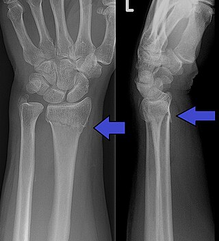
A Colles' fracture is a type of fracture of the distal forearm in which the broken end of the radius is bent backwards. Symptoms may include pain, swelling, deformity, and bruising. Complications may include damage to the median nerve.

A bone fracture is a medical condition in which there is a partial or complete break in the continuity of any bone in the body. In more severe cases, the bone may be broken into several fragments, known as a comminuted fracture. An open fracture is a bone fracture where the broken bone breaks through the skin. A bone fracture may be the result of high force impact or stress, or a minimal trauma injury as a result of certain medical conditions that weaken the bones, such as osteoporosis, osteopenia, bone cancer, or osteogenesis imperfecta, where the fracture is then properly termed a pathologic fracture. Most bone fractures require urgent medical attention to prevent further injury.

Orthopedic casts or just casts are a form of medical treatment used to immobilize and support bones and soft tissues during the healing process after fractures, surgeries, or severe injuries. By restricting movement, casts provide stability to the affected area, enabling proper alignment and healing of bones, ligaments, and tendons. They are commonly applied to the limbs but can also be used for the trunk, neck, or other parts of the body in specific cases. Orthopedic casts come in various types and designs, tailored to the nature and severity of the injury, as well as the patient's needs. Advances in medical techniques have made casts more comfortable, effective, and versatile, allowing for both weight-bearing and non-weight-bearing options.
A distal radius fracture, also known as wrist fracture, is a break of the part of the radius bone which is close to the wrist. Symptoms include pain, bruising, and rapid-onset swelling. The ulna bone may also be broken.

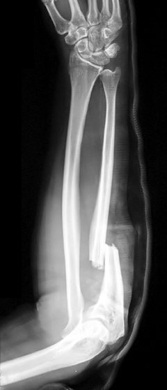
The Galeazzi fracture is a fracture of the distal third of the radius with dislocation of the distal radioulnar joint. It classically involves an isolated fracture of the junction of the distal third and middle third of the radius with associated subluxation or dislocation of the distal radio-ulnar joint; the injury disrupts the forearm axis joint.

Madelung's deformity is usually characterized by malformed wrists and wrist bones and is often associated with Léri-Weill dyschondrosteosis. It can be bilateral or just in the one wrist. It has only been recognized within the past hundred years. Named after Otto Wilhelm Madelung (1846–1926), a German surgeon, who described it in detail, it was noted by others. Guillaume Dupuytren mentioned it in 1834, Auguste Nélaton in 1847, and Joseph-François Malgaigne in 1855.

A patella fracture is a break of the kneecap. Symptoms include pain, swelling, and bruising to the front of the knee. A person may also be unable to walk. Complications may include injury to the tibia, femur, or knee ligaments.
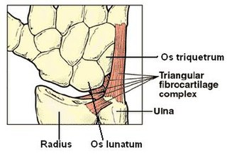
The triangular fibrocartilage complex (TFCC) is formed by the triangular fibrocartilage discus (TFC), the radioulnar ligaments (RULs) and the ulnocarpal ligaments (UCLs).
The Monteggia fracture is a fracture of the proximal third of the ulna with dislocation of the proximal head of the radius. It is named after Giovanni Battista Monteggia.

The elbow is the region between the upper arm and the forearm that surrounds the elbow joint. The elbow includes prominent landmarks such as the olecranon, the cubital fossa, and the lateral and the medial epicondyles of the humerus. The elbow joint is a hinge joint between the arm and the forearm; more specifically between the humerus in the upper arm and the radius and ulna in the forearm which allows the forearm and hand to be moved towards and away from the body. The term elbow is specifically used for humans and other primates, and in other vertebrates it is not used. In those cases, forelimb plus joint is used.
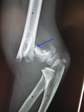
A supracondylar humerus fracture is a fracture of the distal humerus just above the elbow joint. The fracture is usually transverse or oblique and above the medial and lateral condyles and epicondyles. This fracture pattern is relatively rare in adults, but is the most common type of elbow fracture in children. In children, many of these fractures are non-displaced and can be treated with casting. Some are angulated or displaced and are best treated with surgery. In children, most of these fractures can be treated effectively with expectation for full recovery. Some of these injuries can be complicated by poor healing or by associated blood vessel or nerve injuries with serious complications.
A child bone fracture or a pediatric fracture is a medical condition in which a bone of a child is cracked or broken. About 15% of all injuries in children are fracture injuries. Bone fractures in children are different from adult bone fractures because a child's bones are still growing. Also, more consideration needs to be taken when a child fractures a bone since it will affect the child in his or her growth.
An ulna fracture is a break in the ulna bone, one of the two bones in the forearm. It is often associated with a fracture of the other forearm bone, the radius.

Olecranon fracture is a fracture of the bony portion of the elbow. The injury is fairly common and often occurs following a fall or direct trauma to the elbow. The olecranon is the proximal extremity of the ulna which is articulated with the humerus bone and constitutes a part of the elbow articulation. Its location makes it vulnerable to direct trauma.

Radioulnar synostosis is a rare condition where there is an abnormal connection (synostosis) between the radius and ulna bones of the forearm. This can be present at birth (congenital), when it is a result of a failure of the bones to form separately, or following an injury (post-traumatic).

A Torus fracture, also known as a buckle fracture is the most common fracture in children. It is a common occurrence following a fall, as the wrist absorbs most of the impact and compresses the bony cortex on one side and remains intact on the other, creating a bulging effect. As the bulge is only on one side of the bone, this injury can be classified as an incomplete fracture. The compressive force is provided by the trabeculae and is longitudinal to the axis of the long bone, meaning that the fracture itself is orthogonal to that axis. The word "torus" originates from the Latin word "protuberance."
