Femoral fracture

Last updated
Femoral fracture
Medical X-Ray imaging IYN05 nevit.jpg
X-ray image of a femoral shaft fracture
Specialty Orthopedic

A femoral fracture is a bone fracture that involves the femur. They are typically sustained in high-impact trauma, such as car crashes, due to the large amount of force needed to break the bone. Fractures of the diaphysis, or middle of the femur, are managed differently from those at the head, neck, and trochanter; those are conventionally called hip fractures (because they involve the hip joint region). Thus, mentions of femoral fracture in medicine usually refer implicitly to femoral fractures at the shaft or distally.

Contents

Signs and symptoms

Fractures are commonly obvious, since femoral fractures are often caused by high energy trauma. [1] Signs of fracture include swelling, deformity, and shortening of the leg. [2] Extensive soft-tissue injury, bleeding, and shock are common. [3] The most common symptom is severe pain, which prevents movement of the leg. [4]

Diagnosis

Common locations of fracture of femur Common fracture locations of femur.jpg
Common locations of fracture of femur

Physical exam

Femoral shaft fractures occur during extensive trauma, and they can act as distracting injuries, whereby the observer accidentally overlooks other injuries, preventing a thorough exam of the complete body. [4] For example, the ligaments and meniscus of the ipsilateral (same side) knee are also commonly injured. [2] [3]

Radiography

Anterior-posterior (AP) and lateral radiographs are typically obtained. [4] In order to rule out other injuries, hip, pelvis, and knee radiographs are also obtained. [5] The hip radiograph is of particular importance, because femoral neck fractures can lead to osteonecrosis of the femoral head. [4]

Classification

The diaphysis is the midshaft of the femur Structure of a Long Bone.png
The diaphysis is the midshaft of the femur

The fracture may be classed as open, which occurs when the bone fragments protrude through the skin, or there is an overlying wound that penetrates to the bone. These types of fracture cause more damage to the surrounding tissue, are less likely to heal properly, and are at much greater risk of infection.[ citation needed ]

Femoral shaft fractures

Femoral shaft fractures can be classified with the Winquist and Hansen classification, which is based on the amount of comminution. [6]

Distal femur fractures

Fractures of the inferior or distal femur may be complicated by separation of the condyles, resulting in misalignment of the articular surfaces of the knee joint, or by hemorrhage from the large popliteal artery that runs directly on the posterior surface of the bone. This fracture compromises the blood supply to the leg (an occurrence that should always be considered in knee fractures or dislocations). [7]

Treatment

A 2015 Cochrane review (updated in 2022) found that available evidence for treatment options of distal femur fractures is insufficient to inform clinical practice and that there is a priority for a high-quality trial to be undertaken. [8] Open fractures must undergo urgent surgery to clean and repair them, but closed fractures can be maintained until the patient is stable and ready for surgery. [9] [10]

Skeletal traction

Available evidence suggests that treatment depends on the part of the femur that is fractured. Traction may be useful for femoral shaft fractures because it counteracts the force of the muscle pulling the two separated parts together, and thus may decrease bleeding and pain. [11] Traction should not be used in femoral neck fractures or when there is any other trauma to the leg or pelvis. [12] [13] It is typically only a temporary measure used before surgery. It is only considered definitive treatment for patients with significant comorbidities that contraindicate surgical management. [14]

External fixators

External fixators can be used to prevent further damage to the leg until the patient is stable enough for surgery. [14] It is most commonly used as a temporary measure. However, for some select cases it may be used as an alternative to intramedullary nailing for definitive treatment. [15] [16]

Intramedullary nailing

For femoral shaft fractures, reduction and intramedullary nailing is currently recommended. [14] The bone is re-aligned, then a metal rod is placed into the femoral bone marrow, and secured with nails at either end. This method offers less exposure, a 98–99% union rate, lower infection rates (1–2%) and less muscular scarring. [14] [15] [17]

Rehabilitation

After surgery, the patient should be offered physiotherapy and try to walk as soon as possible, and then every day after that to maximise their chances of a good recovery. [18]

Outcomes

These fractures can take at least 4–6 months to heal. [19] Since femoral shaft fractures are associated with violent trauma, there are many adverse outcomes, including fat embolism, acute respiratory distress syndrome (ARDS), multisystem organ failure, and shock associated with severe blood loss. [4] Open fractures can result in infection, osteomyelitis, and sepsis.[ citation needed ]

Epidemiology

Femoral shaft fractures occur in a bimodal distribution, whereby they are most commonly seen in males age 15-24 (due to high energy trauma) and females aged 75 or older (pathologic fractures due to osteoporosis, low-energy falls). [20] [14] In Germany, femoral fractures are the most common type of fracture seen and treated in hospitals. [9] [21]

Related Research Articles

<span class="mw-page-title-main">Knee</span> Leg joint in primates

In humans and other primates, the knee joins the thigh with the leg and consists of two joints: one between the femur and tibia, and one between the femur and patella. It is the largest joint in the human body. The knee is a modified hinge joint, which permits flexion and extension as well as slight internal and external rotation. The knee is vulnerable to injury and to the development of osteoarthritis.

<span class="mw-page-title-main">Orthopedic surgery</span> Branch of surgery concerned with the musculoskeletal system

Orthopedic surgery or orthopedics is the branch of surgery concerned with conditions involving the musculoskeletal system. Orthopedic surgeons use both surgical and nonsurgical means to treat musculoskeletal trauma, spine diseases, sports injuries, degenerative diseases, infections, tumors, and congenital disorders.

An osteotomy is a surgical operation whereby a bone is cut to shorten or lengthen it or to change its alignment. It is sometimes performed to correct a hallux valgus, or to straighten a bone that has healed crookedly following a fracture. It is also used to correct a coxa vara, genu valgum, and genu varum. The operation is done under a general anaesthetic.

<span class="mw-page-title-main">Bone fracture</span> Physical damage to the continuity of a bone

A bone fracture is a medical condition in which there is a partial or complete break in the continuity of any bone in the body. In more severe cases, the bone may be broken into several fragments, known as a comminuted fracture. A bone fracture may be the result of high force impact or stress, or a minimal trauma injury as a result of certain medical conditions that weaken the bones, such as osteoporosis, osteopenia, bone cancer, or osteogenesis imperfecta, where the fracture is then properly termed a pathologic fracture.

<span class="mw-page-title-main">Genu varum</span> Varus deformity marked by (outward) bowing at the knee

Genu varum is a varus deformity marked by (outward) bowing at the knee, which means that the lower leg is angled inward (medially) in relation to the thigh's axis, giving the limb overall the appearance of an archer's bow. Usually medial angulation of both lower limb bones is involved.

<span class="mw-page-title-main">Joint dislocation</span> Medical injury

A joint dislocation, also called luxation, occurs when there is an abnormal separation in the joint, where two or more bones meet. A partial dislocation is referred to as a subluxation. Dislocations are often caused by sudden trauma on the joint like an impact or fall. A joint dislocation can cause damage to the surrounding ligaments, tendons, muscles, and nerves. Dislocations can occur in any major joint or minor joint. The most common joint dislocation is a shoulder dislocation.

<span class="mw-page-title-main">Clavicle fracture</span> Medical condition

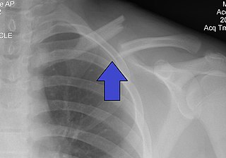
A clavicle fracture, also known as a broken collarbone, is a bone fracture of the clavicle. Symptoms typically include pain at the site of the break and a decreased ability to move the affected arm. Complications can include a collection of air in the pleural space surrounding the lung (pneumothorax), injury to the nerves or blood vessels in the area, and an unpleasant appearance.

<span class="mw-page-title-main">Hip fracture</span> Broken bone in hip joint region

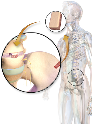
A hip fracture is a break that occurs in the upper part of the femur, at the femoral neck or (rarely) the femoral head. Symptoms may include pain around the hip, particularly with movement, and shortening of the leg. Usually the person cannot walk.

<span class="mw-page-title-main">Hip</span> Anatomical region between the torso and the legs, holding the buttocks and genital region

In vertebrate anatomy, the hip, or coxa in medical terminology, refers to either an anatomical region or a joint on the outer (lateral) side of the pelvis.

<span class="mw-page-title-main">Slipped capital femoral epiphysis</span> Medical condition

Slipped capital femoral epiphysis is a medical term referring to a fracture through the growth plate (physis), which results in slippage of the overlying end of the femur (metaphysis).

A traction splint most commonly refers to a splinting device that uses straps attaching over the pelvis or hip as an anchor, a metal rod(s) to mimic normal bone stability and limb length, and a mechanical device to apply traction to the limb.

<span class="mw-page-title-main">Hip dislocation</span> Orthopedic injury

A hip dislocation is when the thighbone (femur) separates from the hip bone (pelvis). Specifically it is when the ball–shaped head of the femur separates from its cup–shaped socket in the hip bone, known as the acetabulum. The joint of the femur and pelvis is very stable, secured by both bony and soft-tissue constraints. With that, dislocation would require significant force which typically results from significant trauma such as from a motor vehicle collision or from a fall from elevation. Hip dislocations can also occur following a hip replacement or from a developmental abnormality known as hip dysplasia.

<span class="mw-page-title-main">Femoral neck</span> Aspect of human anatomy

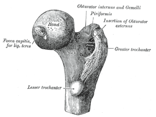
The femoral neck is a flattened pyramidal process of bone, connecting the femoral head with the femoral shaft, and forming with the latter a wide angle opening medialward.

An open fracture, also called a compound fracture, is a type of bone fracture that has an open wound in the skin near the fractured bone. The skin wound is usually caused by the bone breaking through the surface of the skin. An open fracture can be life threatening or limb-threatening due to the risk of a deep infection and/or bleeding. Open fractures are often caused by high energy trauma such as road traffic accidents and are associated with a high degree of damage to the bone and nearby soft tissue. Other potential complications include nerve damage or impaired bone healing, including malunion or nonunion. The severity of open fractures can vary. For diagnosing and classifying open fractures, Gustilo-Anderson open fracture classification is the most commonly used method. This classification system can also be used to guide treatment, and to predict clinical outcomes. Advanced trauma life support is the first line of action in dealing with open fractures and to rule out other life-threatening condition in cases of trauma. The person is also administered antibiotics for at least 24 hours to reduce the risk of an infection.

<span class="mw-page-title-main">Intramedullary rod</span> Metal rod inserted to treat fractures

An intramedullary rod, also known as an intramedullary nail or inter-locking nail or Küntscher nail, is a metal rod forced into the medullary cavity of a bone. IM nails have long been used to treat fractures of long bones of the body. Gerhard Küntscher is credited with the first use of this device in 1939, during World War II, for soldiers with fractures of the femur. Prior to that, treatment of such fractures was limited to traction or plaster, both of which required long periods of inactivity. IM nails resulted in earlier return to activity for the soldiers, sometimes even within a span of a few weeks, since they share the load with the bone, rather than entirely supporting the bone.

Early appropriate care (EAC) is a system in orthopaedic trauma surgery aiming to identify serious major trauma patients and treat the most time-critical injuries without adding to their physiological burden.

<span class="mw-page-title-main">Tibia shaft fracture</span> Medical condition

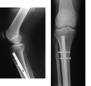
Tibia shaft fracture is a fracture of the proximal (upper) third of the tibia. Due to the location of the tibia, it is frequently injured. Thus it is the most commonly fractured long bone in the body.

<span class="mw-page-title-main">Phemister graft</span>

A Phemister graft is a type of bone graft which uses bone tissue harvested from the patient to treat slow-healing, or delayed union bone fractures. Thus, it is a form of autotransplantation. Typically, the tissue used in the graft is cancellous bone harvested from the patient's Iliac crest and laid in strips across the fracture site. The use of the patient's living bone stimulates osteogenesis, the growth of bones.

Bone malrotation refers to the situation that results when a bone heals out of rotational alignment from another bone, or part of bone. It often occurs as the result of a surgical complication after a fracture where intramedullary nailing (IMN) occurs, especially in the femur and tibial bones, but can also occur genetically at birth. The severity of this complication is often neglected due to its complexity to detect and treat, yet if left untreated, bone malrotation can significantly impact regular bodily functioning, and even lead to severe arthritis. Detection throughout history has become more advanced and accurate, ranging from clinical assessment to ultrasounds to CT scans. Treatment can include an osteotomy, a major surgical procedure where bones are cut and realigned correctly, or compensatory methods, where individuals learn to externally or internally rotate their limb to compensate for the rotation. Further research is currently being examined in this area to reduce occurrences of malrotation, including detailed computer navigation to improve visual accuracy during surgery.

References

  1. Bucholz RW, Jones A (December 1991). "Fractures of the shaft of the femur". The Journal of Bone and Joint Surgery. American Volume. 73 (10): 1561–1566. doi:10.2106/00004623-199173100-00015. PMID   1748704.
  2. 1 2 Rockwood Jr CA, Green DP, Bucholz RW (2010). Rockwood and Green's fractures in adults (7th ed.). Philadelphia, PA: Wolters Kluwer Health/Lippincott Williams & Wilkins. ISBN   9781605476773. OCLC   444336477.
  3. 1 2 Skinner HB, McMahon PJ (2014). Current diagnosis & treatment in orthopedics (5th ed.). New York: McGraw-Hill Medical. ISBN   9780071590754. OCLC   820106991.
  4. 1 2 3 4 5 Sarwark JF (2010). Essentials of musculoskeletal care. Rosemont, Ill.: American Academy of Orthopaedic Surgeons. ISBN   9780892035793. OCLC   706805938.
  5. Committee on Trauma. (2012). Advanced trauma life support : student course manual (9th ed.). Chicago, IL: American College of Surgeons. ISBN   9781880696026. OCLC   846430144.
  6. Stannard JP, Schmidt AH, Kregor PJ (2007). Surgical treatment of orthopaedic trauma. New York, NY: Thieme. p.  612. ISBN   978-1-58890-307-5.
  7. Moore KL, Dalley AF, Agur AM (2014). Clinically Oriented Anatomy (Seventh ed.). Philadelphia. p. 527. ISBN   978-1-4511-8447-1.{{cite book}}: CS1 maint: location missing publisher (link)
  8. Claireaux, Henry A.; Searle, Henry Kc; Parsons, Nick R.; Griffin, Xavier L. (2022-10-05). "Interventions for treating fractures of the distal femur in adults". The Cochrane Database of Systematic Reviews. 2022 (10): CD010606. doi:10.1002/14651858.CD010606.pub3. ISSN   1469-493X. PMC   9534312 . PMID   36197809.
  9. 1 2 Büchele G, Rehm M, Halbgebauer R, Rothenbacher D, Huber-Lang M (2022-02-26). "Trauma-related acute kidney injury during inpatient care of femoral fractures increases the risk of mortality: a claims data analysis". American Journal of Medicine Open. 8: 100009. doi: 10.1016/j.ajmo.2022.100009 . ISSN   2667-0364. PMC   11256273 . S2CID   247152803.
  10. "Femur Shaft Fractures (Broken Thighbone)-OrthoInfo - AAOS". orthoinfo.aaos.org. Retrieved 2016-12-14.
  11. Tintinalli, Judith E. (2010). Emergency Medicine: A Comprehensive Study Guide (Emergency Medicine (Tintinalli)). New York: McGraw-Hill Companies. p. 9. ISBN   978-0-07-148480-0.
  12. AAOS (October 2010). "29". In Andrew N. Pollak MD. FAAOS (ed.). Emergency Care and Transport of the Sick and Injured (Print) (10 ed.). Sudbury, Massachusetts: Jones and Bartlett. pp. 1025–1031. ISBN   978-1-4496-3056-0.
  13. Marx JA (2014). Rosen's emergency medicine : concepts and clinical practice (Eighth ed.). London: Elsevier Health Sciences. p. 680. ISBN   9781455749874.
  14. 1 2 3 4 5 Egol KA, Koval KJ, Zuckerman JD (2015). Handbook of fractures (5th ed.). Philadelphia: Wolters Kluwer Health. ISBN   9781451193626. OCLC   960851324.
  15. 1 2 "Midshaft femur fractures in adults". www.uptodate.com. Retrieved 2017-10-01.
  16. Kovar FM, Jaindl M, Schuster R, Endler G, Platzer P (July 2013). "Incidence and analysis of open fractures of the midshaft and distal femur". Wiener Klinische Wochenschrift. 125 (13–14): 396–401. doi:10.1007/s00508-013-0391-6. PMID   23797531. S2CID   9565227.
  17. el Moumni M, Leenhouts PA, ten Duis HJ, Wendt KW (February 2009). "The incidence of non-union following unreamed intramedullary nailing of femoral shaft fractures". Injury. 40 (2): 205–208. doi:10.1016/j.injury.2008.06.022. PMID   19070840.
  18. Paterno MV, Archdeacon MT (May 2009). "Is there a standard rehabilitation protocol after femoral intramedullary nailing?". Journal of Orthopaedic Trauma. 23 (5 Suppl): S39–S46. doi: 10.1097/BOT.0b013e31819f27c2 . PMID   19390375. S2CID   11239969.
  19. "Femoral Fractures. Information about Femur fractures. Patient | Patient". Patient. Retrieved 2016-12-14.
  20. Hemmann P, Friederich M, Körner D, Klopfer T, Bahrs C (May 2021). "Changing epidemiology of lower extremity fractures in adults over a 15-year period - a National Hospital Discharge Registry study". BMC Musculoskeletal Disorders. 22 (1): 456. doi: 10.1186/s12891-021-04291-9 . PMC   8135150 . PMID   34011331.
  21. Rapp K, Büchele G, Dreinhöfer K, Bücking B, Becker C, Benzinger P (February 2019). "Epidemiology of hip fractures : Systematic literature review of German data and an overview of the international literature". Zeitschrift für Gerontologie und Geriatrie. 52 (1): 10–16. doi:10.1007/s00391-018-1382-z. PMC   6353815 . PMID   29594444.