Autoradiograph

Last updated

Autoradiography of a coronal brain slice, taken from an embryonal rat. GAD67-binding marker is highly expressed in the subventricular zone. Autoradiography of a brain slice from an embryonal rat - PMID19190758 PLoS 0004371.png
Autoradiography of a coronal brain slice, taken from an embryonal rat. GAD67-binding marker is highly expressed in the subventricular zone.

An autoradiograph is an image on an X-ray film or nuclear emulsion produced by the pattern of decay emissions (e.g., beta particles or gamma rays) from a distribution of a radioactive substance. Alternatively, the autoradiograph is also available as a digital image (digital autoradiography), due to the recent development of scintillation gas detectors [1] or rare-earth phosphorimaging systems. [2] The film or emulsion is apposed to the labeled tissue section to obtain the autoradiograph (also called an autoradiogram). The auto- prefix indicates that the radioactive substance is within the sample, as distinguished from the case of historadiography or microradiography, in which the sample is marked using an external source. Some autoradiographs can be examined microscopically for localization of silver grains (such as on the interiors or exteriors of cells or organelles) in which the process is termed micro-autoradiography. For example, micro-autoradiography was used to examine whether atrazine was being metabolized by the hornwort plant or by epiphytic microorganisms in the biofilm layer surrounding the plant. [3]

Contents

Applications

In biology, this technique may be used to determine the tissue (or cell) localization of a radioactive substance, either introduced into a metabolic pathway, bound to a receptor [4] [5] or enzyme, or hybridized to a nucleic acid. [6] Applications for autoradiography are broad, ranging from biomedical to environmental sciences to industry.

Receptor autoradiography

The use of radiolabeled ligands to determine the tissue distributions of receptors is termed either in vivo or in vitro receptor autoradiography if the ligand is administered into the circulation (with subsequent tissue removal and sectioning) or applied to the tissue sections, respectively. [7] Once the receptor density is known, in vitro autoradiography can also be used to determine the anatomical distribution and affinity of a radiolabeled drug towards the receptor. For in vitro autoradiography, radioligand was directly applying on frozen tissue sections without administration to the subject. Thus it cannot follow the distribution, metabolism and degradation situation completely in the living body. But because target in the cryosections is widely exposed and can direct contact with radioligand, in vitro autoradiography is still a quick and easy method to screen drug candidates, PET and SPECT ligands. The ligands are generally labeled with 3H (tritium), 18F (fluorine), 11C (carbon) or 125I (radioiodine). Compare to in vitro, ex vivo autoradiography were performed after administration of radioligand in the body, which can decrease the artifacts and are closer to the inner environment.

The distribution of RNA transcripts in tissue sections by the use of radiolabeled, complementary oligonucleotides or ribonucleic acids ("riboprobes") is called in situ hybridization histochemistry. Radioactive precursors of DNA and RNA, [3H]-thymidine and [3H]-uridine respectively, may be introduced to living cells to determine the timing of several phases of the cell cycle. RNA or DNA viral sequences can also be located in this fashion. These probes are usually labeled with 32P, 33P, or 35S. In the realm of behavioral endocrinology, autoradiography can be used to determine hormonal uptake and indicate receptor location; an animal can be injected with a radiolabeled hormone, or the study can be conducted in vitro.

Rate of DNA replication

The rate of DNA replication in a mouse cell growing in vitro was measured by autoradiography as 33 nucleotides per second. [8] The rate of phage T4 DNA elongation in phage-infected E. coli was also measured by autoradiography as 749 nucleotides per second during the period of exponential DNA increase at 37 °C (99 °F). [9]

Detection of protein phosphorylation

Phosphorylation means the posttranslational addition of a phosphate group to specific amino acids of proteins, and such modification can lead to a drastic change in the stability or the function of a protein in the cell. Protein phosphorylation can be detected on an autoradiograph, after incubating the protein in vitro with the appropriate kinase and γ-32P-ATP. The radiolabeled phosphate of latter is incorporated into the protein which is isolated via SDS-PAGE and visualized on an autoradiograph of the gel. (See figure 3. of a recent study showing that CREB-binding protein is phosphorylated by HIPK2. [10] )

Detection of sugar movement in plant tissue

In plant physiology, autoradiography can be used to determine sugar accumulation in leaf tissue. [11] Sugar accumulation, as it relates to autoradiography, can described the phloem-loading strategy used in a plant. [12] For example, if sugars accumulate in the minor veins of a leaf, it is expected that the leaves have few plasmodesmatal connections which is indicative of apoplastic movement, or an active phloem-loading strategy. Sugars, such as sucrose, fructose, or mannitol, are radiolabeled with [ 14-C], and then absorbed into leaf tissue by simple diffusion. [13] The leaf tissue is then exposed to autoradiographic film (or emulsion) to produce an image. Images will show distinct vein patterns if sugar accumulation is concentrated in leaf veins (apoplastic movement), or images will show a static-like pattern if sugar accumulation is uniform throughout the leaf (symplastic movement).

Other techniques

This autoradiographic approach contrasts to techniques such as PET and SPECT where the exact 3-dimensional localization of the radiation source is provided by careful use of coincidence counting, gamma counters and other devices.

Krypton-85 is used to inspect aircraft components for small defects. Krypton-85 is allowed to penetrate small cracks, and then its presence is detected by autoradiography. The method is called "krypton gas penetrant imaging". The gas penetrates smaller openings than the liquids used in dye penetrant inspection and fluorescent penetrant inspection. [14]

Historical events

Unintentional exposure

A radioactive surgeonfish makes its own X-ray. The bright area is a meal of fresh algae. The rest of the body has absorbed and distributed enough plutonium to make the scales radioactive. The fish was alive and apparently healthy when captured. Crossroads Radioactive Puffy Surgeon Fish.jpg
A radioactive surgeonfish makes its own X-ray. The bright area is a meal of fresh algae. The rest of the body has absorbed and distributed enough plutonium to make the scales radioactive. The fish was alive and apparently healthy when captured.

The task of radioactive decontamination following the Baker nuclear test at Bikini Atoll during Operation Crossroads in 1946 was far more difficult than the U.S. Navy had prepared for. Though the task's futility became apparent and the danger to cleanup crews mounted, Colonel Stafford Warren, in charge of radiation safety, had difficulty persuading Vice Admiral William H. P. Blandy to abandon the cleanup and with it the surviving target ships. On August 10, Warren showed Blandy an autoradiograph made by a surgeonfish from the lagoon that was left on a photographic plate overnight. The film was exposed by alpha radiation produced from the fish's scales, evidence that plutonium, mimicking calcium, had been distributed throughout the fish. Blandy promptly ordered that all further decontamination work be discontinued. Warren wrote home, "A self X ray of a fish ... did the trick." [15]

Related Research Articles

A radioligand is a microscopic particle which consists of a therapeutic radioactive isotope and the cell-targeting compound - the ligand. The ligand is the target binding site, it may be on the surface of the targeted cancer cell for therapeutic purposes. Radioisotopes can occur naturally or be synthesized and produced in a cyclotron/nuclear reactor. The different types of radioisotopes include Y-90, H-3, C-11, Lu-177, Ac-225, Ra-223, In-111, I-131, I-125, etc. Thus, radioligands must be produced in special nuclear reactors for the radioisotope to remain stable. Radioligands can be used to analyze/characterize receptors, to perform binding assays, to help in diagnostic imaging, and to provide targeted cancer therapy. Radiation is a novel method of treating cancer and is effective in short distances along with being unique/personalizable and causing minimal harm to normal surrounding cells. Furthermore, radioligand binding can provide information about receptor-ligand interactions in vitro and in vivo. Choosing the right radioligand for the desired application is important. The radioligand must be radiochemically pure, stable, and demonstrate a high degree of selectivity, and high affinity for their target.

<span class="mw-page-title-main">Ketanserin</span> Antihypertensive agent

Ketanserin (INN, USAN, BAN) (brand name Sufrexal; former developmental code name R41468) is a drug used clinically as an antihypertensive agent and in scientific research to study the serotonergic system; specifically, the 5-HT2 receptor family. It was discovered at Janssen Pharmaceutica in 1980. It is not available in the United States.

IC<sub>50</sub> Half maximal inhibitory concentration

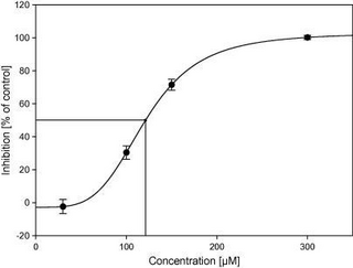
Half maximal inhibitory concentration (IC50) is a measure of the potency of a substance in inhibiting a specific biological or biochemical function. IC50 is a quantitative measure that indicates how much of a particular inhibitory substance (e.g. drug) is needed to inhibit, in vitro, a given biological process or biological component by 50%. The biological component could be an enzyme, cell, cell receptor or microbe. IC50 values are typically expressed as molar concentration.

<span class="mw-page-title-main">Charles Philippe Leblond</span> Canadian cell biology researcher, professor

Charles Philippe Leblond was a pioneer of cell biology and stem cell research and a Canadian former professor of anatomy. Leblond is notable for developing autoradiography and his work showing how cells continuously renew themselves, regardless of age.

<span class="mw-page-title-main">Ligand (biochemistry)</span> Substance that forms a complex with a biomolecule

In biochemistry and pharmacology, a ligand is a substance that forms a complex with a biomolecule to serve a biological purpose. The etymology stems from Latin ligare, which means 'to bind'. In protein-ligand binding, the ligand is usually a molecule which produces a signal by binding to a site on a target protein. The binding typically results in a change of conformational isomerism (conformation) of the target protein. In DNA-ligand binding studies, the ligand can be a small molecule, ion, or protein which binds to the DNA double helix. The relationship between ligand and binding partner is a function of charge, hydrophobicity, and molecular structure.

<span class="mw-page-title-main">PK-11195</span> Chemical compound

PK-11195 is an isoquinoline carboxamide which binds selectively to the peripheral benzodiazepine receptor (PBR). It is one of the most commonly used PBR ligands due to its high affinity for the PBR in all species, although it is starting to be replaced by newer and more selective ligands.

<span class="mw-page-title-main">Radioactivity in the life sciences</span>

Radioactivity is generally used in life sciences for highly sensitive and direct measurements of biological phenomena, and for visualizing the location of biomolecules radiolabelled with a radioisotope.

<span class="mw-page-title-main">Translocator protein</span> Human protein

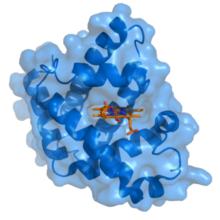
Translocator protein (TSPO) is an 18 kDa protein mainly found on the outer mitochondrial membrane. It was first described as peripheral benzodiazepine receptor (PBR), a secondary binding site for diazepam, but subsequent research has found the receptor to be expressed throughout the body and brain. In humans, the translocator protein is encoded by the TSPO gene. It belongs to a family of tryptophan-rich sensory proteins. Regarding intramitochondrial cholesterol transport, TSPO has been proposed to interact with StAR to transport cholesterol into mitochondria, though evidence is mixed.

<span class="mw-page-title-main">Sigma-2 receptor</span> Protein-coding gene in the species Homo sapiens

The sigma-2 receptor (σ2R) is a sigma receptor subtype that has attracted attention due to its involvement in diseases such as neurological diseases, neurodegenerative, neuro-ophthalmic and cancer. It is currently under investigation for its potential diagnostic and therapeutic uses.

<span class="mw-page-title-main">Corticotropin-releasing hormone receptor 1</span> Protein and coding gene in humans

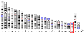
Corticotropin-releasing hormone receptor 1 (CRHR1) is a protein, also known as CRF1, with the latter (CRF1) now being the IUPHAR-recommended name. In humans, CRF1 is encoded by the CRHR1 gene at region 17q21.31, beside micrototubule-associated protein tau MAPT.

<span class="mw-page-title-main">GPR3</span> Protein

G-protein coupled receptor 3 is a protein that in humans is encoded by the GPR3 gene. The protein encoded by this gene is a member of the G protein-coupled receptor family of transmembrane receptors and is involved in signal transduction.

<span class="mw-page-title-main">Altanserin</span> Chemical compound

Altanserin is a compound that binds to the 5-HT2A receptor. Labeled with the isotope fluorine-18 it is used as a radioligand in positron emission tomography (PET) studies of the brain, i.e., studies of the 5-HT2A neuroreceptors. Besides human neuroimaging studies altanserin has also been used in the study of rats.

<span class="mw-page-title-main">Nisoxetine</span> Chemical compound

Nisoxetine, originally synthesized in the Lilly research laboratories during the early 1970s, is a potent and selective inhibitor for the reuptake of norepinephrine (noradrenaline) into synapses. It currently has no clinical applications in humans, although it was originally researched as an antidepressant. Nisoxetine is now widely used in scientific research as a standard selective norepinephrine reuptake inhibitor. It has been used to research obesity and energy balance, and exerts some local analgesia effects.

<span class="mw-page-title-main">5-Fluorowillardiine</span> Chemical compound

5-Fluorowillardiine is a selective agonist for the AMPA receptor, with only limited effects at the kainate receptor. It is an excitotoxic neurotoxin when used in vivo and so is rarely used in intact animals, but it is widely used to selectively stimulate AMPA receptors in vitro. It is structurally similar to the compound willardiine, which is also an agonist for the AMPA and kainate receptors. Willardiine occurs naturally in Mariosousa willardiana and Acacia sensu lato.

<span class="mw-page-title-main">Dihydroalprenolol</span> Chemical compound

Dihydroalprenolol (DHA) is a hydrogenated alprenolol derivative that acts as a beta-adrenergic blocker. When the extra hydrogen atoms are tritium, it is a radiolabeled form of alprenolol, which is used to label beta-adrenergic receptors for isolation.

<span class="mw-page-title-main">Brain positron emission tomography</span> Form of positron emission tomography

Brain positron emission tomography is a form of positron emission tomography (PET) that is used to measure brain metabolism and the distribution of exogenous radiolabeled chemical agents throughout the brain. PET measures emissions from radioactively labeled metabolically active chemicals that have been injected into the bloodstream. The emission data from brain PET are computer-processed to produce multi-dimensional images of the distribution of the chemicals throughout the brain.

A ligand binding assay (LBA) is an assay, or an analytic procedure, which relies on the binding of ligand molecules to receptors, antibodies or other macromolecules. A detection method is used to determine the presence and extent of the ligand-receptor complexes formed, and this is usually determined electrochemically or through a fluorescence detection method. This type of analytic test can be used to test for the presence of target molecules in a sample that are known to bind to the receptor.

<span class="mw-page-title-main">G-less cassette</span>

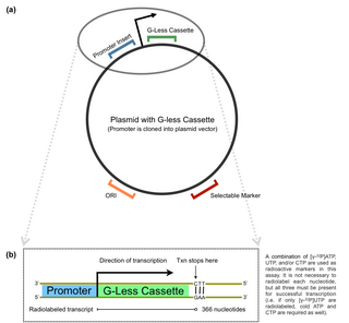
The G-less cassette transcription assay is a method used in molecular biology to determine promoter strength in vitro. The technique involves quantification of an mRNA product with the use of a plasmid. The G-less cassette is part of a pre-constructed vector, usually containing a multiple cloning site (MCS) upstream of the cassette. For this reason, promoters of interest can be inserted directly into the MCS to ultimately measure the accuracy and efficiency of a promoter in recruiting transcription machinery.

Alma Clavering Howard Rolleston Ebert was a Canadian-born English radiobiologist. She was joint editor for many years of the International Journal of Radiation Biology and deputy director of Paterson Laboratories in Manchester. She made a "fundamental contribution to cell biology" in collaboration with physicist Stephen Pelc when they "were the first to ascribe a timeframe to cellular life," creating the concept of the cell cycle. Their nomenclature for the stages of cell replication is used universally and appears in every textbook of biology and pathology.

<span class="mw-page-title-main">Ligand-targeted liposome</span> Ligand-targeted liposomes for use in medical applications

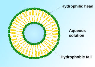
A ligand-targeted liposome (LTL) is a nanocarrier with specific ligands attached to its surface to enhance localization for targeted drug delivery. The targeting ability of LTLs enhances cellular localization and uptake of these liposomes for therapeutic or diagnostic purposes. LTLs have the potential to enhance drug delivery by decreasing peripheral systemic toxicity, increasing in vivo drug stability, enhancing cellular uptake, and increasing efficiency for chemotherapeutics and other applications.

References

  1. Barthe N, Coulon P, Hennion C, Ducassou D, Basse-Cathalinat B, Charpak G (May 1999). "Optimization of a new scintillation gas detector used to localize electrons emitted by 99mTc". J Nucl Med. 40 (5): 868–75. PMID   10319763.
  2. Encyclopedia of Life Sciences: Phosphorimager
  3. Rupassara, S. I., R.A. Larson, G.K. Sims, and K.A. Marley. 2002 Degradation of atrazine by hornwort in aquatic systems. Bioremediation Journal 6(3): 217-224.
  4. Kuhar M, Yamamura HI (July 1976). "Localization of cholinergic muscarinic receptors in rat brain by light microscopic radioautography". Brain Res. 110 (2): 229–43. doi:10.1016/0006-8993(76)90399-1. PMID   938940. S2CID   36648292.
  5. Young WS, Kuhar MJ (December 1979). "A new method for receptor autoradiography: [3H]opioid receptors in rat brain". Brain Res. 179 (2): 255–70. doi:10.1016/0006-8993(79)90442-6. PMID   228806. S2CID   21647100.
  6. Jin L, Lloyd RV (1997). "In situ hybridization: methods and applications". J Clin Lab Anal. 11 (1): 2–9. doi:10.1002/(SICI)1098-2825(1997)11:1<2::AID-JCLA2>3.0.CO;2-F. PMC   6760707 . PMID   9021518.
  7. Davenport, Anthony P. (March 25, 2005). Receptor Binding Techniques. Vol. 306. doi:10.1385/1592599273. ISBN   1-59259-927-3. S2CID   3691391.
  8. Hand R (1975). "Deoxyribonucleic acid fiber autoradiography as a technique for studying the replication of the mammalian chromosome". J. Histochem. Cytochem. 23 (7): 475–81. doi: 10.1177/23.7.1095649 . PMID   1095649.
  9. McCarthy D, Minner C, Bernstein H, Bernstein C (1976). "DNA elongation rates and growing point distributions of wild-type phage T4 and a DNA-delay amber mutant". J Mol Biol. 106 (4): 963–81. doi:10.1016/0022-2836(76)90346-6. PMID   789903.
  10. Kovacs KA, Steinmann M, Halfon O, Magistretti PJ, Cardinaux JR (November 2015). "Complex regulation of CREB-binding protein by homeodomain-interacting protein kinase 2" (PDF). Cell Signaling. 27 (11): 2252–60. doi:10.1016/j.cellsig.2015.08.001. PMID   26247811.
  11. Goggin, Fiona L.; Medville, Richard; Turgeon, Robert (February 1, 2001). "Phloem Loading in the Tulip Tree. Mechanisms and Evolutionary Implications". Plant Physiology. 125 (2): 891–899. doi:10.1104/pp.125.2.891. ISSN   0032-0889. PMC   64890 . PMID   11161046.
  12. Van Bel, A J E (June 1993). "Strategies of Phloem Loading". Annual Review of Plant Physiology and Plant Molecular Biology. 44 (1): 253–281. doi:10.1146/annurev.pp.44.060193.001345. ISSN   1040-2519.
  13. Turgeon, R.; Medville, R. (September 29, 1998). "The absence of phloem loading in willow leaves". Proceedings of the National Academy of Sciences. 95 (20): 12055–12060. Bibcode:1998PNAS...9512055T. doi: 10.1073/pnas.95.20.12055 . ISSN   0027-8424. PMC   21764 . PMID   9751789.
  14. "Krypton Gas Penetrant Imaging - A Valuable Tool for Ensuring Structural Integrity in Aircraft Engine Components". Archived from the original on July 20, 2008.
  15. Weisgall, Jonathan (1994), Operation Crossroads: The Atomic Tests at Bikini Atoll, Annapolis, Maryland: Naval Institute Press, p.  242, ISBN   978-1-55750-919-2

Original publication by sole inventor Askins, Barbara S. (1 November 1976). "Photographic image intensification by autoradiography". Applied Optics. 15 (11): 2860–2865. Bibcode:1976ApOpt..15.2860A. doi:10.1364/ao.15.002860.

Further reading