Brain ischemia | |
---|---|
Other names | Watershed infarct |
T1 MRI of an ischemic stroke in the brain without (left) and with (right) contrast. | |
Specialty | Neurology |
A watershed stroke is defined as a brain ischemia that is localized to the vulnerable border zones between the tissues supplied by the anterior, posterior and middle cerebral arteries. [1] The actual blood stream blockage/restriction site can be located far away from the infarcts. Watershed locations are those border-zone regions in the brain supplied by the major cerebral arteries where blood supply is decreased. Watershed strokes are a concern because they comprise approximately 10% of all ischemic stroke cases. [2] The watershed zones themselves are particularly susceptible to infarction from global ischemia as the distal nature of the vasculature predisposes these areas to be most sensitive to profound hypoperfusion. [1]
Watershed strokes are localized to two primary regions of the brain, and are termed cortical watersheds (CWS) and internal watersheds (IWS). [3] Patients with many different cardiovascular diseases have a higher likelihood of experiencing a blood clot or loss of blood flow in border-zone regions of the brain. The resulting symptoms differ based on the affected area of the brain. A CT scan and MRI are used for diagnosis, and afterward several treatment options are available, including the removal of atherosclerotic plaque and a physical widening of the clogged blood vessel. Long-term care is focused around three areas: rehabilitative therapy, surgical interventions, and prevention of future watershed strokes. Going forward, research to combat watershed strokes is focusing on various topics, such as stem cell research.[ citation needed ]
Watershed stroke symptoms are due to the reduced blood flow to all parts of the body, specifically the brain, thus leading to brain damage. Initial symptoms, as promoted by the American Stroke Association, are FAST, representing F = Facial weakness (droop), A = Arm weakness (drift), S = Speech difficulty (slur), and T = Time to act (priority of intervention). [4]
All strokes are considered a medical emergency. Any one of these symptoms, whether seen alone or in combination, should be assumed to be stroke until proven otherwise. Emergency medical help should be sought IMMEDIATELY if any or all of these symptoms are seen or experienced. Early diagnosis and timely medical intervention can drastically reduce the severity of a stroke, limit damage to the brain, improve the chances of a full recovery and reduce recovery times massively.
After the initial stroke, other symptoms depend on the area of the brain affected. If one of the three central nervous system pathways is affected, symptoms can include numbness, reduced sensation, and hyperreflexia.
Most often, the side of the brain damaged results in body defects on the opposite side. Since the cranial nerves originate from the brainstem, damage to this area can lead to defects in the function of these nerves. Symptoms can include altered breathing, problems with balance, drooping of eyelids, and decreased sensation in the face. [5]
Damage to the cerebral cortex may lead to aphasia or confusion and damage to the cerebellum may lead to lack of motor movement. [5]
Stroke presentations which are particularly suggestive of a watershed stroke include bilateral visual loss, stupor, and weakness of the proximal limbs, sparing the face, hands and feet.
Watershed strokes are caused by ischemia or a lack of blood flow to the brain. [2] There are several causes of ischemia, including embolism and atherosclerosis. There are several conditions that can predispose someone to watershed stroke by increasing the likelihood that insufficient blood supply will be able to reach the brain. Cancer and carcinomatosis as well as complications thereof and the treatments for them can also lead to stroke. [6] People with many different cardiovascular diseases have a higher likelihood of experiencing a clot or a plaque that impedes flow through a blood vessel. [3] Cardiovascular diseases that increase the risk of ischemia include:
Although many imaging techniques are used to document watershed strokes, their pathogenesis remains controversial. It may involve various mechanisms such as systemic hypotension, microemboli, severe arterial stenosis, ICA occlusion or a combination of these. [3]
These events are localized to two primary regions of the brain:
Nonetheless, within the literature itself, there exists confusion over the terminology used to describe cortical (outer brain) infarcts and subcortical (inner brain) infarcts. Besides watershed, border-zone is another common term used to refer to areas of the brain between the ends of two adjacent arteries. Other less used terms include: borderland, end zone, boundary zone, and terminal zone. These varying terms have arisen from the considerable anatomic variability both in the cerebral vascular structure and the territories of the brain that they supply. [10]
A sharp drop in blood pressure is the most frequent cause of watershed infarcts. The most frequent location for a watershed stroke is the region between the anterior cerebral artery and middle cerebral artery. These events caused by hypotension do not usually cause the blood vessel to rupture. [2]
Microemboli have not been experimentally proven to cause watershed strokes. It is unclear whether they are a cause or an effect of a watershed stroke. [11] With watershed strokes, platelet aggregates block the small meningeal arteries in watershed regions creating a microembolism. Microemboli usually form as thrombi, and can block arteries outright. On the other hand, they often detach, move into blood circulation, and eventually block smaller downstream branches of arteries causing a thromboembolism. Generally, emboli travel as far outward as their size permits along the vascular branches of the brain. Using this hypothesis, microemboli are viewed as the cause of the infarct rather than secondary events. Nevertheless, secondary thrombi do form after infarcts, and therefore it has been difficult to distinguish between emboli and thrombi in watershed locations. [2] The best supporting evidence is correlative; patients display subcortical abnormalities on CT scans and present more microembolic signals during a carotid endarterectomy. [11]
Microemboli can be common in some high-risk patients, such as those with carotid stenosis. However, in healthier patients strokes do not usually result from microemboli. [11]
Internal carotid artery | |
---|---|
Anatomical terminology |
Thrombi at the split of the internal carotid artery in the neck may cause watershed infarcts between the territories of the anterior cerebral artery and the middle cerebral artery. The resulting watershed infarcts in carotid artery blockages have mostly been considered to be due to a reduced blood flow, similar to that of hypotension. [2] Imaging studies in severe internal carotid artery (ICA) disease report an incidence of watershed stroke ranging from 19% to 64%. [3] Almost 40% of these watershed infarcts are attributed to narrowing of the carotid artery, which produces the reduced blood flow. [3]
However, a different possible explanation has emerged. Alternatively, the vascular occlusion could be the result of microemboli from the carotid thrombi before the lumen becomes completely blocked. [2] In this scenario, the clotting becomes too severe and the clot breaks free. The resulting traveling clot is known as an embolus (plural emboli). The wall of internal carotid artery just distal to the bifurcation (split) is a common site of atherosclerosis because of the unique hemodynamic effects caused by the blood flow divider. As a result, thrombi formation is more prevalent there. [11] In general, researches have observed that this microembolization is a frequent phenomenon during the build-up of cerebral thrombi. [2] The resulting emboli are pieces of calcified plaque. If these microemboli are 0.1 mm in diameter, they might pass into the small branches of the vascular system. There they may be destroyed by protective cellular defenses, or they may cause a stroke. [11] Altogether, these considerations suggest that the watershed infarcts in carotid thrombosis are caused by microembolization from mural thrombi, thrombi adherent to the vessel wall, rather than by blood flow disturbances. [2]
Diagnosis of a cerebral vascular accident begins with a general neurological examination, used to identify specific areas of resulting injury. A CT scan of the brain is then used to identify any cerebral hemorrhaging. An MRI with special sequences called diffusion-weighted MR imaging (DWI), is very sensitive for locating areas of an ischemic based stroke, such as a watershed stroke.
Further diagnosis and evaluation of a stroke includes evaluation of the blood vessels in the neck using either Doppler ultrasound, MR-angiography or CT-angiography, or formal angiography. An echocardiogram may be performed looking for a cardiac source of emboli. Blood tests for risk factors also may be ordered, including cholesterol levels, triglyceride levels, homocysteine levels, and blood coagulation tests.
Often considered one of the safest ways to treat symptomatic carotid stenosis, carotid endarterectomy is a procedure by which a surgeon gently removes atherosclerotic plaque. Blood flow hopefully then returns to normal, increasing oxygen concentration to normal amounts in watershed areas of the brain.
There is the potential for complications, including disturbing portions of the plaque leading to a stroke or heart attack during or after surgery. Small risks of bleeding and infection exist as well. [15]
In this type of procedure, a narrowed blood vessel is expanded via angioplasty or stenting. A thin angiography catheter is inserted in a large groin blood vessel and advanced to the stenosis. Percutaneous treatment is less invasive than endarterectomy, usually requiring only local anesthesia. Endarterectomy is still considered safer though, as percutaneous treatments can lead to accidental dislodging of plaque or even arterial rupturing. [15]
During carotid angioplasty, an angiography cather with a small deflated balloon attached on the tip is advanced to a carotid stenosis. The ballon is then inflated slowly, forcing the narrowed portion of the vessel to expand.
Carotid stenting follows a similar procedure. Rather than using a balloon, a stent (metal mesh-like tube) is placed over the atherosclerotic plaque in the hopes of stabilizing it and allowing for increased blood flow to watershed portions of the brain.
Watershed strokes are seldom fatal, but they can lead to neuromuscular degeneration, as well as dementia. [3] This degeneration at the watershed regions of the brain can lead to difficulties with movement and motor coordination, as well as speech. Long-term care is focused around three areas: rehabilitative therapy, surgical interventions, and prevention of future watershed strokes.
Long-term rehabilitative therapy for watershed stroke patients involves physical, occupational, and speech and language therapies. Physical therapy can be used to enhance motor function in the legs and arms that has been impacted by stroke. [16] Occupational therapies can be provided to help to alleviate cognitive impairments that result from watershed stroke but its effectiveness remains unclear. [17] Occupational Therapy can also be used in an effort to improve fine motor function that was damaged as a result of the stroke. [18] Stroke can also cause impairments in speech production secondary to neurocognitive and neuromuscular impairments, and therefore speech and language therapies are often a component of long-term care for stroke patients. Intensive speech and language therapy has been shown to improve speech deficits associated with aphasia resulting from stroke. [19]
Endovascular interventions, including surgical revascularization, can increase blood flow in the area of the stroke, thereby decreasing the likelihood that insufficient blood flow to the watershed regions of the brain will result in subsequent strokes. [15] Neuroscientists are currently researching stem cell transplantation therapies to improve recovery of cebreral tissue in affected areas of the brain post-stroke. Should this intervention be proven effective, it will greatly increase the number of neurons in the brain that can recover from a stroke. [20]
There are several interventions that are often used to help prevent the recurrence of a watershed stroke; namely, nutritional interventions, as well as antiplatelet, anticoagulant, and statin drug use. Nutritional interventions, including increased consumption of certain amino acids, antioxidants, B-group vitamins, and zinc, have been shown to increase the recovery of neurocognitive function after a stroke. [21] Antiplatelet drugs, such as aspirin, as well as anticoagulants, are used to help prevent blood clots and therefore embolisms, which can cause watershed strokes. Statin drugs are also used to control hyperlipidemia, another risk factor for watershed stroke.
Ischemic stroke is still a major health concern and studies are being conducted to determine the pathway in which brain damage occurs to identify targets for intervention. Stem cell transplantation may help in intervention to improve cell recovery and regeneration. [20]
Although the mechanism is not entirely understood, the likelihood of a watershed stroke increases after cardiac surgery. An experiment conducted in a five-year span studied the diagnosis, etiology, and outcome of these postoperative strokes. It was observed that intraoperative decrease in blood pressure may lead to these strokes and patients who have undergone aortic procedures are more likely to have bilateral watershed infarcts. Furthermore, bilateral watershed strokes are associated with poor short-term outcomes and are most reliably observed by diffusion-weighted imaging MRI. Thus future clinical research and practice should focus on the identification of bilateral stroke characteristics. This identification can help discover affected areas and increase correct diagnosis. [22]
Hemodynamic impairment is thought to be the cause of deep watershed infarcts, characterized by a rosary-like pattern. However new studies have shown that microembolism might also contribute to the development of deep watershed infarcts. The dual contribution of hemodynamic impairment and microembolism would result in different treatment for patients with these specific infarcts. [23]
While intracranial artery stenting is used for same side stroke prevention, basilar artery stenting may help to improve parallel, accessory blood flow. The stent may also lead to termination of recurrent middle cerebral artery (MCA) strokes. [24]
The area around the damaged ischemia is known as the penumbra. This viable area has the ability to regenerate with the help of pharmacological treatment however most patients with penumbra are left untreated. New research is being conducted in metabolic suppression, direct energy delivery, and selective drug delivery to help salvage this area of the brain after a stroke. [25]
This new drug has been shown to home to ischemic stroke tissue as well as apoptotic neuronal cells of the penumbra region. This discovery may help in creating selective drug delivery for stroke patients. [26]
Nanoliposomes are currently being researched for specific drug delivery due to their ph-sensitive and high blood–brain barrier diffusion characteristics. Many advantages of these drugs include:
Due to the fact that acidic environment and low blood flow are prominent characteristic of the penumbra area, liposomal drugs seem to be well suited. [25]
Watershed strokes are named because they affect the distal watershed areas of the brain. The original terminology came from the German literature, which used the analogy of an irrigation system. The German scholars compared the blood flow in distal arterial territories of the brain to the last field on a farm, which was the area with the least supply of water and therefore most vulnerable to any reduction in flow. [10] In a medical context, the term "watershed" refers to those areas of the brain that receive dual blood supply from the branching ends of two large arteries. [10]
A transient ischemic attack (TIA), commonly known as a mini-stroke, is a minor stroke whose noticeable symptoms usually end in less than an hour. TIA causes the same symptoms associated with strokes, such as weakness or numbness on one side of the body, sudden dimming or loss of vision, difficulty speaking or understanding language, slurred speech, or confusion.
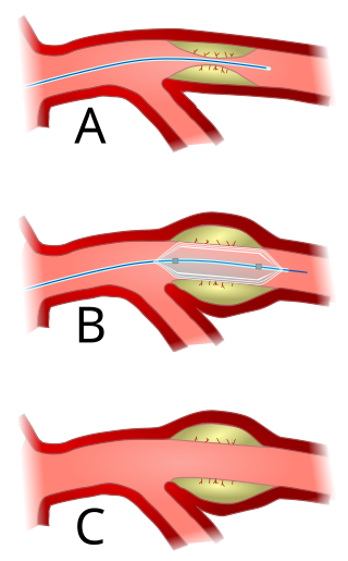
Angioplasty, also known as balloon angioplasty and percutaneous transluminal angioplasty (PTA), is a minimally invasive endovascular procedure used to widen narrowed or obstructed arteries or veins, typically to treat arterial atherosclerosis.
An embolism is the lodging of an embolus, a blockage-causing piece of material, inside a blood vessel. The embolus may be a blood clot (thrombus), a fat globule, a bubble of air or other gas, amniotic fluid, or foreign material.
Cerebrovascular disease includes a variety of medical conditions that affect the blood vessels of the brain and the cerebral circulation. Arteries supplying oxygen and nutrients to the brain are often damaged or deformed in these disorders. The most common presentation of cerebrovascular disease is an ischemic stroke or mini-stroke and sometimes a hemorrhagic stroke. Hypertension is the most important contributing risk factor for stroke and cerebrovascular diseases as it can change the structure of blood vessels and result in atherosclerosis. Atherosclerosis narrows blood vessels in the brain, resulting in decreased cerebral perfusion. Other risk factors that contribute to stroke include smoking and diabetes. Narrowed cerebral arteries can lead to ischemic stroke, but continually elevated blood pressure can also cause tearing of vessels, leading to a hemorrhagic stroke.
Interventional radiology (IR) is a medical specialty that performs various minimally-invasive procedures using medical imaging guidance, such as x-ray fluoroscopy, computed tomography, magnetic resonance imaging, or ultrasound. IR performs both diagnostic and therapeutic procedures through very small incisions or body orifices. Diagnostic IR procedures are those intended to help make a diagnosis or guide further medical treatment, and include image-guided biopsy of a tumor or injection of an imaging contrast agent into a hollow structure, such as a blood vessel or a duct. By contrast, therapeutic IR procedures provide direct treatment—they include catheter-based medicine delivery, medical device placement, and angioplasty of narrowed structures.
Ischemia or ischaemia is a restriction in blood supply to any tissue, muscle group, or organ of the body, causing a shortage of oxygen that is needed for cellular metabolism. Ischemia is generally caused by problems with blood vessels, with resultant damage to or dysfunction of tissue i.e. hypoxia and microvascular dysfunction. It also implies local hypoxia in a part of a body resulting from constriction.
Stroke is a medical condition in which poor blood flow to the brain causes cell death. There are two main types of stroke: ischemic, due to lack of blood flow, and hemorrhagic, due to bleeding. Both cause parts of the brain to stop functioning properly.
Cerebral hypoxia is a form of hypoxia, specifically involving the brain; when the brain is completely deprived of oxygen, it is called cerebral anoxia. There are four categories of cerebral hypoxia; they are, in order of increasing severity: diffuse cerebral hypoxia (DCH), focal cerebral ischemia, cerebral infarction, and global cerebral ischemia. Prolonged hypoxia induces neuronal cell death via apoptosis, resulting in a hypoxic brain injury.
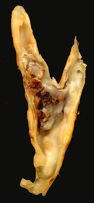
Carotid endarterectomy is a surgical procedure used to reduce the risk of stroke from carotid artery stenosis. In endarterectomy, the surgeon opens the artery and removes the plaque. The plaque forms and thickens the inner layer of the artery, or intima, hence the name of the procedure which simply means removal of part of the internal layers of the artery.
Carotid artery stenosis is a narrowing or constriction of any part of the carotid arteries, usually caused by atherosclerosis.
Intraparenchymal hemorrhage (IPH) is one form of intracerebral bleeding in which there is bleeding within brain parenchyma. The other form is intraventricular hemorrhage (IVH).
Cerebral infarction is the pathologic process that results in an area of necrotic tissue in the brain. It is caused by disrupted blood supply (ischemia) and restricted oxygen supply (hypoxia), most commonly due to thromboembolism, and manifests clinically as ischemic stroke. In response to ischemia, the brain degenerates by the process of liquefactive necrosis.
Vertebrobasilar insufficiency (VBI) describes a temporary set of symptoms due to decreased blood flow (ischemia) in the posterior circulation of the brain. The posterior circulation supplies the medulla, pons, midbrain, cerebellum and supplies the posterior cerebellar artery to the thalamus and occipital cortex. As a result, symptoms vary widely depending which brain region is predominantly affected.
Brain ischemia is a condition in which there is insufficient bloodflow to the brain to meet metabolic demand. This leads to poor oxygen supply or cerebral hypoxia and thus leads to the death of brain tissue or cerebral infarction/ischemic stroke. It is a sub-type of stroke along with subarachnoid hemorrhage and intracerebral hemorrhage.
Animal models of ischemic stroke are procedures inducing cerebral ischemia. The aim is the study of basic processes or potential therapeutic interventions in this disease, and the extension of the pathophysiological knowledge on and/or the improvement of medical treatment of human ischemic stroke. Ischemic stroke has a complex pathophysiology involving the interplay of many different cells and tissues such as neurons, glia, endothelium, and the immune system. These events cannot be mimicked satisfactorily in vitro yet. Thus a large portion of stroke research is conducted on animals.
Fibromuscular dysplasia (FMD) is a non-atherosclerotic, non-inflammatory disease of the blood vessels that causes abnormal growth within the wall of an artery. FMD has been found in nearly every arterial bed in the body, although the most commonly affected are the renal and carotid arteries.
Carotid artery dissection is a separation of the layers of the artery wall in the carotid arteries supplying oxygen-bearing blood to the head. It is the most common cause of stroke in younger adults. The term 'cervical artery dissection' should also be considered in the context of this article.
Carotid artery stenting is an endovascular procedure where a stent is deployed within the lumen of the carotid artery to treat narrowing of the carotid artery and decrease the risk of stroke. It is used to treat narrowing of the carotid artery in high-risk patients, when carotid endarterectomy is considered too risky.
Lacunar stroke or lacunar cerebral infarct (LACI) is the most common type of ischemic stroke, resulting from the occlusion of small penetrating arteries that provide blood to the brain's deep structures. Patients who present with symptoms of a lacunar stroke, but who have not yet had diagnostic imaging performed, may be described as having lacunar stroke syndrome (LACS).
The leptomeningeal collateral circulation is a network of small blood vessels in the brain that connects branches of the middle, anterior and posterior cerebral arteries, with variation in its precise anatomy between individuals. During a stroke, leptomeningeal collateral vessels allow limited blood flow when other, larger blood vessels provide inadequate blood supply to a part of the brain.