Lateral geniculate nucleus | |
---|---|
Details | |
Part of | Thalamus |
System | Visual |
Artery | Anterior choroidal and Posterior cerebral |
Vein | Terminal vein |
Identifiers | |
Latin | corpus geniculatum laterale |
Acronym(s) | LGN |
NeuroNames | 352 |
NeuroLex ID | birnlex_1662 |
TA98 | A14.1.08.302 |
TA2 | 5666 |
FMA | 62209 |
Anatomical terms of neuroanatomy |
In neuroanatomy, the lateral geniculate nucleus (LGN; also called the lateral geniculate body or lateral geniculate complex) is a structure in the thalamus and a key component of the mammalian visual pathway. It is a small, ovoid, ventral projection of the thalamus where the thalamus connects with the optic nerve. There are two LGNs, one on the left and another on the right side of the thalamus. In humans, both LGNs have six layers of neurons (grey matter) alternating with optic fibers (white matter).
The LGN receives information directly from the ascending retinal ganglion cells via the optic tract and from the reticular activating system. Neurons of the LGN send their axons through the optic radiation, a direct pathway to the primary visual cortex. In addition, the LGN receives many strong feedback connections from the primary visual cortex. [1] In humans as well as other mammals, the two strongest pathways linking the eye to the brain are those projecting to the dorsal part of the LGN in the thalamus, and to the superior colliculus. [2]
Both the left and right hemispheres of the brain have a lateral geniculate nucleus, named after its resemblance to a bent knee (genu is Latin for "knee"). In humans as well as in many other primates, the LGN has layers of magnocellular cells and parvocellular cells that are interleaved with layers of koniocellular cells.
In humans the LGN is normally described as having six distinctive layers. The inner two layers, (1 and 2) are magnocellular layers, while the outer four layers, (3, 4, 5 and 6), are parvocellular layers. An additional set of neurons, known as the koniocellular layers, are found ventral to each of the magnocellular and parvocellular layers. [3] : 227ff [4] This layering is variable between primate species, and extra leafleting is variable within species.
The average volume of each LGN in an adult human is about 118mm. (This is the same volume as a 4.9mm-sided cube.) A study of 24 hemispheres from 15 normal individuals with average age 59 years at autopsy found variation from about 91 to 157mm. [5] The same study found that in each LGN, the magnocellular layers comprised about 28mm in total, and the parvocellular layers comprised about 90mm in total.
Type | Size* | RGC Source | Type of Information | Location | Response | Number |
---|---|---|---|---|---|---|
M: Magnocellular cells | Large | Parasol cells | perception of movement, depth, and small differences in brightness | Layers 1 and 2 | rapid and transient | ? |
P: Parvocellular cells (or "parvicellular") | Small | Midget cells | perception of color and form (fine details) | Layers 3, 4, 5 and 6 | slow and sustained | ? |
K: Koniocellular cells (or "interlaminar") | Very small cell bodies | Bistratified cells | Between each of the M and P layers |
*Size describes the cell body and dendritic tree, though also can describe the receptive field
The magnocellular, parvocellular, and koniocellular layers of the LGN correspond with the similarly named types of retinal ganglion cells. Retinal P ganglion cells send axons to a parvocellular layer, M ganglion cells send axons to a magnocellular layer, and K ganglion cells send axons to a koniocellular layer. [6] : 269
Koniocellular cells are functionally and neurochemically distinct from M and P cells and provide a third channel to the visual cortex. They project their axons between the layers of the lateral geniculate nucleus where M and P cells project. Their role in visual perception is presently unclear; however, the koniocellular system has been linked with the integration of somatosensory system-proprioceptive information with visual perception[ citation needed ], and it may also be involved in color perception. [7]
The parvo- and magnocellular fibers were previously thought to dominate the Ungerleider–Mishkin ventral stream and dorsal stream, respectively. However, new evidence has accumulated showing that the two streams appear to feed on a more even mixture of different types of nerve fibers. [8]
The other major retino–cortical visual pathway is the tectopulvinar pathway, routing primarily through the superior colliculus and thalamic pulvinar nucleus onto posterior parietal cortex and visual area MT.
Both the LGN in the right hemisphere and the LGN in the left hemisphere receive input from each eye. However, each LGN only receives information from one half of the visual field. Retinal ganglion cells (RGCs) from the inner halves of each retina (the nasal sides) decussate (cross to the other side of the brain) through the optic chiasma (khiasma means "cross-shaped"). RGCs from the outer half of each retina (the temporal sides) remain on the same side of the brain. Therefore, the right LGN receives visual information from the left visual field, and the left LGN receives visual information from the right visual field. Within one LGN, the visual information is divided among the various layers as follows: [9]
This description applies to the LGN of many primates, but not all. The sequence of layers receiving information from the ipsilateral and contralateral (opposite side of the head) eyes is different in the tarsier. [10] Some neuroscientists suggested that "this apparent difference distinguishes tarsiers from all other primates, reinforcing the view that they arose in an early, independent line of primate evolution". [11]
The LGN receives input from the retina and many other brain structures, especially visual cortex.
The principal neurons in the LGN receive strong inputs from the retina. However, the retina only accounts for a small percentage of LGN input. As much as 95% of input in the LGN comes from the visual cortex, superior colliculus, pretectum, thalamic reticular nuclei, and local LGN interneurons. Regions in the brainstem that are not involved in visual perception also project to the LGN, such as the mesencephalic reticular formation, dorsal raphe nucleus, periaqueuctal grey matter, and the locus coeruleus. [12] The LGN also receives some inputs from the optic tectum (known as the superior colliculus in mammals). [13] These non-retinal inputs can be excitatory, inhibitory, or modulatory. [12]
Information leaving the LGN travels out on the optic radiations, which form part of the retrolenticular portion of the internal capsule.
The axons that leave the LGN go to V1 visual cortex. Both the magnocellular layers 1–2 and the parvocellular layers 3–6 send their axons to layer 4 in V1. Within layer 4 of V1, layer 4cβ receives parvocellular input, and layer 4cα receives magnocellular input. However, the koniocellular layers, intercalated between LGN layers 1–6 send their axons primarily to the cytochrome-oxidase rich blobs of layers 2 and 3 in V1. [14] Axons from layer 6 of visual cortex send information back to the LGN.
Studies involving blindsight have suggested that projections from the LGN travel not only to the primary visual cortex but also to higher cortical areas V2 and V3. Patients with blindsight are phenomenally blind in certain areas of the visual field corresponding to a contralateral lesion in the primary visual cortex; however, these patients are able to perform certain motor tasks accurately in their blind field, such as grasping. This suggests that neurons travel from the LGN to both the primary visual cortex and higher cortex regions. [15]
The output of the LGN serves several functions.
Computations are achieved to determine the position of every major element in object space relative to the principal plane. Through subsequent motion of the eyes, a larger stereoscopic mapping of the visual field is achieved. [16]
It has been shown that while the retina accomplishes spatial decorrelation through center surround inhibition, the LGN accomplishes temporal decorrelation. [17] This spatial–temporal decorrelation makes for much more efficient coding. However, there is almost certainly much more going on.
Like other areas of the thalamus, particularly other relay nuclei, the LGN likely helps the visual system focus its attention on the most important information. That is, if you hear a sound slightly to your left, the auditory system likely "tells" the visual system, through the LGN via its surrounding peri-reticular nucleus, to direct visual attention to that part of space. [18] The LGN is also a station that refines certain receptive fields. [19]
Axiomatically determined functional models of LGN cells have been determined by Lindeberg [20] [21] in terms of Laplacian of Gaussian kernels over the spatial domain in combination with temporal derivatives of either non-causal or time-causal scale-space kernels over the temporal domain. It has been shown that this theory both leads to predictions about receptive fields with good qualitative agreement with the biological receptive field measurements performed by DeAngelis et al. [22] [23] and guarantees good theoretical properties of the mathematical receptive field model, including covariance and invariance properties under natural image transformations. [24] [25] Specifically according to this theory, non-lagged LGN cells correspond to first-order temporal derivatives, whereas lagged LGN cells correspond to second-order temporal derivatives.
The LGN is integral in the early steps of color processing, where opponent channels are created that compare signals between the different Photoreceptor cell types. The output of P-cells comprises red-green opponent signals. The output of M-cells does not include much color opponency, rather a sum of the red-green signal that evokes luminance. The output of K-cells comprises mostly blue-yellow opponent signals. [26]
In rodents, the lateral geniculate nucleus contains the dorsal lateral geniculate nucleus (dLGN), the ventral lateral geniculate nucleus (vLGN), and the region in between called the intergeniculate leaflet (IGL). These are distinct subcortical nuclei with differences in function.
The dorsolateral geniculate nucleus is the main division of the lateral geniculate body. In the mouse, the area of the dLGN is about 0.48mm. The majority of input to the dLGN comes from the retina. It is laminated and shows retinotopic organization. [27]
The ventrolateral geniculate nucleus has been found to be relatively large in several species such as lizards, rodents, cows, cats, and primates. [28] An initial cytoarchitectural scheme, which has been confirmed in several studies, suggests that the vLGN is divided into two parts. The external and internal divisions are separated by a group of fine fibers and a zone of thinly dispersed neurons. Additionally, several studies have suggested further subdivisions of the vLGN in other species. [29] For example, studies indicate that the cytoarchitecture of the vLGN in the cat differs from rodents. Although five subdivisions of the vLGN in the cat have been identified by some, [30] the scheme that divides the vLGN into three regions (medial, intermediate, and lateral) has been more widely accepted.
The intergeniculate leaflet is a relatively small area found dorsal to the vLGN. Earlier studies had referred to the IGL as the internal dorsal division of the vLGN. Several studies have described homologous regions in several species, including humans. [31]
The vLGN and IGL appear to be closely related based on similarities in neurochemicals, inputs and outputs, and physiological properties.
The vLGN and IGL have been reported to share many neurochemicals that are found concentrated in the cells, including neuropeptide Y, GABA, encephalin, and nitric oxide synthase. The neurochemicals serotonin, acetylcholine, histamine, dopamine, and noradrenaline have been found in the fibers of these nuclei.
Both the vLGN and IGL receive input from the retina, locus coreuleus, and raphe. Other connections that have been found to be reciprocal include the superior colliculus, pretectum, and hypothalamus, as well as other thalamic nuclei.
Physiological and behavioral studies have shown spectral-sensitive and motion-sensitive responses that vary with species. The vLGN and IGL seem to play an important role in mediating phases of the circadian rhythms that are not involved with light, as well as phase shifts that are light-dependent. [29]
The visual system is the physiological basis of visual perception. The system detects, transduces and interprets information concerning light within the visible range to construct an image and build a mental model of the surrounding environment. The visual system is associated with the eye and functionally divided into the optical system and the neural system.
In neuroanatomy, the optic radiation are axons from the neurons in the lateral geniculate nucleus to the primary visual cortex. The optic radiation receives blood through deep branches of the middle cerebral artery and posterior cerebral artery.
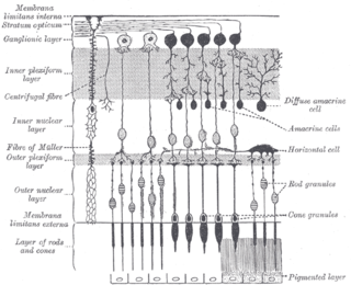
A retinal ganglion cell (RGC) is a type of neuron located near the inner surface of the retina of the eye. It receives visual information from photoreceptors via two intermediate neuron types: bipolar cells and retina amacrine cells. Retina amacrine cells, particularly narrow field cells, are important for creating functional subunits within the ganglion cell layer and making it so that ganglion cells can observe a small dot moving a small distance. Retinal ganglion cells collectively transmit image-forming and non-image forming visual information from the retina in the form of action potential to several regions in the thalamus, hypothalamus, and mesencephalon, or midbrain.
In neuroscience, parvocellular cells, also called P-cells, are neurons located within the parvocellular layers of the lateral geniculate nucleus (LGN) of the thalamus. Their name comes from Latin parvus 'small', due to the small size of the cell compared to the larger magnocellular cells. Phylogenetically, parvocellular neurons are more modern than magnocellular ones.
Magnocellular cells, also called M-cells, are neurons located within the magnocellular layer of the lateral geniculate nucleus of the thalamus. The cells are part of the visual system. They are termed "magnocellular" since they are characterized by their relatively large size compared to parvocellular cells.
In neuroanatomy, the superior colliculus is a structure lying on the roof of the mammalian midbrain. In non-mammalian vertebrates, the homologous structure is known as the optic tectum or optic lobe. The adjective form tectal is commonly used for both structures.
The inferior colliculus (IC) is the principal midbrain nucleus of the auditory pathway and receives input from several peripheral brainstem nuclei in the auditory pathway, as well as inputs from the auditory cortex. The inferior colliculus has three subdivisions: the central nucleus, a dorsal cortex by which it is surrounded, and an external cortex which is located laterally. Its bimodal neurons are implicated in auditory-somatosensory interaction, receiving projections from somatosensory nuclei. This multisensory integration may underlie a filtering of self-effected sounds from vocalization, chewing, or respiration activities.
In neuroanatomy, the pretectal area, or pretectum, is a midbrain structure composed of seven nuclei and comprises part of the subcortical visual system. Through reciprocal bilateral projections from the retina, it is involved primarily in mediating behavioral responses to acute changes in ambient light such as the pupillary light reflex, the optokinetic reflex, and temporary changes to the circadian rhythm. In addition to the pretectum's role in the visual system, the anterior pretectal nucleus has been found to mediate somatosensory and nociceptive information.
In neuroscience, koniocellular cells, also called K-cells, are relatively small neurons located in the koniocellular layer of the lateral geniculate nucleus (LGN) within the thalamus of primates, including humans. The term 'koniocellular' is derived from Greek konio 'dust, poison'.
In neuroanatomy, thalamocortical radiations, also known as thalamocortical fibers, are the efferent fibers that project from the thalamus to distinct areas of the cerebral cortex. They form fiber bundles that emerge from the lateral surface of the thalamus.
The medial geniculate nucleus (MGN) or medial geniculate body (MGB) is part of the auditory thalamus and represents the thalamic relay between the inferior colliculus (IC) and the auditory cortex (AC). It is made up of a number of sub-nuclei that are distinguished by their neuronal morphology and density, by their afferent and efferent connections, and by the coding properties of their neurons. It is thought that the MGN influences the direction and maintenance of attention.
Retinotopy is the mapping of visual input from the retina to neurons, particularly those neurons within the visual stream. For clarity, 'retinotopy' can be replaced with 'retinal mapping', and 'retinotopic' with 'retinally mapped'.
The zona incerta (ZI) is a horizontally elongated small nucleus that separates the larger subthalamic nucleus from the thalamus. Its connections project extensively over the brain from the cerebral cortex down into the spinal cord.
In neuroanatomy, topographic map is the ordered projection of a sensory surface or an effector system to one or more structures of the central nervous system. Topographic maps can be found in all sensory systems and in many motor systems.
The medial dorsal nucleus is a large nucleus in the thalamus. It is separated from the other thalamic nuclei by the internal medullary lamina.
The isothalamus is a division used by some researchers in describing the thalamus.
Blobs are sections of primary visual cortex (V1) above and below layer IV where groups of neurons sensitive to color assemble in cylindrical shapes. They were first identified in 1979 by Margaret Wong-Riley in cats when she used a cytochrome oxidase stain, from which they get their name. These areas receive input from koniocellular cells in the dorsal lateral geniculate nucleus dLGN and output to the thin stripes of area V2. Interblobs are areas between blobs that receive the same input, but are sensitive to orientation instead of color. They output to the pale and thick stripes of area V2.
A midget cell, sometimes called a P cell or P ganglion cell, is one type of retinal ganglion cell (RGC). Midget cells originate in the ganglion cell layer of the retina, and project to the parvocellular layers of the lateral geniculate nucleus (LGN). The axons of midget cells travel through the optic nerve and optic tract, ultimately synapsing with parvocellular cells in the LGN. These cells are known as midget retinal ganglion cells due to the small sizes of their dendritic trees and cell bodies. About 80% of RGCs are midget cells. They receive inputs from relatively few rods and cones. In many cases, they are connected to midget bipolar cells, which are linked to one cone each.
A parasol cell, sometimes called an M cell or M ganglion cell, is one type of retinal ganglion cell (RGC) located in the ganglion cell layer of the retina. These cells project to magnocellular cells in the lateral geniculate nucleus (LGN) as part of the magnocellular pathway in the visual system. They have large cell bodies as well as extensive branching dendrite networks and as such have large receptive fields. Relative to other RGCs, they have fast conduction velocities. While they do show clear center-surround antagonism, they receive no information about color. Parasol ganglion cells contribute information about the motion and depth of objects to the visual system.
Laura Busse is a German neuroscientist and professor of Systemic Neuroscience within the Division of Neurobiology at the Ludwig Maximilian University of Munich. Busse's lab studies context-dependent visual processing in mouse models by performing large scale in vivo electrophysiological recordings in the thalamic and cortical circuits of awake and behaving mice.