WO2023225569A1 - Manufacturing viral particles - Google Patents
Manufacturing viral particles Download PDFInfo
- Publication number
- WO2023225569A1 WO2023225569A1 PCT/US2023/067136 US2023067136W WO2023225569A1 WO 2023225569 A1 WO2023225569 A1 WO 2023225569A1 US 2023067136 W US2023067136 W US 2023067136W WO 2023225569 A1 WO2023225569 A1 WO 2023225569A1
- Authority
- WO
- WIPO (PCT)
- Prior art keywords
- filter
- formulation
- protein
- cell
- less
- Prior art date
Links
- 239000002245 particle Substances 0.000 title claims abstract description 338
- 230000003612 virological effect Effects 0.000 title abstract description 159
- 238000004519 manufacturing process Methods 0.000 title description 44
- 239000000203 mixture Substances 0.000 claims abstract description 482
- 238000000034 method Methods 0.000 claims abstract description 264
- 210000004027 cell Anatomy 0.000 claims description 586
- 238000009472 formulation Methods 0.000 claims description 287
- 238000001914 filtration Methods 0.000 claims description 236
- 108090000623 proteins and genes Proteins 0.000 claims description 165
- 239000013612 plasmid Substances 0.000 claims description 158
- 230000014759 maintenance of location Effects 0.000 claims description 155
- 239000000706 filtrate Substances 0.000 claims description 143
- 102000004169 proteins and genes Human genes 0.000 claims description 139
- 239000000725 suspension Substances 0.000 claims description 111
- 238000004587 chromatography analysis Methods 0.000 claims description 80
- 108010003533 Viral Envelope Proteins Proteins 0.000 claims description 77
- 239000010410 layer Substances 0.000 claims description 71
- 239000013598 vector Substances 0.000 claims description 66
- 239000012510 hollow fiber Substances 0.000 claims description 63
- 239000000356 contaminant Substances 0.000 claims description 60
- 238000000108 ultra-filtration Methods 0.000 claims description 59
- 239000003623 enhancer Substances 0.000 claims description 52
- 102100034349 Integrase Human genes 0.000 claims description 51
- 108090000765 processed proteins & peptides Proteins 0.000 claims description 51
- 241000713666 Lentivirus Species 0.000 claims description 50
- 238000010361 transduction Methods 0.000 claims description 50
- 230000026683 transduction Effects 0.000 claims description 50
- 238000009295 crossflow filtration Methods 0.000 claims description 49
- 229920001184 polypeptide Polymers 0.000 claims description 47
- 102000004196 processed proteins & peptides Human genes 0.000 claims description 47
- 108010019670 Chimeric Antigen Receptors Proteins 0.000 claims description 46
- 101710091045 Envelope protein Proteins 0.000 claims description 45
- 101710188315 Protein X Proteins 0.000 claims description 45
- 241000501789 Cocal virus Species 0.000 claims description 44
- 208000015181 infectious disease Diseases 0.000 claims description 42
- 108010042407 Endonucleases Proteins 0.000 claims description 38
- 102100031780 Endonuclease Human genes 0.000 claims description 37
- 239000003446 ligand Substances 0.000 claims description 37
- 102000005962 receptors Human genes 0.000 claims description 37
- 108020003175 receptors Proteins 0.000 claims description 37
- 210000002865 immune cell Anatomy 0.000 claims description 35
- 230000003213 activating effect Effects 0.000 claims description 34
- 230000002458 infectious effect Effects 0.000 claims description 34
- 108091006629 SLC13A2 Proteins 0.000 claims description 33
- 239000008186 active pharmaceutical agent Substances 0.000 claims description 32
- 239000000427 antigen Substances 0.000 claims description 32
- 102000036639 antigens Human genes 0.000 claims description 32
- 108091007433 antigens Proteins 0.000 claims description 32
- 229940088679 drug related substance Drugs 0.000 claims description 32
- 239000002355 dual-layer Substances 0.000 claims description 32
- 239000000872 buffer Substances 0.000 claims description 31
- -1 R0R1 Proteins 0.000 claims description 29
- 108020001507 fusion proteins Proteins 0.000 claims description 29
- 102000037865 fusion proteins Human genes 0.000 claims description 29
- 150000007523 nucleic acids Chemical class 0.000 claims description 29
- 238000013060 ultrafiltration and diafiltration Methods 0.000 claims description 29
- 230000000139 costimulatory effect Effects 0.000 claims description 28
- 101000914514 Homo sapiens T-cell-specific surface glycoprotein CD28 Proteins 0.000 claims description 27
- 102100027213 T-cell-specific surface glycoprotein CD28 Human genes 0.000 claims description 27
- 150000003839 salts Chemical class 0.000 claims description 25
- 108010067390 Viral Proteins Proteins 0.000 claims description 24
- 239000012634 fragment Substances 0.000 claims description 24
- 210000004779 membrane envelope Anatomy 0.000 claims description 24
- 102000039446 nucleic acids Human genes 0.000 claims description 24
- 108020004707 nucleic acids Proteins 0.000 claims description 24
- 230000001954 sterilising effect Effects 0.000 claims description 23
- 102100025237 T-cell surface antigen CD2 Human genes 0.000 claims description 22
- 102000017420 CD3 protein, epsilon/gamma/delta subunit Human genes 0.000 claims description 20
- 108050005493 CD3 protein, epsilon/gamma/delta subunit Proteins 0.000 claims description 20
- 101000934346 Homo sapiens T-cell surface antigen CD2 Proteins 0.000 claims description 20
- 108091006027 G proteins Proteins 0.000 claims description 19
- 102000030782 GTP binding Human genes 0.000 claims description 19
- 108091000058 GTP-Binding Proteins 0.000 claims description 19
- 101001063392 Homo sapiens Lymphocyte function-associated antigen 3 Proteins 0.000 claims description 18
- 102100030984 Lymphocyte function-associated antigen 3 Human genes 0.000 claims description 18
- 108010027225 gag-pol Fusion Proteins Proteins 0.000 claims description 17
- 101000914484 Homo sapiens T-lymphocyte activation antigen CD80 Proteins 0.000 claims description 16
- 206010028980 Neoplasm Diseases 0.000 claims description 16
- 238000012258 culturing Methods 0.000 claims description 16
- 238000011026 diafiltration Methods 0.000 claims description 15
- 102000040430 polynucleotide Human genes 0.000 claims description 15
- 108091033319 polynucleotide Proteins 0.000 claims description 15
- 239000002157 polynucleotide Substances 0.000 claims description 15
- 102100037904 CD9 antigen Human genes 0.000 claims description 14
- 238000001727 in vivo Methods 0.000 claims description 14
- 238000005571 anion exchange chromatography Methods 0.000 claims description 13
- 230000009467 reduction Effects 0.000 claims description 13
- 102100027222 T-lymphocyte activation antigen CD80 Human genes 0.000 claims description 12
- 230000002829 reductive effect Effects 0.000 claims description 11
- 101000633778 Homo sapiens SLAM family member 5 Proteins 0.000 claims description 10
- 101000946843 Homo sapiens T-cell surface glycoprotein CD8 alpha chain Proteins 0.000 claims description 10
- 101000764622 Homo sapiens Transmembrane and immunoglobulin domain-containing protein 2 Proteins 0.000 claims description 10
- 101000679851 Homo sapiens Tumor necrosis factor receptor superfamily member 4 Proteins 0.000 claims description 10
- 102100029216 SLAM family member 5 Human genes 0.000 claims description 10
- 102100034922 T-cell surface glycoprotein CD8 alpha chain Human genes 0.000 claims description 10
- 102100026224 Transmembrane and immunoglobulin domain-containing protein 2 Human genes 0.000 claims description 10
- 102100022153 Tumor necrosis factor receptor superfamily member 4 Human genes 0.000 claims description 10
- 102100032937 CD40 ligand Human genes 0.000 claims description 9
- 101001109501 Homo sapiens NKG2-D type II integral membrane protein Proteins 0.000 claims description 9
- 101000851370 Homo sapiens Tumor necrosis factor receptor superfamily member 9 Proteins 0.000 claims description 9
- 101100236305 Mus musculus Ly9 gene Proteins 0.000 claims description 9
- 102100022680 NKG2-D type II integral membrane protein Human genes 0.000 claims description 9
- 108010074687 Signaling Lymphocytic Activation Molecule Family Member 1 Proteins 0.000 claims description 9
- 102100036856 Tumor necrosis factor receptor superfamily member 9 Human genes 0.000 claims description 9
- 102100038077 CD226 antigen Human genes 0.000 claims description 8
- 101000884298 Homo sapiens CD226 antigen Proteins 0.000 claims description 8
- 101000589305 Homo sapiens Natural cytotoxicity triggering receptor 2 Proteins 0.000 claims description 8
- 108010004217 Natural Cytotoxicity Triggering Receptor 1 Proteins 0.000 claims description 8
- 102100032870 Natural cytotoxicity triggering receptor 1 Human genes 0.000 claims description 8
- 102100032851 Natural cytotoxicity triggering receptor 2 Human genes 0.000 claims description 8
- 108091008035 T cell costimulatory receptors Proteins 0.000 claims description 8
- 102100038080 B-cell receptor CD22 Human genes 0.000 claims description 7
- 102100027207 CD27 antigen Human genes 0.000 claims description 7
- 101000884305 Homo sapiens B-cell receptor CD22 Proteins 0.000 claims description 7
- 101000914511 Homo sapiens CD27 antigen Proteins 0.000 claims description 7
- 101000738354 Homo sapiens CD9 antigen Proteins 0.000 claims description 7
- 101000971538 Homo sapiens Killer cell lectin-like receptor subfamily F member 1 Proteins 0.000 claims description 7
- 101000884270 Homo sapiens Natural killer cell receptor 2B4 Proteins 0.000 claims description 7
- 101000801234 Homo sapiens Tumor necrosis factor receptor superfamily member 18 Proteins 0.000 claims description 7
- 101000851376 Homo sapiens Tumor necrosis factor receptor superfamily member 8 Proteins 0.000 claims description 7
- 102100021458 Killer cell lectin-like receptor subfamily F member 1 Human genes 0.000 claims description 7
- 108010064548 Lymphocyte Function-Associated Antigen-1 Proteins 0.000 claims description 7
- 241000712079 Measles morbillivirus Species 0.000 claims description 7
- 108010004222 Natural Cytotoxicity Triggering Receptor 3 Proteins 0.000 claims description 7
- 102100032852 Natural cytotoxicity triggering receptor 3 Human genes 0.000 claims description 7
- 102100038082 Natural killer cell receptor 2B4 Human genes 0.000 claims description 7
- 102100033728 Tumor necrosis factor receptor superfamily member 18 Human genes 0.000 claims description 7
- 102100036857 Tumor necrosis factor receptor superfamily member 8 Human genes 0.000 claims description 7
- 102100026122 High affinity immunoglobulin gamma Fc receptor I Human genes 0.000 claims description 6
- 101000913074 Homo sapiens High affinity immunoglobulin gamma Fc receptor I Proteins 0.000 claims description 6
- 101000777628 Homo sapiens Leukocyte antigen CD37 Proteins 0.000 claims description 6
- 101000917858 Homo sapiens Low affinity immunoglobulin gamma Fc region receptor III-A Proteins 0.000 claims description 6
- 101000917839 Homo sapiens Low affinity immunoglobulin gamma Fc region receptor III-B Proteins 0.000 claims description 6
- 101000934338 Homo sapiens Myeloid cell surface antigen CD33 Proteins 0.000 claims description 6
- 101000738771 Homo sapiens Receptor-type tyrosine-protein phosphatase C Proteins 0.000 claims description 6
- 101000934341 Homo sapiens T-cell surface glycoprotein CD5 Proteins 0.000 claims description 6
- 102100031586 Leukocyte antigen CD37 Human genes 0.000 claims description 6
- 239000000232 Lipid Bilayer Substances 0.000 claims description 6
- 102100029185 Low affinity immunoglobulin gamma Fc region receptor III-B Human genes 0.000 claims description 6
- 102100025243 Myeloid cell surface antigen CD33 Human genes 0.000 claims description 6
- 102100037422 Receptor-type tyrosine-protein phosphatase C Human genes 0.000 claims description 6
- 102100025244 T-cell surface glycoprotein CD5 Human genes 0.000 claims description 6
- 230000004936 stimulating effect Effects 0.000 claims description 6
- 108010029697 CD40 Ligand Proteins 0.000 claims description 5
- 101000716102 Homo sapiens T-cell surface glycoprotein CD4 Proteins 0.000 claims description 5
- 241000526636 Nipah henipavirus Species 0.000 claims description 5
- 102100036011 T-cell surface glycoprotein CD4 Human genes 0.000 claims description 5
- 210000005260 human cell Anatomy 0.000 claims description 5
- 210000002966 serum Anatomy 0.000 claims description 5
- 108091027967 Small hairpin RNA Proteins 0.000 claims description 4
- 108020004459 Small interfering RNA Proteins 0.000 claims description 4
- 108091070501 miRNA Proteins 0.000 claims description 4
- 239000002679 microRNA Substances 0.000 claims description 4
- 239000004055 small Interfering RNA Substances 0.000 claims description 4
- 102000006942 B-Cell Maturation Antigen Human genes 0.000 claims description 3
- 108010008014 B-Cell Maturation Antigen Proteins 0.000 claims description 3
- 101150029707 ERBB2 gene Proteins 0.000 claims description 3
- 102100031507 Fc receptor-like protein 5 Human genes 0.000 claims description 3
- 102100021197 G-protein coupled receptor family C group 5 member D Human genes 0.000 claims description 3
- 101001040713 Homo sapiens G-protein coupled receptor family C group 5 member D Proteins 0.000 claims description 3
- 241001465754 Metazoa Species 0.000 claims description 3
- 108010026331 alpha-Fetoproteins Proteins 0.000 claims description 3
- 102000013529 alpha-Fetoproteins Human genes 0.000 claims description 3
- GNBHRKFJIUUOQI-UHFFFAOYSA-N fluorescein Chemical compound O1C(=O)C2=CC=CC=C2C21C1=CC=C(O)C=C1OC1=CC(O)=CC=C21 GNBHRKFJIUUOQI-UHFFFAOYSA-N 0.000 claims description 3
- 108010065816 zeta chain antigen T cell receptor Proteins 0.000 claims description 3
- 239000006227 byproduct Substances 0.000 claims description 2
- BGFTWECWAICPDG-UHFFFAOYSA-N 2-[bis(4-chlorophenyl)methyl]-4-n-[3-[bis(4-chlorophenyl)methyl]-4-(dimethylamino)phenyl]-1-n,1-n-dimethylbenzene-1,4-diamine Chemical compound C1=C(C(C=2C=CC(Cl)=CC=2)C=2C=CC(Cl)=CC=2)C(N(C)C)=CC=C1NC(C=1)=CC=C(N(C)C)C=1C(C=1C=CC(Cl)=CC=1)C1=CC=C(Cl)C=C1 BGFTWECWAICPDG-UHFFFAOYSA-N 0.000 claims 1
- 101710120217 Fc receptor-like protein 5 Proteins 0.000 claims 1
- 102100025390 Integrin beta-2 Human genes 0.000 claims 1
- 102000008115 Signaling Lymphocytic Activation Molecule Family Member 1 Human genes 0.000 claims 1
- 238000011031 large-scale manufacturing process Methods 0.000 abstract description 4
- 239000002773 nucleotide Substances 0.000 description 167
- 125000003729 nucleotide group Chemical group 0.000 description 167
- 150000001413 amino acids Chemical group 0.000 description 150
- 235000018102 proteins Nutrition 0.000 description 122
- 239000013603 viral vector Substances 0.000 description 97
- 230000001177 retroviral effect Effects 0.000 description 72
- 238000001890 transfection Methods 0.000 description 55
- 238000003306 harvesting Methods 0.000 description 46
- 210000001744 T-lymphocyte Anatomy 0.000 description 44
- 241000700605 Viruses Species 0.000 description 40
- 238000011081 inoculation Methods 0.000 description 36
- 241000894007 species Species 0.000 description 35
- 239000012535 impurity Substances 0.000 description 34
- 238000005352 clarification Methods 0.000 description 32
- 239000012528 membrane Substances 0.000 description 31
- 210000004379 membrane Anatomy 0.000 description 29
- 239000011148 porous material Substances 0.000 description 29
- 108091008874 T cell receptors Proteins 0.000 description 28
- 102000016266 T-Cell Antigen Receptors Human genes 0.000 description 28
- 230000008569 process Effects 0.000 description 25
- 101710177291 Gag polyprotein Proteins 0.000 description 24
- 101710125418 Major capsid protein Proteins 0.000 description 24
- 101710150344 Protein Rev Proteins 0.000 description 24
- 238000011143 downstream manufacturing Methods 0.000 description 24
- 238000010828 elution Methods 0.000 description 24
- 230000002297 mitogenic effect Effects 0.000 description 24
- 108010089520 pol Gene Products Proteins 0.000 description 24
- 230000005526 G1 to G0 transition Effects 0.000 description 22
- 238000006471 dimerization reaction Methods 0.000 description 22
- 238000004806 packaging method and process Methods 0.000 description 22
- 238000011144 upstream manufacturing Methods 0.000 description 22
- 108020004414 DNA Proteins 0.000 description 21
- 102000004127 Cytokines Human genes 0.000 description 20
- 108090000695 Cytokines Proteins 0.000 description 20
- 230000003834 intracellular effect Effects 0.000 description 18
- 239000002609 medium Substances 0.000 description 18
- 238000000746 purification Methods 0.000 description 18
- 238000012546 transfer Methods 0.000 description 18
- 241001430294 unidentified retrovirus Species 0.000 description 18
- 230000004927 fusion Effects 0.000 description 14
- 230000001965 increasing effect Effects 0.000 description 14
- 239000000047 product Substances 0.000 description 14
- 125000006850 spacer group Chemical group 0.000 description 14
- YURDCJXYOLERLO-LCYFTJDESA-N (2E)-5-methyl-2-phenylhex-2-enal Chemical compound CC(C)C\C=C(\C=O)C1=CC=CC=C1 YURDCJXYOLERLO-LCYFTJDESA-N 0.000 description 13
- 239000006143 cell culture medium Substances 0.000 description 13
- 238000004659 sterilization and disinfection Methods 0.000 description 13
- 230000000638 stimulation Effects 0.000 description 13
- 102000003886 Glycoproteins Human genes 0.000 description 12
- 108090000288 Glycoproteins Proteins 0.000 description 12
- 230000004913 activation Effects 0.000 description 12
- 238000004255 ion exchange chromatography Methods 0.000 description 12
- 230000006044 T cell activation Effects 0.000 description 11
- 102000003675 cytokine receptors Human genes 0.000 description 11
- 108010057085 cytokine receptors Proteins 0.000 description 11
- 230000002463 transducing effect Effects 0.000 description 11
- 102000027257 transmembrane receptors Human genes 0.000 description 11
- 108091008578 transmembrane receptors Proteins 0.000 description 11
- 241000711975 Vesicular stomatitis virus Species 0.000 description 10
- 239000003795 chemical substances by application Substances 0.000 description 10
- 210000000130 stem cell Anatomy 0.000 description 10
- 238000005349 anion exchange Methods 0.000 description 9
- 238000004191 hydrophobic interaction chromatography Methods 0.000 description 9
- 102100026234 Cytokine receptor common subunit gamma Human genes 0.000 description 8
- 101710189311 Cytokine receptor common subunit gamma Proteins 0.000 description 8
- 206010025323 Lymphomas Diseases 0.000 description 8
- 102100029215 Signaling lymphocytic activation molecule Human genes 0.000 description 8
- 102000018679 Tacrolimus Binding Proteins Human genes 0.000 description 8
- 108010027179 Tacrolimus Binding Proteins Proteins 0.000 description 8
- DTQVDTLACAAQTR-UHFFFAOYSA-N Trifluoroacetic acid Chemical group OC(=O)C(F)(F)F DTQVDTLACAAQTR-UHFFFAOYSA-N 0.000 description 8
- 238000001042 affinity chromatography Methods 0.000 description 8
- 210000001519 tissue Anatomy 0.000 description 8
- 208000010839 B-cell chronic lymphocytic leukemia Diseases 0.000 description 7
- 101800001467 Envelope glycoprotein E2 Proteins 0.000 description 7
- 241000282414 Homo sapiens Species 0.000 description 7
- 101000946860 Homo sapiens T-cell surface glycoprotein CD3 epsilon chain Proteins 0.000 description 7
- 108010003723 Single-Domain Antibodies Proteins 0.000 description 7
- 101800001271 Surface protein Proteins 0.000 description 7
- 102100035794 T-cell surface glycoprotein CD3 epsilon chain Human genes 0.000 description 7
- 238000005119 centrifugation Methods 0.000 description 7
- 239000012141 concentrate Substances 0.000 description 7
- 230000002209 hydrophobic effect Effects 0.000 description 7
- 208000032839 leukemia Diseases 0.000 description 7
- 239000000463 material Substances 0.000 description 7
- 229910021645 metal ion Inorganic materials 0.000 description 7
- 238000004366 reverse phase liquid chromatography Methods 0.000 description 7
- 229960002930 sirolimus Drugs 0.000 description 7
- 238000001542 size-exclusion chromatography Methods 0.000 description 7
- WQZGKKKJIJFFOK-GASJEMHNSA-N Glucose Natural products OC[C@H]1OC(O)[C@H](O)[C@@H](O)[C@@H]1O WQZGKKKJIJFFOK-GASJEMHNSA-N 0.000 description 6
- 102100035943 HERV-H LTR-associating protein 2 Human genes 0.000 description 6
- 101001021491 Homo sapiens HERV-H LTR-associating protein 2 Proteins 0.000 description 6
- 241000725303 Human immunodeficiency virus Species 0.000 description 6
- 102100034980 ICOS ligand Human genes 0.000 description 6
- 102100022339 Integrin alpha-L Human genes 0.000 description 6
- 108010002350 Interleukin-2 Proteins 0.000 description 6
- 102000000588 Interleukin-2 Human genes 0.000 description 6
- 108010042215 OX40 Ligand Proteins 0.000 description 6
- 102000004473 OX40 Ligand Human genes 0.000 description 6
- 108060008682 Tumor Necrosis Factor Proteins 0.000 description 6
- 102000000852 Tumor Necrosis Factor-alpha Human genes 0.000 description 6
- QVGXLLKOCUKJST-UHFFFAOYSA-N atomic oxygen Chemical compound [O] QVGXLLKOCUKJST-UHFFFAOYSA-N 0.000 description 6
- 210000004899 c-terminal region Anatomy 0.000 description 6
- 201000011510 cancer Diseases 0.000 description 6
- 230000010261 cell growth Effects 0.000 description 6
- 230000001086 cytosolic effect Effects 0.000 description 6
- 229940126534 drug product Drugs 0.000 description 6
- 230000000694 effects Effects 0.000 description 6
- 239000008103 glucose Substances 0.000 description 6
- 230000035772 mutation Effects 0.000 description 6
- 229910052760 oxygen Inorganic materials 0.000 description 6
- 239000001301 oxygen Substances 0.000 description 6
- 239000000825 pharmaceutical preparation Substances 0.000 description 6
- ZAHRKKWIAAJSAO-UHFFFAOYSA-N rapamycin Natural products COCC(O)C(=C/C(C)C(=O)CC(OC(=O)C1CCCCN1C(=O)C(=O)C2(O)OC(CC(OC)C(=CC=CC=CC(C)CC(C)C(=O)C)C)CCC2C)C(C)CC3CCC(O)C(C3)OC)C ZAHRKKWIAAJSAO-UHFFFAOYSA-N 0.000 description 6
- 230000010076 replication Effects 0.000 description 6
- 230000011664 signaling Effects 0.000 description 6
- QFJCIRLUMZQUOT-HPLJOQBZSA-N sirolimus Chemical compound C1C[C@@H](O)[C@H](OC)C[C@@H]1C[C@@H](C)[C@H]1OC(=O)[C@@H]2CCCCN2C(=O)C(=O)[C@](O)(O2)[C@H](C)CC[C@H]2C[C@H](OC)/C(C)=C/C=C/C=C/[C@@H](C)C[C@@H](C)C(=O)[C@H](OC)[C@H](O)/C(C)=C/[C@@H](C)C(=O)C1 QFJCIRLUMZQUOT-HPLJOQBZSA-N 0.000 description 6
- 108010082808 4-1BB Ligand Proteins 0.000 description 5
- 102100030886 Complement receptor type 1 Human genes 0.000 description 5
- 101000727061 Homo sapiens Complement receptor type 1 Proteins 0.000 description 5
- 108010021625 Immunoglobulin Fragments Proteins 0.000 description 5
- 102000008394 Immunoglobulin Fragments Human genes 0.000 description 5
- 241000714177 Murine leukemia virus Species 0.000 description 5
- 108091028043 Nucleic acid sequence Proteins 0.000 description 5
- 229920002873 Polyethylenimine Polymers 0.000 description 5
- 108010034546 Serratia marcescens nuclease Proteins 0.000 description 5
- 108700019146 Transgenes Proteins 0.000 description 5
- 102100032101 Tumor necrosis factor ligand superfamily member 9 Human genes 0.000 description 5
- 108091023045 Untranslated Region Proteins 0.000 description 5
- 239000000654 additive Substances 0.000 description 5
- 238000012512 characterization method Methods 0.000 description 5
- 238000012217 deletion Methods 0.000 description 5
- 230000037430 deletion Effects 0.000 description 5
- 150000002500 ions Chemical class 0.000 description 5
- 210000000822 natural killer cell Anatomy 0.000 description 5
- 230000004083 survival effect Effects 0.000 description 5
- 108091032973 (ribonucleotides)n+m Proteins 0.000 description 4
- 241000217815 Bas-Congo tibrovirus Species 0.000 description 4
- 241001481489 Carajas vesiculovirus Species 0.000 description 4
- 102000014914 Carrier Proteins Human genes 0.000 description 4
- 241001481494 Chandipura vesiculovirus Species 0.000 description 4
- 241001481490 Cocal vesiculovirus Species 0.000 description 4
- 241000701022 Cytomegalovirus Species 0.000 description 4
- 102100026879 Interleukin-2 receptor subunit beta Human genes 0.000 description 4
- 101710154942 Interleukin-2 receptor subunit beta Proteins 0.000 description 4
- 241001481491 Isfahan vesiculovirus Species 0.000 description 4
- 208000031422 Lymphocytic Chronic B-Cell Leukemia Diseases 0.000 description 4
- 241001481492 Maraba vesiculovirus Species 0.000 description 4
- 241000713869 Moloney murine leukemia virus Species 0.000 description 4
- VYPSYNLAJGMNEJ-UHFFFAOYSA-N Silicium dioxide Chemical compound O=[Si]=O VYPSYNLAJGMNEJ-UHFFFAOYSA-N 0.000 description 4
- 241001517166 Vesicular stomatitis Alagoas virus Species 0.000 description 4
- 241000711973 Vesicular stomatitis Indiana virus Species 0.000 description 4
- 241000711959 Vesicular stomatitis New Jersey virus Species 0.000 description 4
- 229940024606 amino acid Drugs 0.000 description 4
- 235000001014 amino acid Nutrition 0.000 description 4
- 210000003719 b-lymphocyte Anatomy 0.000 description 4
- 239000011230 binding agent Substances 0.000 description 4
- 108091008324 binding proteins Proteins 0.000 description 4
- 238000004113 cell culture Methods 0.000 description 4
- 239000003153 chemical reaction reagent Substances 0.000 description 4
- 238000010586 diagram Methods 0.000 description 4
- 206010012818 diffuse large B-cell lymphoma Diseases 0.000 description 4
- 230000006870 function Effects 0.000 description 4
- 230000003993 interaction Effects 0.000 description 4
- 210000004698 lymphocyte Anatomy 0.000 description 4
- 239000012466 permeate Substances 0.000 description 4
- 238000003753 real-time PCR Methods 0.000 description 4
- 238000013341 scale-up Methods 0.000 description 4
- 239000000243 solution Substances 0.000 description 4
- 239000000126 substance Substances 0.000 description 4
- 230000008685 targeting Effects 0.000 description 4
- 102000040650 (ribonucleotides)n+m Human genes 0.000 description 3
- WEVYAHXRMPXWCK-UHFFFAOYSA-N Acetonitrile Chemical compound CC#N WEVYAHXRMPXWCK-UHFFFAOYSA-N 0.000 description 3
- 102100038078 CD276 antigen Human genes 0.000 description 3
- 101710185679 CD276 antigen Proteins 0.000 description 3
- 101150013553 CD40 gene Proteins 0.000 description 3
- 108010038940 CD48 Antigen Proteins 0.000 description 3
- 102100036008 CD48 antigen Human genes 0.000 description 3
- 102100025221 CD70 antigen Human genes 0.000 description 3
- 102000000844 Cell Surface Receptors Human genes 0.000 description 3
- 108010001857 Cell Surface Receptors Proteins 0.000 description 3
- 239000006144 Dulbecco’s modified Eagle's medium Substances 0.000 description 3
- 238000002965 ELISA Methods 0.000 description 3
- 101710121417 Envelope glycoprotein Proteins 0.000 description 3
- 101000934356 Homo sapiens CD70 antigen Proteins 0.000 description 3
- 101001019455 Homo sapiens ICOS ligand Proteins 0.000 description 3
- 101000991061 Homo sapiens MHC class I polypeptide-related sequence B Proteins 0.000 description 3
- 101100101727 Homo sapiens RAET1L gene Proteins 0.000 description 3
- 101001132524 Homo sapiens Retinoic acid early transcript 1E Proteins 0.000 description 3
- 101100207070 Homo sapiens TNFSF8 gene Proteins 0.000 description 3
- 101000607316 Homo sapiens UL-16 binding protein 5 Proteins 0.000 description 3
- 101000607306 Homo sapiens UL16-binding protein 1 Proteins 0.000 description 3
- 101000607320 Homo sapiens UL16-binding protein 2 Proteins 0.000 description 3
- 101000607318 Homo sapiens UL16-binding protein 3 Proteins 0.000 description 3
- 101000955999 Homo sapiens V-set domain-containing T-cell activation inhibitor 1 Proteins 0.000 description 3
- 101000666896 Homo sapiens V-type immunoglobulin domain-containing suppressor of T-cell activation Proteins 0.000 description 3
- 108090000144 Human Proteins Proteins 0.000 description 3
- 102000003839 Human Proteins Human genes 0.000 description 3
- 101710093458 ICOS ligand Proteins 0.000 description 3
- 108010064593 Intercellular Adhesion Molecule-1 Proteins 0.000 description 3
- 102100037877 Intercellular adhesion molecule 1 Human genes 0.000 description 3
- 108020003285 Isocitrate lyase Proteins 0.000 description 3
- 208000031671 Large B-Cell Diffuse Lymphoma Diseases 0.000 description 3
- 102100030301 MHC class I polypeptide-related sequence A Human genes 0.000 description 3
- 102100030300 MHC class I polypeptide-related sequence B Human genes 0.000 description 3
- 241000124008 Mammalia Species 0.000 description 3
- 208000025205 Mantle-Cell Lymphoma Diseases 0.000 description 3
- 108010061593 Member 14 Tumor Necrosis Factor Receptors Proteins 0.000 description 3
- 101100207071 Mus musculus Tnfsf8 gene Proteins 0.000 description 3
- 101000597780 Mus musculus Tumor necrosis factor ligand superfamily member 18 Proteins 0.000 description 3
- 102100029527 Natural cytotoxicity triggering receptor 3 ligand 1 Human genes 0.000 description 3
- 101710201161 Natural cytotoxicity triggering receptor 3 ligand 1 Proteins 0.000 description 3
- 101710163270 Nuclease Proteins 0.000 description 3
- 241001504519 Papio ursinus Species 0.000 description 3
- 102100029740 Poliovirus receptor Human genes 0.000 description 3
- 108010076504 Protein Sorting Signals Proteins 0.000 description 3
- 108020004511 Recombinant DNA Proteins 0.000 description 3
- 102100033964 Retinoic acid early transcript 1E Human genes 0.000 description 3
- ZMANZCXQSJIPKH-UHFFFAOYSA-N Triethylamine Chemical compound CCN(CC)CC ZMANZCXQSJIPKH-UHFFFAOYSA-N 0.000 description 3
- 102100035283 Tumor necrosis factor ligand superfamily member 18 Human genes 0.000 description 3
- 102100032100 Tumor necrosis factor ligand superfamily member 8 Human genes 0.000 description 3
- 102100028785 Tumor necrosis factor receptor superfamily member 14 Human genes 0.000 description 3
- 102100040245 Tumor necrosis factor receptor superfamily member 5 Human genes 0.000 description 3
- 102100040010 UL-16 binding protein 5 Human genes 0.000 description 3
- 102100040012 UL16-binding protein 1 Human genes 0.000 description 3
- 102100039989 UL16-binding protein 2 Human genes 0.000 description 3
- 102100040011 UL16-binding protein 3 Human genes 0.000 description 3
- 102100040013 UL16-binding protein 6 Human genes 0.000 description 3
- 102100038929 V-set domain-containing T-cell activation inhibitor 1 Human genes 0.000 description 3
- 210000000612 antigen-presenting cell Anatomy 0.000 description 3
- 230000008901 benefit Effects 0.000 description 3
- 210000000170 cell membrane Anatomy 0.000 description 3
- 230000003833 cell viability Effects 0.000 description 3
- 208000032852 chronic lymphocytic leukemia Diseases 0.000 description 3
- 230000003247 decreasing effect Effects 0.000 description 3
- 238000011118 depth filtration Methods 0.000 description 3
- 108700004025 env Genes Proteins 0.000 description 3
- 101150030339 env gene Proteins 0.000 description 3
- 201000003444 follicular lymphoma Diseases 0.000 description 3
- 239000012737 fresh medium Substances 0.000 description 3
- 230000012010 growth Effects 0.000 description 3
- 239000001963 growth medium Substances 0.000 description 3
- 125000000487 histidyl group Chemical group [H]N([H])C(C(=O)O*)C([H])([H])C1=C([H])N([H])C([H])=N1 0.000 description 3
- RAXXELZNTBOGNW-UHFFFAOYSA-N imidazole Natural products C1=CNC=N1 RAXXELZNTBOGNW-UHFFFAOYSA-N 0.000 description 3
- 210000000428 immunological synapse Anatomy 0.000 description 3
- 230000010354 integration Effects 0.000 description 3
- 238000002955 isolation Methods 0.000 description 3
- 238000005259 measurement Methods 0.000 description 3
- 201000001441 melanoma Diseases 0.000 description 3
- 239000010445 mica Substances 0.000 description 3
- 229910052618 mica group Inorganic materials 0.000 description 3
- 108010048507 poliovirus receptor Proteins 0.000 description 3
- 230000035755 proliferation Effects 0.000 description 3
- 238000005215 recombination Methods 0.000 description 3
- 230000006798 recombination Effects 0.000 description 3
- 230000001105 regulatory effect Effects 0.000 description 3
- 230000000717 retained effect Effects 0.000 description 3
- 238000000926 separation method Methods 0.000 description 3
- 230000009469 supplementation Effects 0.000 description 3
- 238000005406 washing Methods 0.000 description 3
- JLIDBLDQVAYHNE-YKALOCIXSA-N (+)-Abscisic acid Chemical compound OC(=O)/C=C(/C)\C=C\[C@@]1(O)C(C)=CC(=O)CC1(C)C JLIDBLDQVAYHNE-YKALOCIXSA-N 0.000 description 2
- YBJHBAHKTGYVGT-ZKWXMUAHSA-N (+)-Biotin Chemical compound N1C(=O)N[C@@H]2[C@H](CCCCC(=O)O)SC[C@@H]21 YBJHBAHKTGYVGT-ZKWXMUAHSA-N 0.000 description 2
- FWMNVWWHGCHHJJ-SKKKGAJSSA-N 4-amino-1-[(2r)-6-amino-2-[[(2r)-2-[[(2r)-2-[[(2r)-2-amino-3-phenylpropanoyl]amino]-3-phenylpropanoyl]amino]-4-methylpentanoyl]amino]hexanoyl]piperidine-4-carboxylic acid Chemical compound C([C@H](C(=O)N[C@H](CC(C)C)C(=O)N[C@H](CCCCN)C(=O)N1CCC(N)(CC1)C(O)=O)NC(=O)[C@H](N)CC=1C=CC=CC=1)C1=CC=CC=C1 FWMNVWWHGCHHJJ-SKKKGAJSSA-N 0.000 description 2
- 241000714175 Abelson murine leukemia virus Species 0.000 description 2
- 208000024893 Acute lymphoblastic leukemia Diseases 0.000 description 2
- 208000014697 Acute lymphocytic leukaemia Diseases 0.000 description 2
- 241000713840 Avian erythroblastosis virus Species 0.000 description 2
- 208000003950 B-cell lymphoma Diseases 0.000 description 2
- 206010006187 Breast cancer Diseases 0.000 description 2
- 208000026310 Breast neoplasm Diseases 0.000 description 2
- 102000004631 Calcineurin Human genes 0.000 description 2
- 108010042955 Calcineurin Proteins 0.000 description 2
- 241001502567 Chikungunya virus Species 0.000 description 2
- 108020004705 Codon Proteins 0.000 description 2
- 206010009944 Colon cancer Diseases 0.000 description 2
- 102000001493 Cyclophilins Human genes 0.000 description 2
- 108010068682 Cyclophilins Proteins 0.000 description 2
- PMATZTZNYRCHOR-CGLBZJNRSA-N Cyclosporin A Chemical compound CC[C@@H]1NC(=O)[C@H]([C@H](O)[C@H](C)C\C=C\C)N(C)C(=O)[C@H](C(C)C)N(C)C(=O)[C@H](CC(C)C)N(C)C(=O)[C@H](CC(C)C)N(C)C(=O)[C@@H](C)NC(=O)[C@H](C)NC(=O)[C@H](CC(C)C)N(C)C(=O)[C@H](C(C)C)NC(=O)[C@H](CC(C)C)N(C)C(=O)CN(C)C1=O PMATZTZNYRCHOR-CGLBZJNRSA-N 0.000 description 2
- 229930105110 Cyclosporin A Natural products 0.000 description 2
- 108010036949 Cyclosporine Proteins 0.000 description 2
- 102100038132 Endogenous retrovirus group K member 6 Pro protein Human genes 0.000 description 2
- 208000006168 Ewing Sarcoma Diseases 0.000 description 2
- 241000712469 Fowl plague virus Species 0.000 description 2
- 241000714475 Fujinami sarcoma virus Species 0.000 description 2
- 241000713813 Gibbon ape leukemia virus Species 0.000 description 2
- 101000846908 Homo sapiens Fc receptor-like protein 5 Proteins 0.000 description 2
- 101000581981 Homo sapiens Neural cell adhesion molecule 1 Proteins 0.000 description 2
- 108010061833 Integrases Proteins 0.000 description 2
- 108090000172 Interleukin-15 Proteins 0.000 description 2
- 102000003812 Interleukin-15 Human genes 0.000 description 2
- 102100021592 Interleukin-7 Human genes 0.000 description 2
- 108010002586 Interleukin-7 Proteins 0.000 description 2
- 102000015696 Interleukins Human genes 0.000 description 2
- 108010063738 Interleukins Proteins 0.000 description 2
- KFZMGEQAYNKOFK-UHFFFAOYSA-N Isopropanol Chemical compound CC(C)O KFZMGEQAYNKOFK-UHFFFAOYSA-N 0.000 description 2
- 208000008839 Kidney Neoplasms Diseases 0.000 description 2
- 208000006404 Large Granular Lymphocytic Leukemia Diseases 0.000 description 2
- 206010058467 Lung neoplasm malignant Diseases 0.000 description 2
- 241000712899 Lymphocytic choriomeningitis mammarenavirus Species 0.000 description 2
- 201000003791 MALT lymphoma Diseases 0.000 description 2
- 201000005505 Measles Diseases 0.000 description 2
- 102000018697 Membrane Proteins Human genes 0.000 description 2
- 108010052285 Membrane Proteins Proteins 0.000 description 2
- 241000725171 Mokola lyssavirus Species 0.000 description 2
- 241000713862 Moloney murine sarcoma virus Species 0.000 description 2
- 241000713883 Myeloproliferative sarcoma virus Species 0.000 description 2
- 102100027347 Neural cell adhesion molecule 1 Human genes 0.000 description 2
- 108700026244 Open Reading Frames Proteins 0.000 description 2
- 108091005804 Peptidases Proteins 0.000 description 2
- 206010035226 Plasma cell myeloma Diseases 0.000 description 2
- WCUXLLCKKVVCTQ-UHFFFAOYSA-M Potassium chloride Chemical compound [Cl-].[K+] WCUXLLCKKVVCTQ-UHFFFAOYSA-M 0.000 description 2
- 208000006664 Precursor Cell Lymphoblastic Leukemia-Lymphoma Diseases 0.000 description 2
- 206010060862 Prostate cancer Diseases 0.000 description 2
- 208000000236 Prostatic Neoplasms Diseases 0.000 description 2
- 239000004365 Protease Substances 0.000 description 2
- 241000711798 Rabies lyssavirus Species 0.000 description 2
- 206010038389 Renal cancer Diseases 0.000 description 2
- 241000714474 Rous sarcoma virus Species 0.000 description 2
- 241000710961 Semliki Forest virus Species 0.000 description 2
- 241000713311 Simian immunodeficiency virus Species 0.000 description 2
- 241000710960 Sindbis virus Species 0.000 description 2
- VMHLLURERBWHNL-UHFFFAOYSA-M Sodium acetate Chemical compound [Na+].CC([O-])=O VMHLLURERBWHNL-UHFFFAOYSA-M 0.000 description 2
- FAPWRFPIFSIZLT-UHFFFAOYSA-M Sodium chloride Chemical compound [Na+].[Cl-] FAPWRFPIFSIZLT-UHFFFAOYSA-M 0.000 description 2
- 102000013530 TOR Serine-Threonine Kinases Human genes 0.000 description 2
- 108010065917 TOR Serine-Threonine Kinases Proteins 0.000 description 2
- 108010022394 Threonine synthase Proteins 0.000 description 2
- 229940127174 UCHT1 Drugs 0.000 description 2
- XSQUKJJJFZCRTK-UHFFFAOYSA-N Urea Chemical compound NC(N)=O XSQUKJJJFZCRTK-UHFFFAOYSA-N 0.000 description 2
- 241000710951 Western equine encephalitis virus Species 0.000 description 2
- 238000004458 analytical method Methods 0.000 description 2
- 210000004102 animal cell Anatomy 0.000 description 2
- 150000001450 anions Chemical class 0.000 description 2
- 239000003242 anti bacterial agent Substances 0.000 description 2
- 238000013459 approach Methods 0.000 description 2
- 239000012736 aqueous medium Substances 0.000 description 2
- 230000015572 biosynthetic process Effects 0.000 description 2
- 229960003008 blinatumomab Drugs 0.000 description 2
- 239000001506 calcium phosphate Substances 0.000 description 2
- 229910000389 calcium phosphate Inorganic materials 0.000 description 2
- 235000011010 calcium phosphates Nutrition 0.000 description 2
- 230000004663 cell proliferation Effects 0.000 description 2
- 230000001413 cellular effect Effects 0.000 description 2
- 230000003196 chaotropic effect Effects 0.000 description 2
- 239000002738 chelating agent Substances 0.000 description 2
- 238000011210 chromatographic step Methods 0.000 description 2
- 229960001265 ciclosporin Drugs 0.000 description 2
- 238000003776 cleavage reaction Methods 0.000 description 2
- 238000007796 conventional method Methods 0.000 description 2
- 230000004940 costimulation Effects 0.000 description 2
- 238000000432 density-gradient centrifugation Methods 0.000 description 2
- 238000002298 density-gradient ultracentrifugation Methods 0.000 description 2
- 238000001085 differential centrifugation Methods 0.000 description 2
- 230000004069 differentiation Effects 0.000 description 2
- 102000004419 dihydrofolate reductase Human genes 0.000 description 2
- 239000003085 diluting agent Substances 0.000 description 2
- 238000006073 displacement reaction Methods 0.000 description 2
- 239000012636 effector Substances 0.000 description 2
- 239000002158 endotoxin Substances 0.000 description 2
- 239000000835 fiber Substances 0.000 description 2
- MHMNJMPURVTYEJ-UHFFFAOYSA-N fluorescein-5-isothiocyanate Chemical compound O1C(=O)C2=CC(N=C=S)=CC=C2C21C1=CC=C(O)C=C1OC1=CC(O)=CC=C21 MHMNJMPURVTYEJ-UHFFFAOYSA-N 0.000 description 2
- 101150047047 gag-pol gene Proteins 0.000 description 2
- 238000001415 gene therapy Methods 0.000 description 2
- 230000001976 improved effect Effects 0.000 description 2
- 230000006872 improvement Effects 0.000 description 2
- 238000010348 incorporation Methods 0.000 description 2
- 201000010982 kidney cancer Diseases 0.000 description 2
- 230000000670 limiting effect Effects 0.000 description 2
- 238000011068 loading method Methods 0.000 description 2
- 230000007774 longterm Effects 0.000 description 2
- 201000005202 lung cancer Diseases 0.000 description 2
- 208000020816 lung neoplasm Diseases 0.000 description 2
- 230000036210 malignancy Effects 0.000 description 2
- 239000003550 marker Substances 0.000 description 2
- 239000002207 metabolite Substances 0.000 description 2
- 229960003816 muromonab-cd3 Drugs 0.000 description 2
- 201000005962 mycosis fungoides Diseases 0.000 description 2
- 239000003960 organic solvent Substances 0.000 description 2
- 238000012856 packing Methods 0.000 description 2
- OXNIZHLAWKMVMX-UHFFFAOYSA-N picric acid Chemical compound OC1=C([N+]([O-])=O)C=C([N+]([O-])=O)C=C1[N+]([O-])=O OXNIZHLAWKMVMX-UHFFFAOYSA-N 0.000 description 2
- 238000002360 preparation method Methods 0.000 description 2
- 230000037452 priming Effects 0.000 description 2
- 230000001566 pro-viral effect Effects 0.000 description 2
- 238000013064 process characterization Methods 0.000 description 2
- 230000000644 propagated effect Effects 0.000 description 2
- 238000001742 protein purification Methods 0.000 description 2
- 239000012465 retentate Substances 0.000 description 2
- 108700004030 rev Genes Proteins 0.000 description 2
- 101150098213 rev gene Proteins 0.000 description 2
- 201000009410 rhabdomyosarcoma Diseases 0.000 description 2
- 230000007017 scission Effects 0.000 description 2
- 238000012163 sequencing technique Methods 0.000 description 2
- 239000000377 silicon dioxide Substances 0.000 description 2
- 201000000849 skin cancer Diseases 0.000 description 2
- 238000001228 spectrum Methods 0.000 description 2
- 230000001225 therapeutic effect Effects 0.000 description 2
- 238000003146 transient transfection Methods 0.000 description 2
- 102000035160 transmembrane proteins Human genes 0.000 description 2
- 108091005703 transmembrane proteins Proteins 0.000 description 2
- QORWJWZARLRLPR-UHFFFAOYSA-H tricalcium bis(phosphate) Chemical compound [Ca+2].[Ca+2].[Ca+2].[O-]P([O-])([O-])=O.[O-]P([O-])([O-])=O QORWJWZARLRLPR-UHFFFAOYSA-H 0.000 description 2
- 210000004881 tumor cell Anatomy 0.000 description 2
- VQTBINYMFPKLQD-UHFFFAOYSA-N (2,5-dioxopyrrolidin-1-yl) 2-(3-hydroxy-6-oxoxanthen-9-yl)benzoate Chemical compound C=12C=CC(=O)C=C2OC2=CC(O)=CC=C2C=1C1=CC=CC=C1C(=O)ON1C(=O)CCC1=O VQTBINYMFPKLQD-UHFFFAOYSA-N 0.000 description 1
- LRMSQVBRUNSOJL-UHFFFAOYSA-N 2,2,3,3,3-pentafluoropropanoic acid Chemical compound OC(=O)C(F)(F)C(F)(F)F LRMSQVBRUNSOJL-UHFFFAOYSA-N 0.000 description 1
- UFBJCMHMOXMLKC-UHFFFAOYSA-N 2,4-dinitrophenol Chemical compound OC1=CC=C([N+]([O-])=O)C=C1[N+]([O-])=O UFBJCMHMOXMLKC-UHFFFAOYSA-N 0.000 description 1
- 108020003589 5' Untranslated Regions Proteins 0.000 description 1
- 241001664176 Alpharetrovirus Species 0.000 description 1
- 206010073478 Anaplastic large-cell lymphoma Diseases 0.000 description 1
- 244000303258 Annona diversifolia Species 0.000 description 1
- 235000002198 Annona diversifolia Nutrition 0.000 description 1
- 241000713834 Avian myelocytomatosis virus 29 Species 0.000 description 1
- 208000036170 B-Cell Marginal Zone Lymphoma Diseases 0.000 description 1
- 208000032568 B-cell prolymphocytic leukaemia Diseases 0.000 description 1
- 102100024222 B-lymphocyte antigen CD19 Human genes 0.000 description 1
- 206010005949 Bone cancer Diseases 0.000 description 1
- 208000018084 Bone neoplasm Diseases 0.000 description 1
- 108091003079 Bovine Serum Albumin Proteins 0.000 description 1
- 208000003174 Brain Neoplasms Diseases 0.000 description 1
- 208000011691 Burkitt lymphomas Diseases 0.000 description 1
- 208000016778 CD4+/CD56+ hematodermic neoplasm Diseases 0.000 description 1
- 108090000565 Capsid Proteins Proteins 0.000 description 1
- 102100025064 Cellular tumor antigen p53 Human genes 0.000 description 1
- 102100023321 Ceruloplasmin Human genes 0.000 description 1
- 241000711969 Chandipura virus Species 0.000 description 1
- 208000001333 Colorectal Neoplasms Diseases 0.000 description 1
- 108010025905 Cystine-Knot Miniproteins Proteins 0.000 description 1
- 238000007399 DNA isolation Methods 0.000 description 1
- 206010011968 Decreased immune responsiveness Diseases 0.000 description 1
- 108700022150 Designed Ankyrin Repeat Proteins Proteins 0.000 description 1
- 206010059352 Desmoid tumour Diseases 0.000 description 1
- 208000008743 Desmoplastic Small Round Cell Tumor Diseases 0.000 description 1
- 206010064581 Desmoplastic small round cell tumour Diseases 0.000 description 1
- SHIBSTMRCDJXLN-UHFFFAOYSA-N Digoxigenin Natural products C1CC(C2C(C3(C)CCC(O)CC3CC2)CC2O)(O)C2(C)C1C1=CC(=O)OC1 SHIBSTMRCDJXLN-UHFFFAOYSA-N 0.000 description 1
- 238000012286 ELISA Assay Methods 0.000 description 1
- 201000011001 Ebola Hemorrhagic Fever Diseases 0.000 description 1
- 208000001976 Endocrine Gland Neoplasms Diseases 0.000 description 1
- 102000004533 Endonucleases Human genes 0.000 description 1
- 102000004190 Enzymes Human genes 0.000 description 1
- 108090000790 Enzymes Proteins 0.000 description 1
- 206010066919 Epidemic polyarthritis Diseases 0.000 description 1
- 208000000832 Equine Encephalomyelitis Diseases 0.000 description 1
- 206010061850 Extranodal marginal zone B-cell lymphoma (MALT type) Diseases 0.000 description 1
- 241000713859 FBR murine osteosarcoma virus Species 0.000 description 1
- 108010087819 Fc receptors Proteins 0.000 description 1
- 102000009109 Fc receptors Human genes 0.000 description 1
- 241000714165 Feline leukemia virus Species 0.000 description 1
- 241001663880 Gammaretrovirus Species 0.000 description 1
- 208000021309 Germ cell tumor Diseases 0.000 description 1
- 108060003393 Granulin Proteins 0.000 description 1
- 241001326189 Gyrodactylus prostae Species 0.000 description 1
- 208000031886 HIV Infections Diseases 0.000 description 1
- 206010066476 Haematological malignancy Diseases 0.000 description 1
- 208000002250 Hematologic Neoplasms Diseases 0.000 description 1
- 208000017604 Hodgkin disease Diseases 0.000 description 1
- 208000021519 Hodgkin lymphoma Diseases 0.000 description 1
- 208000010747 Hodgkins lymphoma Diseases 0.000 description 1
- 241000282412 Homo Species 0.000 description 1
- 101000980825 Homo sapiens B-lymphocyte antigen CD19 Proteins 0.000 description 1
- 101100220044 Homo sapiens CD34 gene Proteins 0.000 description 1
- 101000721661 Homo sapiens Cellular tumor antigen p53 Proteins 0.000 description 1
- 101000608935 Homo sapiens Leukosialin Proteins 0.000 description 1
- 101001051093 Homo sapiens Low-density lipoprotein receptor Proteins 0.000 description 1
- 101000589301 Homo sapiens Natural cytotoxicity triggering receptor 1 Proteins 0.000 description 1
- 101000633786 Homo sapiens SLAM family member 6 Proteins 0.000 description 1
- 101001001300 Human cytomegalovirus (strain Towne) 65 kDa phosphoprotein Proteins 0.000 description 1
- 241000713772 Human immunodeficiency virus 1 Species 0.000 description 1
- 241000713340 Human immunodeficiency virus 2 Species 0.000 description 1
- 108010067060 Immunoglobulin Variable Region Proteins 0.000 description 1
- 102000017727 Immunoglobulin Variable Region Human genes 0.000 description 1
- 206010061218 Inflammation Diseases 0.000 description 1
- 102100034353 Integrase Human genes 0.000 description 1
- 102000013462 Interleukin-12 Human genes 0.000 description 1
- 108010065805 Interleukin-12 Proteins 0.000 description 1
- 102000003810 Interleukin-18 Human genes 0.000 description 1
- 108090000171 Interleukin-18 Proteins 0.000 description 1
- 108010017411 Interleukin-21 Receptors Proteins 0.000 description 1
- 102100030699 Interleukin-21 receptor Human genes 0.000 description 1
- 108090001005 Interleukin-6 Proteins 0.000 description 1
- 108010038498 Interleukin-7 Receptors Proteins 0.000 description 1
- 102100021593 Interleukin-7 receptor subunit alpha Human genes 0.000 description 1
- 108020004684 Internal Ribosome Entry Sites Proteins 0.000 description 1
- HNDVDQJCIGZPNO-YFKPBYRVSA-N L-histidine Chemical compound OC(=O)[C@@H](N)CC1=CN=CN1 HNDVDQJCIGZPNO-YFKPBYRVSA-N 0.000 description 1
- QIVBCDIJIAJPQS-VIFPVBQESA-N L-tryptophane Chemical compound C1=CC=C2C(C[C@H](N)C(O)=O)=CNC2=C1 QIVBCDIJIAJPQS-VIFPVBQESA-N 0.000 description 1
- 208000032004 Large-Cell Anaplastic Lymphoma Diseases 0.000 description 1
- 208000032420 Latent Infection Diseases 0.000 description 1
- 102100039564 Leukosialin Human genes 0.000 description 1
- 206010024612 Lipoma Diseases 0.000 description 1
- 102100024640 Low-density lipoprotein receptor Human genes 0.000 description 1
- 208000034578 Multiple myelomas Diseases 0.000 description 1
- 241000699729 Muridae Species 0.000 description 1
- 241001529936 Murinae Species 0.000 description 1
- 241000282331 Mustelidae Species 0.000 description 1
- 241000204031 Mycoplasma Species 0.000 description 1
- OKIZCWYLBDKLSU-UHFFFAOYSA-M N,N,N-Trimethylmethanaminium chloride Chemical compound [Cl-].C[N+](C)(C)C OKIZCWYLBDKLSU-UHFFFAOYSA-M 0.000 description 1
- 230000006051 NK cell activation Effects 0.000 description 1
- 208000002454 Nasopharyngeal Carcinoma Diseases 0.000 description 1
- 206010061306 Nasopharyngeal cancer Diseases 0.000 description 1
- 108010077854 Natural Killer Cell Receptors Proteins 0.000 description 1
- 102000010648 Natural Killer Cell Receptors Human genes 0.000 description 1
- 208000034176 Neoplasms, Germ Cell and Embryonal Diseases 0.000 description 1
- 206010029260 Neuroblastoma Diseases 0.000 description 1
- 206010029461 Nodal marginal zone B-cell lymphomas Diseases 0.000 description 1
- 208000015914 Non-Hodgkin lymphomas Diseases 0.000 description 1
- 101710160107 Outer membrane protein A Proteins 0.000 description 1
- 206010033128 Ovarian cancer Diseases 0.000 description 1
- 206010061535 Ovarian neoplasm Diseases 0.000 description 1
- 241000711965 Piry virus Species 0.000 description 1
- 208000007452 Plasmacytoma Diseases 0.000 description 1
- 239000004695 Polyether sulfone Substances 0.000 description 1
- 206010065857 Primary Effusion Lymphoma Diseases 0.000 description 1
- 206010036711 Primary mediastinal large B-cell lymphomas Diseases 0.000 description 1
- 241000288906 Primates Species 0.000 description 1
- 208000037276 Primitive Peripheral Neuroectodermal Tumors Diseases 0.000 description 1
- 208000035416 Prolymphocytic B-Cell Leukemia Diseases 0.000 description 1
- 208000033766 Prolymphocytic Leukemia Diseases 0.000 description 1
- 108010092799 RNA-directed DNA polymerase Proteins 0.000 description 1
- 239000012980 RPMI-1640 medium Substances 0.000 description 1
- 108010008281 Recombinant Fusion Proteins Proteins 0.000 description 1
- 102000007056 Recombinant Fusion Proteins Human genes 0.000 description 1
- 201000000582 Retinoblastoma Diseases 0.000 description 1
- 241000710942 Ross River virus Species 0.000 description 1
- 102100029197 SLAM family member 6 Human genes 0.000 description 1
- 206010039491 Sarcoma Diseases 0.000 description 1
- 208000009359 Sezary Syndrome Diseases 0.000 description 1
- 208000021388 Sezary disease Diseases 0.000 description 1
- 208000000453 Skin Neoplasms Diseases 0.000 description 1
- 208000021712 Soft tissue sarcoma Diseases 0.000 description 1
- 101710172711 Structural protein Proteins 0.000 description 1
- 230000020385 T cell costimulation Effects 0.000 description 1
- 230000006052 T cell proliferation Effects 0.000 description 1
- 201000008717 T-cell large granular lymphocyte leukemia Diseases 0.000 description 1
- 208000000389 T-cell leukemia Diseases 0.000 description 1
- 208000028530 T-cell lymphoblastic leukemia/lymphoma Diseases 0.000 description 1
- 101710165202 T-cell surface antigen CD2 Proteins 0.000 description 1
- 108091036066 Three prime untranslated region Proteins 0.000 description 1
- 208000033781 Thyroid carcinoma Diseases 0.000 description 1
- 208000024770 Thyroid neoplasm Diseases 0.000 description 1
- 208000037280 Trisomy Diseases 0.000 description 1
- QIVBCDIJIAJPQS-UHFFFAOYSA-N Tryptophan Natural products C1=CC=C2C(CC(N)C(O)=O)=CNC2=C1 QIVBCDIJIAJPQS-UHFFFAOYSA-N 0.000 description 1
- 108700005077 Viral Genes Proteins 0.000 description 1
- 108010087302 Viral Structural Proteins Proteins 0.000 description 1
- 208000016025 Waldenstroem macroglobulinemia Diseases 0.000 description 1
- 208000033559 Waldenström macroglobulinemia Diseases 0.000 description 1
- 208000008383 Wilms tumor Diseases 0.000 description 1
- WTIJXIZOODAMJT-WBACWINTSA-N [(3r,4s,5r,6s)-5-hydroxy-6-[4-hydroxy-3-[[5-[[4-hydroxy-7-[(2s,3r,4s,5r)-3-hydroxy-5-methoxy-6,6-dimethyl-4-(5-methyl-1h-pyrrole-2-carbonyl)oxyoxan-2-yl]oxy-8-methyl-2-oxochromen-3-yl]carbamoyl]-4-methyl-1h-pyrrole-3-carbonyl]amino]-8-methyl-2-oxochromen- Chemical compound O([C@@H]1[C@H](C(O[C@H](OC=2C(=C3OC(=O)C(NC(=O)C=4C(=C(C(=O)NC=5C(OC6=C(C)C(O[C@@H]7[C@@H]([C@H](OC(=O)C=8NC(C)=CC=8)[C@@H](OC)C(C)(C)O7)O)=CC=C6C=5O)=O)NC=4)C)=C(O)C3=CC=2)C)[C@@H]1O)(C)C)OC)C(=O)C1=CC=C(C)N1 WTIJXIZOODAMJT-WBACWINTSA-N 0.000 description 1
- 230000002378 acidificating effect Effects 0.000 description 1
- 230000001154 acute effect Effects 0.000 description 1
- 230000000996 additive effect Effects 0.000 description 1
- 239000000853 adhesive Substances 0.000 description 1
- 230000001070 adhesive effect Effects 0.000 description 1
- 208000020990 adrenal cortex carcinoma Diseases 0.000 description 1
- 208000007128 adrenocortical carcinoma Diseases 0.000 description 1
- 230000002411 adverse Effects 0.000 description 1
- 208000015230 aggressive NK-cell leukemia Diseases 0.000 description 1
- 125000000217 alkyl group Chemical group 0.000 description 1
- BFNBIHQBYMNNAN-UHFFFAOYSA-N ammonium sulfate Chemical compound N.N.OS(O)(=O)=O BFNBIHQBYMNNAN-UHFFFAOYSA-N 0.000 description 1
- 229910052921 ammonium sulfate Inorganic materials 0.000 description 1
- 235000011130 ammonium sulphate Nutrition 0.000 description 1
- 229940088710 antibiotic agent Drugs 0.000 description 1
- 239000003125 aqueous solvent Substances 0.000 description 1
- 210000004507 artificial chromosome Anatomy 0.000 description 1
- 230000001580 bacterial effect Effects 0.000 description 1
- 230000002457 bidirectional effect Effects 0.000 description 1
- 238000010364 biochemical engineering Methods 0.000 description 1
- 230000003115 biocidal effect Effects 0.000 description 1
- 229960000074 biopharmaceutical Drugs 0.000 description 1
- 229960002685 biotin Drugs 0.000 description 1
- 235000020958 biotin Nutrition 0.000 description 1
- 239000011616 biotin Substances 0.000 description 1
- 210000004204 blood vessel Anatomy 0.000 description 1
- 210000000234 capsid Anatomy 0.000 description 1
- 239000004202 carbamide Substances 0.000 description 1
- HJMZMZRCABDKKV-UHFFFAOYSA-N carbonocyanidic acid Chemical compound OC(=O)C#N HJMZMZRCABDKKV-UHFFFAOYSA-N 0.000 description 1
- 125000003178 carboxy group Chemical group [H]OC(*)=O 0.000 description 1
- 150000001768 cations Chemical class 0.000 description 1
- 230000020411 cell activation Effects 0.000 description 1
- 230000024245 cell differentiation Effects 0.000 description 1
- 230000032823 cell division Effects 0.000 description 1
- 230000003915 cell function Effects 0.000 description 1
- 239000002458 cell surface marker Substances 0.000 description 1
- 230000019522 cellular metabolic process Effects 0.000 description 1
- 230000006364 cellular survival Effects 0.000 description 1
- 230000008859 change Effects 0.000 description 1
- 238000013375 chromatographic separation Methods 0.000 description 1
- 210000000349 chromosome Anatomy 0.000 description 1
- 239000011248 coating agent Substances 0.000 description 1
- 238000000576 coating method Methods 0.000 description 1
- 208000029742 colonic neoplasm Diseases 0.000 description 1
- 201000010989 colorectal carcinoma Diseases 0.000 description 1
- 238000011109 contamination Methods 0.000 description 1
- 108091008034 costimulatory receptors Proteins 0.000 description 1
- 230000008878 coupling Effects 0.000 description 1
- 238000010168 coupling process Methods 0.000 description 1
- 238000005859 coupling reaction Methods 0.000 description 1
- 238000004132 cross linking Methods 0.000 description 1
- 210000004748 cultured cell Anatomy 0.000 description 1
- 210000000448 cultured tumor cell Anatomy 0.000 description 1
- 208000035250 cutaneous malignant susceptibility to 1 melanoma Diseases 0.000 description 1
- 125000000151 cysteine group Chemical group N[C@@H](CS)C(=O)* 0.000 description 1
- 230000016396 cytokine production Effects 0.000 description 1
- 230000001461 cytolytic effect Effects 0.000 description 1
- 210000000172 cytosol Anatomy 0.000 description 1
- 230000002950 deficient Effects 0.000 description 1
- 238000004925 denaturation Methods 0.000 description 1
- 230000036425 denaturation Effects 0.000 description 1
- 210000004443 dendritic cell Anatomy 0.000 description 1
- 230000001419 dependent effect Effects 0.000 description 1
- 201000006827 desmoid tumor Diseases 0.000 description 1
- FCRACOPGPMPSHN-UHFFFAOYSA-N desoxyabscisic acid Natural products OC(=O)C=C(C)C=CC1C(C)=CC(=O)CC1(C)C FCRACOPGPMPSHN-UHFFFAOYSA-N 0.000 description 1
- 238000001514 detection method Methods 0.000 description 1
- 239000003599 detergent Substances 0.000 description 1
- 239000012538 diafiltration buffer Substances 0.000 description 1
- 238000000502 dialysis Methods 0.000 description 1
- QONQRTHLHBTMGP-UHFFFAOYSA-N digitoxigenin Natural products CC12CCC(C3(CCC(O)CC3CC3)C)C3C11OC1CC2C1=CC(=O)OC1 QONQRTHLHBTMGP-UHFFFAOYSA-N 0.000 description 1
- SHIBSTMRCDJXLN-KCZCNTNESA-N digoxigenin Chemical compound C1([C@@H]2[C@@]3([C@@](CC2)(O)[C@H]2[C@@H]([C@@]4(C)CC[C@H](O)C[C@H]4CC2)C[C@H]3O)C)=CC(=O)OC1 SHIBSTMRCDJXLN-KCZCNTNESA-N 0.000 description 1
- 201000010099 disease Diseases 0.000 description 1
- 208000037771 disease arising from reactivation of latent virus Diseases 0.000 description 1
- 208000037265 diseases, disorders, signs and symptoms Diseases 0.000 description 1
- 238000010494 dissociation reaction Methods 0.000 description 1
- 230000005593 dissociations Effects 0.000 description 1
- 238000009826 distribution Methods 0.000 description 1
- 239000003814 drug Substances 0.000 description 1
- 238000004520 electroporation Methods 0.000 description 1
- 201000011523 endocrine gland cancer Diseases 0.000 description 1
- 238000005516 engineering process Methods 0.000 description 1
- 230000002708 enhancing effect Effects 0.000 description 1
- 208000037902 enteropathy Diseases 0.000 description 1
- 108010078428 env Gene Products Proteins 0.000 description 1
- 229940088598 enzyme Drugs 0.000 description 1
- 239000002532 enzyme inhibitor Substances 0.000 description 1
- 210000002919 epithelial cell Anatomy 0.000 description 1
- 238000011067 equilibration Methods 0.000 description 1
- 210000003743 erythrocyte Anatomy 0.000 description 1
- 150000002148 esters Chemical class 0.000 description 1
- 239000013604 expression vector Substances 0.000 description 1
- 239000012527 feed solution Substances 0.000 description 1
- 239000012894 fetal calf serum Substances 0.000 description 1
- 210000002950 fibroblast Anatomy 0.000 description 1
- 206010016629 fibroma Diseases 0.000 description 1
- 108091006047 fluorescent proteins Proteins 0.000 description 1
- 102000034287 fluorescent proteins Human genes 0.000 description 1
- 210000000285 follicular dendritic cell Anatomy 0.000 description 1
- 239000012537 formulation buffer Substances 0.000 description 1
- 238000002825 functional assay Methods 0.000 description 1
- 125000000524 functional group Chemical group 0.000 description 1
- 230000002068 genetic effect Effects 0.000 description 1
- 208000005017 glioblastoma Diseases 0.000 description 1
- 210000003714 granulocyte Anatomy 0.000 description 1
- 239000005090 green fluorescent protein Substances 0.000 description 1
- 201000005787 hematologic cancer Diseases 0.000 description 1
- 208000024200 hematopoietic and lymphoid system neoplasm Diseases 0.000 description 1
- 210000003958 hematopoietic stem cell Anatomy 0.000 description 1
- 208000006359 hepatoblastoma Diseases 0.000 description 1
- 238000005734 heterodimerization reaction Methods 0.000 description 1
- 239000012145 high-salt buffer Substances 0.000 description 1
- HNDVDQJCIGZPNO-UHFFFAOYSA-N histidine Natural products OC(=O)C(N)CC1=CN=CN1 HNDVDQJCIGZPNO-UHFFFAOYSA-N 0.000 description 1
- 125000002883 imidazolyl group Chemical group 0.000 description 1
- NBZBKCUXIYYUSX-UHFFFAOYSA-N iminodiacetic acid Chemical compound OC(=O)CNCC(O)=O NBZBKCUXIYYUSX-UHFFFAOYSA-N 0.000 description 1
- 239000012642 immune effector Substances 0.000 description 1
- 229940121354 immunomodulator Drugs 0.000 description 1
- 230000001506 immunosuppresive effect Effects 0.000 description 1
- 230000003116 impacting effect Effects 0.000 description 1
- 238000007901 in situ hybridization Methods 0.000 description 1
- 238000000338 in vitro Methods 0.000 description 1
- 229910052738 indium Inorganic materials 0.000 description 1
- 230000001524 infective effect Effects 0.000 description 1
- 230000036512 infertility Effects 0.000 description 1
- 230000004054 inflammatory process Effects 0.000 description 1
- 208000037797 influenza A Diseases 0.000 description 1
- 208000037798 influenza B Diseases 0.000 description 1
- 208000037799 influenza C Diseases 0.000 description 1
- 208000037800 influenza D Diseases 0.000 description 1
- 230000002401 inhibitory effect Effects 0.000 description 1
- 238000002347 injection Methods 0.000 description 1
- 239000007924 injection Substances 0.000 description 1
- 210000004964 innate lymphoid cell Anatomy 0.000 description 1
- 229940047122 interleukins Drugs 0.000 description 1
- 208000028774 intestinal disease Diseases 0.000 description 1
- 230000004068 intracellular signaling Effects 0.000 description 1
- 208000026876 intravascular large B-cell lymphoma Diseases 0.000 description 1
- 125000003010 ionic group Chemical group 0.000 description 1
- 230000002427 irreversible effect Effects 0.000 description 1
- 210000003292 kidney cell Anatomy 0.000 description 1
- 150000002632 lipids Chemical class 0.000 description 1
- 238000004895 liquid chromatography mass spectrometry Methods 0.000 description 1
- 201000007270 liver cancer Diseases 0.000 description 1
- 208000014018 liver neoplasm Diseases 0.000 description 1
- 208000007282 lymphomatoid papulosis Diseases 0.000 description 1
- 201000007919 lymphoplasmacytic lymphoma Diseases 0.000 description 1
- 210000004962 mammalian cell Anatomy 0.000 description 1
- 239000011159 matrix material Substances 0.000 description 1
- 238000011140 membrane chromatography Methods 0.000 description 1
- 238000005374 membrane filtration Methods 0.000 description 1
- 238000001471 micro-filtration Methods 0.000 description 1
- 239000003226 mitogen Substances 0.000 description 1
- 238000010369 molecular cloning Methods 0.000 description 1
- 238000009126 molecular therapy Methods 0.000 description 1
- 201000000050 myeloid neoplasm Diseases 0.000 description 1
- 208000009091 myxoma Diseases 0.000 description 1
- 201000011216 nasopharynx carcinoma Diseases 0.000 description 1
- 239000013642 negative control Substances 0.000 description 1
- 201000008026 nephroblastoma Diseases 0.000 description 1
- 235000015097 nutrients Nutrition 0.000 description 1
- 238000002515 oligonucleotide synthesis Methods 0.000 description 1
- 238000005457 optimization Methods 0.000 description 1
- 210000000056 organ Anatomy 0.000 description 1
- 229920000620 organic polymer Polymers 0.000 description 1
- 201000008968 osteosarcoma Diseases 0.000 description 1
- 230000000737 periodic effect Effects 0.000 description 1
- 230000002093 peripheral effect Effects 0.000 description 1
- 208000016802 peripheral primitive neuroectodermal tumor Diseases 0.000 description 1
- 230000002688 persistence Effects 0.000 description 1
- 125000001997 phenyl group Chemical group [H]C1=C([H])C([H])=C(*)C([H])=C1[H] 0.000 description 1
- 125000002467 phosphate group Chemical group [H]OP(=O)(O[H])O[*] 0.000 description 1
- 229920002492 poly(sulfone) Polymers 0.000 description 1
- 229920006393 polyether sulfone Polymers 0.000 description 1
- 239000001103 potassium chloride Substances 0.000 description 1
- 235000011164 potassium chloride Nutrition 0.000 description 1
- 230000003389 potentiating effect Effects 0.000 description 1
- 210000004986 primary T-cell Anatomy 0.000 description 1
- 208000000814 primary cutaneous anaplastic large cell lymphoma Diseases 0.000 description 1
- 238000011137 process chromatography Methods 0.000 description 1
- 238000004886 process control Methods 0.000 description 1
- 238000012545 processing Methods 0.000 description 1
- 230000002062 proliferating effect Effects 0.000 description 1
- 230000001737 promoting effect Effects 0.000 description 1
- 108020001775 protein parts Proteins 0.000 description 1
- 239000004627 regenerated cellulose Substances 0.000 description 1
- 238000009877 rendering Methods 0.000 description 1
- 230000000284 resting effect Effects 0.000 description 1
- 238000010839 reverse transcription Methods 0.000 description 1
- 230000002441 reversible effect Effects 0.000 description 1
- 210000003705 ribosome Anatomy 0.000 description 1
- 238000001881 scanning electron acoustic microscopy Methods 0.000 description 1
- 230000028327 secretion Effects 0.000 description 1
- 239000012679 serum free medium Substances 0.000 description 1
- 230000019491 signal transduction Effects 0.000 description 1
- 125000005372 silanol group Chemical group 0.000 description 1
- 239000002924 silencing RNA Substances 0.000 description 1
- 201000008261 skin carcinoma Diseases 0.000 description 1
- 150000003384 small molecules Chemical class 0.000 description 1
- 239000001632 sodium acetate Substances 0.000 description 1
- 235000017281 sodium acetate Nutrition 0.000 description 1
- 239000011780 sodium chloride Substances 0.000 description 1
- 239000007787 solid Substances 0.000 description 1
- 239000002904 solvent Substances 0.000 description 1
- 206010062113 splenic marginal zone lymphoma Diseases 0.000 description 1
- 206010041823 squamous cell carcinoma Diseases 0.000 description 1
- 238000006467 substitution reaction Methods 0.000 description 1
- 239000012906 subvisible particle Substances 0.000 description 1
- 235000000346 sugar Nutrition 0.000 description 1
- 150000008163 sugars Chemical class 0.000 description 1
- 239000006228 supernatant Substances 0.000 description 1
- 239000013589 supplement Substances 0.000 description 1
- 230000008093 supporting effect Effects 0.000 description 1
- 238000004114 suspension culture Methods 0.000 description 1
- 210000000225 synapse Anatomy 0.000 description 1
- 238000003786 synthesis reaction Methods 0.000 description 1
- 201000002510 thyroid cancer Diseases 0.000 description 1
- 208000013077 thyroid gland carcinoma Diseases 0.000 description 1
- 238000004448 titration Methods 0.000 description 1
- 231100000331 toxic Toxicity 0.000 description 1
- 230000002588 toxic effect Effects 0.000 description 1
- 238000013518 transcription Methods 0.000 description 1
- 230000035897 transcription Effects 0.000 description 1
- 238000003151 transfection method Methods 0.000 description 1
- 238000013519 translation Methods 0.000 description 1
- 229960004799 tryptophan Drugs 0.000 description 1
- 241000701447 unidentified baculovirus Species 0.000 description 1
- 238000010200 validation analysis Methods 0.000 description 1
- 210000002845 virion Anatomy 0.000 description 1
- 230000001018 virulence Effects 0.000 description 1
- 238000001262 western blot Methods 0.000 description 1
Classifications
-
- C—CHEMISTRY; METALLURGY
- C12—BIOCHEMISTRY; BEER; SPIRITS; WINE; VINEGAR; MICROBIOLOGY; ENZYMOLOGY; MUTATION OR GENETIC ENGINEERING
- C12N—MICROORGANISMS OR ENZYMES; COMPOSITIONS THEREOF; PROPAGATING, PRESERVING, OR MAINTAINING MICROORGANISMS; MUTATION OR GENETIC ENGINEERING; CULTURE MEDIA
- C12N15/00—Mutation or genetic engineering; DNA or RNA concerning genetic engineering, vectors, e.g. plasmids, or their isolation, preparation or purification; Use of hosts therefor
- C12N15/09—Recombinant DNA-technology
- C12N15/63—Introduction of foreign genetic material using vectors; Vectors; Use of hosts therefor; Regulation of expression
- C12N15/79—Vectors or expression systems specially adapted for eukaryotic hosts
- C12N15/85—Vectors or expression systems specially adapted for eukaryotic hosts for animal cells
- C12N15/86—Viral vectors
-
- C—CHEMISTRY; METALLURGY
- C12—BIOCHEMISTRY; BEER; SPIRITS; WINE; VINEGAR; MICROBIOLOGY; ENZYMOLOGY; MUTATION OR GENETIC ENGINEERING
- C12N—MICROORGANISMS OR ENZYMES; COMPOSITIONS THEREOF; PROPAGATING, PRESERVING, OR MAINTAINING MICROORGANISMS; MUTATION OR GENETIC ENGINEERING; CULTURE MEDIA
- C12N2740/00—Reverse transcribing RNA viruses
- C12N2740/00011—Details
- C12N2740/10011—Retroviridae
- C12N2740/16011—Human Immunodeficiency Virus, HIV
- C12N2740/16041—Use of virus, viral particle or viral elements as a vector
- C12N2740/16043—Use of virus, viral particle or viral elements as a vector viral genome or elements thereof as genetic vector
-
- C—CHEMISTRY; METALLURGY
- C12—BIOCHEMISTRY; BEER; SPIRITS; WINE; VINEGAR; MICROBIOLOGY; ENZYMOLOGY; MUTATION OR GENETIC ENGINEERING
- C12N—MICROORGANISMS OR ENZYMES; COMPOSITIONS THEREOF; PROPAGATING, PRESERVING, OR MAINTAINING MICROORGANISMS; MUTATION OR GENETIC ENGINEERING; CULTURE MEDIA
- C12N2740/00—Reverse transcribing RNA viruses
- C12N2740/00011—Details
- C12N2740/10011—Retroviridae
- C12N2740/16011—Human Immunodeficiency Virus, HIV
- C12N2740/16051—Methods of production or purification of viral material
Definitions
- the present disclosure provides methods for large scale production of viral particles, and compositions and methods for using said viral particles.
- Retroviruses are often used as a delivery the transfer of one or more nucleotides of interest to one or more sites of interest.
- retroviruses lentiviral vector systems are of considerable interest because lentiviruses are able to infect non-dividing cells.
- lentiviral vectors allow stable long-term expression of the gene of interest.
- vectors can be concentrated and purified by relatively simple methods using centrifugation techniques.
- scaling up the purification methods for large-scale production for clinical use represents a major challenge.
- the vector purification process is directly linked to safety in terms of purity. There remains a need for a large-scale retroviral vector purification method that yields a product of high purity.
- the disclosure is based, at least in part, on the discovery of a method for manufacturing viral particles (e.g., lentiviral particles) for in vivo administration to a subject.
- subjecting a mixture of host cells and viral particles to at least two filtration steps reduces contaminants (e.g., host cell DNA and protein) prior to concentrating the mixture via chromatography and ultrafiltration.
- contaminants e.g., host cell DNA and protein
- the filtration steps described herein reduce contaminants to an amount sufficient for in vivo administration.
- the disclosure provides a method for preparing a lentivirus formulation, comprising:
- filtering comprises:
- the disclosure provides a method for preparing a lentivirus formulation, comprising
- step (ii) culturing the population of host cells of step (i) for a period of time sufficient to produce a suspension mixture comprising a population of host cells and lentiviral particles;
- the host cell comprises a human cell.
- the human cell comprises a HEK293 cell, a HEK293T cell, a HEK293F cell, a HEK293FT cell, a Te671 cell, a HT1080 cell, or a CEM cell.
- the cell comprises a HEK293 cell.
- the cell comprises a HEK293T cell.
- the first filter has a retention threshold of 1-60 ⁇ m. In some aspects the first filter has a retention threshold of 60 ⁇ m. In some aspects, the second filter has a retention threshold of 0.4-4 ⁇ m. In some aspects, the second filter has a retention threshold of 0.45 ⁇ m. In some embodiments, the third filter has a retention threshold of 0.45 ⁇ m ⁇ 0.2 ⁇ m. In some aspects the third filter has a retention threshold of 0.2-0.3 ⁇ m. In some aspects, the third filter has a retention threshold of 0.2 ⁇ m. In some aspects, the first filter has a retention threshold of 60 ⁇ m, the second filter has a retention threshold of 0.45 ⁇ m, and the third filter has a retention threshold of 0.2 ⁇ m.
- the second filter and the third filter are two layers within a dual- layer filter component.
- the third filter is a dual-layer filter comprising a first layer filter and a second layer filter, wherein the second layer filter has a retention threshold smaller than the first layer filter.
- the retention threshold of the first filter is 60 ⁇ m
- the retention threshold of the second filter is 0.45 ⁇ m
- the retention threshold of the first layer filter is 0.45 ⁇ m
- the retention threshold of the second layer filter is 0.2 ⁇ m.
- an endonuclease is present through steps (i)(a)-(i)(c). In some aspects, the endonuclease is present through steps (iii)(b)-(iii)(d).
- the chromatography is anion exchange chromatography.
- the AEX chromatography comprises eluting the lentiviral particles with a salt buffer.
- the salt buffer comprises NaC1.
- the NaC1 is at a concentration from about 0.5M to 3M.
- the NaC1 is at a concentration from about 0.5 M to 1 M.
- the NaC1 is or about 0.75M.
- the NaC1 is at a concentration from about IM to 3 M.
- the NaC1 is at a concentration from about 1.5 M to 2.5 M.
- the NaC1 is about 2M.
- the chromatography is performed before ultrafiltration.
- the ultrafiltration is ultrafiltration/diafiltration (UF/DF).
- the UF/DF is by one or more tangential flow filtration (TFF) steps.
- the filter of the one or more TFF is a hollow fiber filter.
- the nominal molecular weight cutoff (NMWC) of the hollow fiber filter is or is about 500 kDa.
- the TFF comprises a first tangential flow filtration (TFF) step and second tangential flow filtration (TFF) step.
- the first TFF is performed with a first hollow fiber filter and the second TFF is performed with a second hollow fiber filter.
- the first and second hollow fiber filter have the same nominal molecular weight cutoff (NMWC). In some aspects, the NMWC is 500 kDa. In some aspects, the first hollow fiber filter has a greater nominal molecular weight cutoff (NMWC) than the second hollow fiber filter. In some aspects, the first hollow fiber filter is 500 kDa. In some aspects, the first hollow fiber filter has a greater surface area than the second hollow fiber filter. In some aspects, the first hollow fiber filter is between 790 cm2 to 1600 cm2. In some aspects, the first hollow fiber filter holds a larger volume than the second hollow fiber filter. In some aspects, the volume of the first hollow fiber filter is 300 mL.
- the method comprises sterilizing filtration of the filtered formulation after concentration, thereby producing a sterilized formulation.
- sterilizing filtration comprises filtering the filtered formulation with a fourth filter.
- the fourth filter has a retention threshold of 0.2 ⁇ m.
- the method comprises formulating the sterilized formulation in a buffer, thereby producing a drug substance.
- the method occurs at a pH of 6-8.
- the lentivirus formulation is for in vivo administration to a subject.
- the amount of contaminants in the filtered formulation is reduced compared to the amount of contaminants in the second filtrate.
- the amount of contaminants in the sterilized formulation is at a level acceptable for in vivo administration to a subject.
- the contaminants comprise host cells, host cell DNA (hcDNA), and/or host cell proteins (HCP).
- the amount of hcDNA is less than 10%, less than 9%, less than 8%, less than 7%, less than 6%, less than 5%, less than 4%, less than 3%, less than 2%, less than 1% or less than 0.5% of the sterilized formulation.
- the amount of hcDNA is reduced greater than 90%, greater than 91%, greater than 92%, greater than 93%, greater than 94%, greater than 95%, greater than 96%, greater than 97%, greater than 98%, or greater than 99% in the sterilized formulation.
- the amount of HCP is less than 10%, less than 9%, less than 8%, less than 7%, less than 6%, less than 5%, less than 4%, less than 3%, less than 2%, less than 1% or less than 0.5% of the sterilized formulation.
- the amount of HCP is reduced greater than 90%, greater than 91%, greater than 92%, greater than 93%, greater than 94%, greater than 95%, greater than 96%, greater than 97%, greater than 98%, or greater than 99% in the sterilized formulation.
- the hcDNA amount in the filtered formulation is less than about 2500 ng/lE9 TU. In some aspects, the hcDNA amount in the filtered formulation is at least about 80-fold lower compared to the hcDNA amount in the suspension mixture. In some aspects, the hcDNA amount in the filtered formulation is at least about 5-fold lower compared to the hcDNA amount in the second filtrate. In some aspects, the HCP amount after chromatography is less than about 3000 xg/lE9 TU. In some aspects, the HCP amount after chromatography is at least about 40-fold lower compared to the HCP amount before chromatography.
- the HCP amount after chromatography is at least about 99% lower compared to the HCP amount before chromatography. In some aspects, the HCP amount is less than about 1500 ⁇ g/lE9 TU after a first UF/DF step. In some aspects, the HCP amount is not detectable after a second UF/DF step.
- the suspension mixture comprises a media, provided the media does not contain serum and/or animal by-products.
- the culturing of step (ii) is for 40-48 hours.
- the filtering and concentrating occurs over a time period of 5- 8 hours.
- the suspension mixture has a volume of 3-50 liters.
- the suspension mixture has a volume of 5 L to 200 L.
- the suspension mixture has a volume of 100 L to 200 L.
- the suspension mixture has a volume of at or about 180 L to at or about 200 L.
- the lentiviral particle comprises at least one pay load.
- the payload is at least one nucleic acid.
- the at least one nucleic acid is a non-coding nucleic acid, optionally wherein the non-coding nucleic acid is an siRNA, a miRNA, or a shRNA.
- the at least one nucleic acid is a polynucleotide encoding a polypeptide of interest.
- the at least one plasmid is a polynucleotide encoding a polypeptide of interest.
- the polypeptide of interest is a chimeric antigen receptor (CAR).
- the CAR is specific for a tumor-associated antigen.
- the tumor-associate antigen is CD 19, BCMA, GPRC5D, R0R1, FcRL5, alpha-fetoprotein, or Her2.
- the CAR is a universal CAR.
- the universal CAR comprises a tag binding domain.
- the tag is a fluorescein.
- the CAR comprises a hapten binding domain.
- the lentiviral particle comprises a surface engineered fusion protein exposed on the surface of the lentiviral particle, optionally wherein the surface engineered protein is embedded in the lipid bilayer.
- the surface engineered protein is composed of a single binding domain protein that binds to a target molecule on a target cell.
- the surface engineered protein is composed of a multiple binding domain protein, wherein each binding domain binds to a target molecule on a target cell, optionally wherein each binding domain binds to a different target molecule.
- the single binding domain protein or the multiple binding domain protein is an immune-cell activating protein.
- the surface engineered protein is a fusion protein comprising an immune cell-activating protein and a viral envelope protein.
- the lentiviral particle comprises a viral envelope comprising an immune cell-activating protein and a viral envelope protein.
- the at least one plasmid is a plasmid encoding an immune cell-activating protein and a plasmid encoding a viral envelope protein.
- the immune-cell activating protein is a protein that specifically binds CD2, CD3, CD28H, LFA-1, DNAM-1, CD27, ICOS, LIGHT, GITR, CD30, SLAM, Ly-9, CD84, Lyl08, NKG2D, NKp46, NKp44, NKp30, CD244, TCR ⁇ chain, TCR ⁇ chain, TCR ⁇ chain, TCR ⁇ chain, TCR ⁇ chain, CD3 ⁇ TCR subunit, CD3 ⁇ TCR subunit, CD3 ⁇ TCR subunit, or NKp80.
- the immune-cell activating protein comprises at least one binding domain that binds a target molecule selected from the group consisting of a T cell activation receptor, a costimulatory molecule or an adhesion molecule.
- the immune- cell activating protein comprises a single binding domain that binds to one target molecule selected from the group consisting of a T cell activation receptor, a costimulatory molecule or an adhesion molecule.
- the immune-cell activating protein comprises multiple binding domains that bind to two or more target molecules selected from the group consisting of a T cell activation receptor, a costimulatory molecule and an adhesion molecule.
- the immune-cell activating protein comprises multiple binding domains that each bind to a different target molecule that is a T cell activation receptor, a costimulatory molecule and an adhesion molecule.
- the immune cell activating protein comprises at least one binding domain that binds at least one costimulatory molecule.
- the T cell activation receptor is CD3; the costimulatory molecule is CD28, CD137 or CD134; and/or the adhesion molecule is CD58 or CD2.
- each of the at least one binding domain is independently selected from an antibody or antigen-binding fragment or an ectodomain of a native ligand of the target molecule.
- the viral envelope protein is a VSV-G envelope protein, a measles virus envelope protein, a nipha virus envelope protein, or a cocal virus G protein.
- the viral envelope protein is a VSV-G envelope protein, a measles virus envelope protein, a nipha virus envelope protein, or a cocal virus G protein.
- the viral envelope protein comprises at least one co-stimulatory molecule.
- the at least one plasmid is a plasmid encoding a co-stimulatory molecule.
- the at least one co-stimulatory molecule is CD45, CD2, CD4, CD5, CD8, CD9, CD16, CD22, CD33, CD28, CD37, CD64, CD80, CD86, CD134, CD137, CD154, 0X40, 4- 1BB, CD40L, or any combination thereof.
- the at least one plasmid is a plasmid encoding a helper viral protein.
- the helper viral protein is rev and/or gagpol.
- the population of host cells is contacted with a mixture of plasmids comprising (i) a plasmid encoding a gene of interest; (ii) a plasmid encoding a rev viral protein; (iii) a plasmid encoding a gagpol viral protein; and (iv) a plasmid encoding a viral envelope protein.
- the mixture of plasmids comprises (v) a plasmid encoding an immune cell-activating protein, (vi) a plasmid encoding a co-stimulatory molecule, or (vii) any combination of (v)-(vi).
- the disclosure provides a lentiviral formulation produced by a method described herein.
- the lentiviral formulation comprises an infectious titer of 2.0 to 6x 10 8 TU/mL.
- the formulation has an infectious titer of 2.5 to 4.7 x 10 8 TU/mL.
- the total number of infectious units in the formulation is 4 x 10¹⁰ TU to 8 x 10¹⁰ TU.
- the total number of infectious units in the formulation is 5 x 10¹⁰ TU to 7 x 10¹⁰ TU, optionally at or about 6 x 10¹⁰ TU.
- the formulation comprises less than 10%, less than 9%, less than 8%, less than 7%, less than 6%, less than 5%, less than 4%, less than 3%, less than 2%, less than 1% or less than 0.5% of HCP. In some aspects, the formulation comprises less than 10%, less than 9%, less than 8%, less than 7%, less than 6%, less than 5%, less than 4%, less than 3%, less than 2%, less than 1% or less than 0.5% of hcDNA. In some aspects, the formulation comprises greater than 90%, greater than 91%, greater than 92%, greater than 93%, greater than 94%, greater than 95%, greater than 96%, greater than 97%, greater than 98%, or greater than 99% reduction of HCP.
- the formulation comprises greater than 90%, greater than 91%, greater than 92%, greater than 93%, greater than 94%, greater than 95%, greater than 96%, greater than 97%, greater than 98%, or greater than 99% reduction of hcDNA, optionally reduced compared to the filtered formulation prior to the concentrating. In some aspects, the formulation comprises greater than 99% reduction of hcDNA and greater than 99% reduction in HCP, optionally reduced compared to the filtered formulation prior to the concentrating. In some aspects, the formulation comprises less than 1% hcDNA and less than 1% HCP.
- a lentiviral formulation comprises a lentiviral vector at a titer of 2.5 to 4.7 x 10 8 TU/mL, wherein the formulation comprises less than 1% hcDNA and less than 1% HCP.
- the volume of the formulation is 1 mL to 500 mL, optionally 10 mL to 100 mL.
- FIGs. 1A-1C provide schematic diagrams depicting the upstream, downstream or both the upstream and downstream manufacturing processes of lentiviral particles.
- FIG. 1A shows the upstream process of viral particles production with host cells in suspension
- FIG. IB shows the downstream process of viral particles purification.
- FIG. 1C shows an exemplary lentiviral manufacturing process.
- FIG. 2 provides a box plot showing the scaling up of the upstream process. Consistent product titers were achieved when the bioreactor was scaled up from 3 liters to 10 liters to 40 liters.
- FIG. 3 shows the number of harvested surface engineered lentiviral vectors (LV) produced by either HEK 293 cells or HEK 293T in bioreactors capable of holding 3L, 10L, 40L or 50L of cell culture medium.
- the LVs produced by HEK 293 cells were surface engineered to express a single domain fusion protein, which is referred to as LV-1 in FIG. 3.
- the LVs produced by the HEK 293 T cells were surface engineered to express a multi- domain fusion protein, which is referred to as LV-5 in FIG. 3.
- FIGS. 4A-4B provide a schematic depicting host cell protein (HCP) and host cell DNA (hcDNA) present in the viral particle formulation following the upstream and downstream processes provided herein.
- HCP host cell protein
- hcDNA host cell DNA
- LV-1 refers to a lentivirus expressing a single domain fusion protein.
- LV-5 refers to a lentivirus expressing a multi-domain fusion protein.
- FIG. 5 provides a table showing the run conditions of seven different downstream processes.
- FIG. 6 provides a table showing viral particle titer and impurities. Titer was measured by ddPCR, p24, and particles per transducing unit (TU). Impurities include residual HCP, hcDNA, El A DNA, benzonase, and endotoxin.
- FIG. 7 provides a bar graph showing the amount of host cell DNA after the second filtration step, with or without an endonuclease, and the third filtration step in the downstream process.
- FIG. 8 provides a bar graph showing the amount of host cell protein after harvest clarification, ion exchange chromatography and subsequent ultrafiltration/diafiltration in the downstream process.
- FIG. 9 provides a bar graph showing residual DNA and host cell protein per dose calculated for individual steps in Process No. 6.
- FIG. 10 provides a schematic diagram showing a candidate downstream process.
- TFF refers to tangential flow filtration.
- Ultrafiltration/diafiltration (UF/DF) is performed in two steps, referred to as TFF-A and TFF-B.
- FIGs. 11A-11B provide schematic diagrams depicting the amount of host cell protein after the downstream process.
- FIG. 11A provides a box plot showing HCP ( ⁇ g/TU) in a clarified harvest, a chromatography purified harvest and a UF/DF purified harvest. Protein was measured by an ELISA assay in four different runs, which are labeled.
- FIG. 11A provides a box plot showing HCP ( ⁇ g/TU) in a clarified harvest, a chromatography purified harvest and a UF/DF purified harvest. Protein was measured by an ELISA assay in four different runs, which are labeled.
- 11B provides a box plot showing host cell protein in the clarified harvest, chromatography purified harvest and UF/DF purified harvest, as measured by LC-MS.
- FIG. 12 provides a schematic diagram depicting host cell DNA present after the downstream process. Host cell DNA was measured by qPCR in four different runs, which are labeled.
- FIG. 13 provides a schematic depicting lentiviral particle yield as a percentage of the amount of virus loaded to the column in the chromatography step following 0.75M NaC1 elution compared to 2M NaC1 elution, in a lentivirus expressing a single domain protein (LV-1), a lentivirus expressing three different single domain proteins + GFP (LV-2), a lentivirus expressing three different single domain proteins + CAR (LV-3), a lentivirus expressing a multi-domain fusion protein + CAR (LV-4), or a lentivirus expressing a multi- domain fusion protein (LV-5).
- LV-1 single domain protein
- LV-2 a lentivirus expressing three different single domain proteins + GFP
- LV-3 a lentivirus expressing three different single domain proteins + CAR
- the present disclosure provides methods of preparing a formulation of infectious viral particles suitable for use in drug product manufacturing and/or in vivo use. In some embodiments, the present disclosure provides methods of preparing a formulation of viral particles for direct in vivo administration. In some embodiments, preparing a formulation of viral particles comprises subjecting a mixture of host cells and viral particles to at least two filtration steps to clarify the mixture. In some embodiments, preparing a formulation of viral particles comprises subjecting a mixture of host cells and viral particles to at least two filtration steps to clarify the mixture and subsequently concentrating the clarified mixture via chromatography and ultrafiltration.
- preparing a formulation of viral particles comprises subjecting a mixture of host cells and viral particles to three filtration steps to clarify the mixture. In some embodiments, preparing a formulation of viral particles comprises subjecting a mixture of host cells and viral particles to three filtration steps to clarify the mixture and subsequently concentrating the clarified mixture via chromatography and ultrafiltration.
- Embodiments of the disclosure may employ conventional techniques of chemistry, molecular biology, microbiology, recombinant DNA and immunology, which are within the capabilities of a person of ordinary skill in the art. Such techniques are explained in the literature. See, for example, J. Sambrook, E. F. Fritsch, and T. Maniatis (1989) Molecular Cloning: A Laboratory Manual, Second Edition, Books 1-3, Cold Spring Harbor Laboratory Press; Ausubel, F. M. et al. (1995 and periodic supplements) Current Protocols in Molecular Biology, Ch. 9, 13, and 16, John Wiley & Sons, New York, NY; B. Roe, J. Crabtree, and A.
- Scaling up virus particle production for large-scale manufacturing for clinical use represents a challenge in terms of purity and yield.
- the disclosure aims to overcome this challenge by providing scalable methods of manufacturing viral particles that are sufficiently pure to be used in in vivo direct injection for human subjects.
- the disclosure provides a scalable, suspension cell culture- based manufacture process. Some embodiments of the methods of the disclosure provide consistent cell culture titers for bioreactor scale up from 3 L to 10 L to 40 L.
- the upstream and downstream processes in producing and purifying viral vector particles for in vivo use are described.
- the disclosure also provides methods of effectively removing host cell DNA and protein, which are critical quality attributes that directly correlate with drug product safety.
- the disclosure provides methods that may be transferred to clinical and commercial scale manufacturing of viral particles used for in vivo administration.
- the disclosure provides a method for preparing a viral particle formulation.
- the method for preparing a viral particle formulation comprises a seed train, a bioreactor outgrowth, transfection with a vector and vector production, referred to herein as an upstream process.
- the goal of the upstream process is to increase cell yield.
- the upstream process includes the upstream process steps demonstrated in FIG. 1A.
- the methods described herein comprise an upstream process to generate viral particles.
- the upstream process comprises (i) host cell expansion; (ii) plasmid transfection; (iii) harvesting of viral particles; or (iv) any combination of (i)-(iii).
- the methods described herein generate retroviral particles.
- the methods described herein generate lentiviral particles.
- the disclosure provides a method for preparing a viral particle formulation, comprising (i) contacting a population of host cells in suspension with at least one plasmid encoding a viral protein; (ii) culturing the population of host cells of step (i) for a period of time sufficient to produce a suspension mixture comprising a population of host cells and viral particles; (iii) filtering the suspension mixture to remove contaminants, comprising: (a) contacting the mixture with an endonuclease, (b) filtering the mixture with a first filter, wherein the first filter is a depth filter, resulting in a first filtrate (c) filtering the first filtrate with a second filter, wherein the second filter has a retention threshold smaller than the first filter, resulting in a second filtrate, and (d) filtering the second filtrate with a third filter, wherein the third filter has a retention threshold smaller than the second filter, thereby producing a filtered formulation of viral particles; and (iv) concentrating
- the disclosure provides a method for preparing a retroviral particle formulation, comprising (i) contacting a population of host cells in suspension with at least one plasmid encoding a retroviral protein; (ii) culturing the population of host cells of step (i) for a period of time sufficient to produce a suspension mixture comprising a population of host cells and retroviral particles; (iii) filtering the suspension mixture to remove contaminants, comprising: (a) contacting the mixture with an endonuclease, (b) filtering the mixture with a first filter, wherein the first filter is a depth filter, resulting in a first filtrate (c) filtering the first filtrate with a second filter, wherein the second filter has a retention threshold smaller than the first filter, resulting in a second filtrate, and (d) filtering the second filtrate with a third filter, wherein the third filter has a retention threshold smaller than the second filter, thereby producing a filtered formulation of retroviral particles
- the disclosure provides a method for preparing a lentivirus formulation, comprising (i) contacting a population of host cells in suspension with at least one plasmid encoding a lentiviral protein; (ii) culturing the population of host cells of step (i) for a period of time sufficient to produce a suspension mixture comprising a population of host cells and lentiviral particles; (iii) filtering the suspension mixture to remove contaminants, comprising: (a) contacting the mixture with an endonuclease, (b) filtering the mixture with a first filter, wherein the first filter is a depth filter, resulting in a first filtrate (c) filtering the first filtrate with a second filter, wherein the second filter has a retention threshold smaller than the first filter, resulting in a second filtrate, and (d) filtering the second filtrate with a third filter, wherein the third filter has a retention threshold smaller than the second filter, thereby producing a filtered formulation of lentivirus
- Viral vectors can be suitably propagated in cells (also referred to as “host cells”).
- a cell according to the disclosure can be any cell wherein a desired viral vector can be propagated.
- stable producer/packaging cell lines it is possible to propagate quantities of viral vector particles (e.g., to prepare suitable titers of a viral vector) for subsequent purification.
- the term “producer cell” or “host cell” refers to a cell which contains all the elements necessary for production of lentiviral vector particles.
- the term “packaging cell” refers to a cell which contains those elements necessary for production of infectious recombinant virus which are lacking in the RNA genome.
- packaging cells typically contain one or more producer plasmids which are capable of expressing viral structural proteins (such as codon optimized gag-pol and env) but they do not contain a packaging signal.
- producer plasmids which are capable of expressing viral structural proteins (such as codon optimized gag-pol and env) but they do not contain a packaging signal.
- host cells/packaging cells of the disclosure are derived from a mammalian cell and. Any type of cell that is capable of supporting replication of the virus would be acceptable in the practice of methods of the disclosure.
- the host cell may be selected from any cell allowing production of an enveloped virus.
- the host cell is selected from a human cell (HEK293, HEK293T, HEK293F, HEK293FT, Te671, HT1080, CEM), a muridae cell (NIH-3T3), a mustelidae cell (Mpf), and a canid cell (D17) (Miller and Chen 1996; Miller 2001; Merten 2004; Rodrigues et al. 2011; Stacey and Merten 2011).
- host cells include, but are not limited to, Vero, MDBK, BK-21, CV-1 cells, and mammalian fibroblast or cultured epithelial cells.
- the host cells are readily available from commercial Sources (e.g., ATCC, Rockville, Md.).
- host cells/packaging cells of the disclosure are derived from a primate cell, such as human embryonic kidney cell.
- the cell may be derived from an existing cell line, e.g., from a HEK 293 cell line.
- the host cell is an HEK 293 cell line.
- the cell may be derived from an existing cell line, e.g., from a HEK 293T cell line.
- the host cell is an HEK 293T cell line.
- the cells are adapted for suspension culture.
- the host cells are cultivated in a medium suitable for cultivation of mammal cells and for producing an enveloped virus.
- the medium may be supplemented with additives well known in the field such as antibiotics and serum (notably fetal calf serum, etc.) added in suitable concentrations.
- the medium used may notably comprise serum or be serum- free.
- the culture media for mammal cells are well known in the field, including but not limited to, DMEM (Dulbecco's Modified Eagle's Medium), RPMI1640 or a mixture of various culture media, including for example DMEM/F12, or a serum-free medium like optiMEM®, optiPRO®, optiPRO-SFM®, CD293®, Freestyle F17® (Life Technologies) or Ex-Cell® 293 (Sigma-Aldrich), and LV-MAXTM medium (ThermoFisher Scientific #A3583402).
- the host cells are cultivated in a medium that does not contain serum. In some embodiments, the host cells are cultivated in a medium that does not contain animal products.
- host cells are first diluted into a suitable medium before centrifuging to remove the medium.
- seed train expansion of the host cells is performed following the removal of the medium.
- seed train expansion is performed to achieve a target cell number.
- the series of target cell numbers are achieved.
- different target cell numbers correspond to containers of different sizes.
- the seed expansion is first carried out in a plurality of containers until a sufficient number of cells for a bioreactor inoculation is achieved.
- the seed expansion comprises a target inoculation cell density and a target passage cell density.
- seed train expansion of the host cells is performed to achieve a target inoculation cell density.
- seed train expansion of the host cells is performed to achieve a target passage cell density.
- the seed expansion is initiated by inoculating a container with a target inoculation cell density.
- the target passage cell density is achieved after cultivating the host cells in the container for their expansion.
- the host cells are passaged into another container, one or more times (e.g., serially).
- each passage into a new container is at a target inoculation density and cultivation is maintained under conditions to achieve the target passage density.
- the seed expansion is performed to achieve a target number of cells for bioreactor inoculation.
- cells are inoculated in a container or containers for cultivation in the medium until the target passage cell density is achieved.
- the container comprises a tissue culture flask, dish, or roller bottle.
- the container is a tissue culture flask.
- the tissue culture flask is a shake flask.
- the tissue culture flask is appropriate for suspension cells.
- the tissue culture flask is a non-treated flask.
- the container is a bioreactor. In some embodiments, the bioreactor is appropriate for suspension cells.
- the seed train expansion is performed serially in a plurality of containers.
- the plurality of containers comprises different containers.
- the different containers are different sizes.
- the size of the containers can hold a volume between 0.125 L to 50 L, such as 0.125 L to 10L or 0.125L to 5 L, each with an increasing size compared to the prior container in a series of containers for expansion.
- the size of the containers can hold a volume between 0.125 L to 5 L, each with an increasing size compared to the prior container in a series of containers for expansion.
- the container can hold a volume of 0.125 L, 0.25 L, 0.5 L, I L, 1.6 L, 2 L, or 5 L.
- the plurality of containers comprise two or more shake flasks. In some embodiments, the plurality of containers is for serially passaging the cells. In some embodiments, the cells are inoculated in a container (e.g. shake flask) at a target inoculation density of between 2 x 10 5 cells/mL to 5 x 10 5 cells/mL, such as at or about 3 x 10 5 cells/mL to 5 x 10 5 cells/mL, and are serially passaged for inoculation of a larger container when the cells reach a target passage density of about 3.5 to 6 x 10 6 cells/mL, such as 4.0 to 6 x 10 6 cells/mL.
- a container e.g. shake flask
- the methods for seed expansion before bioreactor inoculation involves 2, 3, 4, 5 or more passages of the cells. In some embodiments, the methods for seed expansion involves passaging the cells at least 5 times. In some embodiments, the cells are passaged into containers for increasing size, such as containers that can hold a volume of 0.125 L, 0.5 L, 1 L, 1.6 L, and 5 L.
- the seed train expansion is performed in a plurality of shake flasks. In some embodiments, seed train expansion is performed in two or more, three or more, or four or more shake flasks. In some embodiments, seed train expansion is performed in at least two, at least three, at least four, or at least five shake flasks.
- the seed train expansion is initiated by inoculating host cells that achieve a target inoculation cell density into a first shake flask.
- the first shake flask holds a volume of 0.125 L.
- the target inoculation cell density for the first shake flask is no lower than 5 x 10 5 cells/mL.
- the host cells are passaged into a second shake flask at a target inoculation cell density.
- the second shake flask holds a volume of 0.5 L.
- the target inoculation cell density for the second shake flask is about 2 x 10 5 cells/mL to 5 x 10 5 cells/mL, such as at or about 3 x 10 5 cells/mL to 5 x 10 5 cells/mL.
- the target inoculation cell density for the second shake flask is no lower than 5 x 10 5 cells/mL.
- the target inoculation cell density for the second shake flask is about 5 x 10 5 cells/mL. In some embodiments, the target passage cell density of the first flask is about 3.5 to 6 x 10 6 cells/mL. In some embodiments, the target passage cell density for the first shake flask is 4 x 10 6 cells/mL. In some embodiments, the target passage cell density for the first shake flask is 5 x 10 6 cells/mL. In some embodiments, the target passage cell density for the first shake flask is 6 x 10 6 cells/mL.
- the host cells are passaged into a third shake flask at a target inoculation cell density.
- the third shake flask holds a volume of 1 L.
- the target inoculation cell density for the third shake flask is about 2 x 10 5 cells/mL to 5 x 10 5 cells/mL, such as at or about 3 x 10 5 cells/mL to 5 x 10 5 cells/mL.
- the target inoculation cell density for the third shake flask is no lower than 5 x 10 5 cells/mL.
- the target inoculation cell density for the third shake flask is about 5 x 10 5 cells/mL. In some embodiments, the target passage cell density of the second shake flask is between about 3.5 to 6 x 10 6 cells/mL. In some embodiments, the target passage cell density for the second shake flask is 4 x 10 6 cells/mL. In some embodiments, the target passage cell density for the second shake flask is 5 x 10 6 cells/mL. In some embodiments, the target passage cell density for the second shake flask is 6 x 10 6 cells/mL.
- the host cells are passaged into a fourth shake flask at a target inoculation cell density.
- the fourth shake flask holds a volume of 1.6 L.
- the target inoculation cell density for the fourth shake flask is about 2 x 10 5 cells/mL to 5 x 10 5 cells/mL, such as at or about 3 x 10 5 cells/mL to 5 x 10 5 cells/mL.
- the target inoculation cell density for the fourth shake flask is no lower than 5 x 10 5 cells/mL.
- the target inoculation cell density for the fourth shake flask is about 5 x 10 5 cells/mL. In some embodiments, the target passage cell density of the third shake flask is between about 3.5 to 6 x 10 6 cells/mL. In some embodiments, the target passage cell density for the third shake flask is 4 x 10 6 cells/mL. In some embodiments, the target passage cell density for the third shake flask is 5 x 10 6 cells/mL. In some embodiments, the target passage cell density for the third shake flask is 6 x 10 6 cells/mL.
- the host cells are passaged into a fifth shake flask at a target inoculation cell density.
- the fifth shake flask holds a volume of 5 L.
- the target inoculation cell density for the fifth shake flask is about 2 x 10 5 cells/mL to 5 x 10 5 cells/mL, such as at or about 3 x 10 5 cells/mL to 5 x 10 5 cells/mL.
- the target inoculation cell density for the fifth shake flask is no lower than 5 x 10 5 cells/mL.
- the target inoculation cell density for the fifth shake flask is about 5 x 10 5 cells/mL. In some embodiments, the target passage cell density of the fourth shake flask is between about 3.5 to 6 x 10 6 cells/mL. In some embodiments, the target passage cell density for the fourth shake flask is 4 x 10 6 cells/mL. In some embodiments, the target passage cell density for the fourth shake flask is 5 x 10 6 cells/mL. In some embodiments, the target passage cell density for the fourth shake flask is 6 x 10 6 cells/mL.
- the seed train expansion is completed when the host cells in the fifth shake flask achieve a target passage cell density.
- the target passage cell density of the fifth shake flask is between about 3.5 to 6 x 10 6 cells/mL. In some embodiments, the target passage cell density for the fifth shake flask is 4 x 10 6 cells/mL. In some embodiments, the target passage cell density for the fifth shake flask is 5 x 10 6 cells/mL. In some embodiments, the target passage cell density for the fifth shake flask is 6 x 10 6 cells/mL. In some embodiments, the target passage cell density of the fifth shake flask is between about 6 to 7 x 10 9 cells/mL.
- the target inoculation cell density for the bioreactor is no lower than 5 x 10 5 cells/mL. In some embodiments, the target inoculation cell density for the bioreactor is about 2 x 10 5 cells/mL to 5 x 10 5 cells/mL, such as at or about 3 x 10 5 cells/mL to 5 x 10 5 cells/mL. In some embodiments, the target inoculation cell density for the second shake flask is about 5 x 10 5 cells/mL.
- the bioreactor can hold a volume between 3 L to 200 L. In some embodiments, the bioreactor can hold a volume of 3 L, 10 L, 40 L, 50 L or 200 L. In some embodiments, the bioreactor can hold a volume of 40 L. In some embodiments, the bioreactor can hold a volume of 200 L. In some embodiments, the bioreactor has a volume of from about 20 mL to about 1500 mL, such as from about 20 mL to about 1000 mL, about 20 mL to about 500 mL, about 20 mL to about 200 mL, about 20 mL to about 100 mL or about 20 mL to about 40 mL.
- the bioreactor has a volume of about 40 mL to about 200 mL. In some embodiments, the bioreactor has a volume of at or about 40 mL. In some embodiments, the bioreactor has a volume of at or about 50 mL. In some embodiments, the bioreactor has a volume of at or about 100 mL. In some embodiments, the bioreactor has a volume of at or about 200 mL.
- the host cells are inoculated and grown in each of the one or more bioreactor under dissolved oxygen and pH control. In some embodiments, the host cells are inoculated in a first bioreactor and grown under dissolved oxygen and pH control. In some embodiments, the host cells are transferred from a first bioreactor to a second bioreactor and grown under dissolved oxygen and pH control. In some embodiments, the bioreactor is suitable for transfection. [0073] For instance, in some embodiments, cells are first inoculated into a shake flask (e.g. 125 mL shake flask) and then are transferred to one or more larger shake flasks (e.g.
- a shake flask e.g. 125 mL shake flask
- the cell number sufficient for bioreactor inoculation is 6 to 7 x 10 9 cells. In some embodiments, the cell density sufficient for bioreactor inoculation is between 3.5 to 6 x 10 6 cells/mL.
- the seed train expansion is initiated with no lower than 5 x 10 5 cells/mL.
- the cells are seeded into a 125 mL flask.
- the cells are seeded into a 500 mL shake flask.
- the cells are seeded into a 1 L shake flask.
- the cells are seeded into a 1.6 L shake flask.
- the cells are seeded into a 5 L shake flask.
- the bioreactor is inoculated after seed train expansion is performed up to the 5 L shake flask.
- a series of target cell numbers are achieved from the cultivation in each of the plurality of containers.
- different target cell numbers correspond to containers of different sizes.
- a series of target cell densities are achieved from the cultivation in each of the plurality of containers.
- different target cell densities correspond to containers of different sizes.
- the expansion is continued in one or more bioreactors until a final target cell density of about 4x 10 6 cells/mL to 6 x 10 6 cells/mL is achieved in a bioreactor at the desired scale for the expansion.
- the bioreactor has a size of 40 mL to 500 mL, such as at or about 40 mL to 200 mL.
- the bioreactor is a size of at or about 40 mL.
- the bioreactor is a size of at or about 50 mL.
- the bioreactor is a size of at or about 100 mL.
- the bioreactor is a size of at or about 200 mL.
- the seed expansion is initiated with a first target cell density of cells of at or about 5 x 10 5 cells/mL.
- a second target cell density is 4 to 6 x 10 6 cells/mL.
- the second target cell density is achieved in a container of 125-500 mL.
- a third target cell number is 6 to 7 x 10 9 cells.
- the third target cell number is achieved at a density of the cells that is at or about 5 x 10 5 cells/mL.
- the third target cell number is achieved in a container of 1-5L.
- the third target cell number is suitable for inoculation in a container of about 50 L, optionally where the container is a bioreactor.
- the bioreactor is a first bioreactor and the cells are transferred to a second bioreactor for full scale expansion until a final target cell density is achieved.
- the host cells are transfected with plasmid when the final target cell density is achieved.
- the host cells are inoculated and grown in a first bioreactor under dissolved oxygen and pH control.
- the first bioreactor is about 50 L.
- the target cell density in the first bioreactor is 4 to 6 x 10 6 cells/mL.
- the host cells are transferred to a second bioreactor once the target cell density is reached in the first bioreactor.
- the second bioreactor is about 200 L.
- the initial cell density in the second bioreactor upon transfer is 1-5 x 10 5 cells/mL.
- the host cells are grown in the second bioreactor until a final target cell density is reached.
- the final target cell density in the second bioreactor is 4 to 6 x 10 6 cells/mL.
- the final target cell density in the second bioreactor is about 5 x 10 6 cells/mL.
- the final target cell density in the second bioreactor is suitable for transfection.
- the seed expansion is initiated with a first cell density that is about 5 x 10 5 cells/mL into a first container.
- the cells are cultivated in the first container until a second target cell density is achieved that is 4 to 6 x 10 6 cells/mL.
- cells from the first container are transferred to a second container, and the cells are cultivated in the second container until expansion of a target number of cells for bioreactor inoculation that is 6 to 7 x 10 9 cells/mL.
- each of the first and second container are containers of increasing size.
- the first and second containers are shake flasks.
- the first container e.g.
- shake flask has a volume of 125-500 mL.
- the second container e.g. shake flask
- the first shake flask holds a volume of 125 mL.
- the first shake flask holds a volume of 500 mL.
- the second shake flask holds a volume of IL.
- the second shake flask holds a volume of 1.6L.
- the second shake flask holds a volume of 5L.
- the target number of cells expanded from the seed train expansion in shake flasks is used to inoculate a bioreactor.
- cells from the target number of cells from the second container are inoculated into a first bioreactor, and the cells are cultivated in the bioreactor until a target cell density is achieved between the range of 4 to 6 x 10 6 cells/mL.
- the cells from the first bioreactor are inoculated into a second bioreactor at scale, and the cells are cultivated in the second bioreactor until a final target cell density is achieved between the range of 4 to 7 x 10 6 cells/mL.
- the final target cell density in the second bioreactor is about 5 x 10 6 cells/mL.
- the first bioreactor has a smaller volume than the second bioreactor.
- the first bioreactor has a volume of 3-50L. In some embodiments, the second bioreactor has a volume of 40-200L. In some embodiments, the first bioreactor holds a volume of 3L. In some embodiments, the first bioreactor holds a volume of 10L. In some embodiments, the first bioreactor holds a volume of 50L. In some embodiments, the first bioreactor holds a volume of 40L. In some embodiments, a second bioreactor holds a volume of 200L.
- the host cells are transfected with plasmid when the final target cell density is achieved.
- the methods described herein comprise transfecting a host cell with at least one plasmid to generate a viral particle. In some embodiments, the methods described herein comprise contacting a population of host cells with at least one plasmid encoding a viral particle for a period of time sufficient to produce a viral particle. In some embodiments, contacting a population of host cells with at least one plasmid encoding a viral protein is performed by transfection.
- transfection is performed in a bioreactor once a target transfection density is reached.
- the target transfection density is the final target density achieved by the seed expansion method for host cell expansion as described above.
- fresh media may be added to the bioreactor to dilute the final target density to the target transfection density.
- the target transfection density is about 0.5 to about 10 x 10 6 cells/mL. In some embodiments, the target transfection density is about 1 to about 3 x 10 6 cells/mL. In some embodiments, the target transfection density is about 3 to 6 x 10 6 cells/mL. In some embodiments, the target transfection density is about 4 to about 7 x 10 6 cells/mL.
- the target transfection density is about 1 x 10 6 cells/mL, about 2 x 10 6 cells/mL, about 3 x 10 6 cells/mL, about 4 x 10 6 cells/mL, about 5 x 10 6 cells/mL or about 6 x 10 6 cells/mL, or any value between any of the foregoing. In some embodiments, the target transfection density is 4 x 10 6 cells/mL.
- the target transfection density is determined by or is dependent on the host cell.
- certain host cells have higher productivity.
- HEK293T cells can exhibit higher productivity than HEK293 cells, such that the target transfection density when using HEK293T cells can be 3-fold or lower, such as 2-3- fold lower, than the target transfection density when using HEK293 cells.
- the ability to achieve a desired yield while using a lower cell density improves the process by providing a lower residual impurity burden to the downstream steps.
- the host cells are HEK293T cells.
- the target transfection density is about 1 to about 3 x 10 6 cells/mL. In some embodiments, the target transfection density is about 1 x 10 6 cells/mL. In some embodiments, the target transfection density is about 2 x 10 6 cells/mL. In some embodiments, the target transfection density is about 3 x 10 6 cells/mL.
- the host cells are HEK293 cells.
- the target transfection density is about 3 to 6 x 10 6 cells/mL. In some embodiments, the target transfection density is about 4 x 10 6 cells/mL. In some embodiments, the target transfection density is about 5 x 10 6 cells/mL. In some embodiments, the target transfection density is about 6 x 10 6 cells/mL.
- the contents of the bioreactor are diluted with fresh media to reach the target transfection density.
- the target transfection density is 4 x 10 6 cells/mL.
- a bolus of glucose is added to the bioreactor with the fresh media to a target glucose level.
- glucose is supplemented in the media for transfection.
- the target glucose level in the media for the transfection is 1 g/L to 10 g/L, such as 2.5 g/L to 7.5 g/L.
- the target glucose level is or is about 5.5 g/L.
- glucose is supplemented at a concentration to target 5.5 g/L.
- transfection is performed shortly after the target transfection density is reached.
- the transfection methods may be performed using methods well known in the art.
- the transfection process may be performed using calcium phosphate or commercially available formulations such as LipofectamineTM 2000CD (Invitrogen, CA) or polyethylenimine (PEI).
- kits may be used for transfection.
- the instruments and reagents used for transfection is specific for large- scale virus production.
- Illustrative examples of such commercially available reagents include PEIpro® (Polyplus-transfection®).
- At least one plasmid used for transfection comprises a non- coding nucleic acid.
- the non-coding nucleic acid is a siRNA, a miRNA, or a shRNA.
- At least one plasmid used for transfection comprises a polynucleotide encoding a polypeptide or gene of interest (e.g. also referred to as a payload gene).
- the at least one plasmid comprising a polynucleotide encoding a gene or polypeptide of interest is a transfer plasmid.
- the gene of interest is not expressed by the host cell and the transfer plasmid is incorporated into the viral particle as a nucleic acid.
- the polypeptide of interest is a chimeric antigen receptor (CAR).
- the chimeric antigen receptor is specific for a tumor- associated antigen.
- the tumor-associate antigen is CD 19, BCMA, GPRC5D, ROR1, FcRL5, alpha-fetoprotein, or Her2.
- the chimeric antigen receptor is specific for a tag.
- the tag is a hapten.
- exemplary haptens include: DNP (2,4-dinitrophenol), TNP (2,4,6- trinitrophenol), biotin, and digoxigenin, along with fluorescein and derivatives thereof, including FITC (fluorescein isothiocyanate), NHS -fluorescein, and pentafluorophenyl ester (PFP) and tetrafluorophenyl ester (TFP) derivatives, a knottin, a centyrin, and a DARPin.
- at least one plasmid used for transfection encodes a viral envelope protein.
- the viral envelope protein is a VSV-G envelope protein, a measles virus envelope protein, a nipah virus envelope protein, or a cocal virus G protein. In some embodiments, the viral envelope protein is a cocal virus G protein.
- the Cocal virus G envelope protein comprises an amino acid sequence at least 80%, 85%, 90%, 95%, 96%, 97%, 98%, 99% or 100% identical to the amino acid sequence of SEQ ID NO: 124. In some embodiments, the Cocal virus G envelope protein comprises an amino acid sequence at least 80% identical to the amino acid sequence of SEQ ID NO: 124. In some embodiments, the Cocal virus G envelope protein comprises an amino acid sequence at least 85% identical to the amino acid sequence of SEQ ID NO: 124. In some embodiments, the Cocal virus G envelope protein comprises an amino acid sequence at least 90% identical to the amino acid sequence of SEQ ID NO: 124.
- the Cocal virus G envelope protein comprises an amino acid sequence at least 95% identical to the amino acid sequence of SEQ ID NO: 124. In some embodiments, the Cocal virus G envelope protein comprises an amino acid sequence at least 96% identical to the amino acid sequence of SEQ ID NO: 124. In some embodiments, the Cocal virus G envelope protein comprises an amino acid sequence at least 97% identical to the amino acid sequence of SEQ ID NO: 124. In some embodiments, the Cocal virus G envelope protein comprises an amino acid sequence at least 98% identical to the amino acid sequence of SEQ ID NO: 124. In some embodiments, the Cocal virus G envelope protein comprises an amino acid sequence at least 99% identical to the amino acid sequence of SEQ ID NO: 124.
- the Cocal virus G envelope protein comprises an amino acid sequence at least 100% identical to the amino acid sequence of SEQ ID NO: 124. In some embodiments, the Cocal virus G envelope protein comprises the amino acid sequence of SEQ ID NO: 124. In some embodiments, the Cocal virus G envelope protein consists the amino acid sequence of SEQ ID NO: 124.
- the Cocal virus G envelope protein is encoded by a nucleotide sequence at least 80%, 85%, 90%, 95%, 96%, 97%, 98%, 99% or 100% identical to the nucleotide sequence of SEQ ID NO: 125. In some embodiments, the Cocal virus G envelope protein is encoded by a nucleotide sequence at least 80% identical to the nucleotide sequence of SEQ ID NO: 125. In some embodiments, the Cocal virus G envelope protein is encoded by a nucleotide sequence at least 85% identical to the nucleotide sequence of SEQ ID NO: 125.
- the Cocal virus G envelope protein is encoded by a nucleotide sequence at least 90% identical to the nucleotide sequence of SEQ ID NO: 125. In some embodiments, the Cocal virus G envelope protein is encoded by a nucleotide sequence at least 95% identical to the nucleotide sequence of SEQ ID NO: 125. In some embodiments, the Cocal virus G envelope protein is encoded by a nucleotide sequence at least 96% identical to the nucleotide sequence of SEQ ID NO: 125. In some embodiments, the Cocal virus G envelope protein is encoded by a nucleotide sequence at least 97% identical to the nucleotide sequence of SEQ ID NO: 125.
- the Cocal virus G envelope protein is encoded by a nucleotide sequence at least 98% identical to the nucleotide sequence of SEQ ID NO: 125. In some embodiments, the Cocal virus G envelope protein is encoded by a nucleotide sequence at least 99% identical to the nucleotide sequence of SEQ ID NO: 125. In some embodiments, the Cocal virus G envelope protein is encoded by a nucleotide sequence at least 100% identical to the nucleotide sequence of SEQ ID NO: 125. In some embodiments, the Cocal virus G envelope protein is encoded by a nucleotide comprising the nucleotide sequence of SEQ ID NO: 125. In some embodiments, the Cocal virus G envelope protein is encoded by a nucleotide consisting of the nucleotide sequence of SEQ ID NO: 125.
- the Cocal virus G envelope protein is encoded by a nucleotide sequence at least 80%, 85%, 90%, 95%, 96%, 97%, 98%, 99% or 100% identical to the nucleotide sequence of SEQ ID NO: 126. In some embodiments, the Cocal virus G envelope protein is encoded by a nucleotide sequence at least 80% identical to the nucleotide sequence of SEQ ID NO: 126. In some embodiments, the Cocal virus G envelope protein is encoded by a nucleotide sequence at least 85% identical to the nucleotide sequence of SEQ ID NO: 126.
- the Cocal virus G envelope protein is encoded by a nucleotide sequence at least 90% identical to the nucleotide sequence of SEQ ID NO: 126. In some embodiments, the Cocal virus G envelope protein is encoded by a nucleotide sequence at least 95% identical to the nucleotide sequence of SEQ ID NO: 126. In some embodiments, the Cocal virus G envelope protein is encoded by a nucleotide sequence at least 96% identical to the nucleotide sequence of SEQ ID NO: 126. In some embodiments, the Cocal virus G envelope protein is encoded by a nucleotide sequence at least 97% identical to the nucleotide sequence of SEQ ID NO: 126.
- the Cocal virus G envelope protein is encoded by a nucleotide sequence at least 98% identical to the nucleotide sequence of SEQ ID NO: 126. In some embodiments, the Cocal virus G envelope protein is encoded by a nucleotide sequence at least 99% identical to the nucleotide sequence of SEQ ID NO: 126. In some embodiments, the Cocal virus G envelope protein is encoded by a nucleotide sequence at least 100% identical to the nucleotide sequence of SEQ ID NO: 126. In some embodiments, the Cocal virus G envelope protein is encoded by a nucleotide comprising the nucleotide sequence of SEQ ID NO: 126. In some embodiments, the Cocal virus G envelope protein is encoded by a nucleotide consisting of the nucleotide sequence of SEQ ID NO: 126.
- the envelope expression cassette may include one of a number of envelopes such as VSV-G or various murine retrovirus envelopes such as 4070A.
- the viral envelope protein is a fusion protein with an immune cell-activating protein.
- the immune-activating protein can be a single binding domain protein or a multiple binding domain protein that binds to one or more target molecules on immune cells to stimulate or activate immune cell activity.
- the immune cell-activating protein contains at least one binding domain that binds to a primary T cell receptor (e.g. CD3), a costimulatory molecule or an adhesion molecule.
- the viral envelope protein comprises a binding domain that binds to at least one co-stimulatory molecule.
- At least one plasmid used for transfection encodes a protein that binds to a co-stimulatory molecule.
- at least one co-stimulatory molecule is CD45, CD2, CD4, CD5, CD8, CD9, CD16, CD22, CD33, CD28, CD37, CD64, CD80, CD86, CD134, CD137, CD154, 0X40, 4-1BB, CD40L, or any combination thereof.
- the costimulatory molecule is any of the costimulatory molecules described in Section ILA. Lb.
- At least one plasmid used for transfection encodes an immune cell activating protein.
- the immune-cell activating protein is a protein that specifically binds CD2, CD3, CD28H, LFA-1, DNAM-1, CD27, ICOS, LIGHT, GITR, CD30, SLAM, Ly-9, CD84, Lyl08, NKG2D, NKp46, NKp44, NKp30, CD244, TCR ⁇ chain, TCR ⁇ chain, TCR ⁇ chain, TCR ⁇ chain, TCR ⁇ chain, TCR ⁇ chain, CD3 ⁇ TCR subunit, CD3 ⁇ TCR subunit, CD3 ⁇ TCR subunit, or NKp80.
- the costimulatory molecule is any of the costimulatory molecules described in Section II. A. La.
- At least one plasmid used for transfection encodes a protein that binds to an adhesion molecule.
- at least one adhesion molecule is CD58, HHLA2, ICAM-1, OX40L, 4-1BBL, CD40, CD155, CD70, HVEM, GITRL, ICOSL, CD30L, SLAM, Ly-9, CD84, Lyl08, MICA, MICB, ULBP1, ULBP2, ULBP3, ULBP4, ULBP5, ULBP6, B7-H6, SLAMF2, B7-H2, B7-H5, B7-H3, B7x, and TMIGD2 or any combination thereof.
- the adhesion molecule is any of the adhesion molecules described in Section II.A.l.c.
- At least one plasmid used for transfection encodes a helper viral protein.
- the helper viral protein is rev and/or gagpol.
- the gag protein comprises an amino acid sequence at least 80%, 85%, 90%, 95%, 96%, 97%, 98%, 99% or 100% identical to the amino acid sequence of SEQ ID NO:46. In some embodiments, the gag protein comprises an amino acid sequence at least 80% identical to the amino acid sequence of SEQ ID NO:46. In some embodiments, the gag protein comprises an amino acid sequence at least 85% identical to the amino acid sequence of SEQ ID NO:46. In some embodiments, the gag protein comprises an amino acid sequence at least 90% identical to the amino acid sequence of SEQ ID NO:46. In some embodiments, the gag protein comprises an amino acid sequence at least 95% identical to the amino acid sequence of SEQ ID NO:46.
- the gag protein comprises an amino acid sequence at least 96% identical to the amino acid sequence of SEQ ID NO:46. In some embodiments, the gag protein comprises an amino acid sequence at least 97% identical to the amino acid sequence of SEQ ID NO:46. In some embodiments, the gag protein comprises an amino acid sequence at least 98% identical to the amino acid sequence of SEQ ID NO:46. In some embodiments, the gag protein comprises an amino acid sequence at least 99% identical to the amino acid sequence of SEQ ID NO:46. In some embodiments, the gag protein comprises an amino acid sequence at least 100% identical to the amino acid sequence of SEQ ID NO:46. In some embodiments, the gag protein comprises the amino acid sequence of SEQ ID NO:46. In some embodiments, the gag protein consists the amino acid sequence of SEQ ID NO:46.
- the pol protein comprises an amino acid sequence at least 80%, 85%, 90%, 95%, 96%, 97%, 98%, 99% or 100% identical to the amino acid sequence of SEQ ID NO:49. In some embodiments, the pol protein comprises an amino acid sequence at least 80% identical to the amino acid sequence of SEQ ID NO:49. In some embodiments, the pol protein comprises an amino acid sequence at least 85% identical to the amino acid sequence of SEQ ID NO:49. In some embodiments, the pol protein comprises an amino acid sequence at least 90% identical to the amino acid sequence of SEQ ID NO:49. In some embodiments, the pol protein comprises an amino acid sequence at least 95% identical to the amino acid sequence of SEQ ID NO:49.
- the pol protein comprises an amino acid sequence at least 96% identical to the amino acid sequence of SEQ ID NO:49. In some embodiments, the pol protein comprises an amino acid sequence at least 97% identical to the amino acid sequence of SEQ ID NO:49. In some embodiments, the pol protein comprises an amino acid sequence at least 98% identical to the amino acid sequence of SEQ ID NO:49. In some embodiments, the pol protein comprises an amino acid sequence at least 99% identical to the amino acid sequence of SEQ ID NO:49. In some embodiments, the pol protein comprises an amino acid sequence at least 100% identical to the amino acid sequence of SEQ ID NO:49. In some embodiments, the pol protein comprises the amino acid sequence of SEQ ID NO:49. In some embodiments, the pol protein consists the amino acid sequence of SEQ ID NO:49.
- the gag and pol protein are encoded by a nucleotide sequence at least 80%, 85%, 90%, 95%, 96%, 97%, 98%, 99% or 100% identical to the nucleotide sequence of SEQ ID NO:52. In some embodiments, the gag and pol protein are encoded by a nucleotide sequence at least 80% identical to the nucleotide sequence of SEQ ID NO:52. In some embodiments, the gag and pol protein are encoded by a nucleotide sequence at least 85% identical to the nucleotide sequence of SEQ ID NO:52.
- the gag and pol protein are encoded by a nucleotide sequence at least 90% identical to the nucleotide sequence of SEQ ID NO:52. In some embodiments, the gag and pol protein are encoded by a nucleotide sequence at least 95% identical to the nucleotide sequence of SEQ ID NO:52. In some embodiments, the gag and pol protein are encoded by a nucleotide sequence at least 96% identical to the nucleotide sequence of SEQ ID NO:52. In some embodiments, the gag and pol protein are encoded by a nucleotide sequence at least 97% identical to the nucleotide sequence of SEQ ID NO:52.
- the gag and pol protein are encoded by a nucleotide sequence at least 98% identical to the nucleotide sequence of SEQ ID NO:52. In some embodiments, the gag and pol protein are encoded by a nucleotide sequence at least 99% identical to the nucleotide sequence of SEQ ID NO:52. In some embodiments, the gag and pol protein are encoded by a nucleotide sequence at least 100% identical to the nucleotide sequence of SEQ ID NO:52. In some embodiments, the gag and pol protein are encoded by the nucleotide sequence of SEQ ID NO:52. In some embodiments, the gag and pol protein consist of the nucleotide sequence of SEQ ID NO:52.
- the rev protein comprises an amino acid sequence at least 80%, 85%, 90%, 95%, 96%, 97%, 98%, 99% or 100% identical to the amino acid sequence of SEQ ID NO: 127. In some embodiments, the rev protein comprises an amino acid sequence at least 80% identical to the amino acid sequence of SEQ ID NO: 127. In some embodiments, the rev protein comprises an amino acid sequence at least 85% identical to the amino acid sequence of SEQ ID NO: 127. In some embodiments, the rev protein comprises an amino acid sequence at least 90% identical to the amino acid sequence of SEQ ID NO: 127. In some embodiments, the rev protein comprises an amino acid sequence at least 95% identical to the amino acid sequence of SEQ ID NO: 127.
- the rev protein comprises an amino acid sequence at least 96% identical to the amino acid sequence of SEQ ID NO: 127. In some embodiments, the rev protein comprises an amino acid sequence at least 97% identical to the amino acid sequence of SEQ ID NO: 127. In some embodiments, the rev protein comprises an amino acid sequence at least 98% identical to the amino acid sequence of SEQ ID NO: 127. In some embodiments, the rev protein comprises an amino acid sequence at least 99% identical to the amino acid sequence of SEQ ID NO: 127. In some embodiments, the rev protein comprises an amino acid sequence at least 100% identical to the amino acid sequence of SEQ ID NO: 127. In some embodiments, the rev protein comprises the amino acid sequence of SEQ ID NO: 127. In some embodiments, the rev protein consists the amino acid sequence of SEQ ID NO: 127.
- the rev protein is encoded by a nucleotide sequence comprising a nucleotide sequence at least 80%, 85%, 90%, 95%, 96%, 97%, 98%, 99% or 100% identical to the nucleotide sequence of SEQ ID NO: 128. In some embodiments, the rev protein is encoded by a nucleotide sequence comprising a nucleotide sequence at least 80% identical to the nucleotide sequence of SEQ ID NO: 128. In some embodiments, the rev protein is encoded by a nucleotide sequence comprising a nucleotide sequence at least 85% identical to the nucleotide sequence of SEQ ID NO: 128.
- the rev protein is encoded by a nucleotide sequence comprising a nucleotide sequence at least 90% identical to the nucleotide sequence of SEQ ID NO: 128. In some embodiments, the rev protein is encoded by a nucleotide sequence comprising a nucleotide sequence at least 95% identical to the nucleotide sequence of SEQ ID NO: 128. In some embodiments, the rev protein is encoded by a nucleotide sequence comprising a nucleotide sequence at least 96% identical to the nucleotide sequence of SEQ ID NO: 128.
- the rev protein is encoded by a nucleotide sequence comprising a nucleotide sequence at least 97% identical to the nucleotide sequence of SEQ ID NO: 128. In some embodiments, the rev protein is encoded by a nucleotide sequence comprising a nucleotide sequence at least 98% identical to the nucleotide sequence of SEQ ID NO: 128. In some embodiments, the rev protein is encoded by a nucleotide sequence comprising a nucleotide sequence at least 99% identical to the nucleotide sequence of SEQ ID NO: 128.
- the rev protein is encoded by a nucleotide sequence comprising a nucleotide sequence at least 100% identical to the nucleotide sequence of SEQ ID NO: 128. In some embodiments, the rev protein is encoded by a nucleotide sequence comprising the nucleotide sequence of SEQ ID NO: 128. In some embodiments, the rev protein is encoded by a nucleotide sequence consisting of the nucleotide sequence of SEQ ID NO: 128.
- a mixture of plasmids is used for transfection.
- the mixture of plasmids comprises a plasmid encoding a gene of interest; a plasmid encoding a rev viral protein; and a plasmid encoding a gagpol viral protein.
- the mixture of plasmids comprises a plasmid encoding a gene of interest; a plasmid encoding a rev viral protein; a plasmid encoding a gagpol viral protein; and a plasmid encoding a viral envelope protein.
- the mixture of plasmids comprises a plasmid encoding a gene of interest; a plasmid encoding a rev viral protein; a plasmid encoding a gagpol viral protein; a plasmid encoding a viral envelope protein; and a plasmid encoding an immune cell activating protein.
- the mixture of plasmids comprises a plasmid encoding a gene of interest; a plasmid encoding a rev viral protein; a plasmid encoding a gagpol viral protein; and a plasmid encoding a viral envelope protein.
- the mixture of plasmids comprises a plasmid encoding a gene of interest; a plasmid encoding a rev viral protein; a plasmid encoding a gagpol viral protein; a plasmid encoding a viral envelope protein; and a plasmid encoding a co -stimulatory molecule.
- the mixture of plasmids comprises a plasmid encoding a gene of interest; a plasmid encoding a rev viral protein; a plasmid encoding a gagpol viral protein; a plasmid encoding a viral envelope protein; and a plasmid encoding an immune cell activating protein.
- the mixture of plasmids comprises a plasmid encoding a gene of interest; a plasmid encoding a rev viral protein; a plasmid encoding a gagpol viral protein; a plasmid encoding a viral envelope protein; a plasmid encoding an immune cell activating protein; and a plasmid encoding a co- stimulatory molecule.
- the mixture of plasmids comprises a plasmid encoding a gene of interest; a plasmid encoding a rev viral protein; a plasmid encoding a gagpol viral protein; a plasmid encoding a viral envelope protein; and a plasmid encoding a co -stimulatory molecule.
- the mixture of plasmids comprises a plasmid encoding a gene of interest; a plasmid encoding a rev viral protein; a plasmid encoding a gagpol viral protein; a plasmid encoding a viral envelope protein; a plasmid encoding an immune cell activating protein; and a plasmid encoding a co- stimulatory molecule.
- a 5-plasmid system is used for transfection.
- the 5-plasmid system includes a plasmid expressing a gene of interest, a plasmid expressing the lentiviral gagpol gene, a plasmid expressing the lentiviral rev gene, a plasmid expressing a viral envelope protein, and a plasmid expressing an immune-activating protein.
- a plasmid system is used for transfection that comprises more than 5 plasmids.
- the plasmid system includes a plasmid expressing a gene of interest, a plasmid expressing the lentiviral gagpol gene, a plasmid expressing the lentiviral rev gene, a plasmid expressing a viral envelope protein, a plasmid expressing an immune-activating protein, and at least one additional plasmid expressing a protein for incorporation into the viral envelope (e.g. a costimulatory molecule or other viral particle surface molecule described herein).
- transient transfection is used to generate the viral producing cells of the disclosure.
- Transient transfection avoids the longer time required to generate stable vector-producing cell lines and can be used if the vector genome or lentiviral packaging components are toxic to cells.
- any agent allowing transfection of plasmids may be used.
- Illustrative examples include calcium phosphate or polyethylenimine, although other agents may be contemplated by one skilled in the art (Ansorge et al. 2010).
- the conditions e.g., the number of plasmids, ratio between the plasmids, ratio between the plasmids and the transfection agent, the type of medium, etc.
- the transfection time may be adapted by one skilled in the art according to the characteristics of the produced virus and/or of the transgene introduced into the transfer plasmid.
- cells transfected with the viral vector are cultured to increase cell and virus numbers and/or virus titers.
- Cell culturing can be accomplished by methods well known to persons skilled in the art, and includes but is not limited to providing nutrients for the cells, for instance in the appropriate culture media. The methods may comprise growth adhering to surfaces, growth in suspension, or combinations thereof.
- culturing is performed in tissue culture flasks, dishes, roller bottles, or in bioreactors, using batch, fed-batch, continuous systems, hollow fiber, and the like, in order to achieve large scale production of virus through cell culture.
- the vector-producing cells are capable of growing in suspension. Suitable conditions for culturing cells are known to one of skill in the art (see e.g. Tissue Culture, Academic Press, Kruse and Paterson, editors (1973), and R.I. Freshney, Culture of animal cells: A manual of basic technique, fourth edition (Wiley- Liss Inc., 2000, ISBN 0-471- 34889-9).
- vector producing cells are cultured to a target density suitable for harvest.
- harvest occurs 40-50 hours after transfection. In some embodiments, harvest occurs about 48 hours after transfection.
- an endonuclease is added to the vector producing cells before harvest. In some embodiments, an endonuclease is added to the vector producing cells 1-5 hours before harvest. In some embodiments, an endonuclease is added to the vector producing cells about 2 hours before harvest. In some embodiments, the endonuclease is a nuclease. In some embodiments, the endonuclease is a salt active nuclease. Various endonucleases are known and can be used. In some embodiments, the endonuclease is benzonase. In some embodiments, the endonuclease is denarase.
- the endonuclease is added to the bioreactor to achieve a concentration of between 1 U/mL and 100 U/mL, such as 1 U/mL and 50 U/mL, 1 U/mL and 20 U/mL, 1 U/mL and 10 U/mL, 10 U/mL and 100 U/mL, 10 U/mL and 50 U/mL, 10 U/mL and 20 U/mL, 20 U/mL and 100 U/mL, 20 U/mL and 50 U/mL or 50 U/mL and 100 U/mL.
- the concentration is between 5 U/mL and 20 U/mL. In some embodiments, the concentration is 20 U/mL.
- a MgC1 2 supplementation is added to the vector producing cells before harvest. In some embodiments, a MgC1 2 supplementation is added to the vector producing cells 1-5 hours before harvest. In some embodiments, a MgCh supplementation is added to the vector producing cells about 2 hours before harvest.
- MgC1 2 added to the bioreactor to achieve a concentration of between 0.5 mM and 5 mM, such as 0.5 mM to 4 mM, 0.5 mM to 2 mM, 0.5 mM to 1 mM, 1 mM to 5 mM, 1 mM to 4 mM, 1 mM to 2 mM, 2 mM to 5 mM, 2 mM to 4 mM or 4 mM to 5 mM.
- the concentration is between 0.5 mM to 4 mM. In some embodiments, the concentration is 2 mM.
- the harvested cell culture may also be referred to as a suspension mixture.
- the suspension mixture has a volume of about 3- 50 liters. In some embodiments, the suspension mixture has a volume of about 50-500 liters. In some embodiments, the suspension mixture has a volume of about 200 liters.
- the upstream process produces a suspension mixture with a harvest of infectious titer comprising 0.5 x 10 5 TU/mL to 5 x 10 6 TU/mL.
- the infectious titer comprises 1 x 10 5 TU/mL to 3 x 10 5 TU/mL, 2 x 10 5 TU/mL to 4 x 10 5 TU/mL, 3 x 10 5 TU/mL to 5 x 10 5 TU/mL, 4 x 10 5 TU/mL to 6 x 10 5
- the infectious titer is 2 x 10 6 TU/mL to 4 x 10 6 TU/mL.
- the infectious titer comprises at least about 2 x 10 6 TU/mL. In some embodiments, the infectious titer comprises at least about 3 x 10 6 TU/mL. In some embodiments, the infectious titer comprises at least about 4 x 10 6 TU/mL. In some embodiments, the infection titer is 2 x 10 6 TU/mL. In some embodiments, the infection titer is 3 x 10 6 TU/mL. In some embodiments, the infection titer is 4 x 10 6 TU/mL.
- the suspension mixture is about 3 L to 200 L.
- the suspension mixture comprises a volume between about 1 to 10 L, 5 to 15 L, 10 to 20 L, 15 to 25 L, 20 to 30 L, 25 to 35 L, 30 to 40 L, 35 to 45 L, 40 to 50 L, 45 to 55L, 50 to 60 L, 55 to 65 L, 60 to 70 L, 65 to 75 L, 70 to 80 L, 75 to 85 L, 80 to 90 L, 85 to 95 L, 90 to 100 L, 95 to 105 L or 100 to 200L.
- the suspension mixture comprises at least 3 L, 10 L, 40 L, 50 L, 100 L or 200 L, or any value between any of the foregoing.
- the suspension mixture comprises at least 3 L, 10 L, 40 L, 50 L, 100 L or 200 L. In some embodiments, the suspension mixture is about 40 L. In some embodiments, the suspension mixture is about 200 L. In some embodiments, the suspension mixture is about 160 L to 200 L, such as at or about 160L, 170L, 180L, 190L or 200L, or any value between any of the foregoing.
- the upstream process produces a suspension mixture with a harvest of infectious titer of 2 x 10 9 TU to 2 x 10 11 TU.
- the infectious titer of 4 x 10 9 TU to 12 x 10 9 TU, 8 x 10 9 TU to 16 x 10 9 TU, 12 x 10 9 TU to 2 x 10 10 TU, 16 x 10 9 TU to 2.4 x 10 10 TU, 2 x 10 10 TU to 2.8 x 10 10 TU, 2.4 x 10 10 TU to 3.2 x 10 10 TU, 2.8 x 10 10 TU to 3.6 x 10 10 TU, 3.2 x 10 10 TU to 4 x 10 10 TU, 3.6 x 10 10 TU to 8 x 10 10 TU, 4 x 10 10 TU to 12 x 10 10 TU, 8 x 10 10 TU to 16 x 10 10 TU, 12 x 10 10 TU to 2 x 10 11 TU.
- the infectious titer comprises at least about 8 x 10 10 TU. In some embodiments, the infectious titer comprises at least about 12 x 10 10 TU. In some embodiments, the infectious titer comprises at least about 16 x 10 10 TU. In some embodiments, the infection titer is 8 x 10 10 TU. In some embodiments, the infection titer is 12 x 10 10 TU. In some embodiments, the infection titer is 16 x 10 10 TU.
- the upstream process produces a suspension mixture with a harvest of infectious titer of 1 x 10 10 TU to 1 x 10 12 TU.
- the infectious titer is 2 x 10 10 TU to 6 x 10 10 TU, 4 x 10 10 TU to 8 x 10 10 TU, 6 x 10 10 TU to 10 x 10 10 TU, 8 x 10 10 TU to 12 x 10 10 TU, 10 x 10 10 TU to 14 x 10 10 TU, 12 x 10 10 TU to 16 x
- the upstream process produces a suspension mixture with a harvest of infectious titer of 4 x 10 11 TU to 8 x 10 11 TU.
- the infectious titer comprises at least about 4 x 10 11 TU. In some embodiments, the infectious titer comprises at least about 6 x 10 11 TU. In some embodiments, the infectious titer comprises at least about 8 x 10 11 TU. In some embodiments, the infectious titer is about 4 x 10 11 TU. In some embodiments, the infection titer is about 6 x 10 11 TU. In some embodiments, the infection titer is about 8 x 10 11 TU.
- the disclosure provides a method for preparing a viral particle formulation.
- the method for preparing a viral particle formulation comprises introduction of a nuclease, harvest clarification, concentration and purification of the harvest and formulation and concentration of the harvest, referred to herein as a downstream process.
- the goal of the downstream process is to increase viral particle yield and increase viral particle purity.
- the downstream process comprises the downstream process demonstrated in FIG. IB.
- the disclosure provides a method for preparing a viral particle formulation from a suspension mixture of a population of host cells and viral particles.
- the suspension mixture is prepared from the upstream processes as described in sections above.
- preparing a viral particle formulation comprises (i) clarifying the mixture of host cells and viral particles to produce a filtered formulation of viral particles, and (ii) concentrating the filtered formulation to produce a concentrated formulation of viral particles.
- the method comprises (iii) sterile filtering the concentrated formulation of viral particles to produce the viral particle formulation.
- the viral particle formulation is suitable for in vivo administration.
- the process of preparing the viral particle formulation purifies the viral vectors to remove impurities.
- the disclosure provides a method for preparing a viral particle formulation, comprising (i) filtering a suspension mixture comprising a population of host cells and viral particles to remove contaminants, wherein filtering comprises: (a) filtering the mixture with a first filter, wherein the first filter is a depth filter, resulting in a first filtrate, (b) filtering the first filtrate with a second filter, wherein the second filter has a retention threshold smaller than the first filter, resulting in a second filtrate, and (c) filtering the second filtrate with a third filter, wherein the third filter has a retention threshold smaller than the second filter, thereby producing a filtered formulation of viral particles; and (ii) concentrating the filtered formulation of viral particles, wherein concentrating comprises chromatography and ultrafiltration.
- the disclosure provides a method for preparing a retroviral particle formulation, comprising (i) filtering a suspension mixture comprising a population of host cells and retroviral particles to remove contaminants, wherein filtering comprises: (a) filtering the mixture with a first filter, wherein the first filter is a depth filter, resulting in a first filtrate, (b) filtering the first filtrate with a second filter, wherein the second filter has a retention threshold smaller than the first filter, resulting in a second filtrate, and (c) filtering the second filtrate with a third filter, wherein the third filter has a retention threshold smaller than the second filter, thereby producing a filtered formulation of retroviral particles; and (ii) concentrating the filtered formulation of retroviral particles, wherein concentrating comprises chromatography and ultrafiltration.
- the disclosure provides a method for preparing a retrovirus (e.g., lentivirus) formulation, comprising (i) filtering a suspension mixture comprising a population of host cells and lentiviral particles to remove contaminants, wherein filtering comprises: (a) filtering the mixture with a first filter, wherein the first filter is a depth filter, resulting in a first filtrate, (b) filtering the first filtrate with a second filter, wherein the second filter has a retention threshold smaller than the first filter, resulting in a second filtrate, and (c) filtering the second filtrate with a third filter, wherein the third filter has a retention threshold smaller than the second filter, thereby producing a filtered formulation of lentiviral particles; and (ii) concentrating the filtered formulation of lentiviral particles, wherein concentrating comprises chromatography and ultrafiltration.
- a retrovirus e.g., lentivirus
- the methods described herein comprise filtering a mixture of a population of host cells and viral particles to remove contaminants. This filtering step is also referred to herein as “clarifying” or “clarification”.
- the clarifying or clarification comprises using a filter.
- the filter is a depth filter.
- the clarifying the suspension mixture of host cells and viral particles is carried out to remove host cells, cell debris and precipitated impurities from the suspension mixture from the bioreactor harvest while retaining the viral vector particles.
- clarification can be carried out using filters or by centrifugation.
- centrifugation can comprise density gradient centrifugation, ultracentrifugation and differential centrifugation.
- the clarifying or clarification comprises centrifugation and depth filtration.
- the clarification is carried out using depth filtration or membrane filtration.
- one or more of the filters is a hollow fiber filter.
- the clarification process step is carried out using a series of filters with progressively smaller pore sizes.
- at least one filter is a depth filter, which captures contaminants within its structure and are useful to filter turbid process pools with high throughputs.
- at least one filter is a membrane filter which typically traps contaminants larger than the pore size on the addressed surface of the membrane.
- the processing steps include a series of filters where the first filter is a depth filter, and is followed by filtering with a membrane filter. In some embodiments, using depth filters to reduce the total number of large particles in the pool allows for improved throughput across membrane filters with smaller pore sizes.
- the clarification process uses primary, secondary and tertiary filtration.
- the clarifying or clarification comprises centrifugation.
- centrifugation can comprise density gradient centrifugation, ultracentrifugation and differential centrifugation.
- the clarifying or clarification comprises centrifugation and depth filtration.
- filtering the mixture comprises (a) filtering the mixture with a first filter, wherein the first filter is a depth filter, resulting in a first filtrate, (b) filtering the first filtrate with a second filter, wherein the second filter has a retention threshold smaller than the first filter, resulting in a second filtrate, and (c) filtering the second filtrate with a third filter, wherein the third filter has a retention threshold smaller than the second filter, thereby producing a filtered formulation of viral particles.
- the methods comprise filtering a mixture of a population of host cells and retroviral particles.
- filtering comprises: (a) filtering the mixture with a first filter, wherein the first filter is a depth filter, resulting in a first filtrate, (b) filtering the first filtrate with a second filter, wherein the second filter has a retention threshold smaller than the first filter, resulting in a second filtrate, and (c) filtering the second filtrate with a third filter, wherein the third filter has a retention threshold smaller than the second filter, thereby producing a filtered formulation of retroviral particles.
- the methods comprise filtering a mixture of a population of host cells and lentiviral particles.
- filtering comprises: (a) filtering the mixture with a first filter, wherein the first filter is a depth filter, resulting in a first filtrate, (b) filtering the first filtrate with a second filter, wherein the second filter has a retention threshold smaller than the first filter, resulting in a second filtrate, and (c) filtering the second filtrate with a third filter, wherein the third filter has a retention threshold smaller than the second filter, thereby producing a filtered formulation of lentiviral particles.
- the first filter has a retention threshold of about 1-60 ⁇ m, about 5-60 pm, about 10-60 ⁇ m, about 15-60 ⁇ m, about 20-60 ⁇ m, about 25-60 ⁇ m, about 30-60 ⁇ m, about 35-60 ⁇ m, about 40-60 ⁇ m, about 45-60 ⁇ m, about 50-60 ⁇ m, about 55-60 ⁇ m, or above 60 pm.
- the first filter has a retention threshold of about 1 ⁇ m, about 5 ⁇ m, about 10 ⁇ m, about 15 ⁇ m, about 20 ⁇ m, about 25 ⁇ m, about 30 ⁇ m, about 35 ⁇ m, about 40 ⁇ m, about 45 ⁇ m, about 50 ⁇ m, about 55 ⁇ m, about 60 ⁇ m, about 65 ⁇ m, about 70 ⁇ m, about 75 ⁇ m, about 80 ⁇ m, about 85 ⁇ m, about 90 ⁇ m, about 95 ⁇ m, or about 100 ⁇ m.
- the first filter has a retention threshold of 60 ⁇ m.
- the first filter is a depth filter.
- the first filter is a commercially available depth filter.
- An illustrative example of a commercially available first filter includes but is not limited to, a Clarisolve® Depth Filter (Millipore®).
- the second filter has a retention threshold of about 0.2-4 ⁇ m, about 0.3-4 ⁇ m, about 0.4-4 ⁇ m, about 0.5-4 ⁇ m, about 0.6-4 ⁇ m, about 0.7-4 ⁇ m, about 0.8-4 ⁇ m, about 0.9-4 ⁇ m, about 1-4 ⁇ m, about 2-4 ⁇ m, or about 3-4 ⁇ m.
- the second filter has a retention threshold of about 0.4-3 ⁇ m, about 0.4-2 ⁇ m, about 0.4-1 ⁇ m, about 0.4-0.9 ⁇ m, about 0.4-0.8 ⁇ m, about 0.4-0.7 ⁇ m, about 0.4-0.6 ⁇ m, or about 0.4-0.5 ⁇ m. In some embodiments, the second filter has a retention threshold of about 0.2 ⁇ m, about 0.25 ⁇ m, about 0.3 ⁇ m, about 0.35 ⁇ m, about 0.4 ⁇ m, about 0.45 ⁇ m, about
- the second filter has a retention threshold of 0.45 ⁇ m.
- the second filter is a commercially available filter.
- An illustrative example of a commercially available second filter includes but is not limited to, a Sartopure® PP3 filter (Sartorius).
- the third filter has a retention threshold of about 0.2-1 ⁇ m, about 0.2-0.9 ⁇ m, about 0.2-0.8 ⁇ m, about 0.2-0.7 ⁇ m, about 0.2-0.6 ⁇ m, about 0.2-0.5 ⁇ m, about 0.2-0.4 ⁇ m, or about 0.2-0.3 ⁇ m. In some embodiments, the third filter has a retention threshold of about 0.2 ⁇ m, about 0.25 ⁇ m, about 0.3 ⁇ m, about 0.35 ⁇ m, about 0.4 ⁇ m, about 0.45 ⁇ m, or about 0.5 ⁇ m. In some embodiments, the third filter has a retention threshold of 0.2 ⁇ m.
- the third filter is a commercially available filter.
- An illustrative example of a commercially available third filter includes but is not limited to, a Sartopure®2 HF filter (Sartorius).
- the first filter has a retention threshold of 60 ⁇ m
- the second filter has a retention threshold of 0.45 ⁇ m
- the third filter has a retention threshold of 0.2 ⁇ m.
- the second filter and the third filter are two layers within a dual-layer filter component.
- the third filter is a dual-filter comprising a first layer filter and a second layer filter.
- each layer of the dual- layer filter component has a different retention threshold.
- a first layer has a retention threshold of about 0.4 ⁇ m, about 0.45 ⁇ m, about 0.5 ⁇ m, about 0.55 ⁇ m, about 0.6 ⁇ m, about 0.65 ⁇ m, about 0.7 ⁇ m, about 0.75 ⁇ m, about 0.8 ⁇ m, about 0.85 ⁇ m, about 0.9 ⁇ m, about 0.95 ⁇ m, about 1 ⁇ m, about 2 ⁇ m, about 3 ⁇ m, or about 4 ⁇ m.
- a second layer has a retention threshold of about 0.2 ⁇ m, about 0.25 ⁇ m, about 0.3 ⁇ m, about 0.35 ⁇ m, about 0.4 ⁇ m, about 0.45 ⁇ m, or about 0.5 ⁇ m.
- a first layer has a retention threshold of about 0.45 ⁇ m and a second layer has a retention threshold of about 0.2 ⁇ m.
- the dual-layer filter component is commercially available.
- Illustrative examples of commercially available dual-layer filter components include but are not limited to, Sartopure®2 (pore size: 0.45
- the disclosure provides a method for preparing a viral particle formulation, comprising (i) filtering a suspension mixture comprising a population of host cells and viral particles to remove contaminants, wherein filtering comprises: (a) filtering the mixture with a first filter, wherein the first filter is a depth filter, resulting in a first filtrate, (b) filtering the first filtrate with a dual-layer filter component comprising a second filter and a third filter, wherein the second filter has a retention threshold smaller than the first filter and the third filter has a retention threshold smaller than the second filter, thereby producing a filtered formulation of viral particles.
- the disclosure provides a method for preparing a retroviral particle formulation, comprising (i) filtering a suspension mixture comprising a population of host cells and retroviral particles to remove contaminants, wherein filtering comprises: (a) filtering the mixture with a first filter, wherein the first filter is a depth filter, resulting in a first filtrate, (b) filtering the first filtrate with a dual-layer filter component comprising a second filter and a third filter, wherein the second filter has a retention threshold smaller than the first filter and the third filter has a retention threshold smaller than the second filter, thereby producing a filtered formulation of retroviral particles.
- the disclosure provides a method for preparing a lentivirus formulation, comprising (i) filtering a suspension mixture comprising a population of host cells and lentiviral particles to remove contaminants, wherein filtering comprises: (a) filtering the mixture with a first filter, wherein the first filter is a depth filter, resulting in a first filtrate, (b) filtering the first filtrate with a dual-layer filter component comprising a second filter and a third filter, wherein the second filter has a retention threshold smaller than the first filter and the third filter has a retention threshold smaller than the second filter, thereby producing a filtered formulation of lentiviral particles.
- the disclosure provides a method for preparing a viral particle formulation, comprising (i) filtering a suspension mixture comprising a population of host cells and viral particles to remove contaminants, wherein filtering comprises: (a) filtering the mixture with a first filter, wherein the first filter is a depth filter, resulting in a first filtrate, (b) filtering the first filtrate with a second filter, wherein the second filter has a retention threshold smaller than the first filter, resulting in a second filtrate, (c) filtering the second filtrate with a dual-layer filter component comprising the second filter and a third filter, wherein the third filter has a retention threshold smaller than the second filter, thereby producing a filtered formulation of viral particles.
- the disclosure provides a method for preparing a retroviral particle formulation, comprising (i) filtering a suspension mixture comprising a population of host cells and retroviral particles to remove contaminants, wherein filtering comprises: (a) filtering the mixture with a first filter, wherein the first filter is a depth filter, resulting in a first filtrate, (b) filtering the first filtrate with a second filter, wherein the second filter has a retention threshold smaller than the first filter, resulting in a second filtrate, (c) filtering the second filtrate with a dual-layer filter component comprising the second filter and a third filter, wherein the third filter has a retention threshold smaller than the second filter, thereby producing a filtered formulation of retroviral particles.
- the disclosure provides a method for preparing a lentivirus formulation, comprising (i) filtering a suspension mixture comprising a population of host cells and lentiviral particles to remove contaminants, wherein filtering comprises: (a) filtering the mixture with a first filter, wherein the first filter is a depth filter, resulting in a first filtrate, (b) filtering the first filtrate with a second filter, wherein the second filter has a retention threshold smaller than the first filter, resulting in a second filtrate, (c) filtering the second filtrate with a dual-layer filter component comprising the second filter and a third filter, wherein the third filter has a retention threshold smaller than the second filter, thereby producing a filtered formulation of lentiviral particles.
- the disclosure provides a method for preparing a viral particle formulation, comprising (i) filtering a suspension mixture comprising a population of host cells and viral particles to remove contaminants, wherein filtering comprises: (a) filtering the mixture with a first filter, wherein the first filter is a depth filter, resulting in a first filtrate, (b) filtering the first filtrate with a second filter, wherein the second filter has a retention threshold smaller than the first filter, resulting in a second filter, and (c) filtering the second filtrated with a third filter, wherein the third filter is a dual-layer filter component comprising a first layer filter and a second layer filter, wherein the second layer filter has a retention threshold smaller than the first layer filter, thereby producing a filtered formulation of viral particles.
- filtering comprises: (a) filtering the mixture with a first filter, wherein the first filter is a depth filter, resulting in a first filtrate, (b) filtering the first filtrate with a second filter, wherein the second filter has a
- the disclosure provides a method for preparing a retroviral particle formulation, comprising (i) filtering a suspension mixture comprising a population of host cells and retroviral particles to remove contaminants, wherein filtering comprises: (a) filtering the mixture with a first filter, wherein the first filter is a depth filter, resulting in a first filtrate, (b) filtering the first filtrate with a second filter, wherein the second filter has a retention threshold smaller than the first filter, resulting in a second filter, and (c) filtering the second filtrated with a third filter, wherein the third filter is a dual-layer filter component comprising a first layer filter and a second layer filter, wherein the second layer filter has a retention threshold smaller than the first layer filter, thereby producing a filtered formulation of retroviral particles.
- the disclosure provides a method for preparing a lentivirus formulation, comprising (i) filtering a suspension mixture comprising a population of host cells and lentiviral particles to remove contaminants, wherein filtering comprises: (a) filtering the mixture with a first filter, wherein the first filter is a depth filter, resulting in a first filtrate, (b) filtering the first filtrate with a second filter, wherein the second filter has a retention threshold smaller than the first filter, resulting in a second filter, and (c) filtering the second filtrated with a third filter, wherein the third filter is a dual-layer filter component comprising a first layer filter and a second layer filter, wherein the second layer filter has a retention threshold smaller than the first layer filter, thereby producing a filtered formulation of lentiviral particles.
- the disclosure provides a method for preparing a viral particle formulation, comprising (i) filtering a suspension mixture comprising a population of host cells and viral particles to remove contaminants, wherein filtering comprises: (a) filtering the mixture with a first filter, wherein the first filter is a depth filter, resulting in a first filtrate, (b) filtering the first filtrate with a second filter, wherein the second filter has a retention threshold smaller than the first filter, resulting in a second filter, and (c) filtering the second filtrated with a third filter, wherein the third filter is a dual-layer filter component comprising a first layer filter and a second layer filter, wherein the first layer filter has the same retention threshold as the second filter, and wherein the second layer filter has a retention threshold smaller than the first layer filter, thereby producing a filtered formulation of viral particles.
- filtering comprises: (a) filtering the mixture with a first filter, wherein the first filter is a depth filter, resulting in a first filtrate, (b) filtering the
- the disclosure provides a method for preparing a retroviral particle formulation, comprising (i) filtering a suspension mixture comprising a population of host cells and retroviral particles to remove contaminants, wherein filtering comprises: (a) filtering the mixture with a first filter, wherein the first filter is a depth filter, resulting in a first filtrate, (b) filtering the first filtrate with a second filter, wherein the second filter has a retention threshold smaller than the first filter, resulting in a second filter, and (c) filtering the second filtrated with a third filter, wherein the third filter is a dual-layer filter component comprising a first layer filter and a second layer filter, wherein the first layer filter has the same retention threshold as the second filter, and wherein the second layer filter has a retention threshold smaller than the first layer filter, thereby producing a filtered formulation of retroviral particles.
- the disclosure provides a method for preparing a lentivirus formulation, comprising (i) filtering a suspension mixture comprising a population of host cells and lentiviral particles to remove contaminants, wherein filtering comprises: (a) filtering the mixture with a first filter, wherein the first filter is a depth filter, resulting in a first filtrate, (b) filtering the first filtrate with a second filter, wherein the second filter has a retention threshold smaller than the first filter, resulting in a second filter, and (c) filtering the second filtrated with a third filter, wherein the third filter is a dual-layer filter component comprising a first layer filter and a second layer filter, wherein the first layer filter has the same retention threshold as the second filter, and wherein the second layer filter has a retention threshold smaller than the first layer filter, thereby producing a filtered formulation of lentiviral particles.
- one or more filter is a hollow fiber filter (also described below as a hollow fiber module).
- the hollow fiber filter has a nominal molecular weight cutoffs (NMWC) between 100 kDa and 1000 kDa.
- NMWC nominal molecular weight cutoffs
- the NMWC is between 100 kDa and 500 kDa.
- the NMWC is between 300 kDa and 750 kDa.
- the NMWC is between 500 kDa and 1000 kDa.
- the NMWC is 500 kDa. In some embodiments, the NMWC is 300 kDa. In some embodiments, the NMWC is 100 kDa.
- the hollow fiber filter has a pore size between 0.1 ⁇ m and 0.65 ⁇ m. In some embodiments, the pore size is 0.1 ⁇ m. In some embodiments, the pore size is 0.2 ⁇ m. In some embodiments, the pore size is 0.45 ⁇ m. In some embodiments, the pore size is 0.65 ⁇ m.
- the hollow fiber filter has a length of 20 cm, 41.5 cm, 65 cm, 50 cm, 68 cm, or 108 cm. In some embodiments, the hollow fiber filter has an inner lumen of 0.50 mm, 0.63 mm, 0.75 mm, 1 mm, 2 mm or 3 mm.
- the hollow fiber filter has a surface area between 235 cm 2 and 1600 cm 2 . In some embodiments, the surface area is 235 cm 2 . In some embodiments, the surface area is 790 cm 2 . In some embodiments, the surface area is 1600 cm 2 .
- the hollow fiber filter holds a volume of 300 mL.
- the hollow fiber filter is a commercially available hollow fiber filter.
- An illustrative example of a commercially available hollow fiber filter includes but is not limited to, a Spectrum® Hollow Fiber Filter (Repligen).
- the filtering is carried out under sterile conditions.
- each of the filter steps are performed in a closed system.
- one or more of the filters can be operably connected in-line in a closed system.
- the first filter is connected in-line to the bioreactor.
- the second or third filter are connected in-line to the first filter, in which all filters are operably connected.
- the filtering is in a closed system and is operably connected in-line to the bioreactor, in which the suspension harvest from the bioreactor is pumped directly through the first filter, and optionally further pumped directly through the second filter and third filter all in a closed system.
- the closed system does not allow transfer of any material out of the system, such as does not allow exposure of material to air or the outside environment.
- an endonuclease is added prior to filtering the suspension mixture. In some embodiments, an endonuclease is added prior to filtering the mixture with a first filter. In some embodiments, an endonuclease is added prior to filtering the mixture with a second filter. In some embodiments, an endonuclease is added prior to filtering the mixture with a third filter. In some embodiments, an endonuclease is added after filtering the mixture with a third filter. In some embodiments, an endonuclease is added after filtering the mixture with a third filter and prior to concentrating the filtered formulation.
- an endonuclease is present through the steps of filtering the mixture with a third filter and concentrating the filtered formulation. In some embodiments, an endonuclease is present through the steps of filtering the mixture with a second filter and concentrating the filtered formulation. In some embodiments, an endonuclease is present through the steps of filtering the mixture with a first filter and concentrating the filtered formulation.
- hcDNA residual host cell DNA
- hcDNA is measured after the filtration steps.
- hcDNA may be measured by qPCR and normalized to the amount of virus particles.
- hcDNA is measured in units per dose, where one dose is defined using the transducing unit (TU) of the virus.
- hcDNA is measured in ng per dose, where one dose is 1E9 TU. A unit of 1E9 is the same as 1 x 10 9 .
- hcDNA is less than about 5000 ng/lE9 TU, less than about 4500 ng/lE9 TU, less than about 4000 ng/lE9 TU, less than about 3500 ng/lE9 TU, less than about 3000 ng/lE9 TU, less than about 2500 ng/lE9 TU, less than about 2000 ng/lE9 TU, less than about 1500 ng/lE9 TU, or less than about 1000 ng/lE9 TU after the third filter. In some embodiments, hcDNA is less than about 2500 ng/lE9 TU after the third filter. In some embodiments, hcDNA is not detectable after the third filter.
- hcDNA after the third filter is at least 5-fold, at least 10-fold, at least 15-fold, at least 20-fold, at least 30-fold, at least 40-fold, at least 50-fold, at least 60- fold, at least 70-fold, at least 80-fold, at least 90-fold, or at least 100-fold lower compared to hcDNA at harvest (in the suspension mixture). In some embodiments, hcDNA after the third filter is at least 80-fold lower compared to hcDNA at harvest.
- hcDNA after the third filter is at least 1.5-fold, at least 2-fold, at least 2.5-fold, at least 3-fold, at least 3.5-fold, at least 4-fold, at least 4.5-fold, at least 5- fold, at least 5.5-fold, at least 6-fold, at least 6.5-fold, at least 7-fold, at least 7.5-fold, at least 8-fold, at least 8.5-fold, at least 9-fold, at least 9.5-fold, or at least 10-fold lower compared to hcDNA after the second filter.
- hcDNA after the third filter is at least 5-fold lower compared to hcDNA after the second filter.
- the methods of the disclosure comprise concentrating a filtered formulation of viral particles.
- concentrating can be by chromatography.
- chromatography also acts to remove impurities, such as HCP and HcDNA, from the filtered formulation of particles.
- the methods of the disclosure include clarifying a suspension mixture by using a series of filters as described above, followed by a step of chromatography.
- the chromatography is used to further remove impurities, such as HCP and hcDNA, from the filtered formulation of particles, as well as concentrate the viral particles in the formulation.
- Chromatography techniques well known to those of skill in the art may be used in the methods of the disclosure.
- chromatographic methods may be used for purification. In some embodiments, chromatography is performed after clarification of the harvest.
- HCP residual host cell protein
- HCP is measured after chromatography.
- HCP may be measured by ELISA and normalized to the amount of virus particles.
- HCP is measured in units per dose, where one dose is defined using the transducing unit (TU) of the virus.
- TU transducing unit
- HCP is measured in ⁇ g per dose, where one dose is 1E9 TU.
- HCP is less than about 5000 ⁇ g/lE9 TU, less than about 4500 ⁇ g/lE9 TU, less than about 4000 ⁇ g/lE9 TU, less than about 3500 ⁇ g/lE9 TU, less than about 3000 ⁇ g/lE9 TU, less than about 2500 ⁇ g/lE9 TU, less than about 2000 ⁇ g/lE9 TU, less than about 1500 ⁇ g/lE9 TU, or less than about 1000 ⁇ g/lE9 TU after chromatography. In some embodiments, HCP is less than about 3000 ⁇ g/lE9 TU after chromatography.
- HCP is at least 3-fold, at least 4-fold, at least 5-fold, at least 6-fold, at least 7-fold, at least 8-fold, at least 9-fold, at least 10-fold, at least 11-fold, at least 12-fold, at least 13 -fold, at least 14-fold, at least 15-fold, at least 20-fold, at least 25-fold, at least 30-fold, at least 35-fold, at least 40-fold, at least 45-fold, or at least 50-fold lower after chromatography compared to before chromatography. In some embodiments, HCP is at least about 40-fold lower after chromatography compared to before chromatography.
- the chromatography includes ion-exchange chromatography.
- Ion-exchange chromatography utilizes the fact that charged species, such as biomolecules and viral vectors, can bind reversibly to a stationary phase (such as a membrane, or else the packing in a column) that has, fixed on its surface, groups that have an opposite charge.
- a stationary phase such as a membrane, or else the packing in a column
- Anion exchangers are stationary phases that bear groups having a positive charge and hence can bind species with a negative charge.
- Cation exchangers bear groups with a negative charge and hence can bind species with positive charge.
- the pH of the medium has an important influence on this, as it can alter the charge on a species. Thus, for a species such as a protein, if the pH is above the pi, the net charge will be negative, whereas below the pi, the net charge will be positive.
- Displacement (elution) of the bound species can be affected by the use of suitable buffers.
- the ionic concentration of the buffer is increased until the species is displaced through competition of buffer ions for the ionic sites on the stationary phase.
- An alternative method of elution entails changing the pH of the buffer until the net charge of the species no longer favors biding to the stationary phase.
- An illustrative example would be reducing the pH until the species assumes a net positive charge and will no longer bind to an anion exchanger.
- ion exchange chromatography is performed after clarification of the harvest. In some embodiments, anion exchange chromatography is performed after clarification of the harvest.
- the chromatography is anion exchange (AEX) chromatography.
- AEX is carried out with a membrane having a surface coating of positively charged functional groups, which allows for convective flow of negatively charged viral vectors across the membrane where they can bind with the positively charged coated surface.
- the membrane has a nominal pore size of about 0.8 pM.
- the AEX utilizes a commercially available membrane.
- An illustrative example of a commercially available for AEX includes but is not limited to, a Mustang® Q chromatography membrane.
- the AEX membrane binds the lentivirus while impurities do not bind. The lentivirus is then able to be eluted by increasing the salt (e.g. NaC1) concentration.
- using AEX allows for larger impurities as well as impurities with different binding capacities than that of the viral vector, primarily positively charged impurities, to flow through the membrane during loading. Loosely bound impurities are then removed with a wash step.
- the wash step is carried out using a salt buffer, which typically includes the same pH and conductivity as that of the filtered formulation added as the bulk load.
- the ion exchange chromatography such as anion exchange chromatography, includes a step of eluting the viral vector bound to the membrane.
- elution can be carried out by increasing the salt concentration across the membrane resulting in dissociation of viral vector from the membrane.
- a low concentration of salt e.g. NaC1 is used during equilibration, loading and wash steps of the anion exchange chromatography and then the concentration of salt (e.g. NaC1) is increased in the buffer added for elution.
- the method provided herein involves eluting the surface engineered lentiviral particle by adding a high salt buffer, such as containing sodium chloride (NaC1), potassium chloride (KC1), sodium acetate (NaOAc), or tetramethylammonium chloride.
- a high salt buffer such as containing sodium chloride (NaC1), potassium chloride (KC1), sodium acetate (NaOAc), or tetramethylammonium chloride.
- the method of eluting the surface engineered lentiviral particle comprises NaC1.
- the NaC1 concentration in the salt buffer for elution is from about 0.5M to 3M. In some embodiments, the NaC1 concentration in the salt buffer for elution is from about 0.75M to 2M. In some embodiments, the NaC1 concentration in the salt buffer for elution is about 0.75M. In some embodiments, the NaC1 concentration in the salt buffer for elution is about IM. In some embodiments, the NaC1 concentration in the salt buffer for elution is about 2M. [0201] In some embodiments, the present disclosure herein surprisingly demonstrates that the concentration of salt buffer for elution can impact the yield of lentivirus produced by the process.
- results show that there is a decreasing trend in the percent yield with the changes to the surface engineering and/or the transgene pay load.
- an improved yield of lentivirus can be achieved by using a higher salt concentration for elution when the surface engineered protein is larger or the transgene payload sequence is longer.
- a lentivirus particle generated to express a surface engineered protein containing a single domain fusion protein can be eluted in a buffer containing 0.5M to 1.5 M NaC1, such as 0.5 M to 0.75 M NaC1.
- a lentivirus particle generated to express a surface engineered protein containing a single domain fusion protein can be eluted in a buffer containing 0.75 M NaC1.
- a lentivirus particle generated to express a surface engineered protein containing a multiple domain fusion protein can be eluted in a buffer containing 1.5M NaC1 to 2.5 M NaC1, such as 1.75 M to 2.25 M NaC1.
- a lentivirus particle generated to express a multi-domain fusion protein is eluted in a buffer containing 2M NaC1.
- the chromatography includes size exclusion chromatography
- Size exclusion chromatography is a technique that separates species according to their size. Typically, it is performed by the use of a column packed with particles having pores of a well- defined size. For the chromatographic separation, particles are chosen that have pore sizes that are appropriate with regard to the sizes of the species in the mixture to be separated.
- the mixture is applied, as a solution (or suspension, in the case of a virus), to the column and then eluted with buffer, the largest particles will elute first as they have limited (or no) access to the pores. Smaller particles will elute later as they can enter the pores and hence take a longer path through the column.
- size exclusion chromatography is performed after clarification of the harvest.
- HIC Hydrophobic Interaction Chromatography
- the chromatography includes hydrophobic interaction chromatography.
- Species such as proteins, have on their surfaces, hydrophobic regions that can bind reversibly to weakly hydrophobic sites on a stationary phase. In media having a relatively high salt concentration, this binding is promoted.
- the sample to be purified is bound to the stationary phase in a high salt environment. Elution is then achieved by the application of a gradient (continuous, or as a series of steps) of decreasing salt concentration.
- a salt that is commonly used is ammonium sulfate.
- Species having differing levels of hydrophobicity will tend to be eluted at different salt concentrations and so the target species can be purified from impurities.
- Viral vectors have on their surface, hydrophobic moieties such as proteins, and thus HIC could be employed as a means of purification.
- HIC is performed after clarification of the harvest.
- RPC Reversed-Phase Chromatography
- the chromatography includes reversed-phase chromatography
- RPC separates species according to differences in their hydrophobicity levels.
- a stationary phase of higher hydrophobicity than that employed in HIC is used.
- the stationary phase often consists of a material, typically silica, to which are bound hydrophobic moieties such as alkyl groups or phenyl groups.
- the stationary phase might be an organic polymer, with no attached groups.
- the sample-containing the mixture of species to be resolved is applied to the stationary phase in an aqueous medium of relatively high polarity which promotes binding.
- Elution is then achieved by reducing the polarity of the aqueous medium by the addition of an organic solvent such as isopropanol or acetonitrile.
- an organic solvent such as isopropanol or acetonitrile.
- a gradient continuously, or as a series of steps of increasing organic solvent concentration is used and the species are eluted in order of their respective hydrophobicity levels.
- TFA trifluoroacetic acid
- ionic groups present on species in the sample, that bear an opposite charge. The interaction tends to mask the charge, increasing the hydrophobicity of the species.
- Anionic ion pairing agents such as TFA and pentafluoropropionic acid interact with positively charged groups on a species.
- Cationic ion pairing agents such, as triethylamine, interact with negatively charged groups.
- Viral vectors have on their surface, hydrophobic moieties such as proteins, and thus RPC, potentially, could be employed as a means of purification.
- RPC is performed after clarification of the harvest. e. Affinity Chromatography
- the chromatography includes affinity chromatography
- Affinity chromatography utilizes the fact that certain ligands that bind specifically with biomolecules such as proteins or nucleotides, can be immobilized on a stationary phase.
- the modified stationary phase can then be used to separate the relevant biomolecule from a mixture.
- highly specific ligands are antibodies, for the purification of target antigens and enzyme inhibitors for the purification of enzymes. More general interactions can also be utilized such as the use of the protein A ligand for the isolation of a wide range of antibodies.
- affinity chromatography is performed by application of a mixture, containing the species of interest, to the stationary phase that has the relevant ligand attached. Under appropriate conditions this will lead to the binding of the species to the stationary phase. Unbound components are then washed away before an eluting medium is applied.
- the eluting medium is chosen to disrupt the binding of the ligand to the target species. This is commonly achieved by choice of an appropriate ionic strength, pH or by the use of substances that will compete with the target species for ligand sites.
- a chaotropic agent such as urea is used to effect displacement from the ligand. This, however, can result in irreversible denaturation of the species.
- Viral vectors have on their surface, moieties such as proteins, which might be capable of binding specifically to appropriate ligands. This means that, potentially, affinity chromatography could be used in their isolation.
- affinity chromatography is performed after clarification of the harvest.
- IMAC Immobilized Metal Ion Affinity Chromatography
- the chromatography includes immobilized metal ion affinity chromatography
- Biomolecules such as proteins, can have on their surface, electron donating moieties that can form coordinate bonds with metal ions. This can facilitate their binding to stationary phases carrying immobilized metal ions such as Ni2+, Cu2+, Zn2+ or Fe3+.
- the stationary phases used in IMAC have chelating agents, typically nitriloacetic acid or iminodiacetic acid covalently attached to their surface and it is the chelating agent that holds the metal ion. It is necessary for the chelated metal ion to have at least one coordination site left available to form a coordinate bond to a biomolecule.
- IMAC is performed by application of a mixture, containing the species of interest, to the stationary phase. Under appropriate conditions this will lead to the coordinate bonding of the species to the stationary phase. Unbound components are then washed away before an eluting medium is applied.
- gradients continuous, or as a series of steps
- a commonly used procedure is the application of a gradient of increasing imidazole concentration. Biomolecules having different donor properties, for example having histidine residues in differing environments, can be separated by the use of gradient elution.
- Viral vectors have on their surface, moieties such as proteins, which might be capable of binding to IMAC stationary phases. This means that, potentially, IMAC could be used in their isolation.
- IMAC is performed after clarification of the harvest.
- the methods of the disclosure comprise concentrating a filtered formulation of viral particles, wherein concentrating comprises ultrafiltration.
- the methods of the disclosure comprise purifying a filtered formulation of viral particles, wherein purifying comprises ultrafiltration.
- ultrafiltration is performed before chromatography.
- ultrafiltration is performed after chromatography.
- ultrafiltration also acts to remove impurities, such as HCP and HcDNA, from the filtered formulation of particles.
- the ultrafiltration is used to further remove impurities, such as HCP and hcDNA, from the filtered formulation of particles and/or from the chromatography eluate, as well as concentrate the viral particles in the formulation.
- the methods of the disclosure include clarifying a suspension mixture by using a series of filters as described above, followed by a step of chromatography, and then a step of ultrafiltration.
- the filtered formulation of viral particles is subjected to ultrafiltration (also referred to as diafiltration when used for buffer exchange) at least once during the process, e.g., for concentrating the vector and/or buffer exchange.
- the process used to concentrate the viral particle according to the method of the disclosure can include any filtration process (e.g., ultrafiltration (UF)) where the concentration of viral particle is increased by forcing diluent to be passed through a filter in such a manner that the diluent is removed from the viral particle preparation whereas the viral particle is unable to pass through the filter and thereby remains, in concentrated form, in the viral particle preparation.
- UF ultrafiltration
- Microfiltration and Ultrafiltration Principles and Applications, L. Zeman and A. Zydney (Marcel Dekker, Inc., New York, NY, 1996); and in: Ultrafiltration Handbook, Munir Cheryan (Technomic Publishing, 1986; ISBN No. 87762-456-9).
- TFF Tangential Flow Filtration
- MILLIPORE catalogue entitled “Pharmaceutical Process Filtration Catalogue” pp. 177-202 (Bedford, Massachusetts, 1995/96) is used.
- TFF is widely used in the bioprocessing industry for cell harvesting, clarification, purification and concentration of products including viruses.
- the system is composed of three distinct process streams: the feed solution, the permeate and the retentate.
- filters with different pore sizes may be used in TFF.
- the retentate contains the product (e.g., lentiviral particle).
- TFF is used to purify and concentrate viral particles using ultrafiltration and diafiltration (UF/DF).
- UF/DF ultrafiltration and diafiltration
- viral vector particles are retained in the system, while smaller components and impurities permeate the membrane and are removed from the system.
- the particular ultrafiltration membrane selected will have a pore size sufficiently small to retain viral particle but large enough to effectively clear impurities.
- nominal molecular weight cutoffs between 100 and 1000 kDa may be appropriate, for instance membranes with 300 kDa or 500 kDa NMWC.
- the membrane composition may be, but is not limited to, regenerated cellulose, polyethersulfone, polysulfone, or derivatives thereof.
- the membranes can be flat sheets (also called flat screens) or hollow fibers.
- UF is generally referred to filtration using filters with a pore size of smaller than 0.1 ⁇ m. Products are generally retained, while volume can be reduced through permeation (or be kept constant during diafiltration by adding buffer with the same speed as the speed with which the permeate, containing buffer and impurities, is removed at the permeate side).
- the two most widely used geometries for TFF in the biopharmaceutical industry are plate & frame (flat screens) and hollow fiber modules.
- the hollow fiber modules are composed of an array of self-supporting fibers with a dense skin layer. Fiber diameters range from 0.5 mm - 3 mm.
- An advantage of hollow fiber modules is the availability of filters from small membrane areas (ca. 16 cm2) to very large membrane areas (ca. 20 m2) allowing linear and simple scale-up.
- hollow fibers are used for TFF. These are reported to give less shear and a better viral particle/infectious unit (VP/IU) ratio than flat screen membranes. Further, the trans membrane pressure is generally lower in hollow fibers than with flat screens.
- VP/IU viral particle/infectious unit
- Ultrafiltration may comprise diafiltration (DF), using ultrafilters and is an ideal way for removal and exchange of salts, sugars, non-aqueous solvents, separation of free from bound species, removal of material of low molecular weight, or rapid change of ionic and/or pH environments. Microsolutes are removed most efficiently by adding solvent to the solution being ultrafiltered at a rate equal to the UF rate. This washes microspecies from the solution at a constant volume, purifying the retained viral particle.
- UF/DF can be used to concentrate and/or buffer exchange the viral particle suspensions according to the present disclosure in different stages of the concentration process.
- methods of the disclosure utilize a DF step to exchange the buffer of the supernatant after chromatography or other purification steps.
- the eluate from the chromatography step according to the disclosure is concentrated and further purified by ultrafiltration-diafiltration. During this process the viral particle is exchanged into formulation buffer.
- the ultrafiltration/diafiltration may be tangential flow diafiltration, stirred cell diafiltration and dialysis.
- the ultrafiltration/diafiltration may comprise a plurality of tangential flow diafiltrations (TFF).
- the plurality can be 1, 2, 3, 4 or more separate tangential flow diafiltrations.
- the process includes two TFF steps and systems.
- a first TFF step can be used to concentrate the chromatography eluate (e.g. AEX pool) and exchange the buffer to the diafiltration buffer, and a concentration.
- a second TFF can be used to further concentrate the pool from the first TFF, such as to achieve a final target titer of the drug substance.
- the first TFF and the second TFF are the same.
- the first and second TFF are different.
- the first TFF and second TFF have the same NMWC but the first TFF has a larger hold up volume or surface area.
- TFF such as each of the TFF individually, be performed with a hollow fiber filter.
- the hollow fiber filter has a nominal molecular weight cutoffs (NMWC) between 100 kDa and 1000 kDa.
- NMWC nominal molecular weight cutoffs
- the NMWC is between 100 kDa and 500 kDa.
- the NMWC is between 300 kDa and 750 kDa.
- the NMWC is between 500 kDa and 1000 kDa.
- the NMWC is 500 kDa. In some embodiments, the NMWC is 300 kDa. In some embodiments, the NMWC is 100 kDa.
- the hollow fiber filter has a pore size between 0.1 ⁇ m and 0.65 ⁇ m. In some embodiments, the pore size is 0.1 ⁇ m. In some embodiments, the pore size is 0.2 ⁇ m. In some embodiments, the pore size is 0.45 ⁇ m. In some embodiments, the pore size is 0.65 ⁇ m. [0236] In some embodiments, the hollow fiber filter has a length of 20 cm, 41.5 cm, 65 cm, 50 cm, 68 cm, or 108 cm. In some embodiments, the hollow fiber filter has an inner lumen of 0.50 mm, 0.63 mm, 0.75 mm, 1 mm, 2 mm or 3 mm.
- the hollow fiber filter has a surface area between 235 cm 2 and 1600 cm 2 . In some embodiments, the surface area is 235 cm 2 . In some embodiments, the surface area is 790 cm 2 . In some embodiments, the surface area is 1600 cm 2 .
- the hollow fiber filter holds a volume of 300 mL.
- the hollow fiber filter is a commercially available hollow fiber filter.
- An illustrative example of a commercially available hollow fiber filter includes but is not limited to, a Spectrum® Hollow Fiber Filter (Repligen).
- each of the TFF is carried out with a hollow fiber filter comprising a different cassette or module that differ in one or more of the batch volume, pore size, membrane cutoff, inner lumen diameter or length.
- the hollow fiber filter of each TFF can have the same nominal molecular weight cutoff (NMWC) or pore size, but differ in the surface area or volume.
- the hollow fiber filter of each TFF can have a different NMWC or pore size and also differ in the surface area or volume.
- HCP is measured after the UF/DF steps.
- HCP may be measured by ELISA and normalized to the amount of virus particles.
- HCP is measured in units per dose, where one dose is defined using the transducing unit (TU) of the virus.
- TU transducing unit
- HCP is measured in ⁇ g per dose, where one dose is 1E9 TU.
- HCP is less than about 5000 ⁇ g/lE9 TU, less than about 4500 ⁇ g/lE9 TU, less than about 4000 ⁇ g/lE9 TU, less than about 3500 ⁇ g/lE9 TU, less than about 3000 ⁇ g/lE9 TU, less than about 2500 ⁇ g/lE9 TU, less than about 2000 ⁇ g/lE9 TU, less than about 1500 ⁇ g/lE9 TU, or less than about 1000 ⁇ g/lE9 TU after UF/DF.
- HCP is less than about 1500 ⁇ g/lE9 TU after a first UF/DF step.
- HCP is not detectable after a second UF/DF step.
- the methods of the disclosure comprise sterile filtering a filtered and concentration viral particle formulation as described herein.
- Filter- sterilization is common in processes for pharmaceutical grade materials, and known to the person skilled in the art. Filter- sterilization renders the resulting formulation substantially free of contaminants. The level of contaminants following filter- sterilization is such that the formulation is suitable for clinical use. Suitable sterilizing filters for use according to the disclosure are well known to those skilled in the art.
- the concentrated mixture after UF/DF is filter sterilized to produce a sterilized formulation.
- the sterilizing filter has a maximum pore size of 0.22 ⁇ m. In some embodiments, the sterilizing filter has a retention threshold of 0.2 ⁇ m.
- the sterilized formulation is formulated in a buffer, thereby producing a drug substance.
- the amount of contaminants in the drug substance is at a level acceptable for in vivo administration to a subject.
- contaminants comprise host cells.
- contaminants comprise host cell DNA.
- contaminants comprise host cell proteins.
- contaminants comprise host cell DNA and host cell proteins.
- the clarification, chromatography, UF/DF, and filter sterilization steps occur over a time period of about 5-10 hours. In some embodiments, the clarification, chromatography, UF/DF, and filter sterilization steps occur over a time period of about 5-8 hours. In some embodiments, the clarification, chromatography, UF/DF, and filter sterilization steps occur over a time period of about 5-6 hours. In some embodiments, the clarification, chromatography, UF/DF, and filter sterilization steps occur over a time period of about 6-7 hours. In some embodiments, the clarification, chromatography, UF/DF, and filter sterilization steps occur over a time period of about 6 hours.
- each of the clarification, chromatography, UF/DF, and filter sterilization steps occurs at a pH of 6-8. In some embodiments, each of the clarification, chromatography, UF/DF, and filter sterilization steps occurs at a pH equal to 6.0, 6.1 6.2, 6.3, 6.4, 6.5, 6.6, 6.7, 6.8, 6.9, 7.0, 7.1, 7.2, 7.3, 7.4, 7.5, 7.6, 7.7, 7.8, 7.9 or 8.0. In some embodiments, each of the clarification, chromatography, UF/DF, and filter sterilization steps occurs at a pH equal to 7.0, 7.1, 7.2, 7.3, 7.4, 7.5, 7.6, 7.7, 7.8, 7.9 or 8.0.
- the viral vector formulation prepared by the above methods lacks impurities.
- impurities comprise host cell DNA (hcDNA).
- impurities comprise host cell protein (HCP).
- the viral vector formulation is substantially pure.
- viral vector formulation provided herein contains less than 10%, less than 9%, less than 8%, less than 7%, less than 6%, less than 5%, less than 4%, less than 3%, less than 2%, less than 1% or less than 0.5% of hcDNA. In some embodiments, viral vector formulation provided herein contains less than 1% of hcDNA.
- a the methods used to prepare the viral vector formulation achieve greater than 90%, greater than 91%, greater than 92%, greater than 93%, greater than 94%, greater than 95%, greater than 96%, greater than 97%, greater than 98%, greater than 99% reduction in hcDNA, such as relative to the suspension mixture. In some embodiments, the methods used to prepare the viral vector formulation achieve a 99% reduction in hcDNA, such as compared to the suspension mixture.
- viral vector formulation provided herein contains less than 10%, less than 9%, less than 8%, less than 7%, less than 6%, less than 5%, less than 4%, less than 3%, less than 2%, less than 1% or less than 0.5% of HCP.
- a viral vector formulation described herein contains less than 1% HCP.
- the methods used to prepare the viral vector formulation achieve greater than 90%, greater than 91%, greater than 92%, greater than 93%, greater than 94%, greater than 95%, greater than 96%, greater than 97%, greater than 98%, greater than 99% reduction in HCP, such as relative to the suspension mixture.
- the methods used to prepare the viral vector formulation achieve a 99% reduction in HCP, such as relative to the suspension mixture.
- the yield of the viral vector achieved by the above methods of preparing the viral vector formulation is at least at or about 10%, at least at or about 12%, at least at or about 15%, at least at or about 18%, at least at or about 20%, at least at or about 22%, at least at or about 25%, at least at or about 28% or at least at or about 30%, In some embodiments, the yield of the viral vector achieved by the above methods of preparing the viral vector formulation is 10% to 30%, such as at or about 10%, 12%, 15%, 18%, 20%, 22%, 25%, 28% or 30%, or any value between any of the foregoing. In some embodiments, the yield of viral vector achieved by the above methods of preparing the viral vector formulation is at least at or about 15% or is about 15%.
- the downstream process produces a viral vector formulation (which may be called a drug substance) with a viral vector titer of 0.5 x 10 7 TU/mL to 5 x 10 8 TU/mL.
- the viral vector titer is 1 x 10 7 TU/mL to 3 x 10 7 TU/mL, 2 x 10 7 TU/mL to 4 x 10 7 TU/mL, 3 x 10 7 TU/mL to 5 x 10 7 TU/mL, 4 x 10 7 TU/mL to 6 x 10 7 TU/mL, 5 x 10 7 TU/mL to 7 x 10 7 TU/mL, 6 x 10 7 TU/mL to 8 x 10 7 TU/mL, 7 x 10 7 TU/mL to 9 x 10 7 TU/mL, 8 x 10 7 TU/mL to 1 x 10 8 TU/mL, 9 x 10 7 TU/mL, 9 x 10 7
- the viral vector titer is 2.5 x 10 8 TU/mL to 4.7 x 10 8 TU/mL.
- the downstream process produces a viral vector formulation (or drug substance) with a viral vector titer of 2.5 x 10 8 TU/mL to 4.7 x 10 8 TU/mL.
- the viral vector titer comprises at least about 2.5 x 10 8 TU/mL.
- the viral vector titer comprises at least about 4.7 x 10 8 TU/mL.
- the viral vector titer is 2.5 x 10 8 TU/mL.
- the viral vector titer is 4.7 x 10 8 TU/mL.
- the viral vector formulation is at a volume between about 1 to 500 mL. In some embodiments, the viral vector formulation comprises a volume between about 1 to 10 mL, 5 to 15 mL, 10 to 20 mL, 15 to 25 mL, 20 to 30 mL, 25 to 35 mL, 30 to 40 mL, 35 to 45 mL, 40 to 50 mL, 50 to 100 mL, 75 to 125 mL, 100 to 300 mL, 200 to 400 mL, or 300 to 500. In some embodiments, the viral vector formulation comprises at least 10 mL, 50 mL, 100 mL, 200 mL, or 300 mL. In some embodiments, the viral vector formulation is 10 mL.
- the viral vector formulation is 100 mL. In some embodiments, the viral vector formulation is 200 mL. In some embodiments, the viral vector formulation is 300 mL. In some embodiments, the viral vector formulation is 500 mL. In some embodiments, the viral vector is a retrovirus (e.g., a lentivirus).
- a retrovirus e.g., a lentivirus
- the downstream process produces a viral vector formulation or drug substance with a viral vector titer of 5 x 10 10 TU to 5 x 10 12 TU.
- the viral vector titer is 1 x 10 11 TU to 3 x 10 11 TU, 2 x 10 11 TU to 4 x 10 11 TU, 3 x 10 11 TU to 5 x 10 11 TU, 4 x 10 11 TU to 6 x 10 11 TU, 5 x 10 11 TU to 7 x 10 11 TU, 6 x
- the viral vector titer is 2.5 x 10 12 TU to 4.7 x 10 12 TU. In some embodiments, the viral vector titer is at least about 2.5 x 10 12 TU. In some embodiments, the viral vector titer is at least about 4.7 x 10 12 TU. In some embodiments, the viral vector titer is 2.5 x 10 12 TU. In some embodiments, the viral vector titer is 4.7 x 10 12 TU.
- the downstream process produces a viral vector formulation or drug substance with a viral vector titer of 5 x 10 8 TU to 5 x 10 10 TU.
- the viral vector titer is 1 x 10 9 TU to 3 x 10 9 TU, 2 x 10 9 TU to 4 x 10 9 TU, 3 x 10 9 TU to 5 x 10 9 TU, 4 x 10 9 TU to 6 x 10 9 TU, 5 x 10 9 TU to 7 x 10 9 TU, 6 x 10 9 TU to 8 x 10 9 TU, 7 x 10 9 TU to 9 x 10 9 TU, 8 x 10 9 TU to 1 x 10 10 TU, 9 x 10 9 TU to 2 x 10 10 TU, 1 x 10 10 TU to 3 x 10 10 TU, 2 x 10 10 TU to 4 x 10 10 TU, 3 x 10 10 TU to 5 x 10 10 TU.
- the viral vector titer is 2.5 x 10 10 TU to 4.7 x 10 10 TU. In some embodiments, the viral vector titer comprises at least about 2.5 x 10 10 TU. In some embodiments, the viral vector titer comprises at least about 4.7 x 10 10 TU. In some embodiments, the viral vector titer is 2.5 x 10 10 TU. In some embodiments, the viral vector titer is 4.7 x 10 10 TU. In some embodiments, the viral vector titer is 6 x 10 10 TU.
- the downstream process produces a viral vector formulation or drug substance with a viral vector titer of 1 x 10 9 TU to 1 x 10 11 TU.
- the viral vector titer is 2 x 10 9 TU to 6 x 10 9 TU, 4 x 10 9 TU to 8 x 10 9 TU, 6 x 10 9 TU to 1 x 10 10 TU, 8 x 10 9 TU to 1.2 x 10 10 TU, 1 x 10 10 TU to 1.4 x 10 10 TU, 1.2 x 10 10 TU to 1.6 x 10 10 TU, 1.4 x 10 10 TU to 1.8 x 10 10 TU, 1.6 x 10 10 TU to 2 x 10 10 TU, 1.8 x 10 10 TU to 4 x 10 10 TU, 2 x 10 10 TU to 6 x 10 10 TU, 4 x 10 10 TU to 8 x 10 10 TU, 6 x 10 10 TU to 1 x
- the viral vector titer is 5 x 10 10 TU to 9.4 x 10 10 TU. In some embodiments, the viral vector titer comprises at least about 5 x 10 10 TU. In some embodiments, the viral vector titer comprises at least about 9.4 x 10 10 TU. In some embodiments, the viral vector titer is 5 x 10 10 TU. In some embodiments, the viral vector titer is 9.4 x 10 10 TU. In some embodiments, the viral vector titer is 6 x 10 10 TU.
- the downstream process produces a viral vector formulation or drug substance with a viral vector titer of 1.5 x 10 9 TU to 1.5 x 10 11 TU.
- the viral vector titer is 3 x 10 9 TU to 9 x 10 9 TU, 6 x 10 9 TU to 1.2 x 10 10 TU, 9 x 10 9 TU to 1.5 x 10 10 TU, 1.2 x 10 10 TU to 1.8 x 10 10 TU, 1.5 x 10 10 TU to 2.1 x 10 10 TU, 1.8 x 10 10 TU to 2.4 x 10 10 TU, 2.1 x 10 10 TU to 2.7 x 10 10 TU, 2.4 x 10 10 TU to 3 x 10 10 TU, 2.7 x 10 10 TU to 6 x 10 10 TU, 3 x 10 10 TU to 9 x 10 10 TU, 6 x 10 10 TU to 1.2 x 10 11 TU, 9 x 10 10 TU to
- the viral vector titer is 7.5 x 10 10 TU to 1.4 x 10 11 TU. In some embodiments, the viral vector titer comprises at least about 7.5 x 10 10 TU. In some embodiments, the viral vector titer comprises at least about 1.4 x 10 11 TU. In some embodiments, the viral vector titer is 7.5 x 10 10 TU. In some embodiments, the viral vector titer is 1.4 x 10 11 TU. In some embodiments, the viral vector titer is 6 x 10 10 TU.
- the method for preparing a viral particle formulation comprises a manufacturing process.
- an exemplary manufacturing process is shown in the upstream and downstream processes in FIG. 1C.
- the manufacturing process provided herein includes assessment of one or more manufacturing process controls as shown in FIG. 1C.
- the manufacturing process controls include measurement of one or more characteristics during the manufacturing process with the purpose of ensuring consistency between different manufacturing runs. For example, determining cell count at the beginning of the manufacturing process can ensure that similar cell yields will be consistent between different manufacturing runs, which will ultimately lead to consistent, predictable and stable virus production.
- the manufacturing process provided herein includes assessment of one or more manufacturing process characterizations as shown in FIG. 1C.
- a manufacturing process characterization is a characteristic measured to provide information about the performance of the manufacturing process in retrospect. For example, measuring process impurities (i.e., impurities introduced due to reagents or other materials used in the manufacturing process) indicates how well certain steps of the manufacturing process are working. Measuring process impurities at the harvest clarification step or the anion exchange chromatography step indicate how well materials (e.g., columns or membranes) are working to remove process impurities.
- the manufacturing process controls in the upstream process include measuring: cell count or cell viability during cell expansion (e.g., seed train); or cell count, cell viability, pH, dissolved oxygen (DO), or temperature in the production bioreactor.
- cell count is used to determine whether cells are growing and also to enable cell passage at non-limiting densities during seed train expansion. In some cases, higher cell densities may induce stress on the cells.
- cell viability is used to confirm whether the cell count primarily contains viable, non-dead cells.
- pH is used to ensure cells remain viable. In some cases, a pH ⁇ 6.8 negatively impacts viral producer cell growth and transfection.
- DO represents the total amount of oxygen available to viral producer cells.
- the manufacturing process controls in the downstream process include measuring: pressure or turbidity of the viral particle product in the harvest clarification step; pH, conductivity or flow rate of the viral particle product in the AEX chromatography step; viral particle count or p24 of the viral particle product in the UF/DF step; pressure or load factor of the viral particle product during the drug substance (DS) filtration step; or fill weight of the frozen drug product.
- turbidity is measured to indicate the presence of cell or other contamination.
- pressure is measured to determine whether larger impurities need to be removed. In some cases, high pressures can cause issues in the membrane chromatography step.
- P24 is a capsid protein expressed by viruses.
- measuring p24 indicates the presence of cells that are producing vial particles.
- fill weight is the amount of drug product in a given container (e.g., a vial).
- a drug product is assessed for whether it conforms to an acceptable weight range, otherwise the drug product could be rejected during clinical use.
- the manufacturing process characterizations in the upstream process include measuring metabolites, p24, titer, turbidity, hcDNA during cell expansion (e.g., seed train) and production bioreactor steps.
- metabolites are used to determine whether cells are healthy.
- the manufacturing process characterizations in the downstream process include measuring: p24, titer, turbidity, or process impurities during the harvest clarification step; p24, titer or process impurities during the AEX and UF/DF steps; or full release, characterization or stability during the DS filtration step.
- an analytical platform can be used to support a rapid scale up of the lentiviral particle formulation.
- the analytical platform comprises methods to assess the: (i) identity of the viral particle formulation; (ii) purity of the viral particle formulation; (iii) potency of the viral particle formulation; and (iv) safety of the viral particle formulation.
- methods employed in identifying the viral particle formulation include sequencing to detect the presence of transgenes expressed by the virus or western blot/ELISA to detect the presence of proteins expressed by the virus.
- methods employed to assess purity of the viral particle formulation include detection of impurities such as endonuclease (a process related impurity), host cell protein, host cell DNA, and polyethyleneimine (PEI; a process related impurity).
- impurities such as endonuclease (a process related impurity), host cell protein, host cell DNA, and polyethyleneimine (PEI; a process related impurity).
- viral particle formulation components that indicate purity include the presence of plasmid DNA, E1A DNA, or SV40 DNA.
- methods employed to assess potency of the viral particle formulation includes physical titer, which is determined through the measurement of p24, RNA genomes, and the number of physical particles.
- another method used to assess potency includes the transducing titer required to generate the viral particle product.
- the transducing titer or infectious titer is any of the infectious titers provided herein.
- further methods used to assess potency include measuring the expression of cell surface markers on the virus particles or measuring cell surface marker functionality (e.g., CAR functionality).
- methods employed to assess safety of the viral particle product includes measuring endotoxins, bioburden (i.e., the number of contaminated organisms found in the viral particle formulation prior to sterilization), sterility, subvisible particles (i.e., particles that are too large for analysis by size exclusion chromatography (SEC) (e.g., ⁇ > 0.1 ⁇ m), but too small to be visible to the unaided eye (e.g., ⁇ 100 ⁇ m)), mycoplasma, adventitious viruses, and replication competent lentivirus.
- SEC size exclusion chromatography
- the disclosure provides a method for preparing a viral particle formulation, comprising (i) filtering a suspension mixture comprising a population of host cells and viral particles to remove contaminants, wherein filtering comprises: (a) filtering the mixture with a first filter, wherein the first filter is a depth filter, resulting in a first filtrate, (b) filtering the first filtrate with a dual-layer filter component comprising a second filter and a third filter, wherein the second filter has a retention threshold smaller than the first filter and the third filter has a retention threshold smaller than the second filter, thereby producing a filtered formulation of viral particles; (ii) purifying the filtered formulation of viral particles; and (iii) concentrating the filtered formulation of viral particles, wherein concentrating comprises chromatography and ultrafiltration.
- the disclosure provides a method for preparing a retroviral particle formulation, comprising (i) filtering a suspension mixture comprising a population of host cells and retroviral particles to remove contaminants, wherein filtering comprises: (a) filtering the mixture with a first filter, wherein the first filter is a depth filter, resulting in a first filtrate, (b) filtering the first filtrate with a dual-layer filter component comprising a second filter and a third filter, wherein the second filter has a retention threshold smaller than the first filter and the third filter has a retention threshold smaller than the second filter, thereby producing a filtered formulation of retroviral particles; (ii) purifying the filtered formulation of retroviral particles; and (iii) concentrating the filtered formulation of retroviral particles, wherein concentrating comprises chromatography and ultrafiltration.
- the disclosure provides a method for preparing a lentivirus formulation, comprising (i) filtering a suspension mixture comprising a population of host cells and lentiviral particles to remove contaminants, wherein filtering comprises: (a) filtering the mixture with a first filter, wherein the first filter is a depth filter, resulting in a first filtrate, (b) filtering the first filtrate with a dual-layer filter component comprising a second filter and a third filter, wherein the second filter has a retention threshold smaller than the first filter and the third filter has a retention threshold smaller than the second filter, thereby producing a filtered formulation of lentiviral particles; (ii) purifying the filtered formulation of lentiviral particles; and (iii) concentrating the filtered formulation of lentiviral particles, wherein concentrating comprises chromatography and ultrafiltration.
- the disclosure provides a method for preparing a viral particle formulation, comprising (i) filtering a suspension mixture comprising a population of host cells and viral particles to remove contaminants, wherein filtering comprises: (a) filtering the mixture with a first filter, wherein the first filter is a depth filter, resulting in a first filtrate, (b) filtering the first filtrate with a second filter, wherein the second filter has a retention threshold smaller than the first filter, resulting in a second filtrate, (c) filtering the second filtrate with a dual-layer filter component comprising the second filter and a third filter, wherein the third filter has a retention threshold smaller than the second filter, thereby producing a filtered formulation of viral particles; (ii) purifying the filtered formulation of viral particles; and (iii) concentrating the filtered formulation of viral particles, wherein concentrating comprises chromatography and ultrafiltration.
- the disclosure provides a method for preparing a retroviral particle formulation, comprising (i) filtering a suspension mixture comprising a population of host cells and retroviral particles to remove contaminants, wherein filtering comprises: (a) filtering the mixture with a first filter, wherein the first filter is a depth filter, resulting in a first filtrate, (b) filtering the first filtrate with a second filter, wherein the second filter has a retention threshold smaller than the first filter, resulting in a second filtrate, (c) filtering the second filtrate with a dual-layer filter component comprising the second filter and a third filter, wherein the third filter has a retention threshold smaller than the second filter, thereby producing a filtered formulation of viral particles; (ii) purifying the filtered formulation of retroviral particles; and (iii) concentrating the filtered formulation of retroviral particles, wherein concentrating comprises chromatography and ultrafiltration.
- the disclosure provides a method for preparing a lentivirus formulation, comprising (i) filtering a suspension mixture comprising a population of host cells and lentiviral particles to remove contaminants, wherein filtering comprises: (a) filtering the mixture with a first filter, wherein the first filter is a depth filter, resulting in a first filtrate, (b) filtering the first filtrate with a second filter, wherein the second filter has a retention threshold smaller than the first filter, resulting in a second filtrate, (c) filtering the second filtrate with a dual-layer filter component comprising the second filter and a third filter, wherein the third filter has a retention threshold smaller than the second filter, thereby producing a filtered formulation of viral particles; (ii) purifying the filtered formulation of lentiviral particles; and (iii) concentrating the filtered formulation of lentiviral particles, wherein concentrating comprises chromatography and ultrafiltration.
- the disclosure provides a method for preparing a viral particle formulation, comprising (i) filtering a suspension mixture comprising a population of host cells and viral particles to remove contaminants, wherein filtering comprises: (a) filtering the mixture with a first filter, wherein the first filter is a depth filter, resulting in a first filtrate, (b) filtering the first filtrate with a second filter, wherein the second filter has a retention threshold smaller than the first filter, resulting in a second filtrate, and (c) filtering the second filtrate with a third filter, wherein the third filter is a dual-layer filter comprising a first layer filter and a second layer filter, wherein the first layer filter has the same retention threshold as the second filter, and wherein the second layer filter has a retention threshold smaller than the first layer filter; (ii) purifying the filtered formulation of viral particles; and (iii) concentrating the filtered formulation of viral particles, wherein concentrating comprises chromatography and ultrafiltration.
- the disclosure provides a method for preparing a retroviral particle formulation, comprising (i) filtering a suspension mixture comprising a population of host cells and retroviral particles to remove contaminants, wherein filtering comprises: (a) filtering the mixture with a first filter, wherein the first filter is a depth filter, resulting in a first filtrate, (b) filtering the first filtrate with a second filter, wherein the second filter has a retention threshold smaller than the first filter, resulting in a second filtrate, and (c) filtering the second filtrate with a third filter, wherein the third filter is a dual-layer filter comprising a first layer filter and a second layer filter, wherein the first layer filter has the same retention threshold as the second filter, and wherein the second layer filter has a retention threshold smaller than the first layer filter; (ii) purifying the filtered formulation of retroviral particles; and (iii) concentrating the filtered formulation of retroviral particles, wherein concentrating comprises chromatography and ultrafiltration
- the disclosure provides a method for preparing a lentivirus formulation, comprising (i) filtering a suspension mixture comprising a population of host cells and lentiviral particles to remove contaminants, wherein filtering comprises: (a) filtering the mixture with a first filter, wherein the first filter is a depth filter, resulting in a first filtrate, ((b) filtering the first filtrate with a second filter, wherein the second filter has a retention threshold smaller than the first filter, resulting in a second filtrate, and (c) filtering the second filtrate with a third filter, wherein the third filter is a dual-layer filter comprising a first layer filter and a second layer filter, wherein the first layer filter has the same retention threshold as the second filter, and wherein the second layer filter has a retention threshold smaller than the first layer filter, thereby producing a filtered formulation of lentiviral particles; (ii) purifying the filtered formulation of lentiviral particles; and (iii) concentrating the filtered
- the disclosure provides a method for preparing a lentivirus formulation, comprising (i) contacting a population of host cells in suspension with at least one plasmid encoding a lentiviral protein; (ii) culturing the population of host cells of step (i) for a period of time sufficient to produce a suspension mixture comprising a population of host cells and lentiviral particles; (iii) filtering the suspension mixture to remove contaminants, comprising: (a) contacting the mixture with an endonuclease, (b) filtering the mixture with a first filter, wherein the first filter is a depth filter with a retention threshold of at least 8 ⁇ m, resulting in a first filtrate (c) filtering the first filtrate with a second filter, wherein the second filter has a retention threshold of 0.45 ⁇ m, resulting in a second filtrate, and (d) filtering the second filtrate with a third filter, wherein the third filter has a retention threshold of 0.2
- the disclosure provides a method for preparing a lentivirus formulation, comprising (i) contacting a population of host cells in suspension with at least one plasmid encoding a lentiviral protein; (ii) culturing the population of host cells of step (i) for a period of time sufficient to produce a suspension mixture comprising a population of host cells and lentiviral particles; (iii) filtering the suspension mixture to remove contaminants, comprising: (a) contacting the mixture with an endonuclease, (b) filtering the mixture with a first filter, wherein the first filter is a depth filter with a retention threshold of at least 8 ⁇ m, resulting in a first filtrate (c) filtering the first filtrate with a second filter, wherein the second filter has a retention threshold of 0.45 ⁇ m, resulting in a second filtrate, and (d) filtering the second filtrate with a third filter, wherein the third filter has a retention threshold of 0.2
- the disclosure provides a method for preparing a lentivirus formulation, comprising (i) contacting a population of host cells in suspension with at least one plasmid encoding a lentiviral protein; (ii) culturing the population of host cells of step (i) for a period of time sufficient to produce a suspension mixture comprising a population of host cells and lentiviral particles; (iii) filtering the suspension mixture to remove contaminants, comprising: (a) contacting the mixture with an endonuclease, (b) filtering the mixture with a first filter, wherein the first filter is a depth filter with a retention threshold of at least 8 ⁇ m, resulting in a first filtrate (c) filtering the first filtrate with a second filter, wherein the second filter has a retention threshold of 0.45 ⁇ m, resulting in a second filtrate, and (d) filtering the second filtrate with a third filter, wherein the third filter has a retention threshold of 0.2
- the disclosure provides a method for preparing a lentivirus formulation, comprising (i) filtering a suspension mixture comprising a population of host cells and lentiviral particles to remove contaminants, wherein filtering comprises: (a) filtering the mixture with a first filter, wherein the first filter is a depth filter with a retention threshold of at least 8 ⁇ m, (b) filtering the first filtrate with a dual-layer filter component comprising a second filter and a third filter, wherein the second filter has a retention threshold of 0.45 ⁇ m and the third filter has a retention threshold of 0.2 ⁇ m, thereby producing a filtered formulation of lentiviral particles; (ii) purifying the filtered formulation of lentiviral particles; and (iii) concentrating the filtered formulation of lentiviral particles, wherein concentrating comprises chromatography and ultrafiltration.
- the disclosure provides a method for preparing a lentivirus formulation, comprising (i) filtering a suspension mixture comprising a population of host cells and lentiviral particles to remove contaminants, wherein filtering comprises: (a) filtering the mixture with a first filter, wherein the first filter is a depth filter with a retention threshold of at least 8 ⁇ m, (b) filtering the first filtrate with a dual-layer filter component comprising a second filter and a third filter, wherein the second filter has a retention threshold of 0.45 ⁇ m and the third filter has a retention threshold of 0.2 ⁇ m, thereby producing a filtered formulation of lentiviral particles; (ii) purifying the filtered formulation of lentiviral particles; and (iii) concentrating the filtered formulation of lentiviral particles, wherein concentrating comprises ion exchange chromatography and two steps of ultrafiltration.
- the disclosure provides a method for preparing a lentivirus formulation, comprising (i) filtering a suspension mixture comprising a population of host cells and lentiviral particles to remove contaminants, wherein filtering comprises: (a) filtering the mixture with a first filter, wherein the first filter is a depth filter with a retention threshold of at least 8 ⁇ m, (b) filtering the first filtrate with a dual-layer filter component comprising a second filter and a third filter, wherein the second filter has a retention threshold of 0.45 ⁇ m and the third filter has a retention threshold of 0.2 ⁇ m, thereby producing a filtered formulation of lentiviral particles; (ii) purifying the filtered formulation of lentiviral particles; and (ii) concentrating the filtered formulation of lentiviral particles, wherein concentrating comprises ion exchange chromatography and two steps of ultrafiltration; and (iii) filter sterilizing the concentrated formulation to produce a sterile drug substance.
- the disclosure provides a method for preparing a lentivirus formulation, comprising (i) contacting a population of host cells in suspension with at least one plasmid encoding a lentiviral protein; (ii) culturing the population of host cells of step (i) for a period of time sufficient to produce a suspension mixture comprising a population of host cells and lentiviral particles; (iii) filtering the suspension mixture to remove contaminants, comprising: (a) contacting the mixture with an endonuclease, (b) filtering the mixture with a first filter, wherein the first filter is a depth filter with a retention threshold of at least 8 ⁇ m, resulting in a first filtrate (c) filtering the first filtrate with a second filter, wherein the second filter has a retention threshold of 0.45 ⁇ m, resulting in a second filtrate, and (d) filtering the second filtrate with a third filter, wherein the third filter has a first layer filter having a
- the disclosure provides a method for preparing a lentivirus formulation, comprising (i) contacting a population of host cells in suspension with at least one plasmid encoding a lentiviral protein; (ii) culturing the population of host cells of step (i) for a period of time sufficient to produce a suspension mixture comprising a population of host cells and lentiviral particles; (iii) filtering the suspension mixture to remove contaminants, comprising: (a) contacting the mixture with an endonuclease, (b) filtering the mixture with a first filter, wherein the first filter is a depth filter with a retention threshold of at least 8 ⁇ m, resulting in a first filtrate (c) filtering the first filtrate with a second filter, wherein the second filter has a retention threshold of 0.45 ⁇ m, resulting in a second filtrate, and (d) filtering the second filtrate with a third filter, wherein the third filter has a first layer filter having a
- the disclosure provides a method for preparing a lentivirus formulation, comprising (i) contacting a population of host cells in suspension with at least one plasmid encoding a lentiviral protein; (ii) culturing the population of host cells of step (i) for a period of time sufficient to produce a suspension mixture comprising a population of host cells and lentiviral particles; (iii) filtering the suspension mixture to remove contaminants, comprising: (a) contacting the mixture with an endonuclease, (b) filtering the mixture with a first filter, wherein the first filter is a depth filter with a retention threshold of at least 8 ⁇ m, resulting in a first filtrate (c) filtering the first filtrate with a second filter, wherein the second filter has a retention threshold of 0.45 ⁇ m, resulting in a second filtrate, and (d) filtering the second filtrate with a third filter, wherein the third filter has a first layer filter having a
- the disclosure provides a method for preparing a viral particle formulation comprising (i) expanding a population of host cells in one or more seed trains in a shake flask; (ii) inoculating a bioreactor with the population of host cells; (iii) transfecting the population of host cells in the bioreactor; (iv) adding an endonuclease (e.g., benzonase);
- an endonuclease e.g., benzonase
- disclosure provides a method for preparing a lentivirus formulation comprising (i) expanding a population of host cells in one or more seed trains in a shake flask; (ii) inoculating a bioreactor with the population of host cells; (iii) transfecting the population of host cells in the bioreactor; (iv) adding an endonuclease (e.g., benzonase);
- an endonuclease e.g., benzonase
- the disclosure provides a method for preparing a retrovirus formulation comprising (i) expanding a population of host cells in one or more seed trains in a shake flask; (ii) inoculating a bioreactor with the population of host cells; (iii) transfecting the population of host cells in the bioreactor; (iv) adding an endonuclease (e.g., benzonase);
- an endonuclease e.g., benzonase
- a viral particle is a tool that allows or facilitates the transfer of an entity from one environment to another.
- some viral particles used in recombinant DNA techniques allow entities, such as a segment of DNA, to be transferred into a host cell.
- vectors used in recombinant DNA techniques include but are not limited to, plasmids, chromosomes, artificial chromosomes or viruses.
- expression vector means a construct capable of in vivo or in vitro/ex vivo expression.
- the disclosure provides a method for preparing a viral formulation.
- the virus is a retrovirus.
- retrovirus A large number of different retroviruses have been identified. Examples of retrovirus include but are not limited to: murine leukemia virus (MLV), human immunodeficiency virus (HIV), human T-cell leukemia virus (HTLV), mouse mammary tumour virus (MMTV), Rous sarcoma virus (RSV), Fujinami sarcoma virus (FuSV), Moloney murine leukemia virus (Mo-MLV), FBR murine osteosarcoma virus (FBR MSV), Moloney murine sarcoma virus (Mo-MSV), Abelson murine leukemia virus (A-MLV), Avian myelocytomatosis virus-29 (MC29), and Avian erythroblastosis virus (AEV).
- MMV murine leukemia virus
- HMV human immunodeficiency virus
- HTLV human T-cell leukemia virus
- Retroviruses include lentiviruses, gamma-retroviruses, and alpha-retroviruses, each of which may be used to deliver polynucleotides to cells using methods known in the art.
- Lentiviruses are complex retroviruses, which, in addition to the common retroviral genes gag, pol, and env, contain other genes with regulatory or structural function. The higher complexity enables the virus to modulate its life cycle, as in the course of latent infection.
- Some examples of lentivirus include the Human Immunodeficiency Viruses (HIV-1 and HIV-2) and the Simian Immunodeficiency Virus (SIV).
- Retroviral vectors have been generated by multiply attenuating the HIV virulence genes, for example, the genes env, vif, vpr, vpu and nef are deleted, making the vector biologically safe.
- a lentiviral vector of the disclosure may be derived from or may be derivable from any suitable lentivirus.
- a recombinant retroviral vector particle is capable of transducing a recipient cell with a nucleotide of interest (NOI). Once within the cell, the RNA genome from the vector particle is reverse transcribed into DNA and integrated into the DNA of the recipient cell.
- NOI nucleotide of interest
- the RNA genome from the vector particle is reverse transcribed into DNA and integrated into the DNA of the recipient cell.
- at least part of one or more protein coding regions essential for replication may be removed from the virus. This makes the viral vector replication defective.
- Portions of the viral genome may also be replaced by an NOI in order to generate a vector comprising an NOI which is capable of transducing a target non- dividing host cell and/or integrating its genome into a host genome.
- Illustrative lentiviral vectors include those described in Naldini et al. (1996) Science 272:263-7; Zufferey et al. (1998) J. Virol. 72:9873-9880; Dull et al. (1998) J. Virol. 72:8463-8471; U.S. Pat. No. 6,013,516; and U.S. Pat. No. 5,994,136, which are each incorporated herein by reference in their entireties.
- these vectors are configured to carry the essential sequences for selection of cells containing the vector, for incorporating foreign nucleic acid into a lentiviral particle, and for transfer of the nucleic acid into a target cell.
- a commonly used lentiviral vector system is the so-called third-generation system.
- Third-generation lentiviral vector systems include four plasmids.
- the “transfer plasmid” encodes the polynucleotide sequence that is delivered by the lentiviral vector system to the target cell.
- the transfer plasmid generally has one or more transgene sequences of interest flanked by long terminal repeat (LTR) sequences, which facilitate integration of the transfer plasmid sequences into the host genome.
- LTR long terminal repeat
- transfer plasmids are generally designed to make the resulting vector replication incompetent.
- the transfer plasmid lacks gene elements necessary for generation of infective particles in the host cell.
- the transfer plasmid may be designed with a deletion of the 3' LTR, rendering the virus “self-inactivating” (SIN). See Dull et al. (1998) J. Virol. 72:8463-71; Miyoshi et al. (1998) J. Virol. 72:8150-57.
- the viral particle may also comprise a 3' untranslated region (UTR) and a 5' UTR.
- the UTRs comprise retroviral regulatory elements that support packaging, reverse transcription and integration of a proviral genome into a cell following contact of the cell by the retroviral particle.
- Third-generation systems also generally include two “packaging plasmids” and an “envelope plasmid.”
- the “envelope plasmid” generally encodes an Env gene operatively linked to a promoter.
- the Env gene is VSV-G and the promoter is the CMV promoter.
- the third-generation system uses two packaging plasmids, one encoding gag and pol and the other encoding rev as a further safety feature; an improvement over the single packaging plasmid of so-called second-generation systems. Although safer, the third-generation system can be more cumbersome to use and result in lower viral titers due to the addition of an additional plasmid.
- Illustrative packing plasmids include, without limitation, pMD2.G, pRSV-rev, pMDLG-pRRE, and pRRL-GOI.
- the packaging cell line is a cell line whose cells are capable of producing infectious retroviral particles when the transfer plasmid, packaging plasmid(s), and envelope plasmid are introduced into the cells.
- Various methods of introducing the plasmids into the cells may be used, including transfection or electroporation.
- a packaging cell line is adapted for high-efficiency packaging of a retroviral vector system into retroviral particles.
- Retroviral vector or “lentiviral vector” is intended to mean a nucleic acid that encodes a retroviral or lentiviral cis nucleic acid sequence required for genome packaging and one or more polynucleotide sequence to be delivered into the target cell.
- Retroviral particles and lentiviral particles generally include an RNA genome (derived from the transfer plasmid), a lipid-bilayer envelope in which the Env protein is embedded, and other accessory proteins including integrase, protease, and matrix protein.
- the terms “retroviral particle” and “lentiviral particle” refers a viral particle that includes an envelope, has one or more characteristics of a lentivirus, and is capable of invading a target host cell. Such characteristics include, for example, infecting non-dividing host cells, transducing non-dividing host cells, infecting or transducing host immune cells, containing a retroviral or lentiviral virion including one or more of the gag structural polypeptides, containing a retroviral or lentiviral envelope including one or more of the env encoded glycoproteins, containing a genome including one or more retrovirus or lentivirus cis-acting sequences functioning in replication, proviral integration or transcription, containing a genome encoding a retroviral or lentiviral protease, reverse transcriptase or integrase, or containing a genome encoding regulatory activities such as Tat or Rev.
- the transfer plasmids may comprise a cPPT sequence, as described in U.S. Patent No. 8,093,042.
- the efficiency of the system is an important concern in vector engineering.
- the efficiency of a retroviral or lentiviral vector system may be assessed in various ways known in the art, including measurement of vector copy number (VCN) or vector genomes (vg) such as by quantitative polymerase chain reaction (qPCR), or titer of the virus in infectious units per milliliter (lU/mL).
- VCN vector copy number
- vg vector genomes
- qPCR quantitative polymerase chain reaction
- titer of the virus in infectious units per milliliter titer may be assessed using a functional assay performed on the cultured tumor cell line HT1080 as described in Humbert et al.
- the retroviral particles and/or lentiviral particles of the disclosure comprise a polynucleotide comprising a sequence encoding a receptor that specifically binds to the gating adaptor.
- a sequence encoding a receptor that specifically binds to the gating adaptor is operatively linked to a promoter.
- Illustrative promoters include, without limitation, a cytomegalovirus (CMV) promoter, a CAG promoter, an SV40 promoter, an SV40/CD43 promoter, and a MND promoter.
- the retroviral particles comprise transduction enhancers. In some embodiments, the retroviral particles comprise tagging proteins.
- each of the retroviral particles comprises a polynucleotide comprising, in 5' to 3' order: (i) a 5' long terminal repeat (LTR) or untranslated region (UTR), (ii) a promoter, (iii) a sequence encoding a receptor that specifically binds to a ligand, and (iv) a 3 ' LTR or UTR.
- LTR 5' long terminal repeat
- UTR untranslated region
- the retroviral particles comprise a cell surface receptor that binds to a surface marker on a target host cell, allowing host cell transduction.
- the viral vector may comprise a heterologous viral envelope glycoprotein giving a pseudotyped viral vector.
- the viral envelope glycoprotein may be derived from RD114 or one of its variants, VSV-G, Gibbon-ape leukaemia virus (GALV), or is the Amphotropic envelope, Measles envelope or baboon retroviral envelope glycoprotein.
- the cell-surface receptor is a VSV G protein from the Cocal strain or a functional variant thereof.
- the viral envelope comprises a viral envelope protein.
- a viral envelope protein is a VSV-G envelope protein, a measles virus envelope protein, a nipah virus envelope protein, or a cocal virus G protein.
- the viral particle comprises a modified VSV G protein that lacks LDLR binding affinity.
- these mutations comprise mutations at positions 47 (for example, K47Q) and/or 354 (for example, R354A).
- the viral envelope protein is a VSV G protein from the Cocal strain (Cocal glycoprotein).
- the VSV G protein is a Cocal envelope protein containing a mutation at position 354 (R354). In some embodiments, the VSV G protein is a Cocal envelope protein containing a mutation at position 47 (K47). In some embodiments, the VSV G protein is a Cocal envelope variant containing a R354Q mutation. In some embodiments, the VSV G protein is a Cocal envelope variant containing a K47Q mutation. In some embodiments, this variant may be referred to as “blinded” Cocal envelope. Illustrative Cocal envelope variants are provided in, e.g., US 2020/0216502 Al, which is incorporated herein by reference in its entirety.
- fusion glycoproteins can be used to pseudotype lentiviral vectors. While the most commonly used example is the envelope glycoprotein from vesicular stomatitis virus (VSVG), many other viral proteins have also been used for pseudotyping of lentiviral vectors. See Joglekar et al. Human Gene Therapy Methods 28:291-301 (2017). The present disclosure contemplates substitution of various fusion glycoproteins. Notably, some fusion glycoproteins result in higher vector efficiency.
- VSVG vesicular stomatitis virus
- pseudotyping a fusion glycoprotein or functional variant thereof facilitates targeted transduction of specific cell types, including, but not limited to, innate lymphoid cells or NK-cells.
- the fusion glycoprotein or functional variant thereof is/are full-length polypeptide(s), functional fragment(s), homolog(s), or functional variant(s) of Human immunodeficiency virus (HIV) gpl60, Murine leukemia virus (MLV) gp70, Gibbon ape leukemia virus (GALV) gp70, Feline leukemia virus (RD114) gp70, Amphotropic retrovirus (Ampho) gp70, 10A1 MLV (10A1) gp70, Ecotropic retrovirus (Eco) gp70, Baboon ape leukemia virus (BaEV) gp70, Measles virus (MV) H and F, Nipah virus (NiV) H and F, Rabies virus (RabV
- the fusion glycoprotein or functional variant thereof is a full- length polypeptide, functional fragment, homolog, or functional variant of the G protein of Vesicular Stomatitis Alagoas Virus (VSAV), Carajas Vesiculovirus (CJSV), Chandipura Vesiculovirus (CHPV), Cocal Vesiculovirus (COCV), Vesicular Stomatitis Indiana Virus (VSIV), Isfahan Vesiculovirus (ISFV), Maraba Vesiculovirus (MARAV), Vesicular Stomatitis New Jersey virus (VSNJV), Bas-Congo Virus (BASV).
- the fusion glycoprotein or functional variant thereof is the Cocal virus G protein.
- the disclosure further provides various retroviral vectors, including but not limited to gamma-retroviral vectors, alpha-retroviral vectors, and lentiviral vectors.
- the vector may be a viral vector, a retroviral vector, a lentiviral vector, a gamma-retroviral vector.
- the viral vector comprises a VSV G-protein or functional variant thereof.
- the viral vector comprises a Cocal G- protein or functional variant thereof.
- the viral envelope protein is engineered to express a surface engineered protein.
- the surface engineered protein is exposed on the surface of the lentiviral particle.
- the surface engineered protein is embedded in the lipid bilayer.
- the surface engineered protein is composed of a single binding domain protein that binds to a target molecule on a target cell.
- the surface engineered protein is composed of a multiple binding domain protein, wherein each binding domain binds to a target molecule on a target cell.
- each binding domain of the multiple binding protein binds to a different target molecule.
- the binding domains of the multiple binding protein are connected by a linker.
- the target cell is an immune cell, such as a T cell.
- the surface engineered protein is a transduction enhancer.
- the surface engineered protein part of the viral envelope as a fusion protein with a viral envelope protein.
- the viral envelope comprises a transduction enhancer.
- the viral envelope comprises an immune cell-activating protein.
- the viral envelope comprises a co-stimulation molecule.
- the viral envelope comprises an immune cell-activating protein, and a co- stimulation molecule.
- the viral envelope comprises an adhesion molecule.
- the transduction enhancer is a single domain binding protein or is a multiple domain binding protein. In some embodiments, the transduction enhancer comprises at least one binding domain that binds a target molecule selected from an immune cell activating receptor, a T cell costimulatory receptor or an adhesion molecule. In some embodiments, the immune cell activating receptor is a T cell activating receptor or an NK cell activating receptor.
- the transduction enhancer comprises a single binding domain that binds to one target molecule selected from an immune cell activating receptor (e.g. T cell activating receptor), a T cell costimulatory receptor or an adhesion molecule.
- the single domain fusion protein is encoded by a nucleic acid sequence of 600-900 nucleotides. In some embodiments, the single domain fusion protein is encoded by a nucleic acid sequence of about 720 nucleotides. In some embodiments, the single fusion protein is 200-300 amino acids in length. In some embodiments, the single fusion protein is about 240 amino acids in length.
- the transduction enhancer comprises multiple binding domains that bind to two or more target molecules selected from an immune cell activating receptor (e.g. a T cell activating receptor), a T cell costimulatory receptor or an adhesion molecule.
- the transduction enhancer comprises multiple binding domains that each bind to a different target molecule that is an immune cell activating receptor (e.g. T cell activating receptor), a T cell costimulatory receptor and an adhesion molecule.
- the viral envelope comprises an immune cell-activating protein, a co-stimulation molecule and an adhesion molecule.
- the multi-domain fusion protein is encoded by a nucleic acid sequence of 1,000 to 3,000 nucleotides. In some embodiments, the multi-domain fusion protein is encoded by a protein that is at least 2,000, nucleotides. In some embodiments, the multi-domain fusion protein is at least 700 amino acids in length.
- the viral envelope comprises one or more transduction enhancers. In some embodiments, the transduction enhancers include T cell activation receptors, NK cell activation receptors, and/or co- stimulatory molecules. In some embodiments, one or more transduction enhancers comprise one or more of anti-CD3scFv, CD86, CD80, and/or CD58.
- the transduction enhancers comprise at least an anti-CD3 scFv, and CD58. In some embodiments, the transduction enhancers comprise at least an anti-CD3 scFv, and CD80. In some embodiments, the transduction enhancers comprise at least an anti-CD3 scFv, and CD86. In some embodiments, the transduction enhancers comprise at least an anti-CD3 scFv, a CD80, and CD58. In some embodiments, the transduction enhancers comprise at least an anti-CD3 scFv, a CD86, and CD58.
- the viral particle is surface engineered with a protein that binds to a target molecule on a target cell.
- the surface engineered protein is a fusion of one or more binding domains that bind to a target molecule on a target cell with a viral envelope protein.
- the viral envelope protein is a heterologous viral envelope protein.
- the viral particle comprises a cell surface receptor that binds to a ligand on a target host cell, allowing host cell transduction.
- the viral particle comprises a heterologous viral envelope glycoprotein yielding a pseudotyped viral particle.
- the viral envelope glycoprotein may be derived from RD114 or one of its variants, VSV-G, Gibbon-ape leukemia virus (GALV), or is the Amphotropic envelope, Measles envelope or baboon retroviral envelope glycoprotein.
- the viral envelope glycoprotein is a VSV G protein from the Cocal strain (Cocal glycoprotein) or a functional variant thereof.
- the viral envelope comprises more than one polypeptide on the surface. In some embodiments, the more than one polypeptide binds to a target immune cells and replicates an immunological synapse.
- the viral envelope comprises an immune cell-activating protein, a co-stimulatory molecule, and an adhesion molecule, wherein the immune cell-activating protein, co-stimulatory molecule, and adhesion molecule each bind a target immune cell.
- the transduction enhancer comprises a mitogenic stimulus, which is incorporated into a retroviral or lentiviral capsid, such that the virus both activates and transduces T cells. This removes the need to add vector and mitogen.
- the transduction enhancer comprises a mitogenic transmembrane protein and/or one or more costimulatory molecules, which get(s) incorporated into the retrovirus when it buds from the producer/packaging cell membrane.
- the transduction enhancers are expressed as separate cell surface molecules on the producer cell rather than being part of the viral envelope glycoprotein.
- the viral vector described herein comprises a mitogenic transduction enhancer in the viral envelope.
- the mitogenic transduction enhancer is derived from the host cell during retroviral vector production.
- the mitogenic transduction enhancer is made by the packaging cell and expressed at the cell surface. When the nascent retroviral vector buds from the host cell membrane, the mitogenic transduction enhancer may be incorporated in the viral envelope as part of the packaging cell-derived lipid bilayer.
- the mitogenic enhancer is an antibody or fragment thereof.
- the mitogenic enhancer is a single domain antibody, for example, a camelid antibody.
- the mitogenic enhancer is an scFv.
- the mitogenic enhancer is a nanobody.
- the transduction enhancer is host-cell derived.
- host-cell derived indicates that the mitogenic transduction enhancer is derived from the host cell as described above and is not produced as a fusion or chimera from one of the viral genes, such as gag, which encodes the main structural proteins; or env, which encodes the envelope protein.
- Envelope proteins are formed by two subunits, the transmembrane (TM) that anchors the protein into the lipid membrane and the surface (SU) which binds to the cellular receptors.
- the packaging-cell derived mitogenic transduction enhancer of the present invention does not comprise the surface envelope subunit (SU).
- the mitogenic transduction enhancer has the structure: M-S- TM, in which M is a mitogenic domain; S is an optional spacer domain and TM is a transmembrane domain.
- the mitogenic domain is the part of the mitogenic transduction enhancer which causes T-cell activation. It may bind or otherwise interact, directly or indirectly, with a T cell, leading to T cell activation. In some embodiments, the mitogenic domain binds a T cell surface antigen, such as CD3, CD28, CD134 and CD137.
- CD3 is a T-cell co-receptor. It is a protein complex composed of four distinct chains. In mammals, the complex contains a CD3y chain, a CD35 chain, and two CD3e chains. These chains associate with the T-cell receptor (TCR) and the z-chain to generate an activation signal in T lymphocytes. The TCR, z-chain, and CD3 molecules together comprise the TCR complex. In some embodiments, the mitogenic domain binds to a CD3 e chain.
- the mitogenic domain comprises all or part of an antibody or other molecule which specifically binds a T-cell surface antigen.
- the antibody activates the TCR or CD28.
- the antibody binds the TCR, CD3 or CD28. Examples of such antibodies include: OKT3, 15E8 and TGN1412.
- Other suitable antibodies include:
- Anti-CD28 CD28.2, 10F3
- Anti-CD3/TCR UCHT1 , YTH12.5, TR66
- the mitogenic domain comprises the binding domain from OKT3, 15E8, TGN1412, CD28.2, 10F3, UCHT1, YTH12.5 or TR66.
- the mitogenic domain comprises all or part of a co- stimulatory molecule such as OX40L and 41BBL.
- the mitogenic domain may comprise the binding domain from OX40L or 41BBL.
- OKT3 also known as Muromonab-CD3 is a monoclonal antibody targeted at the CD3e chain. It is clinically used to reduce acute rejection in patients with organ transplants. It was the first monoclonal antibody to be approved for clinical use in humans.
- the viral envelope comprises an immune cell-activating protein.
- the immune cell-activating protein specifically binds a receptor on an immune cell.
- the immune cell-activating protein provides signal one for T cell activation.
- the immune cell-activating protein specifically binds CD2, CD3, CD28H, LFA-1, DNAM-1, CD27, ICOS, EIGHT, GITR, CD30, SEAM, Ly-9, CD84, Lyl08, NKG2D, NKp46, NKp44, NKp30, CD244, or NKp80. In some embodiments, the immune cell-activating protein specifically binds CD3 ⁇ , CD35, or CD3 ⁇ .
- the immune cell-activating protein specifically binds CD3 ⁇ , CD35, CD3 ⁇ , CD9, CD5, CD22, CD33, CD37, CD64, CD45, CD28H, LFA-1, DNAM-1, CD27, ICOS, LIGHT, GITR, CD30, SLAM, Ly-9, CD84, Ly108, CD16, CD56, NKG2D, NKp46, NKp44, NKp30, CD244, NKp80, TCRa chain, TCR ⁇ chain, TCR ⁇ chain, or TCR5 chain.
- the immune cell-activating protein specifically binds CD3y, CD35, or CD3 ⁇ .
- the immune cell-activating protein specifically binds CD3.
- the immune cell-activating protein is an antibody or antigen binding fragment thereof that specifically binds a receptor on an immune cell.
- the immune cell-activating protein is an antibody or antigen binding fragment thereof that specifically binds CD28, CD2, CD3, CD28H, LFA-1, 0X40, 4-1BB, CD40L, DNAM-1, CD27, ICOS, LIGHT, GITR, CD30, SLAM, Ly-9, CD84, Lyl08, NKG2D, NKp46, NKp44, NKp30, CD244, or NKp80.
- the immune cell-activating protein is an antibody or antigen binding fragment thereof that specifically binds CD28, CD2, CD3y, CD35, CD3 ⁇ , CD4, CD8, CD9, CD5, CD22, CD33, CD37, CD64, CD45, CD28H, LFA-1, 0X40, 4-1BB, CD40L, DNAM-1, CD27, ICOS, LIGHT, GITR, CD30, SLAM, Ly-9, CD84, Lyl08, CD16, CD56, NKG2D, NKp46, NKp44, NKp30, CD244, NKp80, TCR ⁇ chain, TCR ⁇ chain, TCR ⁇ chain, or TCR5 chain.
- the immune cell-activating protein is an antibody or antigen binding fragment thereof that specifically binds CD3 ⁇ , CD3 ⁇ , or CD3 ⁇ . In some embodiments, immune cell- activating protein is an antibody or antigen binding fragment thereof that specifically binds CD3.
- Antibodies targeting the polypeptides described herein are known to those of skill in the art. Methods for generating antibodies are known to those of skill in the art.
- the viral envelope comprises an anti- CD3 ⁇ antibody, or antigen-binding fragment thereof.
- the anti-CD3 ⁇ antibody, or antigen-binding fragment thereof is coupled to a transmembrane domain.
- An illustrative anti-CD3 ⁇ antibody is OKT3.
- OKT3, also known as Muromonab-CD3, is a monoclonal antibody targeted at the CD3 ⁇ chain.
- the viral envelope comprises a single chain Fv fragment (scFv) of an anti-CD3 antibody.
- scFv single chain Fv fragment
- the anti-CD3 scFv comprises an amino acid sequence at least 80%, 85%, 90%, 95%, 96%, 97%, 98%, 99% or 100% identical to the amino acid sequence of SEQ ID NO: 120. In some embodiments, the anti-CD3 scFv comprises an amino acid sequence at least 80% identical to the amino acid sequence of SEQ ID NO: 120. In some embodiments, the anti-CD3 scFv comprises an amino acid sequence at least 85% identical to the amino acid sequence of SEQ ID NO: 120. In some embodiments, the anti-CD3 scFv comprises an amino acid sequence at least 90% identical to the amino acid sequence of SEQ ID NO: 120.
- the anti-CD3 scFv comprises an amino acid sequence at least 95% identical to the amino acid sequence of SEQ ID NO: 120. In some embodiments, the anti-CD3 scFv comprises an amino acid sequence at least 96% identical to the amino acid sequence of SEQ ID NO: 120. In some embodiments, the anti-CD3 scFv comprises an amino acid sequence at least 97% identical to the amino acid sequence of SEQ ID NO: 120. In some embodiments, the anti-CD3 scFv comprises an amino acid sequence at least 98% identical to the amino acid sequence of SEQ ID NO: 120. In some embodiments, the anti-CD3 scFv comprises an amino acid sequence at least 99% identical to the amino acid sequence of SEQ ID NO: 120.
- the anti-CD3 scFv comprises an amino acid sequence at least 100% identical to the amino acid sequence of SEQ ID NO: 120. In some embodiments, the anti-CD3 scFv comprises the amino acid sequence of SEQ ID NO: 120. In some embodiments, the anti-CD3 scFv consists the amino acid sequence of SEQ ID NO: 120.
- the anti-CD3 scFv comprises an amino acid sequence at least 80%, 85%, 90%, 95%, 96%, 97%, 98%, 99% or 100% identical to the amino acid sequence of SEQ ID NO: 122. In some embodiments, the anti-CD3 scFv comprises an amino acid sequence at least 80% identical to the amino acid sequence of SEQ ID NO: 122. In some embodiments, the anti-CD3 scFv comprises an amino acid sequence at least 85% identical to the amino acid sequence of SEQ ID NO: 122. In some embodiments, the anti-CD3 scFv comprises an amino acid sequence at least 90% identical to the amino acid sequence of SEQ ID NO: 122.
- the anti-CD3 scFv comprises an amino acid sequence at least 95% identical to the amino acid sequence of SEQ ID NO: 122. In some embodiments, the anti-CD3 scFv comprises an amino acid sequence at least 96% identical to the amino acid sequence of SEQ ID NO: 122. In some embodiments, the anti-CD3 scFv comprises an amino acid sequence at least 97% identical to the amino acid sequence of SEQ ID NO: 122. In some embodiments, the anti-CD3 scFv comprises an amino acid sequence at least 98% identical to the amino acid sequence of SEQ ID NO: 122.
- the anti-CD3 scFv comprises an amino acid sequence at least 99% identical to the amino acid sequence of SEQ ID NO: 122. In some embodiments, the anti-CD3 scFv comprises an amino acid sequence at least 100% identical to the amino acid sequence of SEQ ID NO: 122. In some embodiments, the anti-CD3 scFv comprises the amino acid sequence of SEQ ID NO: 122. In some embodiments, the anti-CD3 scFv consists the amino acid sequence of SEQ ID NO: 122.
- the anti-CD3 scFV is encoded by a nucleotide sequence at least 80%, 85%, 90%, 95%, 96%, 97%, 98%, 99% or 100% identical to the nucleotide sequence of SEQ ID NO: 121. In some embodiments, the anti-CD3 scFv is encoded by a nucleotide sequence at least 80% identical to the nucleotide sequence of SEQ ID NO: 121. In some embodiments, the anti-CD3 scFv is encoded by a nucleotide sequence at least 85% identical to the nucleotide sequence of SEQ ID NO: 121.
- the anti-CD3 scFv is encoded by a nucleotide sequence at least 90% identical to the nucleotide sequence of SEQ ID NO: 121. In some embodiments, the anti-CD3 scFv is encoded by a nucleotide sequence at least 95% identical to the nucleotide sequence of SEQ ID NO: 121. In some embodiments, the anti-CD3 scFv is encoded by a nucleotide sequence at least 96% identical to the nucleotide sequence of SEQ ID NO: 121.
- the anti-CD3 scFv is encoded by a nucleotide sequence at least 97% identical to the nucleotide sequence of SEQ ID NO: 121. In some embodiments, the anti-CD3 scFv is encoded by a nucleotide sequence at least 98% identical to the nucleotide sequence of SEQ ID NO: 121. In some embodiments, the anti-CD3 scFv is encoded by a nucleotide sequence at least 99% identical to the nucleotide sequence of SEQ ID NO: 121.
- the anti-CD3 scFv is encoded by a nucleotide sequence at least 100% identical to the nucleotide sequence of SEQ ID NO: 121. In some embodiments, the anti-CD3 scFv is encoded by a nucleotide sequence comprising the nucleotide sequence of SEQ ID NO: 121. In some embodiments, the anti-CD3 scFv is encoded by a nucleotide sequence consisting of the nucleotide sequence of SEQ ID NO: 121.
- the anti-CD3 scFV is encoded by a nucleotide sequence at least 80%, 85%, 90%, 95%, 96%, 97%, 98%, 99% or 100% identical to the nucleotide sequence of SEQ ID NO: 123. In some embodiments, the anti-CD3 scFv is encoded by a nucleotide sequence at least 80% identical to the nucleotide sequence of SEQ ID NO: 123. In some embodiments, the anti-CD3 scFv is encoded by a nucleotide sequence at least 85% identical to the nucleotide sequence of SEQ ID NO: 123.
- the anti-CD3 scFv is encoded by a nucleotide sequence at least 90% identical to the nucleotide sequence of SEQ ID NO: 123. In some embodiments, the anti-CD3 scFv is encoded by a nucleotide sequence at least 95% identical to the nucleotide sequence of SEQ ID NO: 123. In some embodiments, the anti-CD3 scFv is encoded by a nucleotide sequence at least 96% identical to the nucleotide sequence of SEQ ID NO: 123.
- the anti-CD3 scFv is encoded by a nucleotide sequence at least 97% identical to the nucleotide sequence of SEQ ID NO: 123. In some embodiments, the anti-CD3 scFv is encoded by a nucleotide sequence at least 98% identical to the nucleotide sequence of SEQ ID NO: 123. In some embodiments, the anti-CD3 scFv is encoded by a nucleotide sequence at least 99% identical to the nucleotide sequence of SEQ ID NO: 123.
- the anti-CD3 scFv is encoded by a nucleotide sequence at least 100% identical to the nucleotide sequence of SEQ ID NO: 123. In some embodiments, the anti-CD3 scFv is encoded by a nucleotide sequence comprising the nucleotide sequence of SEQ ID NO: 123. In some embodiments, the anti-CD3 scFv is encoded by a nucleotide sequence consisting of the nucleotide sequence of SEQ ID NO: 123.
- the viral envelope comprises at least one co-stimulatory molecule.
- the co-stimulatory molecule specifically binds a receptor on an immune cell.
- the co-stimulatory provides signal two for cell activation.
- co stimulatory molecule refers to a molecule capable of generating a costimulatory signal to T cells.
- Lymphocytes such as T cells and natural killer (NK) cells, typically require several signals and interactions with antigen presenting cells (APCs) for optimal priming to gain full effector functions.
- APCs antigen presenting cells
- T cells these include signaling through the T cell receptor (TCR), costimulatory molecules (such as CD28 and CD2), cytokines, as well as various adhesion molecules necessary to allow sufficient time for proper synapse formation and signal transduction.
- TCR T cell receptor
- costimulatory molecules such as CD28 and CD2
- cytokines as well as various adhesion molecules necessary to allow sufficient time for proper synapse formation and signal transduction.
- NK cells require similar types of stimulation but may rely on different activating receptors, such as NKG2D, NKp46, and DNAM-1.
- costimulation in addition to TCR stimulation, is especially important for effective priming and many studies have shown that TCR stimulation alone can lead to functional anergy and unresponsiveness.
- Costimulatory signals augment T and NK cell function by enhancing cell metabolism, cytokine production, differentiation, and long-term persistence. Costimulation is an important factor for cell proliferation, differentiation and survival.
- costimulatory molecules include, but are not limited to, CD45, CD2, CD4, CD5, CD8, CD9, CD16, CD22, CD33, CD28, CD37, CD64, CD80, CD86, CD134, CD137, and CD154.
- the costimulatory molecule includes, but is not limited to, binding agents, such as scFvs, antibodies, single- domain antibodies, antibody fragments, nanobodies that bind to any of the costimulatory molecules described herein.
- these binding agents may include anti- CD28, anti-CD2, anti-CD45, anti-CD4, anti-CD5, anti-CD8, anti-CD9, anti-CD16, anti- CD22, anti-CD33, anti-CD37, anti-CD64, anti-CD80, anti-CD86, anti-CD137, anti-CD154, anti-CD28H, anti-LFA-1, anti-OX40, anti-4-lBB, anti-CD40L, anti-DNAM-1, anti-CD27, anti-ICOS, anti-LIGHT, anti-GITR, anti-CD30, anti-SLAM, anti-Ly-9, anti-CD84, anti- LylO8, anti-NKG2D, anti-NKp46, anti-NKp44, anti-NKp30
- the co-stimulation molecule is a ligand for CD28.
- CD28 is one of the proteins expressed on T cells that provide co-stimulatory signals required for T cell activation and survival. T cell stimulation through CD28 in addition to the T-cell receptor (TCR) can provide a potent signal for the production of various interleukins (IL-6 in particular).
- the co-stimulation molecule is an antibody, or fragment thereof, that binds to CD28. Examples of such antibodies include: 15E8 and TGN1412. Other suitable antibodies include: CD28.2 and 10F3.
- the co-stimulation molecule is CD86.
- CD86 also known as B7-2, is a ligand for CD28.
- the ligand for CD28 is CD86.
- the co-stimulation molecule is CD80.
- CD80 is an additional ligand for CD28.
- the ligand for CD28 is CD80.
- the ligand for CD28 is an anti-CD28 antibody or an anti-CD28 scFv.
- the anti- CD28 antibody or an anti-CD28 scFv is coupled to a transmembrane domain for display on the surface of the viral envelope.
- the co-stimulation molecule is a CD86 polypeptide comprising the amino acid sequence of SEQ ID NO: 5.
- the co- stimulation molecule is a CD86 polypeptide comprising an amino acid sequence at least 80%, at least 85%, at least 90%, at least 95%, at least 96%, at least 97%, at least 98% or at least 99% identical to SEQ ID NO: 5.
- the CD86 polypeptide is encoded by the nucleotide sequence of SEQ ID NO: 6. In some embodiments, the CD86 polypeptide is encoded by a nucleotide sequence at least 80%, at least 85%, at least 90%, at least 95%, at least 96%, at least 97%, at least 98% or at least 99% identical to SEQ ID NO: 6.
- the co-stimulation molecule is a CD80 polypeptide comprising the amino acid sequence of SEQ ID NO: 3.
- the co- stimulation molecule is a CD80 polypeptide comprising an amino acid sequence at least 80%, at least 85%, at least 90%, at least 95%, at least 96%, at least 97%, at least 98% or at least 99% identical to SEQ ID NO: 3.
- the CD80 polypeptide is encoded by the nucleotide sequence of SEQ ID NO: 4. In some embodiments, the CD80 polypeptide is encoded by a nucleotide sequence at least 80%, at least 85%, at least 90%, at least 95%, at least 96%, at least 97%, at least 98% or at least 99% identical to SEQ ID NO: 4.
- the co-stimulation molecule is a CD80 extracellular domain polypeptide comprising the amino acid sequence of SEQ ID NO: 7. In some embodiments, the co-stimulation molecule is a CD80 extracellular domain polypeptide comprising an amino acid sequence at least 80%, at least 85%, at least 90%, at least 95%, at least 96%, at least 97%, at least 98% or at least 99% identical to SEQ ID NO: 7. In some embodiments, the co-stimulation molecule is a CD86 extracellular domain polypeptide comprising the amino acid sequence of SEQ ID NO: 8.
- the co-stimulation molecule is a CD86 extracellular domain polypeptide comprising an amino acid sequence at least 80%, at least 85%, at least 90%, at least 95%, at least 96%, at least 97%, at least 98% or at least 99% identical to SEQ ID NO: 8.
- CD134 also known as 0X40, is a member of the TNFR- superfamily of receptors which is expressed on activated T cells. 0X40 may promote cell division and survival. 0X40 is a secondary costimulatory molecule, expressed after 24 to 72 hours following activation; its ligand, OX40L, is also not expressed on resting antigen presenting cells, but is following their activation.
- the viral particle comprises a ligand for 0X40, or functional fragment thereof, coupled to its native transmembrane domain or a heterologous transmembrane domain.
- CD 137 also known as 4- IBB, is a member of the tumor necrosis factor (TNF) receptor family. CD137 is expressed on activated T cells. In addition, CD137 expression is found on dendritic cells, follicular dendritic cells, natural killer cells, granulocytes and cells of blood vessel walls at sites of inflammation. The best characterized activity of CD137 is its costimulatory activity for activated T cells. Crosslinking of CD137 enhances T cell proliferation, IL-2 secretion survival and cytolytic activity.
- the viral particle comprises a ligand for 4- IBB, or functional fragment thereof, coupled to its native transmembrane domain or a heterologous transmembrane domain.
- 4-1BBL is a cytokine that belongs to the tumor necrosis factor (TNF) ligand family. This transmembrane cytokine is a bidirectional signal transducer that acts as a ligand for 4- IBB, which is a costimulatory receptor molecule in T lymphocytes. 4-1BBL has been shown to reactivate anergic T lymphocytes in addition to promoting T lymphocyte proliferation.
- TNF tumor necrosis factor
- Viral particles comprising one or more activation or co-stimulation molecule(s) may be made by engineering the packaging cell line by methods provided by WO 2016/139463; or by expression of the T-cell activation or co- stimulation molecule(s) from a polycistronic helper vector as described in IntT Pat. Pub. No. WO 2020/106992 Al, both of which are incorporated herein by reference in their entireties.
- Adhesion Molecules may be made by engineering the packaging cell line by methods provided by WO 2016/139463; or by expression of the T-cell activation or co- stimulation molecule(s) from a polycistronic helper vector as described in IntT Pat. Pub. No. WO 2020/106992 Al, both of which are incorporated herein by reference in their entireties.
- the viral particle comprises an adhesion molecule.
- adhesion molecule refers to a subset of cell surface molecules involved in the binding of cells with other cells. Adhesion cells may help to form more stable interactions, such as an immunological synapse, between immune cells.
- the immunological synapse is a stable adhesive junction between a polarized immune effector cell and an antigen-bearing cell.
- the adhesion molecule may provide a costimulatory signal to the target cell.
- adhesion molecules include, but are not limited to, CD58, HHLA2, ICAM-1, OX40L, 4-1BBL, CD40, CD155, CD70, HVEM, GITRL, ICOSL, CD30L, SLAM, Ly-9, CD84, Lyl08, MICA, MICB, ULBP1, ULBP2, ULBP3, ULBP4, ULBP5, ULBP6, and B7-H6.
- the adhesion molecule is a fragment or ectodomain of CD58, HHLA2, ICAM-1, OX40L, 4-1BBL, CD40, CD155, CD70, HVEM, GITRL, ICOSL, CD30L, SLAM, Ly-9, CD84, Lyl08, MICA, MICB, ULBP1, ULBP2, ULBP3, ULBP4, ULBP5, ULBP6, and B7-H6.
- adhesion molecules include but are not limited to SLAMF2, B7-H2, B7-H5, B7-H3, B7x, and TMIGD2.
- the adhesion molecule is a fragment or ectodomain of SLAMF2, B7-H2, B7-H5, B7-H3, B7x, and TMIGD2.
- the adhesion molecule includes, but is not limited to, binding agents, such as scFvs, antibodies, single-domain antibodies, antibody fragments, and nanobodies that bind to any of the adhesion or costimulatory molecules described herein.
- these binding agents may include anti-CD28, anti-CD2, anti-CD28H, anti-LFA-1, anti-OX40, anti-4-lBB, anti-CD40L, anti-DNAM-1, anti-CD27, anti-ICOS, anti-LIGHT, anti-GITR, anti-CD30, anti-SLAM, anti-Ly-9, anti-CD84, anti-Lyl08, anti- NKG2D, anti-NKp46, anti-NKp44, anti-NKp30, anti-CD244, anti-NKp80, anti-TCR ⁇ chain, anti-TCR ⁇ chain, anti-TCR ⁇ chain, and anti-TCR ⁇ chain agents.
- the adhesion molecule binds to CD2.
- CD2 is also known as Til, LFA-2, and the erythrocyte rosette receptor and is a surface protein expressed on T lymphocytes and NK cells.
- CD2 is a natural ligand for CD58.
- engagement of CD2 provides a costimulatory signal that may enhance activation and effector functions.
- the lentiviral particle comprises a molecule that binds to CD2.
- the lentiviral particle comprises an antibody, single domain antibody, antibody fragment, and/or nanobody specific for CD2.
- the lentiviral particle comprises CD58, or a functional portion thereof, that binds to CD2.
- the adhesion molecule is CD58. In some embodiments, the adhesion molecule is a CD58 polypeptide comprising the amino acid sequence of SEQ ID NO: 1. In some embodiments, the adhesion molecule is a CD58 polypeptide comprising an amino acid sequence at least 80%, at least 85%, at least 90%, at least 95%, at least 96%, at least 97%, at least 98% or at least 99% identical to SEQ ID NO: 1.
- the CD58 polypeptide is encoded by the nucleotide sequence of SEQ ID NO: 2. In some embodiments, the CD58 polypeptide is encoded by a nucleotide sequence at least 80%, at least 85%, at least 90%, at least 95%, at least 96%, at least 97%, at least 98% or at least 99% identical to SEQ ID NO: 2.
- the adhesion molecule is a CD58 extracellular domain polypeptide comprising the amino acid sequence of SEQ ID NO: 9. In some embodiments, the adhesion molecule is a CD58 extracellular domain polypeptide comprising an amino acid sequence at least 80%, at least 85%, at least 90%, at least 95%, at least 96%, at least 97%, at least 98% or at least 99% identical to SEQ ID NO: 9. d. Additional Non-Viral Proteins
- the viral particle comprises at least one non-viral protein. In some embodiments, the viral particle comprises at least one non-viral protein in addition to those described supra.
- the viral particle comprises a targeting ligand.
- the viral particle comprises CD 19, or a functional fragment thereof, coupled to its native transmembrane domain or a heterologous transmembrane domain.
- CD 19 acts as a ligand for blinatumomab, thus providing an adapter for coupling the particle to T-cells via the anti-CD3 moiety of blinatumomab.
- another type of particle surface ligand can serve to couple an appropriately surface engineered lentiviral particle to a T-cell using a multispecific antibody comprising a binding moiety for the particle surface ligand.
- the multispecific antibody is a bispecific antibody, for example, a Bispecific T-cell engager (BiTE).
- the non-viral protein is a cytokine.
- the cytokine may be selected from the group consisting of IL-2, IL-7, IL-12, IL-15, IL-18, IL- 21, and any combination thereof.
- the cytokine is IL- 12 ⁇ . In some embodiments, the cytokine is IL-12 ⁇ .
- non-viral protein used is a soluble protein (such as an scFv or a cytokine) it may be tethered to the surface of the viral particle by fusion to a transmembrane domain, such as the transmembrane domain of CD8. Alternatively, it may be indirectly tethered to the lentiviral particle by use of a transmembrane protein engineered to bind the soluble protein. Further inclusion of one or more cytoplasmic residues may increase the stability of the fusion protein.
- the mitogenic transduction enhancer and/or cytokine- based transduction enhancer may comprise a “spacer sequence” to connect the antigen-binding domain with the transmembrane domain.
- a flexible spacer allows the antigen-binding domain to orient in different directions to facilitate binding.
- the term “coupled to” refers to a chemical linkage, a direct C-terminal to N-terminal fusion of two protein; chemical linkage to a non-peptide space; chemical linkage to a polypeptide space; and C-terminal to N- terminal fusion of two protein via peptide bonds to a polypeptide spacer, e.g., a spacer sequence.
- the spacer sequence may, for example, comprise an IgGl Fc region, an IgGl hinge or a human CD8 stalk or the mouse CD8 stalk.
- the spacer may alternatively comprise an alternative linker sequence which has similar length and/or domain spacing properties as an IgGl Fc region, an IgGl hinge or a CD8 stalk.
- a human IgGl spacer may be altered to remove Fc binding motifs.
- the spacer sequence may be derived from a human protein.
- the spacer sequence comprises a CD8 derived hinge.
- the spacer sequence comprises a ‘short’ hinge.
- the short hinge is described as hinge region comprising fewer nucleotides relative to CAR hinge regions known in the art.
- the transmembrane domain is the sequence of the mitogenic transduction enhancer and/or cytokine-based transduction enhancer that spans the membrane.
- the transmembrane domain may comprise a hydrophobic alpha helix.
- the transmembrane domain may be derived from CD28.
- the transmembrane domain is derived from a human protein.
- the viral particle of the present invention may comprise a cytokine-based transduction enhancer in the viral envelope.
- the cytokine-based transduction enhancer is derived from the host cell during viral particle production.
- the cytokine-based transduction enhancer is made by the host cell and expressed at the cell surface. When the nascent viral particle buds from the host cell membrane, the cytokine-based transduction enhancer may be incorporated in the viral envelope as part of the packaging cell-derived lipid bilayer.
- the cytokine-based transduction enhancer may comprise a cytokine domain and a transmembrane domain. It may have the structure C-S-TM, where C is the cytokine domain, S is an optional spacer domain (e.g., a spacer sequence) and TM is the transmembrane domain.
- C is the cytokine domain
- S is an optional spacer domain (e.g., a spacer sequence)
- TM is the transmembrane domain.
- the spacer domain and transmembrane domains are as defined above.
- the cytokine domain may comprise a T-cell activating cytokine, such as from IL2, IL7 and IL15, or a functional fragment thereof.
- a “functional fragment” of a cytokine is a fragment of a polypeptide that retains the capacity to bind its particular receptor and activate T-cells.
- IL2 is one of the factors secreted by T cells to regulate the growth and differentiation of T cells and certain B cells.
- IL2 is a lymphokine that induces the proliferation of responsive T cells. It is secreted as a single glycosylated polypeptide, and cleavage of a signal sequence is required for its activity.
- Solution NMR suggests that the structure of IL2 comprises a bundle of 4 helices (termed A-D), flanked by 2 shorter helices and several poorly defined loops. Residues in helix A, and in the loop region between helices A and B, are important for receptor binding. . PAYLOAD
- the viral particle comprises a payload.
- the payload is conjugated to the surface of the particle.
- the payload is encapsulated by the particle.
- the viral particle delivers a payload to a target cell.
- the payload is a nucleic acid.
- the nucleic acid is a coding nucleic acid.
- the nucleic acid encodes a polypeptide of interest.
- the polypeptide of interest is a therapeutic polypeptide.
- the polypeptide of interest is a chimeric antigen receptor.
- the nucleic acid is transduced into a target cell and the polypeptide of interest is expressed in the target cell.
- the nucleic acid is a non-coding nucleic acid. In some embodiments, then nucleic acid is a therapeutic non- coding nucleic acid.
- Non-coding nucleic acids are known to those of skill in the art and include but are not limited to siRNA, miRNA, and shRNA.
- the promoter is the MND promoter (myeloproliferative sarcoma virus enhancer, negative control region deleted, dl587rev primer-binding site substituted), which is a viral-derived synthetic promoter that contains the U3 region of a modified Moloney murine leukemia virus (MoMuLV) LTR with myeloproliferative sarcoma virus enhancer 13 and has high expression in human CD34+ stem cells, lymphocytes, and other tissues.
- MoMuLV Moloney murine leukemia virus
- separate proteins are expressed, separated by 2A peptide sequences that induce ribosomal skipping and cleavage during translation.
- the promoter is a CMV promoter.
- the promoter is the EFla promoter.
- the promoter is an HTLV promoter.
- the retroviral vector comprises a nucleotide sequence encoding a synthetic cytokine receptor complex.
- the synthetic cytokine receptors of the present disclosure comprise a synthetic gamma chain and a synthetic beta chain, each comprising a dimerization domain.
- the dimerization domains controllable dimerize in the present of a non-physiological ligand, thereby activating signaling the synthetic cytokine receptor.
- the non-physiological ligand is rapamycin or a rapalog, such synthetic cytokine receptor termed a rapamycin-activated cytokine receptor (RACR).
- the synthetic gamma chain polypeptide comprises a first dimerization domain, a first transmembrane domain, and an interleukin-2 receptor subunit gamma (IL-2RG) intracellular domain.
- the dimerization domain may be extracellular (N-terminal to the transmembrane domain) or intracellular (C-terminal to the transmembrane domain and N- or C-terminal to the IL-2G intracellular domain.
- the synthetic beta chain polypeptide comprises a second dimerization domain, a second transmembrane domain, and an intracellular domain selected from an interleukin-2 receptor subunit beta (IL-2RB) intracellular domain, an interleukin-7 receptor subunit beta (IL-7RB) intracellular domain, or an interleukin-21 receptor subunit beta (IL-21RB) intracellular domain.
- the synthetic gamma chain polypeptide comprises a first dimerization domain, a first transmembrane domain, and an interleukin-2 receptor subunit gamma (IL- 2RG) intracellular domain).
- the dimerization domain may be extracellular (N-terminal to the transmembrane domain) or intracellular (C-terminal to the transmembrane domain and N- or C-terminal to the IL-2RB or IL-7RB intracellular domain).
- the intracellular signaling domain of the first transmembrane receptor protein comprises an interleukin-2 receptor subunit gamma (IL2Rg) domain.
- IL2Rg interleukin-2 receptor subunit gamma
- the synthetic cytokine receptor comprises a first transmembrane receptor protein comprising an IL-2RG intracellular domain, a first dimerization domain, a second transmembrane receptor protein comprising an IL-2RB intracellular domain, and a second dimerization domain.
- the synthetic cytokine receptor comprises a first transmembrane receptor protein comprising an IL-2RG intracellular domain, a first dimerization domain, a second transmembrane receptor protein comprising an IL-7RB intracellular domain, and a second dimerization domain.
- the synthetic cytokine receptor comprises a first transmembrane receptor protein comprising an IL-2RG intracellular domain, a first dimerization domain, a second transmembrane receptor protein comprising an IL-21RB intracellular domain, and a second dimerization domain.
- the dimerization domains may be heterodimerization domains, including but not limited to FK506-Binding Protein of size 12 kD (FKBP) and a FKBP12-rapamycin binding (FRB) domain, which are known in the art to dimerize in the presence of rapamycin or a rapalog.
- FKBP FK506-Binding Protein of size 12 kD
- FKBP12-rapamycin binding domain FKBP12-rapamycin binding domain
- the first dimerization domain and the second dimerization domain may be a FK506-Binding Protein of size 12 kD (FKBP) and a calcineurin domain, which are known in the art to dimerize in the presence of FK506 or an analogue thereof.
- FKBP FK506-Binding Protein of size 12 kD
- calcineurin domain which are known in the art to dimerize in the presence of FK506 or an analogue thereof.
- the dimerization domains are homodimerization domains selected from: i) FK506-Binding Protein of size 12 kD (FKBP); ii) cyclophiliA (CypA); or iii) gyrase B (CyrB); with the corresponding non-physiological ligands being, respectively i) FK1012, AP1510, AP1903, or AP20187; ii) cyclosporin- A (CsA); or iii) coumermycin or analogs thereof.
- FKBP FK506-Binding Protein of size 12 kD
- CypA cyclophiliA
- CyrB gyrase B
- the first and second dimerization domains of the transmembrane receptor proteins are a FKBP domain and a cyclophilin domain.
- the first and second dimerization domains of the transmembrane receptor proteins are a FKBP domain and a bacterial dihydrofolate reductase (DHFR) domain.
- the first and second dimerization domains of the transmembrane receptor proteins are a calcineurin domain and a cyclophilin domain.
- the first and second dimerization domains of the transmembrane receptor proteins are PYRl-like 1 (PYL1) and abscisic acid insensitive 1 (ABI1). iii) Transmembrane domains
- the transmembrane domain is the sequence of the synthetic cytokine receptor that spans the membrane.
- the transmembrane domain may comprise a hydrophobic alpha helix.
- the transmembrane domain is derived from a human protein.
- the FRB domain is an approximately 100 amino acid domain derived from the mTOR protein kinase. It may be expressed in the cytosol as a freely diffusible soluble protein.
- the FRB domain reduces the inhibitory effects of rapamycin on mTOR in the transduced cells and promote consistent activation of transduced cells giving the cells a proliferative advantage over native cells.
- synthetic cytokine receptor complex comprises a cytosolic polypeptide that binds to the ligand or a complex comprising the ligand.
- the cytosolic polypeptide comprises an FRB domain.
- the cytosolic polypeptide comprises an FRB domain and the ligand is rapamycin.
- the cytosolic FRB confers resistance to the immunosuppressive effect of the non-physiological ligand (e.g., rapamycin or rapalog).
- the viral particles described herein are used to transduce a nucleic acid sequence (polynucleotide) encoding one or more chimeric antigen receptor (CARs) into a cell (e.g., a T lymphocyte).
- a cell e.g., a T lymphocyte
- the transduction of the viral particle results in expression of one or more CARs in the transduced cells.
- CARs are generated by fusing a polynucleotide encoding a VL, VH, or scFv to the 5' end of a polynucleotide encoding transmembrane and intracellular domains, and transducing cells with that polynucleotide as well as with the corresponding VH or VL, if needed.
- VL/VH pairs and scFvs for innumerable haptens are known in the art or can be generated by conventional methods routinely. Accordingly, the present disclosure contemplates using any known hapten-binding domain.
- the binding portion of the CAR can be, for example, a single chain fragment variable region (scFv) of an antibody, a Fab, Fv, Fc, or (Fab’)2 fragment, and the like.
- scFv single chain fragment variable region
- Fab fragment variable region
- Fc Fc
- Fab fragment variable region
- a co- stimulation domain serves to enhance the proliferation and survival of the lymphocytes upon binding of the CAR to a targeted moiety.
- the identity of the co- stimulation domain is limited only in that it has the ability to enhance cellular proliferation and survival activation upon binding of the targeted moiety by the CAR.
- Suitable co- stimulation domains include, but are not limited to: CD28 (see, e.g., Alvarez- Vallina, L. et al., Eur J Immunol. 1996. 26(10):2304-9); CD137 (4-1BB), a member of the tumor necrosis factor (TNF) receptor family (see, e.g., Imai, C. et al., Leukemia. 2004.
- sequence variants of these co- stimulation domains can be used, where the variants have the same or similar activity as the domain on which they are modeled. In various embodiments, such variants have at least about 80%, at least about 90%, at least about 95%, at least about 97%, at least about 98%, at least about 99%, or at least about 99.5% sequence identity to the amino acid sequence of the domain from which they are derived.
- the CAR constructs comprise two co- stimulation domains. While the particular combinations include all possible variations of the four noted domains, specific examples include: 1) CD28+CD137 (4-1BB) and 2) CD28+CD134 (0X40).
- the activation signaling domain serves to activate cells upon binding of the CAR to a targeted moiety.
- the identity of the activation signaling domain is limited only in that it has the ability to induce activation of the selected cell upon binding of the targeted moiety by the CAR.
- Suitable activation signaling domains include the CD3 ⁇ chain and Fc receptor ⁇ . The skilled artisan will understand that sequence variants of these noted activation signaling domains can be used without adversely impacting the invention, where the variants have the same or similar activity as the domain on which they are modeled.
- Such variants may have at least about 80%, at least about 90%, at least about 95%, at least about 97%, at least about 98%, at least about 99%, or at least about 99.5% sequence identity to the amino acid sequence of the domain from which they are derived.
- the CARs may include additional elements, such a signal peptide to ensure proper export of the fusion protein to the cells surface, a transmembrane domain to ensure the fusion protein is maintained as an integral membrane protein, and a hinge domain that imparts flexibility to the recognition region and allows strong binding to the targeted moiety.
- the payload may comprise a CAR-like construct.
- the payload may comprise a nucleic acid that encodes both a receptor and a reporter.
- this construct may comprise the structure S-ETD-MBD- IRES-R where S is a signal sequence, ETD comprises an extracellular targeting domain, MBD comprises a membrane — bound domain, IRES comprises an internal ribosome entry site, and R encodes a reporter.
- the reporter may be a fluorescent protein or an antibiotic resistance marker.
- the extracellular targeting domain may comprise an antibody, an antibody fragment, a small molecule ligand, or peptide.
- the peptide may comprise some or all of a cytokine or interleukin.
- SCFV-NKP44 TM -DAP10IC-CD3 ⁇ s see, e.g., Li Y, Hermanson DL, Moriarity BS Kaufman DS, Cell Stem Cell. 2018; 23: 181-192);
- SCFV-NKG2DTM-CD3 ⁇ s see, e.g., Li Y, Hermanson DL, Moriarity BS Kaufman DS, Cell Stem Cell. 2018; 23: 181-192).
- the binding affinity of the CARs to the targeted ligand will generally be at least about 100 nM, 1 pM, or 10 pM, preferably at least about 100 pM, 1 fM or 10 fM, even more preferably at least about 100 fM.
- the CAR comprises an extracellular domain that binds to an antigen of interest.
- the extracellular domain comprises a receptor, or a portion of a receptor, that binds to said antigen.
- the extracellular domain comprises, or is, an antibody or an antigen-binding portion thereof.
- the extracellular domain comprises, or is, a single-chain Fv domain.
- the single-chain Fv domain can comprise, for example, a VL linked to VH by a flexible linker, wherein said VL and VH are from an antibody that binds said antigen.
- the extracellular domain of CAR may contain any polypeptide that binds the desired antigen (e.g. prostate neoantigen).
- the extracellular domain may comprise a scFv, a portion of an antibody or an alternative scaffold.
- CARs may also be engineered to bind two or more desired antigens that may be arranged in tandem and separated by linker sequences. For example, one or more domain antibodies, scFvs, llama VHH antibodies or other VH only antibody fragments may be organized in tandem via a linker to provide bispecificity or multispecificity to the CAR.
- the antigen to which the extracellular domain of the polypeptide binds can be any antigen of interest, e.g., can be an antigen on a tumor cell.
- the tumor cell may be, e.g., a cell in a solid tumor, or a cell of a blood cancer.
- the antigen can be any antigen that is expressed on a cell of any tumor or cancer type, e.g., cells of a lymphoma, a lung cancer, a breast cancer, a prostate cancer, an adrenocortical carcinoma, a thyroid carcinoma, a nasopharyngeal carcinoma, a melanoma, e.g., a malignant melanoma, a skin carcinoma, a colorectal carcinoma, a desmoid tumor, a desmoplastic small round cell tumor, an endocrine tumor, an Ewing sarcoma, a peripheral primitive neuroectodermal tumor, a solid germ cell tumor, a hepatoblastoma, a neuroblastoma, a non-rhabdomyosarcoma soft tissue sarcoma, an osteosarcoma, a retinoblastoma, a rhabdomyosarcoma, a Wilms tumor, a glio
- said lymphoma can be chronic lymphocytic leukemia (small lymphocytic lymphoma), B-cell prolymphocytic leukemia, lymphoplasmacytic lymphoma, Waldenstrom macroglobulinemia, splenic marginal zone lymphoma, plasma cell myeloma, plasmacytoma, extranodal marginal zone B cell lymphoma, MALT lymphoma, nodal marginal zone B cell lymphoma, follicular lymphoma, mantle cell lymphoma, diffuse large B cell lymphoma, mediastinal (thymic) large B cell lymphoma, intravascular large B cell lymphoma, primary effusion lymphoma, Burkitt's lymphoma, T lymphocyte prolymphocytic leukemia, T lymphocyte large granular lymphocytic leukemia, aggressive NK cell leukemia, adult T lymphocyte leukemia/lymphoma, extranodal NK/T lymph
- the B cells of the CLL have a normal karyotype. In some embodiments, in which the cancer is chronic lymphocytic leukemia (CLL), the B cells of the CLL carry a 17p deletion, an 1 Iq deletion, a 12q trisomy, a 13q deletion or a p53 deletion.
- the antigen is expressed on a B-cell malignancy cell, relapsed/refractory CD19-expressing malignancy cell, diffuse large B-cell lymphoma (DLBCL) cell, Burkitt’s type large B-cell lymphoma (B-LBL) cell, follicular lymphoma (FL) cell, chronic lymphocytic leukemia (CLL) cell, acute lymphocytic leukemia (ALL) cell, mantle cell lymphoma (MCL) cell, hematological malignancy cell, colon cancer cell, lung cancer cell, liver cancer cell, breast cancer cell, renal cancer cell, prostate cancer cell, ovarian cancer cell, skin cancer cell, melanoma cell, bone cancer cell, brain cancer cell, squamous cell carcinoma cell, leukemia cell, myeloma cell, B cell lymphoma cell, kidney cancer cell, uterine cancer cell, adenocarcinoma cell, pancreatic cancer cell, chronic myelogen
- the antigen is a tumor-associated antigen (TAA) or a tumor- specific antigen (TSA).
- TAA tumor-associated antigen
- TSA tumor-specific antigen
- the tumor-associated antigen or tumor- specific antigen is B cell maturation antigen (BCMA), B cell Activating Factor (BAFF), GPRC5D, FCRL5, ROR1, Ll-CAM, CD22, folate receptor, carboxy anhydrase IX (CAIX), claudin 18.2, FAP, mesothelin, IL13Ra2, Lewis Y, CCNA1, WT-1, TACI, CD38, SLAMF7, CD138, DLL3, transmembrane 4 L six family member 1 (TM4SF1), epithelial cell adhesion molecule (EpCAM), PD-1, PD-L1, CTLA-4, AXL, ROR2, glypican-3 (GPC3), CD133, CD147, EGFR, MUC1, GD2, Her2, prostate stem
- the CAR is a universal CAR and does not itself specifically target a tumor antigen.
- the CAR could comprise a tag-specific scFv such that an exogenous agent comprising the tag and a tumor-targeting domain could direct the universal CAR T cell to the target tumor.
- the CAR is a second-generation CAR comprised of an anti- fluorescein scFv linked to the 4- IBB costimulatory domain and the CD3zeta intracellular signaling domain.
- the antigen is CD 19.
- CARs targeting CD 19 are described, for example, in US Publication No. 20160152723, US Patent No. 10,736,918, US Patent No. 10,357,514, and US Patent No. 7,446,190, each incorporated by reference.
- a CAR comprises an extracellular domain comprising a FMC63 scFv binding domain for CD 19 binding.
- the CAR is a second-generation CAR comprised of the FMC63 mouse anti-human CD 19 scFv linked to the 4- IBB costimulatory domain and the CD3zeta intracellular signaling domain.
- a CAR comprises a binding domain for CD 19, a CD8a hinge, a CD8a transmembrane domain, a 4- IBB costimulatory domain, and a CD3zeta signaling domain.
- a CAR comprises a binding domain for CD 19, an IgG4 hinge, a CD28 transmembrane domain, a 4- IBB costimulatory domain, and a CD3zeta signaling domain.
- a CAR comprises a binding domain for CD 19, a CD28 hinge, a CD28 transmembrane domain, a CD28 costimulatory domain, and CD3zeta signaling domain.
- a CAR comprises an extracellular domain comprising a FMC63 scFv binding domain for CD 19 binding, a CD8a hinge, a CD8a transmembrane domain, a 4- IBB costimulatory domain, and a CD3zeta signaling domain.
- a CAR comprises an extracellular domain comprising a FMC63 scFv binding domain for CD 19 binding, an IgG4 hinge, a CD28 transmembrane domain, a 4- IBB costimulatory domain, and a CD3zeta signaling domain.
- a CAR comprises an extracellular domain comprising a FMC63 scFv binding domain for CD 19 binding, a CD28 hinge, a CD28 transmembrane domain, a CD28 costimulatory domain, and CD3zeta signaling domain.
- the CAR is a second-generation CAR comprised of the FMC63 mouse anti-human CD 19 scFv linked to the CD28 costimulatory domain and the CD3zeta intracellular signaling domain.
- the CAR is a second- generation CAR comprised of the FMC63 mouse anti-human CD 19 scFv linked to a CD8 transmembrane domain, 4- IBB costimulatory domain, and the CD3zeta intracellular signaling domain.
- the antigen is BCMA.
- CAR T therapies targeting BCMA have been approved by the FDA and include Abecma and Carvykti.
- CARs targeting BCMA are described, for example, in US Publication No. 2020/0246381; US Patent No. 10,918,665; US Publication No. 2019/0161553, each of which is herein incorporated by reference.
- a CAR comprises a binding domain for BCMA, a CD8a hinge, a CD8a transmembrane domain, a 4- IBB costimulatory domain, and a CD3zeta signaling domain.
- a CAR comprises a binding domain for BCMA, an IgG4 hinge, a CD28 transmembrane domain, a 4- IBB costimulatory domain, and a CD3zeta signaling domain.
- a CAR comprises a binding domain for BCMA, a CD28 hinge, a CD28 transmembrane domain, a CD28 costimulatory domain, and CD3zeta signaling domain.
- the antigen is G protein-coupled receptor class C group 5 member D (GPRC5D).
- GPRC5D G protein-coupled receptor class C group 5 member D
- CARs targeting GRC5D are described, for example, in US Publication Nos. 2018/0118803 and 2021/10393689, each of which is herein incorporated by reference.
- a CAR comprises a binding domain for GRC5D, a CD8a hinge, a CD8a transmembrane domain, a 4- IBB costimulatory domain, and a CD3zeta signaling domain.
- a CAR comprises a binding domain for GRC5D, an IgG4 hinge, a CD28 transmembrane domain, a 4- IBB costimulatory domain, and a CD3zeta signaling domain.
- a CAR comprises a binding domain for GRC5D, a CD28 hinge, a CD28 transmembrane domain, a CD28 costimulatory domain, and CD3zeta signaling domain.
- the antigen is Fc Receptor-like 5 (FcRL5). CARs targeting FcRL5 are described, for example, in US Publication No. US 2017/0275362, which is herein incorporated by reference.
- a CAR comprises a binding domain for FcRL5, a CD8a hinge, a CD8a transmembrane domain, a 4- IBB costimulatory domain, and a CD3zeta signaling domain.
- a CAR comprises a binding domain for FcRL5, an IgG4 hinge, a CD28 transmembrane domain, a 4- IBB costimulatory domain, and a CD3zeta signaling domain.
- a CAR comprises a binding domain for FcRL5, a CD28 hinge, a CD28 transmembrane domain, a CD28 costimulatory domain, and CD3zeta signaling domain.
- the antigen is receptor tyrosine kinase-like orphan receptor 1 (ROR1).
- ROR1 receptor tyrosine kinase-like orphan receptor 1
- CARs targeting ROR1 are described, for example, in US Publication No. 2022/0096651, which is herein incorporated by reference.
- a CAR comprises a binding domain for ROR1, a CD8a hinge, a CD8a transmembrane domain, a 4- 1BB costimulatory domain, and a CD3zeta signaling domain.
- a CAR comprises a binding domain for ROR1, an IgG4 hinge, a CD28 transmembrane domain, a 4- 1BB costimulatory domain, and a CD3zeta signaling domain.
- a CAR comprises a binding domain for ROR1, a CD28 hinge, a CD28 transmembrane domain, a CD28 costimulatory domain, and CD3zeta signaling domain.
- the CAR is a second-generation CAR comprised an anti- BCMA scFv linked to the 4- IBB costimulatory domain and the CD3zeta intracellular signaling domain.
- the CAR is a second-generation CAR comprised an anti-GPRC5D scFv linked to the 4- IBB costimulatory domain and the CD3zeta intracellular signaling domain.
- the CAR is a second-generation CAR comprised an anti-RORl scFv linked to the 4- IBB costimulatory domain and the CD3zeta intracellular signaling domain.
- the TAA or TSA is a cancer/testis (CT) antigen, e.g., BAGE, CAGE, CTAGE, FATE, GAGE, HCA661, HOM-TES-85, MAGEA, MAGEB, MAGEC, NA88, NY-ESO-1, NY-SAR-35, OY-TES-1, SPANXB1, SPA17, SSX, SYCP1, or TPTE.
- CT cancer/testis
- the TAA or TSA is a carbohydrate or ganglioside, e.g., fuc- GM1, GM2 (oncofetal antigen-immunogenic- 1 ; OFA-I-1); GD2 (OFA-I-2), GM3, GD3, and the like.
- fuc- GM1, GM2 oncofetal antigen-immunogenic- 1 ; OFA-I-1); GD2 (OFA-I-2), GM3, GD3, and the like.
- the TAA or TSA is alpha- actinin-4, Bage-1, BCR-ABL, Bcr- Abl fusion protein, beta-catenin, CA 125, CA 15-3 (CA 27.29VBCAA), CA 195, CA 242, CA-50, CAM43, Casp-8, cdc27, cdk4, cdkn2a, CEA, coa-1, dek-can fusion protein, EBNA, EF2, Epstein Barr virus antigens, ETV6-AML1 fusion protein, HLA-A2, HLA-A11, hsp70-2, KIAAO205, Mart2, Mum-1, 2, and 3, neo-PAP, myosin class I, OS-9, ⁇ ml-RARa fusion protein, PTPRK, K-ras, N-ras, triosephosphate isomerase, Gage 3, 4, 5, 6, 7, GnTV, Herv-K- mel, Lü-1,
- said tumor- associated antigen or tumor- specific antigen is integrin ⁇ vP3 (CD61), galactin, K-Ras (V-Ki- ras2 Kirsten rat sarcoma viral oncogene), or Ral-B.
- integrin ⁇ vP3 CD61
- galactin galactin
- K-Ras V-Ki- ras2 Kirsten rat sarcoma viral oncogene
- Ral-B tumor-associated and tumor- specific antigens are known to those in the art.
- Antibodies, and scFvs, that bind to TSAs and TAAs include antibodies and scFVs that are known in the art, as are nucleotide sequences that encode them.
- the antigen is an antigen not considered to be a TSA or a TAA, but which is nevertheless associated with tumor cells, or damage caused by a tumor.
- the antigen is, e.g., a growth factor, cytokine or interleukin, e.g., a growth factor, cytokine, or interleukin associated with angiogenesis or vasculogenesis.
- Such growth factors, cytokines, or interleukins can include, e.g., vascular endothelial growth factor (VEGF), basic fibroblast growth factor (bFGF), platelet-derived growth factor (PDGF), hepatocyte growth factor (HGF), insulin-like growth factor (IGF), or interleukin-8 (IL-8).
- VEGF vascular endothelial growth factor
- bFGF basic fibroblast growth factor
- PDGF platelet-derived growth factor
- HGF hepatocyte growth factor
- IGF insulin-like growth factor
- IL-8 interleukin-8
- Tumors can also create a hypoxic environment local to the tumor.
- the antigen is a hypoxia-associated factor, e.g., HIF-1 ⁇ , HIF- 1 ⁇ , HIF-2a, HIF-2 ⁇ , HIF-3 ⁇ , or HIF-3 ⁇ .
- the antigen is a DAMP, e.g., a heat shock protein, chromatin-associated protein high mobility group box 1 (HMGB 1), S100A8 (MRP8, calgranulin A), S100A9 (MRP14, calgranulin B), serum amyloid A (SAA), or can be a deoxyribonucleic acid, adenosine triphosphate, uric acid, or heparin sulfate.
- DAMP damage associated molecular pattern molecules
- the extracellular domain is joined to said transmembrane domain directly or by a linker, spacer or hinge polypeptide sequence, e.g., a sequence from CD28 or a sequence from CTLA4.
- the extracellular domain that binds the desired antigen may be derived from antibodies or their antigen binding fragments generated using the technologies described herein.
- the CAR is an anti-CD20 CAR and the extracellular binding domain of the CD20 CAR is specific to CD20, for example, human CD20.
- the extracellular binding domain of the CD20 CAR is derived from an antibody specific to CD20, including, for example, Leul6, IF5, 1.5.3, rituximab, obinutuzumab, ibritumomab, ofatumumab, tositumumab, odronextamab, veltuzumab, ublituximab, and ocrelizumab.
- the extracellular binding domain of the CD20 CAR can comprise or consist of the VH, the VL, and/or one or more CDRs of any of the antibodies.
- An exemplary anti-CD20 CAR is shown in Table 2 and Table 3 with its different portions including its extracellular domain.
- the CAR targets a moiety that is not produced or expressed by cells of the subject being treated.
- This CAR thus allows for focused targeting of the cells to target cells, such as cancer cells.
- target cells such as cancer cells.
- the CAR cell response can be targeted to only those cells expressing the tumor receptor, thereby reducing off-target toxicity, and the activation of CAR cells can be more easily controlled due to the rapid clearance of the small conjugate molecule.
- the CAR-expressing cells can be used as a “universal” cytotoxic cell to target a wide variety of tumors without the need to prepare separate CAR constructs.
- the targeted moiety recognized by the CAR may also remain constant. It is only the ligand portion of the small conjugate molecule that needs to be altered to allow the system to target cancer cells of different identity.
- a fluorescein or fluorescein isothiocyanate (FITC) moiety may be conjugated to an agent that binds to a desired target cell (such as a cancer cell), and thereby a CAR cell expressing an anti-fluorescein/FITC chimeric antigen receptor may be selectively targeted to the target cell labeled by the conjugate.
- a fluorescein or fluorescein isothiocyanate (FITC) moiety may be conjugated to an agent that binds to a desired target cell (such as a cancer cell), and thereby a CAR cell expressing an anti-fluorescein/FITC chimeric antigen receptor may be selectively targeted to the target cell labeled by the conjugate.
- a desired target cell such as a cancer cell
- haptens recognized by CARs may be used in place of fluorescein/FITC.
- the CAR may be generated using various scFv sequences known in the art, or scFv sequences generated by conventional and routine methods. Further illustrative scFv sequences for fluorescein/FITC and for other haptens are provided in, for example, WO 2021/076788, the disclosure of which is incorporated by reference herein.
- the CAR is a anti-FITC CAR and the ligand is composed of a fluorescein or fluorescein isothiocyanate (FITC) moiety conjugated to an agent that binds to a desired target cell (such as a cancer cell).
- FITC fluorescein or fluorescein isothiocyanate
- Exemplary ligands are described below.
- the ligand is FITC-folate.
- the CAR comprises an scFv domain.
- the scFv domain comprises anti-fluorescein isothiocyanate (FITC) E2.
- FITC fluorescein isothiocyanate
- the scFv domain comprises a light chain variable domain (VL), a linker, and a heavy chain variable domain (VH).
- the E2 scFv VL comprises a nucleotide sequence at least 80%, 85%, 90%, 95%, 96%, 97%, 98%, 99% or 100% identical to the nucleotide sequence of SEQ ID NO: 45. In some embodiments, the E2 scFv VL comprises a nucleotide sequence at least 80% identical to the nucleotide sequence of SEQ ID NO:45. In some embodiments, the E2 scFv VL comprises a nucleotide sequence at least 85% identical to the nucleotide sequence of SEQ ID NO:45.
- the E2 scFv VL comprises a nucleotide sequence at least 90% identical to the nucleotide sequence of SEQ ID NO:45. In some embodiments, the E2 scFv VL comprises a nucleotide sequence at least 95% identical to the nucleotide sequence of SEQ ID NO:45. In some embodiments, the E2 scFv VL comprises a nucleotide sequence at least 96% identical to the nucleotide sequence of SEQ ID NO:45. In some embodiments, the E2 scFv VL comprises a nucleotide sequence at least 97% identical to the nucleotide sequence of SEQ ID NO:45.
- the E2 scFv VL comprises a nucleotide sequence at least 98% identical to the nucleotide sequence of SEQ ID NO:45. In some embodiments, the E2 scFv VL comprises a nucleotide sequence at least 99% identical to the nucleotide sequence of SEQ ID NO:45. In some embodiments, the E2 scFv VL comprises a nucleotide sequence at least 100% identical to the nucleotide sequence of SEQ ID NO:45. In some embodiments, the E2 scFv VL comprises the nucleotide sequence of SEQ ID NO:45.
- the E2 scFv VL consists of the nucleotide sequence of SEQ ID NO:45. [0432] In some embodiments, the E2 scFv VL comprises an amino acid sequence at least 80%, 85%, 90%, 95%, 96%, 97%, 98%, 99% or 100% identical to the amino acid sequence of SEQ ID NO:47. In some embodiments, the E2 scFv VL comprises an amino acid sequence at least 80% identical to the amino acid sequence of SEQ ID NO:47. In some embodiments, the E2 scFv VL comprises an amino acid sequence at least 85% identical to the amino acid sequence of SEQ ID NO:47.
- the E2 scFv VL comprises an amino acid sequence at least 90% identical to the amino acid sequence of SEQ ID NO:47. In some embodiments, the E2 scFv VL comprises an amino acid sequence at least 95% identical to the amino acid sequence of SEQ ID NO:47. In some embodiments, the E2 scFv VL comprises an amino acid sequence at least 96% identical to the amino acid sequence of SEQ ID NO:47. In some embodiments, the E2 scFv VL comprises an amino acid sequence at least 97% identical to the amino acid sequence of SEQ ID NO:47. In some embodiments, the E2 scFv VL comprises an amino acid sequence at least 98% identical to the amino acid sequence of SEQ ID NO:47.
- the E2 scFv VL comprises an amino acid sequence at least 99% identical to the amino acid sequence of SEQ ID NO:47. In some embodiments, the E2 scFv VL comprises an amino acid sequence at least 100% identical to the amino acid sequence of SEQ ID NO:47. In some embodiments, the E2 scFv VL comprises the amino acid sequence of SEQ ID NO:47. In some embodiments, the E2 scFv VL consists the amino acid sequence of SEQ ID NO:47.
- the E2 scFv VH comprises a nucleotide sequence at least 80%, 85%, 90%, 95%, 96%, 97%, 98%, 99% or 100% identical to the nucleotide sequence of SEQ ID NO:48. In some embodiments, the E2 scFv VH comprises a nucleotide sequence at least 80% identical to the nucleotide sequence of SEQ ID NO:48. In some embodiments, the E2 scFv VH comprises a nucleotide sequence at least 85% identical to the nucleotide sequence of SEQ ID NO:48.
- the E2 scFv VH comprises a nucleotide sequence at least 90% identical to the nucleotide sequence of SEQ ID NO:48. In some embodiments, the E2 scFv VH comprises a nucleotide sequence at least 95% identical to the nucleotide sequence of SEQ ID NO:48. In some embodiments, the E2 scFv VH comprises a nucleotide sequence at least 96% identical to the nucleotide sequence of SEQ ID NO:48. In some embodiments, the E2 scFv VH comprises a nucleotide sequence at least 97% identical to the nucleotide sequence of SEQ ID NO:48.
- the E2 scFv VH comprises a nucleotide sequence at least 98% identical to the nucleotide sequence of SEQ ID NO:48. In some embodiments, the E2 scFv VH comprises a nucleotide sequence at least 99% identical to the nucleotide sequence of SEQ ID NO:48. In some embodiments, the E2 scFv VH comprises a nucleotide sequence at least 100% identical to the nucleotide sequence of SEQ ID NO:48. In some embodiments, the E2 scFv VH comprises the nucleotide sequence of SEQ ID NO:48. In some embodiments, the E2 scFv VH consists of the nucleotide sequence of SEQ ID NO:48.
- the E2 scFv VH comprises an amino acid sequence at least 80%, 85%, 90%, 95%, 96%, 97%, 98%, 99% or 100% identical to the amino acid sequence of SEQ ID NO:50. In some embodiments, the E2 scFv VH comprises an amino acid sequence at least 80% identical to the amino acid sequence of SEQ ID NO:50. In some embodiments, the E2 scFv VH comprises an amino acid sequence at least 85% identical to the amino acid sequence of SEQ ID NO: 50. In some embodiments, the E2 scFv VH comprises an amino acid sequence at least 90% identical to the amino acid sequence of SEQ ID NO:50.
- the E2 scFv VH comprises an amino acid sequence at least 95% identical to the amino acid sequence of SEQ ID NO:50. In some embodiments, the E2 scFv VH comprises an amino acid sequence at least 96% identical to the amino acid sequence of SEQ ID NO:50. In some embodiments, the E2 scFv VH comprises an amino acid sequence at least 97% identical to the amino acid sequence of SEQ ID NO:50. In some embodiments, the E2 scFv VH comprises an amino acid sequence at least 98% identical to the amino acid sequence of SEQ ID NO:50. In some embodiments, the E2 scFv VH comprises an amino acid sequence at least 99% identical to the amino acid sequence of SEQ ID NO:50.
- the E2 scFv VH comprises an amino acid sequence at least 100% identical to the amino acid sequence of SEQ ID NO:50. In some embodiments, the E2 scFv VH comprises the amino acid sequence of SEQ ID NO:50. In some embodiments, the E2 scFv VH consists the amino acid sequence of SEQ ID NO:50.
- the E2 scFv linker comprises a nucleotide sequence at least 80%, 85%, 90%, 95%, 96%, 97%, 98%, 99% or 100% identical to the nucleotide sequence of SEQ ID NO:51. In some embodiments, the E2 scFv linker comprises a nucleotide sequence at least 80% identical to the nucleotide sequence of SEQ ID NO:51. In some embodiments, the E2 scFv linker comprises a nucleotide sequence at least 85% identical to the nucleotide sequence of SEQ ID NO:51.
- the E2 scFv linker comprises a nucleotide sequence at least 90% identical to the nucleotide sequence of SEQ ID NO:51. In some embodiments, the E2 scFv linker comprises a nucleotide sequence at least 95% identical to the nucleotide sequence of SEQ ID NO:51. In some embodiments, the E2 scFv linker comprises a nucleotide sequence at least 96% identical to the nucleotide sequence of SEQ ID NO:51. In some embodiments, the E2 scFv linker comprises a nucleotide sequence at least 97% identical to the nucleotide sequence of SEQ ID NO:51.
- the E2 scFv linker comprises a nucleotide sequence at least 98% identical to the nucleotide sequence of SEQ ID NO:51. In some embodiments, the E2 scFv linker comprises a nucleotide sequence at least 99% identical to the nucleotide sequence of SEQ ID NO:51. In some embodiments, the E2 scFv linker comprises a nucleotide sequence at least 100% identical to the nucleotide sequence of SEQ ID NO:51. In some embodiments, the E2 scFv linker comprises the nucleotide sequence of SEQ ID NO:51. In some embodiments, the E2 scFv linker consists the nucleotide sequence of SEQ ID NO:51.
- the E2 scFv linker comprises an amino acid sequence at least 80%, 85%, 90%, 95%, 96%, 97%, 98%, 99% or 100% identical to the amino acid sequence of SEQ ID NO:53. In some embodiments, the E2 scFv linker comprises an amino acid sequence at least 80% identical to the amino acid sequence of SEQ ID NO:53. In some embodiments, the E2 scFv linker comprises an amino acid sequence at least 85% identical to the amino acid sequence of SEQ ID NO:53. In some embodiments, the E2 scFv linker comprises an amino acid sequence at least 90% identical to the amino acid sequence of SEQ ID NO: 53.
- the E2 scFv linker comprises an amino acid sequence at least 95% identical to the amino acid sequence of SEQ ID NO:53. In some embodiments, the E2 scFv linker comprises an amino acid sequence at least 96% identical to the amino acid sequence of SEQ ID NO:53. In some embodiments, the E2 scFv linker comprises an amino acid sequence at least 97% identical to the amino acid sequence of SEQ ID NO:53. In some embodiments, the E2 scFv linker comprises an amino acid sequence at least 98% identical to the amino acid sequence of SEQ ID NO:53. In some embodiments, the E2 scFv linker comprises an amino acid sequence at least 99% identical to the amino acid sequence of SEQ ID NO: 53.
- the E2 scFv linker comprises an amino acid sequence at least 100% identical to the amino acid sequence of SEQ ID NO:53. In some embodiments, the E2 scFv linker comprises the amino acid sequence of SEQ ID NO:53. In some embodiments, the E2 scFv linker consists the amino acid sequence of SEQ ID NO:53.
- the E2 scFv comprises a nucleotide sequence at least 80%, 85%, 90%, 95%, 96%, 97%, 98%, 99% or 100% identical to the nucleotide sequence of SEQ ID NO:54. In some embodiments, the E2 scFv comprises a nucleotide sequence at least 80% identical to the nucleotide sequence of SEQ ID NO:54. In some embodiments, the E2 scFv comprises a nucleotide sequence at least 85% identical to the nucleotide sequence of SEQ ID NO:54.
- the E2 scFv comprises a nucleotide sequence at least 90% identical to the nucleotide sequence of SEQ ID NO:54. In some embodiments, the E2 scFv comprises a nucleotide sequence at least 95% identical to the nucleotide sequence of SEQ ID NO:54. In some embodiments, the E2 scFv comprises a nucleotide sequence at least 96% identical to the nucleotide sequence of SEQ ID NO:54. In some embodiments, the E2 scFv comprises a nucleotide sequence at least 97% identical to the nucleotide sequence of SEQ ID NO:54.
- the E2 scFv comprises a nucleotide sequence at least 98% identical to the nucleotide sequence of SEQ ID NO:54. In some embodiments, the E2 scFv comprises a nucleotide sequence at least 99% identical to the nucleotide sequence of SEQ ID NO:54. In some embodiments, the E2 scFv comprises a nucleotide sequence at least 100% identical to the nucleotide sequence of SEQ ID NO:54. In some embodiments, the E2 scFv comprises the nucleotide sequence of SEQ ID NO:54. In some embodiments, the E2 scFv consists of the nucleotide sequence of SEQ ID NO:54.
- the E2 scFv comprises an amino acid sequence at least 80%, 85%, 90%, 95%, 96%, 97%, 98%, 99% or 100% identical to the amino acid sequence of SEQ ID NO:56. In some embodiments, the E2 scFv comprises an amino acid sequence at least 80% identical to the amino acid sequence of SEQ ID NO:56. In some embodiments, the E2 scFv comprises an amino acid sequence at least 85% identical to the amino acid sequence of SEQ ID NO:56. In some embodiments, the E2 scFv comprises an amino acid sequence at least 90% identical to the amino acid sequence of SEQ ID NO:56.
- the E2 scFv comprises an amino acid sequence at least 95% identical to the amino acid sequence of SEQ ID NO:56. In some embodiments, the E2 scFv comprises an amino acid sequence at least 96% identical to the amino acid sequence of SEQ ID NO:56. In some embodiments, the E2 scFv comprises an amino acid sequence at least 97% identical to the amino acid sequence of SEQ ID NO:56. In some embodiments, the E2 scFv comprises an amino acid sequence at least 98% identical to the amino acid sequence of SEQ ID NO:56. In some embodiments, the E2 scFv comprises an amino acid sequence at least 99% identical to the amino acid sequence of SEQ ID NO:56.
- the E2 scFv comprises an amino acid sequence at least 100% identical to the amino acid sequence of SEQ ID NO:56. In some embodiments, the E2 scFv comprises the amino acid sequence of SEQ ID NO:56. In some embodiments, the E2 scFv consists of the amino acid sequence of SEQ ID NO:56.
- the CAR system utilizes conjugate molecules as the bridge between CAR-expressing cells and targeted cancer cells.
- the conjugate molecules are conjugates comprising a hapten and a cell-targeting moiety, such as any suitable tumor cell- specific ligand.
- Illustrative haptens that can be recognized and bound by CARs include small molecular weight organic molecules such as DNP (2,4-dinitrophenol), TNP (2,4,6- trinitrophenol), biotin, and digoxigenin, along with fluorescein and derivatives thereof, including FITC (fluorescein isothiocyanate), NHS -fluorescein, and pentafluorophenyl ester (PFP) and tetrafluorophenyl ester (TFP) derivatives, a knottin, a centyrin, and a DARPin.
- Suitable cell-targeting moiety that may themselves act as a hapten for a CAR include knottins (see Kolmar H. et al., The FEBS Journal. 2008. 275(11):26684-90), centyrins, and DARPins (see Reichert, J.M. MAbs 2009. 1(3): 190-209).
- a DUPA derivative can be the ligand of the small molecule ligand linked to a targeting moiety, and DUPA derivatives are described in WO 2015/057852, incorporated herein by reference.
- the cell-targeting moiety is CCK2R ligand, a ligand bound by CCK2R-positive cancer cells (e.g., cancers of the thyroid, lung, pancreas, ovary, brain, stomach, gastrointestinal stroma, and colon; see Wayua. C. et al., Molecular Pharmaceutics. 2013. ePublication).
- CCK2R ligand a ligand bound by CCK2R-positive cancer cells (e.g., cancers of the thyroid, lung, pancreas, ovary, brain, stomach, gastrointestinal stroma, and colon; see Wayua. C. et al., Molecular Pharmaceutics. 2013. ePublication).
- the cell-targeting moiety is folate, folic acid, or an analogue thereof, a ligand bound by the folate receptor on cells of cancers that include cancers of the ovary, cervix, endometrium, lung, kidney, brain, breast, colon, and head and neck cancers; see Sega, E.I. et al., Cancer Metastasis Rev. 2008. 27(4):655-64).
- the cell-targeting moiety is an NK-1R ligand.
- Receptors for NK-1R the ligand are found, for example, on cancers of the colon and pancreas.
- the NK-1R ligand may be synthesized according the method disclosed in Int’l Patent Appl. No. PCT/US2015/044229, incorporated herein by reference.
- the cell-targeting moiety may be a peptide ligand, for example, the ligand may be a peptide ligand that is the endogenous ligand for the NK1 receptor.
- the small conjugate molecule ligand may be a regulatory peptide that belongs to the family of tachykinins which target tachykinin receptors. Such regulatory peptides include Substance P (SP), neurokinin A (substance K), and neurokinin B (neuromedin K), (see Hennig et al., International Journal of Cancer: 61, 786-792).
- the cell-targeting moiety is a CAIX ligand.
- Receptors for the CAIX ligand found, for example, on renal, ovarian, vulvar, and breast cancers.
- the CAIX ligand may also be referred to herein as CA9.
- the cell-targeting moiety is a ligand of gamma glutamyl transpeptidase.
- the transpeptidase is overexpressed, for example, in ovarian cancer, colon cancer, liver cancer, astrocytic gliomas, melanomas, and leukemias.
- the cell-targeting moiety is a CCK2R ligand.
- Receptors for the CCK2R ligand found on cancers of the thyroid, lung, pancreas, ovary, brain, stomach, gastrointestinal stroma, and colon, among others.
- the cell-targeting moiety may have a mass of less than about 10,000 Daltons, less than about 9000 Daltons, less than about 8,000 Daltons, less than about 7000 Daltons, less than about 6000 Daltons, less than about 5000 Daltons, less than about
- the small molecule ligand may have a mass of about 1 to about 10,000 Daltons, about 1 to about 9000 Daltons, about 1 to about 8,000 Daltons, about 1 to about 7000 Daltons, about 1 to about 6000 Daltons, about 1 to about 5000 Daltons, about 1 to about 4500 Daltons, about 1 to about 4000 Daltons, about 1 to about 3500 Daltons, about 1 to about 3000 Daltons, about 1 to about 2500 Daltons, about 1 to about 2000 Daltons, about 1 to about 1500 Daltons, about 1 to about 1000 Daltons, or about 1 to about 500 Daltons.
- the linkage in a conjugate described herein can be a direct linkage (e.g., a reaction between the isothiocyanate group of FITC and a free amine group of a small molecule ligand) or the linkage can be through an intermediary linker.
- an intermediary linker can be any biocompatible linker known in the art, such as a divalent linker.
- the divalent linker can comprise about 1 to about 30 carbon atoms. In another illustrative embodiment, the divalent linker can comprise about 2 to about 20 carbon atoms.
- linkers lengths that are suitable include, but are not limited to, linkers having 2, 3, 4, 5, 6, 7, 8, 9. 10, 11, 12, 13, 14, 15, 16, 17, 18, 19, 20, 21, 22, 23, 24, 25, 26, 27, 28, 29, 30, 31, 32, 33, 34, 35, 36, 37. 38, 39 or 40, or more atoms.
- the hapten and the cell-targeting moiety can be directly conjugated through such means as reaction between the isothiocyanate group of FITC and free amine group of small ligands (e.g., folate, DUPA, and CCK2R ligand).
- small ligands e.g., folate, DUPA, and CCK2R ligand.
- suitable linking domains include: 1) polyethylene glycol (PEG); 2) polyproline; 3) hydrophilic amino acids; 4) sugars; 5) unnatural peptideoglycans; 6) polyvinylpyrrolidone; 7) pluronic F-127.
- Linker lengths that are suitable include, but are not limited to, linkers having 2, 3, 4, 5, 6, 7, 8, 9, 10, 11, 12, 13, 14, 15, 16, 17, 18, 19, 20, 21, 22, 23, 24, 25, 26, 27, 28, 29, 30, 31, 32, 33, 34, 35, 36, 37, 38, 39 or 40, or more atoms.
- the linker may be a divalent linker that may include one or more spacers.
- Illustrative conjugates of the disclosure include the following molecules: FITC- (PEG)i2-Folate, FITC-(PEG) 20 -Folate, FITC-(PEG) 108 -Folate, FITC-DUPA, FITC-(PEG) 12 - DUPA, FITC-CCK2R ligand, FITC-(PEG)i 2 -CCK2R ligand, FITC-(PEG) 11 -NKlR ligand and FITC-(PEG) 2 -CA9.
- the affinity at which the ligands and cancer cell receptors bind can vary, and in some cases low affinity binding may be preferable (such as about 1 pM), the binding affinity of the ligands and cancer cell receptors will generally be at least about 100 pM, 1 nM, 10 nM, or 100 nM, preferably at least about 1 pM or 10 pM, even more preferably at least about 100 pM.
- the viral particle described herein comprises a nucleotide sequence encoding a universal, modular, anti-tag chimeric antigen receptor (UniCAR).
- UniCAR universal, modular, anti-tag chimeric antigen receptor
- the viral particle described herein comprises a nucleotide sequence encoding a switchable CAR and/or CAR effector cell (CAR-EC) switches.
- the CAR -EC switches have a first region that is bound by a CAR on the CAR -EC and a second region that binds a cell surface molecule on a target cell, thereby stimulating an immune response from the CAR -EC that is cytotoxic to the bound target cell.
- the CAR -EC switch may act as an “on-switch” for CAR -EC activity. Activity may be “turned off’ by reducing or ceasing administration of the switch.
- CAR-EC switches may be used with CAR-ECs disclosed herein, as well as existing CAR T-cells, for the treatment of a disease or condition, such as cancer, wherein the target cell is a malignant cell.
- a disease or condition such as cancer
- Such treatment may be referred to herein as switchable immunotherapy (US Patent Publication US9624276 B2 incorporated herein by reference in its entirety).
- the viral particle comprises a nucleotide sequence encoding a universal immune receptor (e.g., switchable CAR, sCAR) that binds a peptide neo-epitope (PNE).
- a universal immune receptor e.g., switchable CAR, sCAR
- PNE peptide neo-epitope
- the peptide neo-epitope (PNE) has been incorporated at defined different locations within an antibody targeting an antigen (antibody switch). Therefore, sCAR-T-cell specificity is redirected only against PNE, not occurring in the human proteome, thus allowing an orthogonal interaction between the sCAR-T-cell and the antibody switch.
- sCAR-T cells are strictly dependent on the presence of the antibody switch to become fully activated, thus excluding CAR T-cell off-target recognition of endogenous tissues or antigens in the absence of the antibody switch (Arcangeli et al., (2016) Transl Cancer Res 5(Suppl 2):S174-S177 incorporated herein by reference in its entirety).
- Other examples of switchable CARs is provided by US Patent Application US20160272718A1 incorporated herein by reference in its entirety.
- a viral particle comprises a nucleotide sequence encoding a CAR comprising a tag binding domain.
- the CAR binds fluorescein isothiocyanate (FITC), streptavidin, biotin, dinitrophenol, peridinin chlorophyll protein complex, green fluorescent protein, phycoerythrin (PE), horse radish peroxidase, palmitoylation, nitrosylation, alkalanine phosphatase, glucose oxidase, or maltose binding protein.
- FITC fluorescein isothiocyanate
- streptavidin biotin
- biotin dinitrophenol
- peridinin chlorophyll protein complex green fluorescent protein
- PE phycoerythrin
- horse radish peroxidase palmitoylation, nitrosylation, alkalanine phosphatase, glucose oxidase, or maltose binding protein.
- the optional linker of CAR positioned between the extracellular domain and the transmembrane domain may be a polypeptide of about 2 to 100 amino acids in length.
- the linker can include or be composed of flexible residues such as glycine and serine so that the adjacent protein domains are free to move relative to one another. Longer linkers may be used, e.g., when it is desirable to ensure that two adjacent domains do not sterically interfere with one another.
- the linker is derived from a hinge region or portion of the hinge region of any immunoglobulin. In some embodiments, the linker is derived from IgG4. In some embodiments, the linker is derived from the extracellular domain of CD28. In other embodiments, the linker is derived from the extracellular domain of CD8. In some embodiments, the IgG hinge comprises an IgGl hinge. In some embodiments, the hinge domain comprises a PD1 hinge.
- the CD8 ⁇ hinge comprises a nucleotide sequence at least 80%, 85%, 90%, 95%, 96%, 97%, 98%, 99% or 100% identical to the nucleotide sequence of SEQ ID NO:57.
- the CD8a hinge comprises a nucleotide sequence at least 80% identical to the nucleotide sequence of SEQ ID NO:57.
- the CD8a hinge comprises a nucleotide sequence at least 85% identical to the nucleotide sequence of SEQ ID NO:57.
- the CD8a hinge comprises a nucleotide sequence at least 90% identical to the nucleotide sequence of SEQ ID NO:57.
- the CD8a hinge comprises a nucleotide sequence at least 95% identical to the nucleotide sequence of SEQ ID NO:57. In some embodiments, the CD8a hinge comprises a nucleotide sequence at least 96% identical to the nucleotide sequence of SEQ ID NO:57. In some embodiments, the CD8a hinge comprises a nucleotide sequence at least 97% identical to the nucleotide sequence of SEQ ID NO:57. In some embodiments, the CD8a hinge comprises a nucleotide sequence at least 98% identical to the nucleotide sequence of SEQ ID NO:57.
- the CD8a hinge comprises a nucleotide sequence at least 99% identical to the nucleotide sequence of SEQ ID NO:57. In some embodiments, the CD8a hinge comprises a nucleotide sequence at least 100% identical to the nucleotide sequence of SEQ ID NO:57. In some embodiments, the CD8a hinge comprises the nucleotide sequence of SEQ ID NO:57. In some embodiments, the CD8a hinge consists of the nucleotide sequence of SEQ ID NO:57. [0462] In some embodiments, the CD8a hinge comprises an amino acid sequence at least 80%, 85%, 90%, 95%, 96%, 97%, 98%, 99% or 100% identical to the amino acid sequence of SEQ ID NO:58.
- the CD8a hinge comprises an amino acid sequence at least 80% identical to the amino acid sequence of SEQ ID NO:58. In some embodiments, the CD8a hinge comprises an amino acid sequence at least 85% identical to the amino acid sequence of SEQ ID NO:58. In some embodiments, the CD8a hinge comprises an amino acid sequence at least 90% identical to the amino acid sequence of SEQ ID NO:58. In some embodiments, the CD8a hinge comprises an amino acid sequence at least 95% identical to the amino acid sequence of SEQ ID NO:58. In some embodiments, the CD8a hinge comprises an amino acid sequence at least 96% identical to the amino acid sequence of SEQ ID NO:58.
- the CD8a hinge comprises an amino acid sequence at least 97% identical to the amino acid sequence of SEQ ID NO:58. In some embodiments, the CD8a hinge comprises an amino acid sequence at least 98% identical to the amino acid sequence of SEQ ID NO:58. In some embodiments, the CD8a hinge comprises an amino acid sequence at least 99% identical to the amino acid sequence of SEQ ID NO:58. In some embodiments, the CD8a hinge comprises an amino acid sequence at least 100% identical to the amino acid sequence of SEQ ID NO:58. In some embodiments, the CD8a hinge comprises the amino acid sequence of SEQ ID NO:58. In some embodiments, the CD8a hinge consists of the amino acid sequence of SEQ ID NO:58.
- the CD8 hinge comprises a nucleotide sequence at least 80%, 85%, 90%, 95%, 96%, 97%, 98%, 99% or 100% identical to the nucleotide sequence of SEQ ID NO:59. In some embodiments, the CD8 hinge comprises a nucleotide sequence at least 80% identical to the nucleotide sequence of SEQ ID NO:59. In some embodiments, the CD8 hinge comprises a nucleotide sequence at least 85% identical to the nucleotide sequence of SEQ ID NO:59. In some embodiments, the CD8 hinge comprises a nucleotide sequence at least 90% identical to the nucleotide sequence of SEQ ID NO:59.
- the CD8 hinge comprises a nucleotide sequence at least 95% identical to the nucleotide sequence of SEQ ID NO:59. In some embodiments, the CD8 hinge comprises a nucleotide sequence at least 96% identical to the nucleotide sequence of SEQ ID NO:59. In some embodiments, the CD8 hinge comprises a nucleotide sequence at least 97% identical to the nucleotide sequence of SEQ ID NO:59. In some embodiments, the CD8 hinge comprises a nucleotide sequence at least 98% identical to the nucleotide sequence of SEQ ID NO:59.
- the CD8 hinge comprises a nucleotide sequence at least 99% identical to the nucleotide sequence of SEQ ID NO:59. In some embodiments, the CD8 hinge comprises a nucleotide sequence at least 100% identical to the nucleotide sequence of SEQ ID NO:59. In some embodiments, the CD8 hinge comprises the nucleotide sequence of SEQ ID NO:59. In some embodiments, the CD8 hinge consists of the nucleotide sequence of SEQ ID NO:59.
- the modified IgG4 hinge comprises an amino acid sequence at least 80%, 85%, 90%, 95%, 96%, 97%, 98%, 99% or 100% identical to the amino acid sequence of SEQ ID NO:60. In some embodiments, the modified IgG4 hinge comprises an amino acid sequence at least 80% identical to the amino acid sequence of SEQ ID NO:60. In some embodiments, the modified IgG4 hinge comprises an amino acid sequence at least 85% identical to the amino acid sequence of SEQ ID NO:60. In some embodiments, the modified IgG4 hinge comprises an amino acid sequence at least 90% identical to the amino acid sequence of SEQ ID NO:60.
- the modified IgG4 hinge comprises an amino acid sequence at least 95% identical to the amino acid sequence of SEQ ID NO:60. In some embodiments, the modified IgG4 hinge comprises an amino acid sequence at least 96% identical to the amino acid sequence of SEQ ID NO:60. In some embodiments, the modified IgG4 hinge comprises an amino acid sequence at least 97% identical to the amino acid sequence of SEQ ID NO:60. In some embodiments, the modified IgG4 hinge comprises an amino acid sequence at least 98% identical to the amino acid sequence of SEQ ID NO:60. In some embodiments, the modified IgG4 hinge comprises an amino acid sequence at least 99% identical to the amino acid sequence of SEQ ID NO:60.
- the modified IgG4 hinge comprises an amino acid sequence at least 100% identical to the amino acid sequence of SEQ ID NO:60. In some embodiments, the modified IgG4 hinge comprises the amino acid sequence of SEQ ID NO:60. In some embodiments, the modified IgG4 hinge consists of the amino acid sequence of SEQ ID NO:60.
- the modified IgG4 hinge comprises an amino acid sequence at least 80%, 85%, 90%, 95%, 96%, 97%, 98%, 99% or 100% identical to the amino acid sequence of SEQ ID NO:61. In some embodiments, the modified IgG4 hinge comprises an amino acid sequence at least 80% identical to the amino acid sequence of SEQ ID NO:61. In some embodiments, the modified IgG4 hinge comprises an amino acid sequence at least 85% identical to the amino acid sequence of SEQ ID NO:61. In some embodiments, the modified IgG4 hinge comprises an amino acid sequence at least 90% identical to the amino acid sequence of SEQ ID NO:61.
- the modified IgG4 hinge comprises an amino acid sequence at least 95% identical to the amino acid sequence of SEQ ID NO:61. In some embodiments, the modified IgG4 hinge comprises an amino acid sequence at least 96% identical to the amino acid sequence of SEQ ID NO:61. In some embodiments, the modified IgG4 hinge comprises an amino acid sequence at least 97% identical to the amino acid sequence of SEQ ID NO:61. In some embodiments, the modified IgG4 hinge comprises an amino acid sequence at least 98% identical to the amino acid sequence of SEQ ID NO:61. In some embodiments, the modified IgG4 hinge comprises an amino acid sequence at least 99% identical to the amino acid sequence of SEQ ID NO:61.
- the modified IgG4 hinge comprises an amino acid sequence at least 100% identical to the amino acid sequence of SEQ ID NO:61. In some embodiments, the modified IgG4 hinge comprises the amino acid sequence of SEQ ID NO:61. In some embodiments, the modified IgG4 hinge consists of the amino acid sequence of SEQ ID NO:61.
- the IgGl hinge comprises an amino acid sequence at least 80%, 85%, 90%, 95%, 96%, 97%, 98%, 99% or 100% identical to the amino acid sequence of SEQ ID NO:62. In some embodiments, the IgGl hinge comprises an amino acid sequence at least 80% identical to the amino acid sequence of SEQ ID NO:62. In some embodiments, the IgGl hinge comprises an amino acid sequence at least 85% identical to the amino acid sequence of SEQ ID NO:62. In some embodiments, the IgGl hinge comprises an amino acid sequence at least 90% identical to the amino acid sequence of SEQ ID NO:62.
- the IgGl hinge comprises an amino acid sequence at least 95% identical to the amino acid sequence of SEQ ID NO:62. In some embodiments, the IgGl hinge comprises an amino acid sequence at least 96% identical to the amino acid sequence of SEQ ID NO:62. In some embodiments, the IgGl hinge comprises an amino acid sequence at least 97% identical to the amino acid sequence of SEQ ID NO:62. In some embodiments, the IgGl hinge comprises an amino acid sequence at least 98% identical to the amino acid sequence of SEQ ID NO:62. In some embodiments, the IgGl hinge comprises an amino acid sequence at least 99% identical to the amino acid sequence of SEQ ID NO:62.
- the IgGl hinge comprises an amino acid sequence at least 100% identical to the amino acid sequence of SEQ ID NO:62. In some embodiments, the IgGl hinge comprises the amino acid sequence of SEQ ID NO:62. In some embodiments, the IgGl hinge consists of the amino acid sequence of SEQ ID NO:62.
- the PD1 hinge comprises an amino acid sequence at least 80%, 85%, 90%, 95%, 96%, 97%, 98%, 99% or 100% identical to the amino acid sequence of SEQ ID NO:63. In some embodiments, the PD1 hinge comprises an amino acid sequence at least 80% identical to the amino acid sequence of SEQ ID NO:63. In some embodiments, the PD1 hinge comprises an amino acid sequence at least 85% identical to the amino acid sequence of SEQ ID NO:63. In some embodiments, the PD1 hinge comprises an amino acid sequence at least 90% identical to the amino acid sequence of SEQ ID NO:63. In some embodiments, the PD1 hinge comprises an amino acid sequence at least 95% identical to the amino acid sequence of SEQ ID NO:63.
- the PD1 hinge comprises an amino acid sequence at least 96% identical to the amino acid sequence of SEQ ID NO:63. In some embodiments, the PD1 hinge comprises an amino acid sequence at least 97% identical to the amino acid sequence of SEQ ID NO:63. In some embodiments, the PD1 hinge comprises an amino acid sequence at least 98% identical to the amino acid sequence of SEQ ID NO:63. In some embodiments, the PD1 hinge comprises an amino acid sequence at least 99% identical to the amino acid sequence of SEQ ID NO:63. In some embodiments, the PD1 hinge comprises an amino acid sequence at least 100% identical to the amino acid sequence of SEQ ID NO:63. In some embodiments, the PD1 hinge comprises the amino acid sequence of SEQ ID NO:63.
- the PD1 hinge consists of the amino acid sequence of SEQ ID NO:63.
- the CD28 hinge comprises an amino acid sequence at least 80%, 85%, 90%, 95%, 96%, 97%, 98%, 99% or 100% identical to the amino acid sequence of SEQ ID NO:64.
- the CD28 hinge comprises an amino acid sequence at least 80% identical to the amino acid sequence of SEQ ID NO:64.
- the CD28 hinge comprises an amino acid sequence at least 85% identical to the amino acid sequence of SEQ ID NO: 64.
- the CD28 hinge comprises an amino acid sequence at least 90% identical to the amino acid sequence of SEQ ID NO:64.
- the CD28 hinge comprises an amino acid sequence at least 95% identical to the amino acid sequence of SEQ ID NO:64. In some embodiments, the CD28 hinge comprises an amino acid sequence at least 96% identical to the amino acid sequence of SEQ ID NO:64. In some embodiments, the CD28 hinge comprises an amino acid sequence at least 97% identical to the amino acid sequence of SEQ ID NO: 64. In some embodiments, the CD28 hinge comprises an amino acid sequence at least 98% identical to the amino acid sequence of SEQ ID NO:64. In some embodiments, the CD28 hinge comprises an amino acid sequence at least 99% identical to the amino acid sequence of SEQ ID NO:64. In some embodiments, the CD28 hinge comprises an amino acid sequence at least 100% identical to the amino acid sequence of SEQ ID NO:64. In some embodiments, the CD28 hinge comprises the amino acid sequence of SEQ ID NO:64. In some embodiments, the CD28 hinge consists of the amino acid sequence of SEQ ID NO:64 iii) CAR Transmembrane Region
- the transmembrane region can be any transmembrane region that can be incorporated into a functional CAR, e.g., a transmembrane region from a CD28, CD4, or a CD8 molecule.
- the transmembrane domain of CAR may be derived from the transmembrane domain of CD8, an alpha, beta or zeta chain of a T-cell receptor, CD28, CD3 epsilon, CD45, CD4, CD5, CD8, CD9, CD16, CD22, CD33, CD37, CD64, CD80, CD86, CD134, CD137, CD154, KIRDS2, 0X40, CD2, CD27, LFA-1 (CDl la, CD18), ICOS (CD278), 4-1 BB (CD137), 4-1 BBL, GITR, CD40, BAFFR, HVEM (LIGHTR), SLAMF7, NKp80 (KLRFI), CD160, CD19, IL2R beta, IL2R gamma, IL7R a, ITGA1, VLA1, CD49a, ITGA4, IA4, CD49D, ITGA6, VLA-6, CD49f, ITGAD, CDl ld,
- the transmembrane domain of CAR may be derived from the transmembrane domain of CD28. In some embodiments, the transmembrane domain of a CAR may be derived from the transmembrane domain of CD8, for example, CD8a.
- the CD8 transmembrane domain comprises a nucleotide sequence at least 80%, 85%, 90%, 95%, 96%, 97%, 98%, 99% or 100% identical to the nucleotide sequence of SEQ ID NO:65. In some embodiments, the CD8 transmembrane domain comprises a nucleotide sequence at least 80% identical to the nucleotide sequence of SEQ ID NO:65. In some embodiments, the CD8 transmembrane domain comprises a nucleotide sequence at least 85% identical to the nucleotide sequence of SEQ ID NO:65.
- the CD8 transmembrane domain comprises a nucleotide sequence at least 90% identical to the nucleotide sequence of SEQ ID NO:65. In some embodiments, the CD8 transmembrane domain comprises a nucleotide sequence at least 95% identical to the nucleotide sequence of SEQ ID NO:65. In some embodiments, the CD8 transmembrane domain comprises a nucleotide sequence at least 96% identical to the nucleotide sequence of SEQ ID NO:65. In some embodiments, the CD8 transmembrane domain comprises a nucleotide sequence at least 97% identical to the nucleotide sequence of SEQ ID NO:65.
- the CD8 transmembrane domain comprises a nucleotide sequence at least 98% identical to the nucleotide sequence of SEQ ID NO:65. In some embodiments, the CD8 transmembrane domain comprises a nucleotide sequence at least 99% identical to the nucleotide sequence of SEQ ID NO:65. In some embodiments, the CD8 transmembrane domain comprises a nucleotide sequence at least 100% identical to the nucleotide sequence of SEQ ID NO:65. In some embodiments, the CD8 transmembrane domain comprises the nucleotide sequence of SEQ ID NO:65. In some embodiments, the CD8 transmembrane domain consists of the nucleotide sequence of SEQ ID NO:65.
- the CD8 transmembrane domain comprises an amino acid sequence at least 80%, 85%, 90%, 95%, 96%, 97%, 98%, 99% or 100% identical to the amino acid sequence of SEQ ID NO:66. In some embodiments, the CD8 transmembrane domain comprises an amino acid sequence at least 80% identical to the amino acid sequence of SEQ ID NO:66. In some embodiments, the CD8 transmembrane domain comprises an amino acid sequence at least 85% identical to the amino acid sequence of SEQ ID NO:66. In some embodiments, the CD8 transmembrane domain comprises an amino acid sequence at least 90% identical to the amino acid sequence of SEQ ID NO:66.
- the CD8 transmembrane domain comprises an amino acid sequence at least 95% identical to the amino acid sequence of SEQ ID NO:66. In some embodiments, the CD8 transmembrane domain comprises an amino acid sequence at least 96% identical to the amino acid sequence of SEQ ID NO:66. In some embodiments, the CD8 transmembrane domain comprises an amino acid sequence at least 97% identical to the amino acid sequence of SEQ ID NO:66. In some embodiments, the CD8 transmembrane domain comprises an amino acid sequence at least 98% identical to the amino acid sequence of SEQ ID NO:66. In some embodiments, the CD8 transmembrane domain comprises an amino acid sequence at least 99% identical to the amino acid sequence of SEQ ID NO:66.
- the CD8 transmembrane domain comprises an amino acid sequence at least 100% identical to the amino acid sequence of SEQ ID NO:66. In some embodiments, the CD8 transmembrane domain comprises the amino acid sequence of SEQ ID NO: 66. In some embodiments, the CD8 transmembrane domain consists of the amino acid sequence of SEQ ID NO:66.
- the intracellular domain of the CAR is or comprises an intracellular domain or motif of a protein that is expressed on the surface of T lymphocytes and triggers activation and/or proliferation of said T lymphocytes.
- a domain or motif is able to transmit a primary antigen-binding signal that is necessary for the activation of a T lymphocyte in response to the antigen's binding to the CAR's extracellular portion.
- this domain or motif comprises, or is, an IT AM (immunoreceptor tyrosine- based activation motif).
- ITAM-containing polypeptides suitable for CARs include, for example, the zeta CD3 chain (CD3 ⁇ ) or ITAM-containing portions thereof.
- the intracellular domain is a CD3 ⁇ intracellular signaling domain.
- the intracellular domain is from a lymphocyte receptor chain, a TCR/CD3 complex protein, an Fc receptor subunit or an IL-2 receptor subunit.
- the intracellular signaling domain of CAR may be derived from the signaling domains of for example CD3 ⁇ , CD3 ⁇ , CD22, CD79a, CD66d or CD39.
- “Intracellular signaling domain,” refers to the part of a CAR polypeptide that participates in transducing the message of effective CAR binding to a target antigen into the interior of the immune effector cell to elicit effector cell function, e.g., activation, cytokine production, proliferation and cytotoxic activity, including the release of cytotoxic factors to the CAR-bound target cell, or other cellular responses elicited following antigen binding to the extracellular CAR domain.
- the intracellular domain of the CAR is the zeta CD3 chain (CD3zeta).
- the CAR additionally comprises one or more co-stimulatory domains or motifs, e.g., as part of the intracellular domain of the polypeptide.
- Co- stimulatory molecules are well-known cell surface molecules other than antigen receptors or Fc receptors that provide a second signal required for efficient activation and function of T lymphocytes upon binding to antigen.
- the one or more co-stimulatory domains or motifs can, for example, be, or comprise, one or more of a co-stimulatory CD27 polypeptide sequence, a co-stimulatory CD28 polypeptide sequence, a co-stimulatory OX40 (CD 134) polypeptide sequence, a co-stimulatory 4-1BB (CD137) polypeptide sequence, or a co- stimulatory inducible T-cell costimulatory (ICOS) polypeptide sequence, or other costimulatory domain or motif, or any combination thereof.
- a co-stimulatory CD27 polypeptide sequence a co-stimulatory CD28 polypeptide sequence
- a co-stimulatory OX40 (CD 134) polypeptide sequence a co-stimulatory 4-1BB (CD137) polypeptide sequence
- a co-stimulatory 4-1BB (CD137) polypeptide sequence or a co- stimulatory inducible T-cell costimulatory (ICOS) polypeptide sequence, or other costimulatory domain
- the one or more co-stimulatory domains are selected from the group consisting of intracellular domains of 4-1BB, CD2, CD7, CD27, CD28, CD30, CD40, CD54 (ICAM), CD83, CD134 (0X40), CD150 (SLAMF1), CD152 (CTLA4), CD223 (LAG3), CD270 (HVEM), CD278 (ICOS), DAP10, LAT, NKD2C SLP76, TRIM, and ZAP70.
- the co-stimulatory domain is an intracellular domain of 4- 1BB, CD28, or 0X40.
- Exemplary CAR constructs comprising a CD28 signaling domain are disclosed in US Patent No. 7,446,190, incorporated by reference.
- Exemplary CAR constructs comprising a 4- IBB signaling domain are disclosed in US Patent No. 9,856,322 and US Patent No. 8,399,964, each incorporated by reference.
- the viral particle encodes a CAR comprising an IgG4 linker operatively linked to a CD28 transmembrane domain operatively linked to a 4- IBB co- stimulatory domain operatively linked to a CD3zeta signaling domain.
- the viral particle encodes a CAR comprising an CD8a linker operatively linked to a CD8a transmembrane domain operatively linked to a 4- IBB co- stimulatory domain operatively linked to a CD3zeta signaling domain.
- the viral particle encodes a CAR comprising an CD28 linker operatively linked to a CD28 transmembrane domain operatively linked to a CD28 co- stimulatory domain operatively linked to a CD3zeta signaling domain.
- the intracellular domain can be further modified to encode a detectable, for example, a fluorescent, protein (e.g., green fluorescent protein) or any variants known thereof.
- a detectable for example, a fluorescent, protein (e.g., green fluorescent protein) or any variants known thereof.
- the 4- IBB endodomain comprises a nucleotide sequence at least 80%, 85%, 90%, 95%, 96%, 97%, 98%, 99% or 100% identical to the nucleotide sequence of SEQ ID NO:67. In some embodiments, the 4- IBB endodomain comprises a nucleotide sequence at least 80% identical to the nucleotide sequence of SEQ ID NO:67. In some embodiments, the 4- IBB endodomain comprises a nucleotide sequence at least 85% identical to the nucleotide sequence of SEQ ID NO:67.
- the 4- IBB endodomain comprises a nucleotide sequence at least 90% identical to the nucleotide sequence of SEQ ID NO:67. In some embodiments, the 4- IBB endodomain comprises a nucleotide sequence at least 95% identical to the nucleotide sequence of SEQ ID NO:67. In some embodiments, the 4- IBB endodomain comprises a nucleotide sequence at least 96% identical to the nucleotide sequence of SEQ ID NO:67. In some embodiments, the 4-1BB endodomain comprises a nucleotide sequence at least 97% identical to the nucleotide sequence of SEQ ID NO:67.
- the 4- IBB endodomain comprises a nucleotide sequence at least 98% identical to the nucleotide sequence of SEQ ID NO:67. In some embodiments, the 4- IBB endodomain comprises a nucleotide sequence at least 99% identical to the nucleotide sequence of SEQ ID NO:67. In some embodiments, the 4- IBB endodomain comprises a nucleotide sequence at least 100% identical to the nucleotide sequence of SEQ ID NO:67. In some embodiments, the 4- IBB endodomain comprises the nucleotide sequence of SEQ ID NO:67. In some embodiments, the 4-1BB endodomain consists the nucleotide sequence of SEQ ID NO:67.
- the 4- IBB endodomain comprises an amino acid sequence at least 80%, 85%, 90%, 95%, 96%, 97%, 98%, 99% or 100% identical to the amino acid sequence of SEQ ID NO:68.
- the 4-1BB endodomain comprises an amino acid sequence at least 80% identical to the amino acid sequence of SEQ ID NO:68.
- the 4- IBB endodomain comprises an amino acid sequence at least 85% identical to the amino acid sequence of SEQ ID NO:68.
- the 4-1BB endodomain comprises an amino acid sequence at least 90% identical to the amino acid sequence of SEQ ID NO:68.
- the 4-1BB endodomain comprises an amino acid sequence at least 95% identical to the amino acid sequence of SEQ ID NO:68. In some embodiments, the 4- IBB endodomain comprises an amino acid sequence at least 96% identical to the amino acid sequence of SEQ ID NO:68. In some embodiments, the 4-1BB endodomain comprises an amino acid sequence at least 97% identical to the amino acid sequence of SEQ ID NO:68. In some embodiments, the 4-1BB endodomain comprises an amino acid sequence at least 98% identical to the amino acid sequence of SEQ ID NO:68. In some embodiments, the 4- IBB endodomain comprises an amino acid sequence at least 99% identical to the amino acid sequence of SEQ ID NO:68.
- the 4-1BB endodomain comprises an amino acid sequence at least 100% identical to the amino acid sequence of SEQ ID NO:68. In some embodiments, the 4-1BB endodomain comprises the amino acid sequence of SEQ ID NO:68. In some embodiments, the 4- IBB endodomain consists of the amino acid sequence of SEQ ID NO:68.
- the CD3 ⁇ endodomain comprises a nucleotide sequence at least 80%, 85%, 90%, 95%, 96%, 97%, 98%, 99% or 100% identical to the nucleotide sequence of SEQ ID NO:69. In some embodiments, the CD3 ⁇ endodomain comprises a nucleotide sequence at least 80% identical to the nucleotide sequence of SEQ ID NO:69. In some embodiments, the CD3 ⁇ endodomain comprises a nucleotide sequence at least 85% identical to the nucleotide sequence of SEQ ID NO:69.
- the CD3 ⁇ endodomain comprises a nucleotide sequence at least 90% identical to the nucleotide sequence of SEQ ID NO:69. In some embodiments, the CD3 ⁇ endodomain comprises a nucleotide sequence at least 95% identical to the nucleotide sequence of SEQ ID NO:69. In some embodiments, the CD3 ⁇ endodomain comprises a nucleotide sequence at least 96% identical to the nucleotide sequence of SEQ ID NO:69. In some embodiments, the CD3 ⁇ endodomain comprises a nucleotide sequence at least 97% identical to the nucleotide sequence of SEQ ID NO:69.
- the CD3 ⁇ endodomain comprises a nucleotide sequence at least 98% identical to the nucleotide sequence of SEQ ID NO:69. In some embodiments, the CD3 ⁇ endodomain comprises a nucleotide sequence at least 99% identical to the nucleotide sequence of SEQ ID NO:69. In some embodiments, the CD3 ⁇ endodomain comprises a nucleotide sequence at least 100% identical to the nucleotide sequence of SEQ ID NO:69. In some embodiments, the CD3 ⁇ endodomain comprises the nucleotide sequence of SEQ ID NO:69. In some embodiments, the CD3 ⁇ endodomain consists the nucleotide sequence of SEQ ID NO:69.
- the CD3 ⁇ endodomain comprises an amino acid sequence at least 80%, 85%, 90%, 95%, 96%, 97%, 98%, 99% or 100% identical to the amino acid sequence of SEQ ID NO:70. In some embodiments, the CD3 ⁇ endodomain comprises an amino acid sequence at least 80% identical to the amino acid sequence of SEQ ID NO:70. In some embodiments, the CD3 ⁇ endodomain comprises an amino acid sequence at least 85% identical to the amino acid sequence of SEQ ID NO:70. In some embodiments, the CD3 ⁇ endodomain comprises an amino acid sequence at least 90% identical to the amino acid sequence of SEQ ID NO:70.
- the CD3 ⁇ endodomain comprises an amino acid sequence at least 95% identical to the amino acid sequence of SEQ ID NO:70. In some embodiments, the CD3 ⁇ endodomain comprises an amino acid sequence at least 96% identical to the amino acid sequence of SEQ ID NO:70. In some embodiments, the CD3 ⁇ endodomain comprises an amino acid sequence at least 97% identical to the amino acid sequence of SEQ ID NO:70. In some embodiments, the CD3 ⁇ endodomain comprises an amino acid sequence at least 98% identical to the amino acid sequence of SEQ ID NO:70. In some embodiments, the CD3 ⁇ endodomain comprises an amino acid sequence at least 99% identical to the amino acid sequence of SEQ ID NO:70.
- the CD3 ⁇ endodomain comprises an amino acid sequence at least 100% identical to the amino acid sequence of SEQ ID NO:70. In some embodiments, the CD3 ⁇ endodomain comprises the amino acid sequence of SEQ ID NO:70. In some embodiments, the CD3 ⁇ endodomain consists of the amino acid sequence of SEQ ID NO:70.
- the CAR is an anti-CD19 CAR.
- the CD 19 CAR may comprise a signal peptide, an extracellular binding domain that specifically binds CD 19, a hinge domain, a transmembrane domain, an intracellular costimulatory domain, and an intracellular activation signaling domain.
- the anti-CD19 CAR comprises the features set forth in Table 1.
- the anti-CD19 CAR is encoded by a nucleotide that is at least 80%, 85%, 90%, 95%, 96%, 97%, 98%, 99% or 100% identical to the nucleotide sequence of SEQ ID NO:73. In some embodiments, the anti-CD19 CAR is encoded by a nucleotide sequence that is at least 80% identical to the nucleotide sequence of SEQ ID NO:73. In some embodiments, the anti-CD19 CAR is encoded by a nucleotide sequence that is at least 85% identical to the nucleotide sequence of SEQ ID NO:73.
- the anti-CD19 CAR is encoded by a nucleotide sequence that is at least 90% identical to the nucleotide sequence of SEQ ID NO:73. In some embodiments, the anti-CD19 CAR is encoded by a nucleotide sequence that is at least 95% identical to the nucleotide sequence of SEQ ID NO:73. In some embodiments, the anti-CD19 CAR is encoded by a nucleotide sequence that is at least 96% identical to the nucleotide sequence of SEQ ID NO:73. In some embodiments, the anti- CD 19 CAR is encoded by a nucleotide sequence that is at least 97% identical to the nucleotide sequence of SEQ ID NO:73.
- the anti-CD19 CAR is encoded by a nucleotide sequence that is at least 98% identical to the nucleotide sequence of SEQ ID NO:73. In some embodiments, the anti-CD19 CAR is encoded by a nucleotide sequence that is at least 99% identical to the nucleotide sequence of SEQ ID NO:73. In some embodiments, the anti- CD 19 CAR is encoded by a nucleotide sequence that is at least 100% identical to the nucleotide sequence of SEQ ID NO:73. In some embodiments, the anti-CD19 CAR is encoded by a nucleotide sequence comprising the nucleotide sequence of SEQ ID NO:73. In some embodiments, the anti-CD19 CAR is encoded by a nucleotide sequence consisting of the nucleotide sequence of SEQ ID NO: 73.
- the anti-CD19 CAR comprises an amino acid sequence at least 80%, 85%, 90%, 95%, 96%, 97%, 98%, 99% or 100% identical to the amino acid sequence of SEQ ID NO:74. In some embodiments, the anti-CD19 CAR comprises an amino acid sequence at least 80% identical to the amino acid sequence of SEQ ID NO:74. In some embodiments, the anti-CD19 CAR comprises an amino acid sequence at least 85% identical to the amino acid sequence of SEQ ID NO:74. In some embodiments, the anti- CD 19 CAR comprises an amino acid sequence at least 90% identical to the amino acid sequence of SEQ ID NO:74.
- the anti-CD19 CAR comprises an amino acid sequence at least 95% identical to the amino acid sequence of SEQ ID NO:74. In some embodiments, the anti-CD19 CAR comprises an amino acid sequence at least 96% identical to the amino acid sequence of SEQ ID NO:74. In some embodiments, the anti- CD 19 CAR comprises an amino acid sequence at least 97% identical to the amino acid sequence of SEQ ID NO:74. In some embodiments, the anti-CD19 CAR comprises an amino acid sequence at least 98% identical to the amino acid sequence of SEQ ID NO:74. In some embodiments, the anti-CD19 CAR comprises an amino acid sequence at least 99% identical to the amino acid sequence of SEQ ID NO:74.
- the anti- CD 19 CAR comprises an amino acid sequence at least 100% identical to the amino acid sequence of SEQ ID NO:74. In some embodiments, the anti-CD19 CAR comprises an amino acid sequence comprising the sequence of SEQ ID NO:74. In some embodiments, the anti-CD19 CAR comprises an amino acid sequence consisting of the sequence of SEQ ID NO:74.
- the CAR is an anti-CD20 CAR.
- the CD20 CAR may comprise a signal peptide, an extracellular binding domain that specifically binds CD20, a flag domain, a hinge domain, a transmembrane domain, an intracellular costimulatory domain, and an intracellular activation signaling domain.
- the CD20 CAR may comprise a signal peptide, an extracellular binding domain that specifically binds CD20, a hinge domain, a transmembrane domain, an intracellular costimulatory domain, and an intracellular activation signaling domain.
- the an anti-CD20 CAR comprises the features set forth in Table 2 (anti-CD20 CAR with flag) or Table 3 (anti-CD20 CAR without flag).
- the anti-CD20 CAR is encoded by a nucleotide that is at least 80%, 85%, 90%, 95%, 96%, 97%, 98%, 99% or 100% identical to the nucleotide sequence of SEQ ID NO: 87. In some embodiments, the anti-CD20 CAR is encoded by a nucleotide sequence that is at least 80% identical to the nucleotide sequence of SEQ ID NO: 87. In some embodiments, the anti-CD20 CAR is encoded by a nucleotide sequence that is at least 85% identical to the nucleotide sequence of SEQ ID NO:87.
- the anti-CD20 CAR is encoded by a nucleotide sequence that is at least 90% identical to the nucleotide sequence of SEQ ID NO:87. In some embodiments, the anti-CD20 CAR is encoded by a nucleotide sequence that is at least 95% identical to the nucleotide sequence of SEQ ID NO:87. In some embodiments, the anti-CD20 CAR is encoded by a nucleotide sequence that is at least 96% identical to the nucleotide sequence of SEQ ID NO:87. In some embodiments, the anti- CD20 CAR is encoded by a nucleotide sequence that is at least 97% identical to the nucleotide sequence of SEQ ID NO:87.
- the anti-CD20 CAR is encoded by a nucleotide sequence that is at least 98% identical to the nucleotide sequence of SEQ ID NO: 87. In some embodiments, the anti-CD20 CAR is encoded by a nucleotide sequence that is at least 99% identical to the nucleotide sequence of SEQ ID NO:87. In some embodiments, the anti- CD20 CAR is encoded by a nucleotide sequence that is at least 100% identical to the nucleotide sequence of SEQ ID NO: 87. In some embodiments, the anti-CD20 CAR is encoded by a nucleotide sequence comprising the nucleotide sequence of SEQ ID NO: 87. In some embodiments, the anti-CD20 CAR is encoded by a nucleotide sequence consisting of the nucleotide sequence of SEQ ID NO: 87.
- the anti-CD20 CAR comprises an amino acid sequence at least 80%, 85%, 90%, 95%, 96%, 97%, 98%, 99% or 100% identical to the amino acid sequence of SEQ ID NO:88. In some embodiments, the anti-CD20 CAR comprises an amino acid sequence at least 80% identical to the amino acid sequence of SEQ ID NO: 88. In some embodiments, the anti-CD20 CAR comprises an amino acid sequence at least 85% identical to the amino acid sequence of SEQ ID NO:88. In some embodiments, the anti- CD20 CAR comprises an amino acid sequence at least 90% identical to the amino acid sequence of SEQ ID NO:88.
- the anti-CD20 CAR comprises an amino acid sequence at least 95% identical to the amino acid sequence of SEQ ID NO:88. In some embodiments, the anti-CD20 CAR comprises an amino acid sequence at least 96% identical to the amino acid sequence of SEQ ID NO:88. In some embodiments, the anti- CD20 CAR comprises an amino acid sequence at least 97% identical to the amino acid sequence of SEQ ID NO:88. In some embodiments, the anti-CD20 CAR comprises an amino acid sequence at least 98% identical to the amino acid sequence of SEQ ID NO: 88. In some embodiments, the anti-CD20 CAR comprises an amino acid sequence at least 99% identical to the amino acid sequence of SEQ ID NO:88.
- the anti- CD20 CAR comprises an amino acid sequence at least 100% identical to the amino acid sequence of SEQ ID NO:88. In some embodiments, the anti-CD20 CAR comprises an amino acid sequence comprising the sequence of SEQ ID NO: 88. In some embodiments, the anti-CD20 CAR comprises an amino acid sequence consisting of the sequence of SEQ ID NO:88.
- the anti-CD20 CAR is encoded by a nucleotide that is at least 80%, 85%, 90%, 95%, 96%, 97%, 98%, 99% or 100% identical to the nucleotide sequence of SEQ ID NO: 129. In some embodiments, the anti-CD20 CAR is encoded by a nucleotide sequence that is at least 80% identical to the nucleotide sequence of SEQ ID NO: 129. In some embodiments, the anti-CD20 CAR is encoded by a nucleotide sequence that is at least 85% identical to the nucleotide sequence of SEQ ID NO: 129.
- the anti-CD20 CAR is encoded by a nucleotide sequence that is at least 90% identical to the nucleotide sequence of SEQ ID NO: 129. In some embodiments, the anti-CD20 CAR is encoded by a nucleotide sequence that is at least 95% identical to the nucleotide sequence of SEQ ID NO: 129. In some embodiments, the anti-CD20 CAR is encoded by a nucleotide sequence that is at least 96% identical to the nucleotide sequence of SEQ ID NO: 129.
- the anti-CD20 CAR is encoded by a nucleotide sequence that is at least 97 % identical to the nucleotide sequence of SEQ ID NO: 129. In some embodiments, the anti-CD20 CAR is encoded by a nucleotide sequence that is at least 98% identical to the nucleotide sequence of SEQ ID NO: 129. In some embodiments, the anti-CD20 CAR is encoded by a nucleotide sequence that is at least 99% identical to the nucleotide sequence of SEQ ID NO: 129.
- the anti-CD20 CAR is encoded by a nucleotide sequence that is at least 100% identical to the nucleotide sequence of SEQ ID NO: 129. In some embodiments, the anti- CD20 CAR is encoded by a nucleotide sequence comprising the nucleotide sequence of SEQ ID NO: 129. In some embodiments, the anti-CD20 CAR is encoded by a nucleotide sequence consisting of the nucleotide sequence of SEQ ID NO: 129.
- the anti-CD20 CAR comprises an amino acid sequence at least 80%, 85%, 90%, 95%, 96%, 97%, 98%, 99% or 100% identical to the amino acid sequence of SEQ ID NO: 130. In some embodiments, the anti-CD20 CAR comprises an amino acid sequence at least 80% identical to the amino acid sequence of SEQ ID NO: 130. In some embodiments, the anti-CD20 CAR comprises an amino acid sequence at least 85% identical to the amino acid sequence of SEQ ID NO: 130. In some embodiments, the anti-CD20 CAR comprises an amino acid sequence at least 90% identical to the amino acid sequence of SEQ ID NO: 130.
- the anti-CD20 CAR comprises an amino acid sequence at least 95% identical to the amino acid sequence of SEQ ID NO: 130. In some embodiments, the anti-CD20 CAR comprises an amino acid sequence at least 96% identical to the amino acid sequence of SEQ ID NO: 130. In some embodiments, the anti-CD20 CAR comprises an amino acid sequence at least 97% identical to the amino acid sequence of SEQ ID NO: 130. In some embodiments, the anti-CD20 CAR comprises an amino acid sequence at least 98% identical to the amino acid sequence of SEQ ID NO: 130. In some embodiments, the anti-CD20 CAR comprises an amino acid sequence at least 99% identical to the amino acid sequence of SEQ ID NO: 130.
- the anti-CD20 CAR comprises an amino acid sequence at least 100% identical to the amino acid sequence of SEQ ID NO: 130. In some embodiments, the anti-CD20 CAR comprises an amino acid sequence comprising the sequence of SEQ ID NO: 130. In some embodiments, the anti-CD20 CAR comprises an amino acid sequence consisting of the sequence of SEQ ID NO: 130.
- the CAR is a universal CAR with the features set forth in Table
- the CAR is an anti-FITC CAR.
- the anti-FITC CAR is encoded by a nucleotide that is at least 80%, 85%, 90%, 95%, 96%, 97%, 98%, 99% or 100% identical to the nucleotide sequence of SEQ ID NOs: 102 or 103. In some embodiments, the anti-FITC CAR is encoded by a nucleotidenucleotide sequence that is at least 80% identical to the nucleotide sequence of SEQ ID NOs: 102 or 103. In some embodiments, the anti-FITC CAR is encoded by a nucleotide sequence that is at least 85% identical to the nucleotide sequence of SEQ ID NOs: 102 or 103.
- the anti-FITC CAR is encoded by a nucleotide sequence that is at least 90% identical to the nucleotide sequence of SEQ ID NOs: 102 or 103. In some embodiments, the anti-FITC CAR is encoded by a nucleotide sequence that is at least 95% identical to the nucleotide sequence of SEQ ID NOs: 102 or 103. In some embodiments, the anti-FITC CAR is encoded by a nucleotide sequence that is at least 96% identical to the nucleotide sequence of SEQ ID NOs: 102 or 103.
- the anti-FITC CAR is encoded by a nucleotide sequence that is at least 97% identical to the nucleotide sequence of SEQ ID NOs: 102 or 103. In some embodiments, the anti-FITC CAR is encoded by a nucleotide sequence that is at least 98% identical to the nucleotide sequence of SEQ ID NOs: 102 or 103. In some embodiments, the anti-FITC CAR is encoded by a nucleotide sequence that is at least 99% identical to the nucleotide sequence of SEQ ID NOs: 102 or 103.
- the anti-FITC CAR is encoded by a nucleotide sequence that is at least 100% identical to the nucleotide sequence of SEQ ID NOs: 102 or 103. In some embodiments, the anti-FITC CAR is encoded by a nucleotide sequence comprising the nucleotide sequence of SEQ ID NOs: 102 or 103. In some embodiments, the anti-FITC CAR is encoded by a nucleotide sequence consisting of the nucleotide sequence of SEQ ID NOs: 102 or 103.
- the anti-FITC CAR comprises an amino acid sequence at least 80%, 85%, 90%, 95%, 96%, 97%, 98%, 99% or 100% identical to the amino acid sequence of SEQ ID NOs: 104 or 105. In some embodiments, the anti-FITC CAR comprises an amino acid sequence at least 80% identical to the amino acid sequence of SEQ ID NOs: 104 or 105. In some embodiments, the anti-FITC CAR comprises an amino acid sequence at least 85% identical to the amino acid sequence of SEQ ID NOs: 104 or 105. In some embodiments, the anti-FITC CAR comprises an amino acid sequence at least 90% identical to the amino acid sequence of SEQ ID NOs: 104 or 105.
- the anti-FITC CAR comprises an amino acid sequence at least 95% identical to the amino acid sequence of SEQ ID NOs: 104 or 105. In some embodiments, the anti-FITC CAR comprises an amino acid sequence at least 96% identical to the amino acid sequence of SEQ ID NOs: 104 or 105. In some embodiments, the anti-FITC CAR comprises an amino acid sequence at least 97% identical to the amino acid sequence of SEQ ID NOs: 104 or 105. In some embodiments, the anti-FITC CAR comprises an amino acid sequence at least 98% identical to the amino acid sequence of SEQ ID NOs: 104 or 105.
- the anti-FITC CAR comprises an amino acid sequence at least 99% identical to the amino acid sequence of SEQ ID NOs: 104 or 105. In some embodiments, the anti-FITC CAR comprises an amino acid sequence at least 100% identical to the amino acid sequence of SEQ ID NOs: 104 or 105. In some embodiments, the anti-FITC CAR comprises an amino acid sequence comprising the sequence of SEQ ID NOs: 104 or 105. In some embodiments, the anti-FITC CAR comprises an amino acid sequence consisting of the sequence of SEQ ID NOs: 104 or 105.
- the disclosure provides a formulation (pharmaceutical composition) for treating an individual by gene therapy, wherein the composition comprises a therapeutically effective amount of a viral particle prepared by the methods disclosed herein.
- the pharmaceutical composition may be for human or animal usage. Typically, a physician will determine the actual dosage which will be most suitable for an individual subject and it will vary with the age, weight and response of the particular individual.
- the composition may optionally comprise a pharmaceutically acceptable carrier, diluent, excipient or adjuvant. The choice of pharmaceutical carrier, excipient or diluent can be selected with regard to the intended route of administration and standard pharmaceutical practice.
- compositions may comprise, or in addition to, the carrier, excipient or diluent any suitable binders), lubricant(s), suspending agent(s), coating agent(s), solubilising agent(s), and other carrier agents that may aid or increase the viral entry into the target site (such as for example a lipid delivery system).
- compositions of the present disclosure may comprise a combination of any number of viral particles, and optionally one or more additional pharmaceutical agents (polypeptides, polynucleotides, compounds etc.) formulated in pharmaceutically acceptable or physiologically-acceptable compositions for administration to a cell, tissue, organ, or an animal, either alone, or in combination with one or more other modalities of therapy.
- additional pharmaceutical agent polypeptides, polynucleotides, compounds etc.
- the one or more additional pharmaceutical agent further increases transduction efficiency of vectors.
- compositions comprising a therapeutically-effective amount of a viral particle, as described herein, formulated together with one or more pharmaceutically acceptable carriers (additives) and/or diluents.
- the composition further comprises other agents, such as, e.g., cytokines, growth factors, hormones, small molecules or various pharmaceutically active agents.
- compositions and formulations of the viral particles used in accordance with the present disclosure may be prepared for storage by mixing a viral particle having the desired degree of purity with optional pharmaceutically acceptable carriers, excipients or stabilizers (Remington's Pharmaceutical Sciences 16th edition, Osol, A. Ed.
- lyophilized formulations or aqueous solutions in the form of lyophilized formulations or aqueous solutions.
- Acceptable carriers, excipients, or stabilizers are nontoxic to recipients at the dosages and concentrations employed.
- one or more pharmaceutically acceptable surface-active agents surfactant, buffers, isotonicity agents, salts, amino acids, sugars, stabilizers and/or antioxidant are used in the formulation.
- Suitable pharmaceutically acceptable surfactants comprise but are not limited to polyethylen-sorbitan-fatty acid esters, polyethylene-polypropylene glycols, polyoxyethylene-stearates and sodium dodecyl sulphates.
- Suitable buffers comprise but are not limited to histidine-buff ers, citrate-buffers, succinate-buffers, acetate-buffers and pho sphate-buffer s .
- Isotonicity agents are used to provide an isotonic formulation.
- An isotonic formulation is liquid, or liquid reconstituted from a solid form, e.g. a lyophilized form and denotes a solution having the same tonicity as some other solution with which it is compared, such as physiologic salt solution and the blood serum.
- Suitable isotonicity agents comprise but are not limited to salts, including but not limited to sodium chloride (NaC1) or potassium chloride, sugars including but not limited to glucose, sucrose, trehalose or and any component from the group of amino acids, sugars, salts and combinations thereof.
- isotonicity agents are generally used in a total amount of about 5 mM to about 350 mM.
- Non-limiting examples of salts include salts of any combinations of the cations sodium potassium, calcium or magnesium with anions chloride, phosphate, citrate, succinate, sulphate or mixtures thereof.
- Non-limiting examples of amino acids comprise arginine, glycine, ornithine, lysine, histidine, glutamic acid, asparagic acid, isoleucine, leucine, alanine, phenylalanine, tyrosine, tryptophane, methionine, serine, proline.
- Non- limiting examples of sugars according to the invention include trehalose, sucrose, mannitol, sorbitol, lactose, glucose, mannose, maltose, galactose, fructose, sorbose, raffinose, glucosamine, N-methylglucosamine (also referred to as “meglumine”), galactosamine and neuraminic acid and combinations thereof.
- Non-limiting examples of stabilizer includes amino acids and sugars as described above as well as commercially available cyclodextrins and dextrans of any kind and molecular weight as known in the art.
- Non-limiting examples of antioxidants include excipients such as methionine, benzylalcohol or any other excipient used to minimize oxidation.
- compositions that do not produce an allergic or similar untoward reaction when administered to a human.
- pharmaceutically acceptable refers to molecular entities and compositions that do not produce an allergic or similar untoward reaction when administered to a human.
- the preparation of an aqueous composition that contains a protein as an active ingredient is well understood in the art.
- such compositions are prepared as injectables, either as liquid solutions or suspensions; solid forms suitable for solution in, or suspension in, liquid prior to injection can also be prepared.
- the preparation can also be emulsified.
- carrier includes any and all solvents, dispersion media, vehicles, coatings, diluents, antibacterial and antifungal agents, isotonic and absorption delaying agents, buffers, carrier solutions, suspensions, colloids, and the like.
- solvents dispersion media, vehicles, coatings, diluents, antibacterial and antifungal agents, isotonic and absorption delaying agents, buffers, carrier solutions, suspensions, colloids, and the like.
- the use of such media and agents for pharmaceutical active substances is well known in the art. Except insofar as any conventional media or agent is incompatible with the active ingredient, its use in the therapeutic compositions is contemplated. Supplementary active ingredients can also be incorporated into the compositions.
- compositions comprising a carrier are suitable for parenteral administration, e.g., intravascular (intravenous or intraarterial), intraperitoneal or intramuscular administration.
- pharmaceutically acceptable carriers include sterile aqueous solutions or dispersions and sterile powders for the extemporaneous preparation of sterile injectable solutions or dispersion. The use of such media and agents for pharmaceutically active substances is well known in the art. Except insofar as any conventional media or agent is incompatible with the transduced cells, use thereof in the pharmaceutical compositions of the present disclosure is contemplated.
- compositions may further comprise one or more polypeptides, polynucleotides, vectors comprising same, compounds that increase the transduction efficiency of vectors, formulated in pharmaceutically acceptable or physiologically-acceptable solutions for administration to a cell or an animal, either alone, or in combination with one or more other modalities of therapy.
- compositions of the present disclosure may be administered in combination with other agents as well, such as, e.g., cytokines, growth factors, hormones, small molecules or various pharmaceutically active agents.
- agents such as, e.g., cytokines, growth factors, hormones, small molecules or various pharmaceutically active agents.
- compositions comprising an expression cassette or vector (e.g., therapeutic vector) disclosed herein and one or more pharmaceutically acceptable carriers, diluents or excipients.
- the pharmaceutical composition comprises a lentiviral vector comprising an expression cassette disclosed herein, e.g., wherein the expression cassette comprises one or more polynucleotide sequences encoding one or more chimeric antigen receptor (CARs) and variants thereof.
- CARs chimeric antigen receptor
- the pharmaceutical compositions that contain the expression cassette or vector may be in any form that is suitable for the selected mode of administration, for example, for intraventricular, intramyocardial, intracoronary, intravenous, intra-arterial, intra-renal, intraurethral, epidural, intrathecal, intraperitoneal, or intramuscular administration.
- the vector can be administered, as sole active agent, or in combination with other active agents, in a unit administration form, as a mixture with conventional pharmaceutical supports, to animals and human beings.
- the pharmaceutical composition comprises cells transduced ex vivo with any of the vectors according to the present disclosure.
- the viral particle e.g., lentiviral particle
- a pharmaceutical composition comprising that viral particle is effective when administered systemically.
- the viral vectors of the disclosure demonstrate efficacy when administered intravenously to subject (e.g., a primate, such as a non-human primate or a human).
- the viral vectors of the disclosure are capable of inducing expression of CAR in various immune cells when administered systemically (e.g., in T-cells, dendritic cells, NK cells).
- the pharmaceutical compositions contain vehicles (e.g., carriers, diluents and excipients) that are pharmaceutically acceptable for a formulation capable of being injected.
- vehicles e.g., carriers, diluents and excipients
- exemplary excipients include a poloxamer.
- Formulation buffers for viral vectors general contains salts to prevent aggregation and other excipients (e.g., poloxamer) to reduce stickiness of the viral particle.
- the formulation is stable for storage and use when frozen (e.g., at less than 0°C, about -60°C, or about -72°C). In some embodiments, the formulation is a cryopreserved solution.
- compositions of the present disclosure formulation of pharmaceutically acceptable excipients and carrier solutions is well-known to those of skill in the art, as is the develo ⁇ ment of suitable dosing and treatment regimens for using the particular compositions described herein in a variety of treatment regimens, including e.g., oral, parenteral, intravenous, intranasal, intraperitoneal, and intramuscular administration and formulation.
- compositions disclosed herein parenterally, intravenously, intramuscularly, or intraperitoneally for example, in U.S. Pat. Nos. 5,543,158; 5,641,515 and 5,399,363 (each specifically incorporated herein by reference in its entirety).
- Solutions of the active compounds as free base or pharmacologically acceptable salts may be prepared in water suitably mixed with a surfactant, such as hydroxypropylcellulose.
- Dispersions may also be prepared in glycerol, liquid polyethylene glycols, and mixtures thereof and in oils. Under ordinary conditions of storage and use, these preparations contain a preservative to prevent the growth of microorganisms.
- the pharmaceutical forms suitable for injectable use include sterile aqueous solutions or dispersions and sterile powders for the extemporaneous preparation of sterile injectable solutions or dispersions (U.S. Pat. No. 5,466,468, specifically incorporated herein by reference in its entirety).
- the form should be sterile and should be fluid to the extent that easy syringability exists. It should be stable under the conditions of manufacture and storage and should be preserved against the contaminating action of microorganisms, such as bacteria and fungi.
- the carrier can be a solvent or dispersion medium containing, for example, water, ethanol, polyol (e.g., glycerol, propylene glycol, and liquid polyethylene glycol, and the like), suitable mixtures thereof, and/or vegetable oils.
- polyol e.g., glycerol, propylene glycol, and liquid polyethylene glycol, and the like
- suitable mixtures thereof e.g., vegetable oils
- vegetable oils e.g., glycerol, propylene glycol, and liquid polyethylene glycol, and the like
- suitable mixtures thereof e.g., glycerol, propylene glycol, and liquid polyethylene glycol, and the like
- vegetable oils e.g., glycerol, propylene glycol, and liquid polyethylene glycol, and the like
- Proper fluidity may be maintained, for example, by the use of a coating, such as lecithin, by the maintenance of the required particle size in the case of dispersion
- isotonic agents for example, sugars or sodium chloride
- Prolonged absorption of the injectable compositions can be brought about by the use in the compositions of agents delaying absorption, for example, aluminum monostearate and gelatin.
- aqueous solution for parenteral administration in an aqueous solution, for example, the solution should be suitably buffered if necessary and the liquid diluent first rendered isotonic with sufficient saline or glucose.
- aqueous solutions are especially suitable for intravenous, intramuscular, subcutaneous and intraperitoneal administration.
- a sterile aqueous medium that can be employed will be known to those of skill in the art in light of the present disclosure.
- one dosage may be dissolved in 1 ml of isotonic NaC1 solution and either added to 1000 ml of hypodermoclysis fluid or injected at the proposed site of infusion (see, e.g., Remington: The Science and Practice of Pharmacy, 20th Edition.
- the present disclosure provides formulations or compositions suitable for the delivery of viral vector systems (z.e., viral-mediated transduction) including, but not limited to, retroviral (e.g., lentiviral) vectors.
- viral vector systems z.e., viral-mediated transduction
- retroviral e.g., lentiviral
- the present disclosure further contemplates that one or more additional agents that improve the transduction efficiency of viral particle may be used.
- purified viral particles of the disclosure may be administered to a human subject.
- purified viral particles of the disclosure is used as a method of treatment for a disease.
- purified viral particles of the disclosure is part of one or more anti-cancer therapies.
- the lentivirus formulation is for in vivo administration to a subject.
- the one or more anti-cancer therapies is selected from the group consisting of an autologous stem cell transplant (ASCT), radiation, surgery, a chemotherapeutic agent, an immunomodulatory agent and a targeted cancer therapy.
- the one or more anti-cancer therapies is selected from the group consisting of lenalidomide, thalidomide, pomalidomide, bortezomib, carfilzomib, elotozumab, ixazomib, melphalan, dexamethasone, vincristine, cyclophosphamide, hydroxy daunorubicin, prednisone, rituximab, imatinib, dasatinib, nilotinib, bosutinib, ponatinib, bafetinib, saracatinib, tozasertib or danusertib, cytar
- the one or more agents to be administered with or after the viral particle comprises one or more adaptor molecules.
- these adaptor molecules may comprise a targeting moiety and a hapten.
- the viral particle may comprise a sequence encoding a hapten- specific CAR. Exemplary combinations are disclosed in WO 2021/076788 and US 20170290900, each of which is incorporated herein in its entirety.
- the viral particles of the disclosure may be used in delivering a nucleic acid to a cell ex vivo.
- the disclosure provides viral particles for delivering a nucleic acid to an immune cell ex vivo.
- the viral particles of the disclosure activate and transduce an immune cell ex vivo.
- the disclosure provides viral particles for delivering a nucleic acid to a cell in an ex vivo CAR T manufacturing process.
- Such methods typically involve the isolation of PBMCs from a patient via leukapheresis. These cells are washed and optionally further purified via one or more selection steps to isolate particular T cell populations of interest. In some aspects, these might include CD4+ and/or CD8+ T cells.
- the washed and/or purified cells may be activated and then transduced using a lentiviral vector.
- the activation step may comprise contacting the cells with an exogenous activation agent such as anti-CD3 and anti-CD28 antibodies bound to a substrate or using unbound antibodies.
- Exemplary activation agents include anti-CD3 and anti-CD28-presenting beads and/or soluble polymers. After transduction, the cells may be optionally further washed and cultured until harvest.
- Methods of manufacturing engineered cell therapies, including CAR T cells are known in the art (see e.g., Abou-el-Enein, M. et al. Blood Cancer Discov (2021), Vol 2(5): 408-422; Arcangeli, S. et al. Front. Immunol (19 Jun 2020), Vol. 11 (1217) 1-13; Ghassemi, S. et al. Nat Biomed Eng (Feb 2022), Vol 6(2): 118-128; Vormittag, P. et al.
- the disclosure provides viral particles for delivering a nucleic acid to a cell in an ex-vivo closed-loop manufacturing process.
- the lentiviral vectors disclosed herein permit delivery of a nucleic acid to a target cell during a closed-loop process. Exemplary methods of closed-loop processes are disclosed in US Patent Publication No. 2021/0244871 and WO2022072885, both of which are incorporated herein in their entirety.
- the lentiviral vectors as disclosed herein eliminate the need for an ex-vivo activation step.
- the isolated cells could be transduced directly after leukapheresis, washing, or selection.
- the disclosure also provides a viral particle that can be used for treatment of diseases, disorders or conditions.
- the disease or disorder is cancer.
- the cancer is a hematological malignancy or a solid tumor.
- the subject is relapsed or refractory to treatment with a prior anti-cancer therapeutic.
- a therapeutic application of the viral particles disclosed herein is to treat malignancies that have failed other non-CAR T-cell treatment options.
- the cancer is a hematological malignancy.
- the hematological malignancy is lymphoma, a B cell malignancy, Hodgkin's lymphoma, non-Hodgkin's lymphoma, a DLBLC, a FL, a MCL, a marginal zone B-cell lymphoma (MZL), a mucosa-associated lymphatic tissue lymphoma (MALT), a CLL, an ALL, an AML, Waldenstrom's Macroglobulinemia or a T-cell lymphoma.
- the hematological malignancy is a multiple myeloma, a smoldering multiple myeloma, a monoclonal gammopathy of undetermined significance (MGUS), an acute lymphoblastic leukemia (ALL), a diffuse large B-cell lymphoma (DLBCL), a Burkitt's lymphoma (BL), a follicular lymphoma (FL), a mantle-cell lymphoma (MCL), Waldenstrom's macroglobulinema, a plasma cell leukemia, a light chain amyloidosis (AL), a precursor B-cell lymphoblastic leukemia, a precursor B-cell lymphoblastic leukemia, an acute myeloid leukemia (AML), a myelodysplastic syndrome (MDS), a chronic lymphocytic leukemia (CLL), a B cell malignancy, a chronic myeloid leukemia (CML), a
- MUS mantle-
- the at least one genetic abnormality is a translocation between chromosomes 8 and 21, a translocation or an inversion in chromosome 16, a translocation between chromosomes 15 and 17, changes in chromosome 11, or mutation in fins-related tyrosine kinase 3 (FLT3), nucleophosmin (NPM1), isocitrate dehydrogenase 1 (IDH1), isocitrate dehydrogenase 2 (IDH2), DNA (cytosine-5)-methyltransferase 3 (DNMT3A), CCAAT/enhancer binding protein alpha (CEB PA), U2 small nuclear RNA auxiliary factor 1 (U2AF1), enhancer of zeste 2 polycomb repressive complex 2 subunit (EZH2), structural maintenance of chromosomes 1A (SMC1A) or structural maintenance of chromosomes 3 (SMC3).
- NPM1 nucleophosmin
- IDH1 isocitrate dehydrogenase
- the hematological malignancy is an ALL.
- the ALL is B-cell lineage ALL, T-cell lineage ALL, adult ALL or pediatric ALL.
- the subject with ALL has a Philadelphia chromosome or is resistant or has acquired resistance to treatment with a BCR-ABL kinase inhibitor.
- the Ph chromosome is present in about 20% of adults with ALL and a small percentage of children with ALL and is associated with poor prognosis.
- patients with Ph+ positive ALL may be on tyrosine kinase inhibitor (TKI) regimen and may have therefore become resistant to the TKI.
- TKI tyrosine kinase inhibitor
- the viral particles as described herein may thus be administered to a subject who has become resistant to selective or partially selective BCR- ABL inhibitors.
- Exemplary BCR-ABL inhibitors are for example imatinib, dasatinib, nilotinib, bosutinib, ponatinib, bafetinib, saracatinib, tozasertib or danusertib.
- the subject has ALL with t(v;l lq23) (MLL rearranged), t(l;19)(q23;pl3.3); TCF3-PBX1 (E2A-PBX1), t(12;21)(pl3;q22); ETV6-RUNX1 (TEL- AML1) or t(5; 14)(q31 ;q32); IL3-IGH chromosomal rearrangement. Chromosomal rearrangements can be identified using well known methods, for example fluorescent in situ hybridization, karyotyping, pulsed field gel electrophoresis, or sequencing.
- the hematological malignancy is the smoldering multiple myeloma, MGUS, ALL, DLBLC, BL, FL, MCL, Waldenstrom's macroglobulinema, plasma cell leukemia, AL, precursor B-cell lymphoblastic leukemia, precursor B-cell lymphoblastic leukemia, myelodysplastic syndrome (MDS), CLL, B cell malignancy, CML, HCL, blastic plasmacytoid dendritic cell neoplasm, Hodgkin's lymphoma, non-Hodgkin's lymphoma, MZL, MALT, plasma cell leukemia, ALCL, leukemia, or lymphoma.
- MDS myelodysplastic syndrome
- the cancer is diffuse large B-cell lymphoma (DLBCL).
- the cancer is Burkitt’s type large B-cell lymphoma (B-LBL).
- the cancer is follicular lymphoma (FL).
- the cancer is chronic lymphocytic leukemia (CLL).
- the cancer is acute lymphocytic leukemia (ALL).
- the cancer is mantle cell lymphoma (MCL).
- the cancer is a solid tumor.
- the solid tumor is a lung cancer, a non-small cell lung cancer (NSCLC), a liver cancer, a cervical cancer, a colon cancer, a breast cancer, an ovarian cancer, an endometrial cancer, a pancreatic cancer, a melanoma, an esophageal cancer, a gastric cancer, a stomach cancer, a renal carcinoma, a bladder cancer, a hepatocellular carcinoma, a renal cell carcinoma, an urothelial carcinoma, a head and neck cancer, a glioma, a glioblastoma, a colorectal cancer, a thyroid cancer, epithelial cancers, or adenocarcinomas.
- NSCLC non-small cell lung cancer
- a liver cancer a cervical cancer, a colon cancer
- breast cancer an ovarian cancer
- an endometrial cancer a pancreatic cancer
- a melanoma an esophageal cancer
- a gastric cancer a stomach cancer
- the solid tumor is a prostate cancer
- the prostate cancer is a relapsed prostate cancer.
- the prostate cancer is a refractory prostate cancer.
- the prostate cancer is a malignant prostate cancer.
- the prostate cancer is a castration resistant prostate cancer.
- the term “about” or “approximately” refers to a quantity, level, value, number, frequency, percentage, dimension, size, amount, weight or length that varies by as much as 15%, 10%, 9%, 8%, 7%, 6%, 5%, 4%, 3%, 2% or 1% to a reference quantity, level, value, number, frequency, percentage, dimension, size, amount, weight or length.
- the term “about” or “approximately” refers a range of quantity, level, value, number, frequency, percentage, dimension, size, amount, weight or length ⁇ 15%, ⁇ 10%, ⁇ 9%, ⁇ 8%, ⁇ 7%, ⁇ 6%, ⁇ 5%, ⁇ 4%, ⁇ 3%, ⁇ 2%, or ⁇ 1% about a reference quantity, level, value, number, frequency, percentage, dimension, size, amount, weight or length.
- isolated means material that is substantially or essentially free from components that normally accompany it in its native state.
- obtained or derived is used synonymously with isolated.
- a “subject,” “individual,” or “patient” as used in the disclosure includes any animal that exhibits a symptom of a condition that can be detected or identified with compositions of the disclosure. Suitable subjects include laboratory animals (such as mouse, rat, rabbit, or guinea pig), farm animals (such as horses, cows, sheep, pigs), and domestic animals or pets (such as a cat or dog). In some embodiments, the subject is a mammal. In certain embodiments, the subject is a non-human primate and, in preferred embodiments, the subject is a human.
- a method for preparing a lentivirus formulation comprising
- filtering comprises:
- a method for preparing a lentivirus formulation comprising
- step (ii) culturing the population of host cells of step (i) for a period of time sufficient to produce a suspension mixture comprising a population of host cells and lentiviral particles;
- the human cell comprises a HEK293 cell, a HEK293T cell, a HEK293F cell, a HEK293FT cell, a Te671 cell, a HT1080 cell, or a CEM cell.
- the third filter is a dual-layer filter comprising a first layer filter and a second layer filter, wherein the second layer filter has a retention threshold smaller than the first layer filter.
- the retention threshold of the first filter is 60 ⁇ m
- the retention threshold of the second filter is 0.45 ⁇ m
- the retention threshold of the first layer filter is 0.45 ⁇ m
- the retention threshold of the second layer filter is 0.2 ⁇ m.
- NMWC nominal molecular weight cutoff
- TFF tangential flow filtration
- TFF tangential flow filtration
- sterilizing filtration comprises filtering the filtered formulation with a fourth filter.
- the suspension mixture comprises a media, wherein the media does not contain serum and/or animal by-products.
- step (ii) is for 40-48 hours.
- the at least one payload comprises a non- coding nucleic acid, optionally wherein the non-coding nucleic acid is an siRNA, a miRNA, or a shRNA.
- tumor- associated antigen is CD19, BCMA, GPRC5D, ROR1, FcRL5, alpha-fetoprotein, or Her2.
- the lentiviral particle comprises a surface engineered fusion protein exposed on the surface of the lentiviral particle, optionally wherein the surface engineered protein is embedded in the lipid bilayer.
- the surface engineered protein is composed of a single binding domain protein that binds to a target molecule on a target cell.
- the surface engineered protein is composed of a multiple binding domain protein, wherein each binding domain binds to a target molecule on a target cell, optionally wherein each binding domain binds to a different target molecule.
- the lentiviral particle comprises a viral envelope comprising an immune cell-activating protein and a viral envelope protein.
- the at least one plasmid is a plasmid encoding a fusion protein comprising an immune cell-activating protein and a viral envelope protein.
- the immune-cell activating protein comprises at least one binding domain that specifically binds CD2, CD3, CD28H, LFA- 1, DNAM-1, CD27, ICOS, LIGHT, GITR, CD30, SLAM, Ly-9, CD84, Ly108, NKG2D, NKp46, NKp44, NKp30, CD244, TCR ⁇ chain, TCR ⁇ chain, TCR ⁇ chain, TCR y chain, TCR ⁇ chain, CD3 ⁇ TCR subunit, CD3 ⁇ TCR subunit, CD3 ⁇ TCR subunit, or NKp80, or combinations thereof.
- the immune-cell activating protein comprises at least one binding domain that binds a target molecule selected from the group consisting of a T cell activation receptor, a costimulatory molecule or an adhesion molecule.
- the immune-cell activating protein comprises a single binding domain that binds to one target molecule selected from the group consisting of a T cell activation receptor, a costimulatory molecule or an adhesion molecule.
- the immune-cell activating protein comprises multiple binding domains that bind to two or more target molecules selected from the group consisting of a T cell activation receptor, a costimulatory molecule and an adhesion molecule.
- the immune-cell activating protein comprises multiple binding domains that each bind to a different target molecule that is a T cell activation receptor, a costimulatory molecule and an adhesion molecule.
- the immune cell activating protein comprises at least one binding domain that binds at least one costimulatory molecule.
- the T cell activation receptor is CD3
- the costimulatory molecule is CD28, CD137 or CD134
- the adhesion molecule is CD58 or CD2.
- each of the at least one binding domain is independently selected from an antibody or antigen-binding fragment or an ectodomain of a native ligand of the target molecule.
- the viral envelope protein is a VSV-G envelope protein, a measles virus envelope protein, a nipha virus envelope protein, or a cocal virus G protein.
- the mixture of plasmids comprises (v) a plasmid encoding an immune cell-activating protein, (vi) a plasmid encoding a co- stimulatory molecule, or (vii) any combination of (v)-(vi).
- a lentiviral formulation produced by the method of any one of embodiments 1-102.
- the lentiviral formulation of any of embodiments 104-106 wherein the total number of infectious units in the formulation is 5 x 10 10 TU to 7 x 10 10 TU, optionally at or about 6 x 10 10 TU. 108.
- lentiviral formulation of any of embodiments 104-108 wherein the formulation comprises less than 10%, less than 9%, less than 8%, less than 7%, less than 6%, less than 5%, less than 4%, less than 3%, less than 2%, less than 1% or less than 0.5% of hcDNA.
- 111 The lentiviral formulation of any of embodiments 104-110, wherein the formulation comprises greater than 90%, greater than 91%, greater than 92%, greater than 93%, greater than 94%, greater than 95%, greater than 96%, greater than 97%, greater than 98%, or greater than 99% reduction of hcDNA, optionally reduced compared to the filtered formulation prior to the concentrating.
- a lentiviral formulation comprising a lentiviral vector at a titer of 2.5 to 4.7 x 10 8 TU/mL, wherein the formulation comprises less than 1% hcDNA and less than 1% HCP.
- lentiviral formulation of any of embodiments 103-114, wherein the volume of the formulation is 1 mL to 500 mL, optionally 10 mL to 100 mL.
- any method described in the disclosure may be rewritten into Swiss-type format for the use of any agent described in the disclosure, for the manufacture of a medicament, in treating any of the disorders described in the disclosure.
- any method described in the disclosure to be rewritten as a compound for use claim, or as a use of a compound claim.
- a process for producing and purifying lentiviral particles in suspension culture for use in a bioreactor was designed.
- the purpose of the upstream process is to expand lentiviral production through a series of viral producer cell (VPC) expansion cell culture steps.
- VPC viral producer cell
- VPCs were expanded to 200 L and transiently transfected at an optimized ratio with a 5-plasmid system. After approximately 2 days of virus production, the cell culture was treated with an endonuclease to reduce DNA impurities prior to harvest and transferred to the downstream process for purification and concentration.
- An exemplary upstream production process is detailed below and depicted in FIG. 1A.
- Step 1 Cell Bank Vial Thaw
- HEK293 VPCs were thawed in a cryovial at a 37 °C set point bead bath for a maximum of 2 minutes until a small ice crystal remains.
- the entire contents of the thawed cryovial were diluted into pre-warmed LV-MAXTM medium (ThermoFisher Scientific #A3583402) before centrifuging at 250 x g for 3 minutes to remove the DMSO containing cryopreservation medium.
- VPCs were inoculated into a shake flask seed train targeting 5 x 10 5 cells/mL using LV-max media. The seed train was periodically maintained with fresh media. When cells reach 4 to 6 x 10 6 cells/mL, the cells are transferred into larger shake flasks (e.g., 1.6 L or 5 L) until the target cell number for a 50 L bioreactor inoculation was achieved. In some embodiments, the transfer occurs when the cells have a target passage density of 4 to 6 x 10 6 cells/mL. In some embodiments, the target cell number may optionally be 6 to 7 x 10 9 cells.
- the seed train expansion was initiated by inoculating 5 x 10 5 cells/mL into a 0.125 L shake flask.
- the cells were then serially passaged for seed expansion and bioreactor inoculation.
- cells were passaged into a larger 0.5 L shake flask.
- cells were passaged into a larger 1 L shake flask.
- the inoculation of each shake flask was performed with about 5 x 10 5 cells/mL.
- Step 3 50 L Seed Bioreactor Inoculation and Growth Stage
- the 50 L seed bioreactor was inoculated from the seed train and grown under dissolved oxygen and pH control.
- the seed bioreactor was a 3 L, 10 L or 40L seed bioreactor. Once a sufficient cell density was achieved (4 to 6 x 10 6 cells/mL), the seed bioreactor was transferred to a 200 L bioreactor for outgrowth stage.
- Step 4 200 L Production Bioreactor Inoculation and Growth Stage
- the 200 L bioreactor was prefilled with fresh media to target 5 x 10 5 cells/mL upon transfer of the 50 L seed bioreactor.
- the seed bioreactor was a 40 L seed bioreactor.
- the cells were again grown up to 4 to 6 x 10 6 cells/mL using the same conditions described for the 50 L Seed Bioreactor Growth stage.
- Step 5 200 L Production Bioreactor Transfection
- the bioreactor contents were diluted with fresh media to the 4 x 10 6 cells/mL target transfection density, including a one-time bolus addition of glucose to replenish the LV-max medium back to its initial 5.5 g/L concentration.
- the bioreactor was transfected shortly after fresh media addition using PEIpro® (Polyplus-transfection®) and a 5-plasmid system.
- the 5- plasmid system included a plasmid expressing a gene of interest, a plasmid expressing the lentiviral gagpol gene, a plasmid expressing the lentiviral rev gene, a plasmid expressing a viral envelope protein, and a plasmid expressing an immune-activating protein.
- Step 6 200 L Production Bioreactor Endonuclease Addition and Harvest
- a one-time bolus addition of an endonuclease and MgC1 2 supplementation was performed 2 hours before batch harvest, targeting 44 to 48 hours after transfection, before proceeding to the first step of the downstream process (harvest clarification).
- the upstream manufacturing platform described above was used to produce engineered lentiviral vectors (LVs) in different producer cell lines (e.g. HEK293 or HEK293T) with transfer plasmids containing different transgenes encoding either single domain proteins (LV-1) or multi-domain fusion proteins (LV-5).
- the upstream manufacturing platform also was carried out using different seed bioreactors and utilizing different laboratory sites.
- FIG. 3 depicts the harvested virus concentration as transduction units (TU) per mL prepared for exemplary upstream process runs under different conditions.
- the results show production of a potent LV engineered product across varied process runs, and demonstrate consistency of the upstream process whether the processes differed in reagent source (e.g. plasmids), producer cell lines, bioreactor and scale, and particular laboratory site.
- HCP host cell protein
- hcDNA host cell DNA
- the purpose of the downstream process is recovering the lentivirus from the upstream bioreactor through a series of unit operations to purify, concentrate, and formulate the lentivirus to produce a lentiviral drug substance.
- the harvest clarification step removed host cells, cell debris, and precipitated impurities.
- the clarified harvest pool was processed using anion exchange (AEX) chromatography to concentrate and purify the lentivirus.
- the AEX chromatography pool was processed using ultrafiltration/diafiltration (UF/DF) to exchange buffer and further concentrate lentivirus to the meet the lentiviral drug substance transducing titer specification.
- the resulting pool was 0.2 ⁇ m-filtered to produce bulk drug substance (BDS), which was stored frozen at ⁇ -60°C.
- BDS bulk drug substance
- HEK293 host cells, cell debris, and precipitates present in the production bioreactor at harvest were removed using a series of depth and membrane filters. Bioreactor harvest solution was fed through the filters at a controlled flow rate and differential pressure to clarify the harvest. The clarified harvest pool was forward processed to the AEX chromatography step.
- the main purification function of this step is host cell protein reduction.
- the AEX chromatography unit operation captured and concentrated lentivirus from the clarified harvest pool and reduced process-related impurities.
- the step was performed with a Mustang QTM (Pall Corporation) AEX membrane chromatography capsule, which is composed of a PES pleated membrane (e.g. 0.8 ⁇ m nominal pore size) and a surface coating of positively charged functional groups. This structure allows for convective flow of the negatively charged lentivirus across the membrane where it can then bind with the positively charged coated surface. Larger impurities as well as impurities with different binding capacities than that of the lentivirus, primarily positively charged impurities, flow through the membrane during loading.
- Loosely bound impurities are then removed with a wash step carried out with a salt buffer with the same pH and conductivity as that of the bulk load. Lentivirus bound to the membrane was then eluted by increasing the salt concentration (e.g. to 0.75 M) across the membrane resulting in dissociation of lentivirus from the membrane. During the salt elution, the lentivirus was collected directly into a lower salt dilution buffer. Prior to starting the step, the clarified harvest pool was conditioned by adding sodium chloride and adjusting to approximately pH 8 with Tris. The conditioned clarified harvest was loaded onto the AEX capsule to bind lentivirus while unbound or loosely bound impurities flowed through the capsule.
- a salt buffer with the same pH and conductivity as that of the bulk load. Lentivirus bound to the membrane was then eluted by increasing the salt concentration (e.g. to 0.75 M) across the membrane resulting in dissociation of lentivirus from the membrane. During the salt e
- lentivirus was eluted from the capsule using a pH 8 elution buffer.
- the eluted lentivirus was diluted five- fold with a pH 8 buffer to reduce the sodium chloride, and the diluted AEX pool was forward processed to the UF/DF step.
- the UF/DF unit operation concentrated and performed buffer exchange (diafiltration) of the diluted AEX chromatography pool to achieve the pH, osmolality, and transducing titer specified for the BDS.
- This step used a ⁇ 500 kD molecular- weight cut-off tangential flow ultrafiltration membrane to produce a UF/DF pool in a pH neutral buffer.
- lentivirus was recovered, and the UF/DF pool was forward processed to the drug substance filtration step.
- Step 10 and 11 Drug Substance Filtration and Storage
- the UF/DF pool in the pH neutral buffer (10 mM Tris, 10% sucrose, pH 7.1) was filtered through a 0.2 ⁇ m pore-size, sterilizing grade filter into a container.
- the lentiviral drug substance was then stored at ⁇ -60°C until further processing to drug product.
- FIG. 2 provides a box plot showing the scaling up of the upstream process.
- hcDNA was measured by qPCR
- HCP was measured by ELISA
- E1DNA with 215bp amplification region was measured by qPCR
- endotoxin levels were measured using the Charles River Endosafe method
- titer was measured by a SUP-T1 cell transduction assay and a p24 ELISA method (bio-techne®) according to manufacturer’s instructions.
- the end product after downstream processing is also referred to herein as research drug substance (RDS).
- Process No. 1 had the lowest harvest volume (20L) and high levels of endotoxin in the end product (FIG. 6).
- Process No. 5 did not include a third filtration step in the harvest clarification process, while both Process No. 6 and Process No. 7 included a third filtration step using a 0.2 ⁇ m filter (for example, a Sartopore®2 0.2 ⁇ m). Impurities analysis showed Process No. 6 had significantly reduced amounts of hcDNA compared to Process No. 5 (FIG. 6).
- Process No. 5 did not include a final concentration step using UF/DF (step TFF-B in FIG. 5), while both Process No. 6 and Process No. 7 included the TFF-B step. Impurities analysis of the RDS showed the HCP levels between the Process No. 5 and Process No. 6 remain similar, while the hcDNA amount in Process No. 5 was about 3-fold higher than that of Process No. 6 (FIG. 5). Furthermore, while Process No. 5 did not include a third filtration step in the clarification process, it also used a 0.2 ⁇ m threshold filter (for example, a Sartopore®2 Pore size: 0.45
- TFF- B final concentration
- the residual DNA (also referred to as resDNA) and HCP per dose was calculated for individual steps in Process No. 6, and results are presented in FIG. 9.
- the amount of resDNA was 168,223 ng/dose.
- the amount of resDNA was 2,098 ng/dose, approximately 80-fold decrease.
- the largest decrease in HCP amount occurred after chromatography: from 112,750 ⁇ g/dose to 2,632 ⁇ g/dose, approximately 40-fold decrease.
- HCP amount further decreased to 1,200 ⁇ g/dose after the first UF/DF concentration step and was undetectable after the second UF/DF concentration.
- FIG. 10 provides a schematic drawing of an exemplary downstream process.
- the lentiviral process purification method described in the above examples was assessed for purity of the preparation by monitoring the presence of host cell DNA and protein levels. Briefly, the upstream process described in Example 1 was performed, with the exception that in Step 3 a 5 L (for downstream Process Nos. 2 and 5) or 10 L (for downstream Process Nos. 6 and 7) seed bioreactor was inoculated from the seed train and grown under dissolved oxygen and pH control. Once a sufficient cell density was achieved (4 to 6 x 10 6 cells/mL), the seed bioreactor was transferred to a 40 L bioreactor for the outgrowth stage.
- FIG. 11A and FIG. 11B depict the amount ( ⁇ g/TU) of HCP protein present by ELISA and LC-MS, respectively.
- “Clarified Harvest” refers to a bioreactor harvest that was treated with an endonuclease, isolated from the bioreactor, filtered and treated with endonuclease again.
- Chrromatography refers to a bioreactor harvest that was treated with an endonuclease, isolated from the bioreactor, filtered and purified by AEX chromatography.
- UFDF refers to a bioreactor harvest that was treated with an endonuclease, isolated from the bioreactor, filtered, purified by AEX chromatography and filtered by UF/DF.
- FIG. 12 depicts the amount (ng/TU) of hcDNA present by qPCR. From left to right, hcDNA (ng/TU) is provided for: bioreactor sample that was not treated with an endonuclease (“Bioreactor pre-Endo”); bioreactor harvest that was treated with an endonuclease (“Bioreactor Harvest”); bioreactor harvest that was treated with an endonuclease, isolated from the bioreactor, filtered and treated with endonuclease again (“Clarified Harvest”); bioreactor harvest that was treated with an endonuclease, isolated from the bioreactor, filtered and purified by AEX chromatography (“Chromatography”); and bioreactor harvest that was treated with an endonuclease, isolated from the bioreactor, filtered, purified by AEX chromatography and filtered by UF/DF (“UF/DF”).
- Bioreactor pre-Endo bioreactor harvest that was treated with an endonuclea
- Lentivirus engineered to express surface molecules were assessed in the downstream process to determine the role of salt concentration in the elution step of the chromatography method. Specifically, a lentivirus expressing a single domain protein (LV-1), a lentivirus expressing at least three different single domain proteins + GFP (LV-2), a lentivirus expressing at least three different single domain proteins + CAR (LV-3), a lentivirus expressing a multi-domain fusion protein + CAR (LV-4), or a lentivirus expressing a multi- domain fusion protein (LV-5) were generated in virus producer cells.
- LV-1 single domain protein
- LV-2 a lentivirus expressing at least three different single domain proteins + GFP
- LV-3 a lentivirus expressing at least three different single domain proteins + CAR
- LV-4 lentivirus expressing a multi-domain fusion protein + CAR
- LV-5 were generated in virus producer cells.
- Example 2 The downstream process #5 described in Example 2 was performed to purify and concentrate the LVs using AEX chromatography as described in step 8 of Example 1, except that the salt elution step of the chromatography method was performed at different salt (NaC1) concentrations (0.75M vs 2M).
Landscapes
- Life Sciences & Earth Sciences (AREA)
- Health & Medical Sciences (AREA)
- Genetics & Genomics (AREA)
- Engineering & Computer Science (AREA)
- Zoology (AREA)
- Bioinformatics & Cheminformatics (AREA)
- Biomedical Technology (AREA)
- Organic Chemistry (AREA)
- Biotechnology (AREA)
- General Engineering & Computer Science (AREA)
- Chemical & Material Sciences (AREA)
- Wood Science & Technology (AREA)
- Microbiology (AREA)
- Physics & Mathematics (AREA)
- Plant Pathology (AREA)
- Virology (AREA)
- Molecular Biology (AREA)
- Biochemistry (AREA)
- General Health & Medical Sciences (AREA)
- Biophysics (AREA)
- Medicines Containing Material From Animals Or Micro-Organisms (AREA)
Abstract
Provided are methods for large scale production of viral particles, and compositions and methods for using said viral particles.
Description
MANUFACTURING VIRAL PARTICLES
CROSS-REFERENCE TO RELATED APPLICATIONS
[0001] This application claims priority from U.S. provisional application No. 63/342,975, filed May 17, 2022, U.S. provisional application No. 63/371,756, filed August 17, 2022, U.S. provisional application No. 63/371,864, filed August 18, 2022, U.S. provisional application No. 63/440,093, filed January 19, 2023, the contents of which are incorporated by reference in their entirety.
INCORPORATION BY REFERENCE OF SEQUENCE LISTING
[0002] The present application is being filed along with a Sequence Listing in electronic format. The Sequence Listing is provided as a file entitled 260132000440SeqList.xml created May 17, 2023 which is 207,707 bytes in size. The information in the electronic format of the Sequence Listing is incorporated by reference in its entirety.
FIELD
[0003] The present disclosure provides methods for large scale production of viral particles, and compositions and methods for using said viral particles.
BACKGROUND
[0004] Gene therapies have shown significant clinical success in treating cancers and other diseases. However, access to these lifesaving therapeutics has been limited due to the critical challenges in cost, supply chain, and manufacturing. Retroviruses are often used as a delivery the transfer of one or more nucleotides of interest to one or more sites of interest. Among retroviruses, lentiviral vector systems are of considerable interest because lentiviruses are able to infect non-dividing cells. In addition, lentiviral vectors allow stable long-term expression of the gene of interest. In most small-scale applications, vectors can be concentrated and purified by relatively simple methods using centrifugation techniques. However, scaling up the purification methods for large-scale production for clinical use represents a major challenge.
[0005] In particular, when considering production of a viral vector for human use, the vector purification process is directly linked to safety in terms of purity. There remains a need for a large-scale retroviral vector purification method that yields a product of high purity.
SUMMARY
[0006] The disclosure is based, at least in part, on the discovery of a method for manufacturing viral particles (e.g., lentiviral particles) for in vivo administration to a subject. Specifically, as demonstrated herein, subjecting a mixture of host cells and viral particles to at least two filtration steps reduces contaminants (e.g., host cell DNA and protein) prior to concentrating the mixture via chromatography and ultrafiltration. Without wishing to be bound by theory, the filtration steps described herein reduce contaminants to an amount sufficient for in vivo administration. Accordingly, in some aspects, the disclosure provides a method for preparing a lentivirus formulation, comprising:
(i) filtering a suspension mixture comprising a population of host cells and lentiviral particles to remove contaminants, wherein filtering comprises:
(a) filtering the mixture with a first filter, wherein the first filter is a depth filter, resulting in a first filtrate,
(b) filtering the first filtrate with a second filter, wherein the second filter has a retention threshold smaller than the first filter, resulting in a second filtrate, and
(c) filtering the second filtrate, with a third filter, wherein the third filter has a retention threshold smaller than the second filter, thereby producing a filtered formulation of lentiviral particles; and
(ii) concentrating the filtered formulation of lentiviral particles, wherein concentrating comprises chromatography and ultrafiltration.
[0007] In other aspects, the disclosure provides a method for preparing a lentivirus formulation, comprising
(i) contacting a population of host cells in suspension with at least one plasmid encoding a lentiviral protein;
(ii) culturing the population of host cells of step (i) for a period of time sufficient to produce a suspension mixture comprising a population of host cells and lentiviral particles;
(iii) filtering the suspension mixture to remove contaminants, comprising:
(a) contacting the mixture with an endonuclease,
(b) filtering the mixture with a first filter, wherein the first filter is a depth filter, resulting in a first filtrate
(c) filtering the first filtrate with a second filter, wherein the second filter has a retention threshold smaller than the first filter, resulting in a second filtrate, and
(d) filtering the second filtrate with a third filter, wherein the third filter has a retention threshold smaller than the second filter, thereby producing a filtered formulation of lentiviral particles; and
(iv) concentrating the filtered formulation of lentiviral particles, wherein concentrating comprises chromatography and ultrafiltration.
[0008] In some aspects, the host cell comprises a human cell. In some aspects, the human cell comprises a HEK293 cell, a HEK293T cell, a HEK293F cell, a HEK293FT cell, a Te671 cell, a HT1080 cell, or a CEM cell. In some aspects, the cell comprises a HEK293 cell. In some aspects, the cell comprises a HEK293T cell.
[0009] In some aspects, the first filter has a retention threshold of 1-60 μm. In some aspects the first filter has a retention threshold of 60 μm. In some aspects, the second filter has a retention threshold of 0.4-4 μm. In some aspects, the second filter has a retention threshold of 0.45 μm. In some embodiments, the third filter has a retention threshold of 0.45 μm ± 0.2 μm. In some aspects the third filter has a retention threshold of 0.2-0.3 μm. In some aspects, the third filter has a retention threshold of 0.2 μm. In some aspects, the first filter has a retention threshold of 60 μm, the second filter has a retention threshold of 0.45 μm, and the third filter has a retention threshold of 0.2 μm.
[0010] In some aspects, the second filter and the third filter are two layers within a dual- layer filter component. In some aspects, the third filter is a dual-layer filter comprising a first layer filter and a second layer filter, wherein the second layer filter has a retention threshold smaller than the first layer filter. In some aspects, the retention threshold of the first filter is 60 μm, the retention threshold of the second filter is 0.45 μm, the retention threshold of the first layer filter is 0.45 μm, and the retention threshold of the second layer filter is 0.2 μm.
[0011] In some aspects, an endonuclease is present through steps (i)(a)-(i)(c). In some aspects, the endonuclease is present through steps (iii)(b)-(iii)(d).
[0012] In some aspects, the chromatography is anion exchange chromatography. In some embodiments, the AEX chromatography comprises eluting the lentiviral particles with a salt buffer. In some embodiments, the salt buffer comprises NaC1. In some embodiments, the NaC1 is at a concentration from about 0.5M to 3M. In some embodiments, the NaC1 is at a
concentration from about 0.5 M to 1 M. In some embodiments, the NaC1 is or about 0.75M. In some embodiments, the NaC1 is at a concentration from about IM to 3 M. In some embodiments, the NaC1 is at a concentration from about 1.5 M to 2.5 M. In some embodiments, the NaC1 is about 2M.
[0013] In some aspects, the chromatography is performed before ultrafiltration. In some aspects, the ultrafiltration is ultrafiltration/diafiltration (UF/DF). In some aspects, the UF/DF is by one or more tangential flow filtration (TFF) steps. In some aspects, the filter of the one or more TFF is a hollow fiber filter. In some aspects, the nominal molecular weight cutoff (NMWC) of the hollow fiber filter is or is about 500 kDa. In some aspects, the TFF comprises a first tangential flow filtration (TFF) step and second tangential flow filtration (TFF) step. In some aspects, the first TFF is performed with a first hollow fiber filter and the second TFF is performed with a second hollow fiber filter. In some aspects, the first and second hollow fiber filter have the same nominal molecular weight cutoff (NMWC). In some aspects, the NMWC is 500 kDa. In some aspects, the first hollow fiber filter has a greater nominal molecular weight cutoff (NMWC) than the second hollow fiber filter. In some aspects, the first hollow fiber filter is 500 kDa. In some aspects, the first hollow fiber filter has a greater surface area than the second hollow fiber filter. In some aspects, the first hollow fiber filter is between 790 cm2 to 1600 cm2. In some aspects, the first hollow fiber filter holds a larger volume than the second hollow fiber filter. In some aspects, the volume of the first hollow fiber filter is 300 mL.
[0014] In some aspects, the method comprises sterilizing filtration of the filtered formulation after concentration, thereby producing a sterilized formulation. In some aspects, sterilizing filtration comprises filtering the filtered formulation with a fourth filter. In some aspects, the fourth filter has a retention threshold of 0.2 μm. In some aspects, the method comprises formulating the sterilized formulation in a buffer, thereby producing a drug substance.
[0015] In some aspects, the method occurs at a pH of 6-8. In some aspects, the lentivirus formulation is for in vivo administration to a subject.
[0016] In some aspects, the amount of contaminants in the filtered formulation is reduced compared to the amount of contaminants in the second filtrate. In some aspects, the amount of contaminants in the sterilized formulation is at a level acceptable for in vivo administration to a subject. In some aspects, the contaminants comprise host cells, host cell DNA (hcDNA), and/or host cell proteins (HCP). In some aspects, the amount of hcDNA is
less than 10%, less than 9%, less than 8%, less than 7%, less than 6%, less than 5%, less than 4%, less than 3%, less than 2%, less than 1% or less than 0.5% of the sterilized formulation. In some aspects, the amount of hcDNA is reduced greater than 90%, greater than 91%, greater than 92%, greater than 93%, greater than 94%, greater than 95%, greater than 96%, greater than 97%, greater than 98%, or greater than 99% in the sterilized formulation. In some aspects, the amount of HCP is less than 10%, less than 9%, less than 8%, less than 7%, less than 6%, less than 5%, less than 4%, less than 3%, less than 2%, less than 1% or less than 0.5% of the sterilized formulation. In some aspects, the amount of HCP is reduced greater than 90%, greater than 91%, greater than 92%, greater than 93%, greater than 94%, greater than 95%, greater than 96%, greater than 97%, greater than 98%, or greater than 99% in the sterilized formulation.
[0017] In some aspects, the hcDNA amount in the filtered formulation is less than about 2500 ng/lE9 TU. In some aspects, the hcDNA amount in the filtered formulation is at least about 80-fold lower compared to the hcDNA amount in the suspension mixture. In some aspects, the hcDNA amount in the filtered formulation is at least about 5-fold lower compared to the hcDNA amount in the second filtrate. In some aspects, the HCP amount after chromatography is less than about 3000 xg/lE9 TU. In some aspects, the HCP amount after chromatography is at least about 40-fold lower compared to the HCP amount before chromatography. In some aspects, the HCP amount after chromatography is at least about 99% lower compared to the HCP amount before chromatography. In some aspects, the HCP amount is less than about 1500 μg/lE9 TU after a first UF/DF step. In some aspects, the HCP amount is not detectable after a second UF/DF step.
[0018] In some aspects, the suspension mixture comprises a media, provided the media does not contain serum and/or animal by-products. In some aspects, the culturing of step (ii) is for 40-48 hours. In some aspects, the filtering and concentrating occurs over a time period of 5- 8 hours. In some aspects, the suspension mixture has a volume of 3-50 liters. In some aspects, the suspension mixture has a volume of 5 L to 200 L. In some aspects, the suspension mixture has a volume of 100 L to 200 L. In some aspects, the suspension mixture has a volume of at or about 180 L to at or about 200 L.
[0019] In some aspects, the lentiviral particle comprises at least one pay load. In some aspects, the payload is at least one nucleic acid. In some aspects, the at least one nucleic acid is a non-coding nucleic acid, optionally wherein the non-coding nucleic acid is an siRNA, a miRNA, or a shRNA. In some aspects, the at least one nucleic acid is a polynucleotide
encoding a polypeptide of interest. In some aspects, the at least one plasmid is a polynucleotide encoding a polypeptide of interest. In some aspects, the polypeptide of interest is a chimeric antigen receptor (CAR). In some aspects, the CAR is specific for a tumor-associated antigen. In some aspects, the tumor-associate antigen is CD 19, BCMA, GPRC5D, R0R1, FcRL5, alpha-fetoprotein, or Her2. In some aspects, the CAR is a universal CAR. In some aspects, the universal CAR comprises a tag binding domain. In some aspects, the tag is a fluorescein. In some aspects, the CAR comprises a hapten binding domain.
[0020] In some aspects, the lentiviral particle comprises a surface engineered fusion protein exposed on the surface of the lentiviral particle, optionally wherein the surface engineered protein is embedded in the lipid bilayer. In some aspects, the surface engineered protein is composed of a single binding domain protein that binds to a target molecule on a target cell. In some aspects, the surface engineered protein is composed of a multiple binding domain protein, wherein each binding domain binds to a target molecule on a target cell, optionally wherein each binding domain binds to a different target molecule. In some aspects, the single binding domain protein or the multiple binding domain protein is an immune-cell activating protein. In some aspects, the surface engineered protein is a fusion protein comprising an immune cell-activating protein and a viral envelope protein.
[0021] In some aspects, the lentiviral particle comprises a viral envelope comprising an immune cell-activating protein and a viral envelope protein. In some aspects, the at least one plasmid is a plasmid encoding an immune cell-activating protein and a plasmid encoding a viral envelope protein. In some aspects, the immune-cell activating protein is a protein that specifically binds CD2, CD3, CD28H, LFA-1, DNAM-1, CD27, ICOS, LIGHT, GITR, CD30, SLAM, Ly-9, CD84, Lyl08, NKG2D, NKp46, NKp44, NKp30, CD244, TCR α chain, TCR β chain, TCR ζ chain, TCR γ chain, TCR δ chain, CD3 ε TCR subunit, CD3 γ TCR subunit, CD3 δ TCR subunit, or NKp80.
[0022] In some aspects, the immune-cell activating protein comprises at least one binding domain that binds a target molecule selected from the group consisting of a T cell activation receptor, a costimulatory molecule or an adhesion molecule. In some aspects, the immune- cell activating protein comprises a single binding domain that binds to one target molecule selected from the group consisting of a T cell activation receptor, a costimulatory molecule or an adhesion molecule. In some aspects, the immune-cell activating protein comprises multiple binding domains that bind to two or more target molecules selected from the group consisting
of a T cell activation receptor, a costimulatory molecule and an adhesion molecule. In some aspects, the immune-cell activating protein comprises multiple binding domains that each bind to a different target molecule that is a T cell activation receptor, a costimulatory molecule and an adhesion molecule. In some aspects, the immune cell activating protein comprises at least one binding domain that binds at least one costimulatory molecule. In some aspects, the T cell activation receptor is CD3; the costimulatory molecule is CD28, CD137 or CD134; and/or the adhesion molecule is CD58 or CD2. In some aspects, each of the at least one binding domain is independently selected from an antibody or antigen-binding fragment or an ectodomain of a native ligand of the target molecule. In some aspects, the viral envelope protein is a VSV-G envelope protein, a measles virus envelope protein, a nipha virus envelope protein, or a cocal virus G protein.
[0023] In some aspects, the viral envelope protein is a VSV-G envelope protein, a measles virus envelope protein, a nipha virus envelope protein, or a cocal virus G protein. In some aspects, the viral envelope protein comprises at least one co-stimulatory molecule. In some aspects, the at least one plasmid is a plasmid encoding a co-stimulatory molecule. In some aspects, the at least one co-stimulatory molecule is CD45, CD2, CD4, CD5, CD8, CD9, CD16, CD22, CD33, CD28, CD37, CD64, CD80, CD86, CD134, CD137, CD154, 0X40, 4- 1BB, CD40L, or any combination thereof. In some aspects, the at least one plasmid is a plasmid encoding a helper viral protein. In some aspects, the helper viral protein is rev and/or gagpol.
[0024] In some aspects, the population of host cells is contacted with a mixture of plasmids comprising (i) a plasmid encoding a gene of interest; (ii) a plasmid encoding a rev viral protein; (iii) a plasmid encoding a gagpol viral protein; and (iv) a plasmid encoding a viral envelope protein. In some aspects, the mixture of plasmids comprises (v) a plasmid encoding an immune cell-activating protein, (vi) a plasmid encoding a co-stimulatory molecule, or (vii) any combination of (v)-(vi).
[0025] In some aspects, the disclosure provides a lentiviral formulation produced by a method described herein. In some aspects, the lentiviral formulation comprises an infectious titer of 2.0 to 6x 10⁸ TU/mL. In some aspects, the formulation has an infectious titer of 2.5 to 4.7 x 10⁸ TU/mL. In some aspects, the total number of infectious units in the formulation is 4 x 10¹⁰ TU to 8 x 10¹⁰ TU. In some aspects, the total number of infectious units in the formulation is 5 x 10¹⁰ TU to 7 x 10¹⁰ TU, optionally at or about 6 x 10¹⁰ TU. In some aspects, the formulation comprises less than 10%, less than 9%, less than 8%, less than 7%,
less than 6%, less than 5%, less than 4%, less than 3%, less than 2%, less than 1% or less than 0.5% of HCP. In some aspects, the formulation comprises less than 10%, less than 9%, less than 8%, less than 7%, less than 6%, less than 5%, less than 4%, less than 3%, less than 2%, less than 1% or less than 0.5% of hcDNA. In some aspects, the formulation comprises greater than 90%, greater than 91%, greater than 92%, greater than 93%, greater than 94%, greater than 95%, greater than 96%, greater than 97%, greater than 98%, or greater than 99% reduction of HCP. In some aspects, the formulation comprises greater than 90%, greater than 91%, greater than 92%, greater than 93%, greater than 94%, greater than 95%, greater than 96%, greater than 97%, greater than 98%, or greater than 99% reduction of hcDNA, optionally reduced compared to the filtered formulation prior to the concentrating. In some aspects, the formulation comprises greater than 99% reduction of hcDNA and greater than 99% reduction in HCP, optionally reduced compared to the filtered formulation prior to the concentrating. In some aspects, the formulation comprises less than 1% hcDNA and less than 1% HCP. In some aspects, a lentiviral formulation comprises a lentiviral vector at a titer of 2.5 to 4.7 x 10⁸ TU/mL, wherein the formulation comprises less than 1% hcDNA and less than 1% HCP. In some aspects, the volume of the formulation is 1 mL to 500 mL, optionally 10 mL to 100 mL.
BRIEF DESCRIPTION OF THE DRAWINGS
[0026] Various objects and advantages and a more complete understanding of the disclosure are apparent and more readily appreciated by reference to the following Detailed Description and to the appended claims when taken in conjunction with the accompanying Drawing wherein:
[0027] FIGs. 1A-1C provide schematic diagrams depicting the upstream, downstream or both the upstream and downstream manufacturing processes of lentiviral particles. FIG. 1A shows the upstream process of viral particles production with host cells in suspension, and FIG. IB shows the downstream process of viral particles purification. FIG. 1C shows an exemplary lentiviral manufacturing process.
[0028] FIG. 2 provides a box plot showing the scaling up of the upstream process. Consistent product titers were achieved when the bioreactor was scaled up from 3 liters to 10 liters to 40 liters.
[0029] FIG. 3 shows the number of harvested surface engineered lentiviral vectors (LV) produced by either HEK 293 cells or HEK 293T in bioreactors capable of holding 3L, 10L,
40L or 50L of cell culture medium. The LVs produced by HEK 293 cells were surface engineered to express a single domain fusion protein, which is referred to as LV-1 in FIG. 3. The LVs produced by the HEK 293 T cells were surface engineered to express a multi- domain fusion protein, which is referred to as LV-5 in FIG. 3.
[0030] FIGS. 4A-4B provide a schematic depicting host cell protein (HCP) and host cell DNA (hcDNA) present in the viral particle formulation following the upstream and downstream processes provided herein. LV-1 refers to a lentivirus expressing a single domain fusion protein. LV-5 refers to a lentivirus expressing a multi-domain fusion protein. [0031] FIG. 5 provides a table showing the run conditions of seven different downstream processes.
[0032] FIG. 6 provides a table showing viral particle titer and impurities. Titer was measured by ddPCR, p24, and particles per transducing unit (TU). Impurities include residual HCP, hcDNA, El A DNA, benzonase, and endotoxin.
[0033] FIG. 7 provides a bar graph showing the amount of host cell DNA after the second filtration step, with or without an endonuclease, and the third filtration step in the downstream process.
[0034] FIG. 8 provides a bar graph showing the amount of host cell protein after harvest clarification, ion exchange chromatography and subsequent ultrafiltration/diafiltration in the downstream process.
[0035] FIG. 9 provides a bar graph showing residual DNA and host cell protein per dose calculated for individual steps in Process No. 6.
[0036] FIG. 10 provides a schematic diagram showing a candidate downstream process. TFF refers to tangential flow filtration. Ultrafiltration/diafiltration (UF/DF) is performed in two steps, referred to as TFF-A and TFF-B.
[0037] FIGs. 11A-11B provide schematic diagrams depicting the amount of host cell protein after the downstream process. FIG. 11A provides a box plot showing HCP (μg/TU) in a clarified harvest, a chromatography purified harvest and a UF/DF purified harvest. Protein was measured by an ELISA assay in four different runs, which are labeled. FIG.
11B provides a box plot showing host cell protein in the clarified harvest, chromatography purified harvest and UF/DF purified harvest, as measured by LC-MS.
[0038] FIG. 12 provides a schematic diagram depicting host cell DNA present after the downstream process. Host cell DNA was measured by qPCR in four different runs, which are labeled.
[0039] FIG. 13 provides a schematic depicting lentiviral particle yield as a percentage of the amount of virus loaded to the column in the chromatography step following 0.75M NaC1 elution compared to 2M NaC1 elution, in a lentivirus expressing a single domain protein (LV-1), a lentivirus expressing three different single domain proteins + GFP (LV-2), a lentivirus expressing three different single domain proteins + CAR (LV-3), a lentivirus expressing a multi-domain fusion protein + CAR (LV-4), or a lentivirus expressing a multi- domain fusion protein (LV-5).
DETAILED DESCRIPTION
[0040] In some embodiments, the present disclosure provides methods of preparing a formulation of infectious viral particles suitable for use in drug product manufacturing and/or in vivo use. In some embodiments, the present disclosure provides methods of preparing a formulation of viral particles for direct in vivo administration. In some embodiments, preparing a formulation of viral particles comprises subjecting a mixture of host cells and viral particles to at least two filtration steps to clarify the mixture. In some embodiments, preparing a formulation of viral particles comprises subjecting a mixture of host cells and viral particles to at least two filtration steps to clarify the mixture and subsequently concentrating the clarified mixture via chromatography and ultrafiltration. [0041] In some embodiments, preparing a formulation of viral particles comprises subjecting a mixture of host cells and viral particles to three filtration steps to clarify the mixture. In some embodiments, preparing a formulation of viral particles comprises subjecting a mixture of host cells and viral particles to three filtration steps to clarify the mixture and subsequently concentrating the clarified mixture via chromatography and ultrafiltration.
[0042] Embodiments of the disclosure may employ conventional techniques of chemistry, molecular biology, microbiology, recombinant DNA and immunology, which are within the capabilities of a person of ordinary skill in the art. Such techniques are explained in the literature. See, for example, J. Sambrook, E. F. Fritsch, and T. Maniatis (1989) Molecular Cloning: A Laboratory Manual, Second Edition, Books 1-3, Cold Spring Harbor Laboratory Press; Ausubel, F. M. et al. (1995 and periodic supplements) Current Protocols in Molecular Biology, Ch. 9, 13, and 16, John Wiley & Sons, New York, NY; B. Roe, J. Crabtree, and A. Kahn (1996) DNA Isolation and Sequencing: Essential Techniques, John Wiley & Sons; J. M. Polak and James O'D. McGee (1990) In Situ Hybridization: Principles
and Practice-, Oxford University Press; M. J. Gait (ed.) (1984) Oligonucleotide Synthesis: A Practical Approach, IRL Press; and, D. M. J. Lilley and J. E. Dahlberg (1992) Methods of Enzymology: DNA Structure Part A: Synthesis and Physical Analysis of DNA ;Methods in Enzymology, Academic Press. Roe, Simon, ed. Protein Purification Techniques. 2nd ed. Oxford: Oxford University Press, 2001, Sofer, Gail and Hagel, Lars. Handbook of Process Chromatography. A Guide to Optimization, Scale-up, and Validation. London and San Diego: Academic Press, 1997, Janson, Jan-Christer and Ryden, Lars, eds. Protein Purification: Principles, High Resolution Methods, and Applications . 2nd ed. New York: John Wiley & Sons, Inc., 1998, Masters, John R.W., ed. Animal Cell Culture: A Practical Approach. 3rd ed. Oxford: Oxford University Press, 2000. Each of these general texts is herein incorporated by reference.
I. Methods for Producing Viral Particles
[0043] Scaling up virus particle production for large-scale manufacturing for clinical use represents a challenge in terms of purity and yield. The disclosure aims to overcome this challenge by providing scalable methods of manufacturing viral particles that are sufficiently pure to be used in in vivo direct injection for human subjects.
[0044] In some embodiments, the disclosure provides a scalable, suspension cell culture- based manufacture process. Some embodiments of the methods of the disclosure provide consistent cell culture titers for bioreactor scale up from 3 L to 10 L to 40 L.
[0045] In some embodiments, the upstream and downstream processes in producing and purifying viral vector particles for in vivo use are described. The disclosure also provides methods of effectively removing host cell DNA and protein, which are critical quality attributes that directly correlate with drug product safety. The disclosure provides methods that may be transferred to clinical and commercial scale manufacturing of viral particles used for in vivo administration.
A. UPSTREAM PROCESS
[0046] In some embodiments, the disclosure provides a method for preparing a viral particle formulation.
[0047] In some embodiments, the method for preparing a viral particle formulation comprises a seed train, a bioreactor outgrowth, transfection with a vector and vector
production, referred to herein as an upstream process. The goal of the upstream process is to increase cell yield. In some embodiments, the upstream process includes the upstream process steps demonstrated in FIG. 1A.
[0048] In some embodiments, the methods described herein comprise an upstream process to generate viral particles. In some embodiments, the upstream process comprises (i) host cell expansion; (ii) plasmid transfection; (iii) harvesting of viral particles; or (iv) any combination of (i)-(iii). In some embodiments, the methods described herein generate retroviral particles. In some embodiments, the methods described herein generate lentiviral particles.
[0049] In some embodiments, the disclosure provides a method for preparing a viral particle formulation, comprising (i) contacting a population of host cells in suspension with at least one plasmid encoding a viral protein; (ii) culturing the population of host cells of step (i) for a period of time sufficient to produce a suspension mixture comprising a population of host cells and viral particles; (iii) filtering the suspension mixture to remove contaminants, comprising: (a) contacting the mixture with an endonuclease, (b) filtering the mixture with a first filter, wherein the first filter is a depth filter, resulting in a first filtrate (c) filtering the first filtrate with a second filter, wherein the second filter has a retention threshold smaller than the first filter, resulting in a second filtrate, and (d) filtering the second filtrate with a third filter, wherein the third filter has a retention threshold smaller than the second filter, thereby producing a filtered formulation of viral particles; and (iv) concentrating the filtered formulation of viral particles, wherein concentrating comprises chromatography and ultrafiltration.
[0050] In some embodiments, the disclosure provides a method for preparing a retroviral particle formulation, comprising (i) contacting a population of host cells in suspension with at least one plasmid encoding a retroviral protein; (ii) culturing the population of host cells of step (i) for a period of time sufficient to produce a suspension mixture comprising a population of host cells and retroviral particles; (iii) filtering the suspension mixture to remove contaminants, comprising: (a) contacting the mixture with an endonuclease, (b) filtering the mixture with a first filter, wherein the first filter is a depth filter, resulting in a first filtrate (c) filtering the first filtrate with a second filter, wherein the second filter has a retention threshold smaller than the first filter, resulting in a second filtrate, and (d) filtering the second filtrate with a third filter, wherein the third filter has a retention threshold smaller than the second filter, thereby producing a filtered formulation of retroviral particles; and
(iv) concentrating the filtered formulation of viral particles, wherein concentrating comprises chromatography and ultrafiltration.
[0051] In some embodiments, the disclosure provides a method for preparing a lentivirus formulation, comprising (i) contacting a population of host cells in suspension with at least one plasmid encoding a lentiviral protein; (ii) culturing the population of host cells of step (i) for a period of time sufficient to produce a suspension mixture comprising a population of host cells and lentiviral particles; (iii) filtering the suspension mixture to remove contaminants, comprising: (a) contacting the mixture with an endonuclease, (b) filtering the mixture with a first filter, wherein the first filter is a depth filter, resulting in a first filtrate (c) filtering the first filtrate with a second filter, wherein the second filter has a retention threshold smaller than the first filter, resulting in a second filtrate, and (d) filtering the second filtrate with a third filter, wherein the third filter has a retention threshold smaller than the second filter, thereby producing a filtered formulation of lentiviral particles; and (iv) concentrating the filtered formulation of lentiviral particles, wherein concentrating comprises chromatography and ultrafiltration.
1. Host Cell Expansion
[0052] Viral vectors can be suitably propagated in cells (also referred to as “host cells”). A cell according to the disclosure can be any cell wherein a desired viral vector can be propagated. By using stable producer/packaging cell lines, it is possible to propagate quantities of viral vector particles (e.g., to prepare suitable titers of a viral vector) for subsequent purification.
[0053] As used herein, the term “producer cell” or “host cell” refers to a cell which contains all the elements necessary for production of lentiviral vector particles. As used herein, the term “packaging cell” refers to a cell which contains those elements necessary for production of infectious recombinant virus which are lacking in the RNA genome.
Typically, such packaging cells contain one or more producer plasmids which are capable of expressing viral structural proteins (such as codon optimized gag-pol and env) but they do not contain a packaging signal.
[0054] In some embodiments, host cells/packaging cells of the disclosure are derived from a mammalian cell and. Any type of cell that is capable of supporting replication of the virus would be acceptable in the practice of methods of the disclosure.
[0055] In some embodiments, the host cell may be selected from any cell allowing production of an enveloped virus. In some embodiments, the host cell is selected from a
human cell (HEK293, HEK293T, HEK293F, HEK293FT, Te671, HT1080, CEM), a muridae cell (NIH-3T3), a mustelidae cell (Mpf), and a canid cell (D17) (Miller and Chen 1996; Miller 2001; Merten 2004; Rodrigues et al. 2011; Stacey and Merten 2011). Other non-limiting examples of host cells include, but are not limited to, Vero, MDBK, BK-21, CV-1 cells, and mammalian fibroblast or cultured epithelial cells. In some embodiments, the host cells are readily available from commercial Sources (e.g., ATCC, Rockville, Md.).
[0056] In some embodiments, host cells/packaging cells of the disclosure are derived from a primate cell, such as human embryonic kidney cell. In some embodiments, the cell may be derived from an existing cell line, e.g., from a HEK 293 cell line. In some embodiments, the host cell is an HEK 293 cell line. In some embodiments, the cell may be derived from an existing cell line, e.g., from a HEK 293T cell line. In some embodiments, the host cell is an HEK 293T cell line. In some embodiments, the cells are adapted for suspension culture. [0057] In some embodiments, the host cells are cultivated in a medium suitable for cultivation of mammal cells and for producing an enveloped virus. The medium may be supplemented with additives well known in the field such as antibiotics and serum (notably fetal calf serum, etc.) added in suitable concentrations. The medium used may notably comprise serum or be serum- free. The culture media for mammal cells are well known in the field, including but not limited to, DMEM (Dulbecco's Modified Eagle's Medium), RPMI1640 or a mixture of various culture media, including for example DMEM/F12, or a serum-free medium like optiMEM®, optiPRO®, optiPRO-SFM®, CD293®, Freestyle F17® (Life Technologies) or Ex-Cell® 293 (Sigma-Aldrich), and LV-MAX™ medium (ThermoFisher Scientific #A3583402).
[0058] In some embodiments, the host cells are cultivated in a medium that does not contain serum. In some embodiments, the host cells are cultivated in a medium that does not contain animal products.
[0059] In some embodiments, host cells are first diluted into a suitable medium before centrifuging to remove the medium. In some embodiments, seed train expansion of the host cells is performed following the removal of the medium. In some embodiments, seed train expansion is performed to achieve a target cell number. In some embodiments, the series of target cell numbers are achieved. In some embodiments, different target cell numbers correspond to containers of different sizes.
[0060] In some embodiments, the seed expansion is first carried out in a plurality of containers until a sufficient number of cells for a bioreactor inoculation is achieved.
[0061] In some embodiments, the seed expansion comprises a target inoculation cell density and a target passage cell density. In some embodiments, seed train expansion of the host cells is performed to achieve a target inoculation cell density. In some embodiments, seed train expansion of the host cells is performed to achieve a target passage cell density. In some embodiments, the seed expansion is initiated by inoculating a container with a target inoculation cell density. In some embodiments, the target passage cell density is achieved after cultivating the host cells in the container for their expansion. In some embodiments, once the target passage cell density is achieved, the host cells are passaged into another container, one or more times (e.g., serially). In some embodiments, each passage into a new container is at a target inoculation density and cultivation is maintained under conditions to achieve the target passage density. In some embodiments, the seed expansion is performed to achieve a target number of cells for bioreactor inoculation.
[0062] In some embodiments, cells are inoculated in a container or containers for cultivation in the medium until the target passage cell density is achieved. In some embodiments, the container comprises a tissue culture flask, dish, or roller bottle. In some embodiments, the container is a tissue culture flask. In some embodiments, the tissue culture flask is a shake flask. In some embodiments, the tissue culture flask is appropriate for suspension cells. In some embodiments, the tissue culture flask is a non-treated flask. In some embodiments, the container is a bioreactor. In some embodiments, the bioreactor is appropriate for suspension cells.
[0063] In some embodiments, the seed train expansion is performed serially in a plurality of containers. In some embodiments, the plurality of containers comprises different containers. In some embodiments, the different containers are different sizes. In some embodiments, the size of the containers can hold a volume between 0.125 L to 50 L, such as 0.125 L to 10L or 0.125L to 5 L, each with an increasing size compared to the prior container in a series of containers for expansion. In some embodiments, the size of the containers can hold a volume between 0.125 L to 5 L, each with an increasing size compared to the prior container in a series of containers for expansion. In some embodiments, the container can hold a volume of 0.125 L, 0.25 L, 0.5 L, I L, 1.6 L, 2 L, or 5 L. In some embodiments, the plurality of containers comprise two or more shake flasks. In some embodiments, the plurality of containers is for serially passaging the cells. In some embodiments, the cells are inoculated in a container (e.g. shake flask) at a target inoculation density of between 2 x 10⁵ cells/mL to 5 x 10⁵ cells/mL, such as at or about 3 x 10⁵ cells/mL to 5 x 10⁵ cells/mL, and are serially
passaged for inoculation of a larger container when the cells reach a target passage density of about 3.5 to 6 x 10⁶ cells/mL, such as 4.0 to 6 x 10⁶ cells/mL. In some embodiments, the methods for seed expansion before bioreactor inoculation involves 2, 3, 4, 5 or more passages of the cells. In some embodiments, the methods for seed expansion involves passaging the cells at least 5 times. In some embodiments, the cells are passaged into containers for increasing size, such as containers that can hold a volume of 0.125 L, 0.5 L, 1 L, 1.6 L, and 5 L.
[0064] In some embodiments, the seed train expansion is performed in a plurality of shake flasks. In some embodiments, seed train expansion is performed in two or more, three or more, or four or more shake flasks. In some embodiments, seed train expansion is performed in at least two, at least three, at least four, or at least five shake flasks.
[0065] In some embodiments, the seed train expansion is initiated by inoculating host cells that achieve a target inoculation cell density into a first shake flask. In some embodiments, the first shake flask holds a volume of 0.125 L. In some embodiments, the target inoculation cell density for the first shake flask is no lower than 5 x 10⁵ cells/mL.
[0066] In some embodiments, once the inoculated cells achieve a target passage cell density in the first shake flask, the host cells are passaged into a second shake flask at a target inoculation cell density. In some embodiments, the second shake flask holds a volume of 0.5 L. In some embodiments, the target inoculation cell density for the second shake flask is about 2 x 10⁵ cells/mL to 5 x 10⁵ cells/mL, such as at or about 3 x 10⁵ cells/mL to 5 x 10⁵ cells/mL. In some embodiments, the target inoculation cell density for the second shake flask is no lower than 5 x 10⁵ cells/mL. In some embodiments, the target inoculation cell density for the second shake flask is about 5 x 10⁵ cells/mL. In some embodiments, the target passage cell density of the first flask is about 3.5 to 6 x 10⁶ cells/mL. In some embodiments, the target passage cell density for the first shake flask is 4 x 10⁶ cells/mL. In some embodiments, the target passage cell density for the first shake flask is 5 x 10⁶ cells/mL. In some embodiments, the target passage cell density for the first shake flask is 6 x 10⁶ cells/mL.
[0067] In some embodiments, once the inoculated cells achieve a target passage cell density in the second shake flask, the host cells are passaged into a third shake flask at a target inoculation cell density. In some embodiments, the third shake flask holds a volume of 1 L. In some embodiments, the target inoculation cell density for the third shake flask is about 2 x 10⁵ cells/mL to 5 x 10⁵ cells/mL, such as at or about 3 x 10⁵ cells/mL to 5 x 10⁵ cells/mL.
In some embodiments, the target inoculation cell density for the third shake flask is no lower than 5 x 10⁵ cells/mL. In some embodiments, the target inoculation cell density for the third shake flask is about 5 x 10⁵ cells/mL. In some embodiments, the target passage cell density of the second shake flask is between about 3.5 to 6 x 10⁶ cells/mL. In some embodiments, the target passage cell density for the second shake flask is 4 x 10⁶ cells/mL. In some embodiments, the target passage cell density for the second shake flask is 5 x 10⁶ cells/mL. In some embodiments, the target passage cell density for the second shake flask is 6 x 10⁶ cells/mL.
[0068] In some embodiments, once the inoculated cells achieve a target passage cell density in the third shake flask, the host cells are passaged into a fourth shake flask at a target inoculation cell density. In some embodiments, the fourth shake flask holds a volume of 1.6 L. In some embodiments, the target inoculation cell density for the fourth shake flask is about 2 x 10⁵ cells/mL to 5 x 10⁵ cells/mL, such as at or about 3 x 10⁵ cells/mL to 5 x 10⁵ cells/mL. In some embodiments, the target inoculation cell density for the fourth shake flask is no lower than 5 x 10⁵ cells/mL. In some embodiments, the target inoculation cell density for the fourth shake flask is about 5 x 10⁵ cells/mL. In some embodiments, the target passage cell density of the third shake flask is between about 3.5 to 6 x 10⁶ cells/mL. In some embodiments, the target passage cell density for the third shake flask is 4 x 10⁶ cells/mL. In some embodiments, the target passage cell density for the third shake flask is 5 x 10⁶ cells/mL. In some embodiments, the target passage cell density for the third shake flask is 6 x 10⁶ cells/mL.
[0069] In some embodiments, once the inoculated cells achieve a target passage cell density in the fourth shake flask, the host cells are passaged into a fifth shake flask at a target inoculation cell density. In some embodiments, the fifth shake flask holds a volume of 5 L. In some embodiments, the target inoculation cell density for the fifth shake flask is about 2 x 10⁵ cells/mL to 5 x 10⁵ cells/mL, such as at or about 3 x 10⁵ cells/mL to 5 x 10⁵ cells/mL. In some embodiments, the target inoculation cell density for the fifth shake flask is no lower than 5 x 10⁵ cells/mL. In some embodiments, the target inoculation cell density for the fifth shake flask is about 5 x 10⁵ cells/mL. In some embodiments, the target passage cell density of the fourth shake flask is between about 3.5 to 6 x 10⁶ cells/mL. In some embodiments, the target passage cell density for the fourth shake flask is 4 x 10⁶ cells/mL. In some embodiments, the target passage cell density for the fourth shake flask is 5 x 10⁶ cells/mL. In
some embodiments, the target passage cell density for the fourth shake flask is 6 x 10⁶ cells/mL.
[0070] In some embodiments, the seed train expansion is completed when the host cells in the fifth shake flask achieve a target passage cell density. In some embodiments, the target passage cell density of the fifth shake flask is between about 3.5 to 6 x 10⁶ cells/mL. In some embodiments, the target passage cell density for the fifth shake flask is 4 x 10⁶ cells/mL. In some embodiments, the target passage cell density for the fifth shake flask is 5 x 10⁶ cells/mL. In some embodiments, the target passage cell density for the fifth shake flask is 6 x 10⁶ cells/mL. In some embodiments, the target passage cell density of the fifth shake flask is between about 6 to 7 x 10⁹ cells/mL. In some embodiments, when the seed train expansion is complete, host cells are passaged into a bioreactor at a target inoculation cell density. In some embodiments, the target inoculation cell density for the bioreactor is no lower than 5 x 10⁵ cells/mL. In some embodiments, the target inoculation cell density for the bioreactor is about 2 x 10⁵ cells/mL to 5 x 10⁵ cells/mL, such as at or about 3 x 10⁵ cells/mL to 5 x 10⁵ cells/mL. In some embodiments, the target inoculation cell density for the second shake flask is about 5 x 10⁵ cells/mL.
[0071] In some embodiments, the bioreactor can hold a volume between 3 L to 200 L. In some embodiments, the bioreactor can hold a volume of 3 L, 10 L, 40 L, 50 L or 200 L. In some embodiments, the bioreactor can hold a volume of 40 L. In some embodiments, the bioreactor can hold a volume of 200 L. In some embodiments, the bioreactor has a volume of from about 20 mL to about 1500 mL, such as from about 20 mL to about 1000 mL, about 20 mL to about 500 mL, about 20 mL to about 200 mL, about 20 mL to about 100 mL or about 20 mL to about 40 mL. In some embodiments, the bioreactor has a volume of about 40 mL to about 200 mL. In some embodiments, the bioreactor has a volume of at or about 40 mL. In some embodiments, the bioreactor has a volume of at or about 50 mL. In some embodiments, the bioreactor has a volume of at or about 100 mL. In some embodiments, the bioreactor has a volume of at or about 200 mL.
[0072] In some embodiments, the host cells are inoculated and grown in each of the one or more bioreactor under dissolved oxygen and pH control. In some embodiments, the host cells are inoculated in a first bioreactor and grown under dissolved oxygen and pH control. In some embodiments, the host cells are transferred from a first bioreactor to a second bioreactor and grown under dissolved oxygen and pH control. In some embodiments, the bioreactor is suitable for transfection.
[0073] For instance, in some embodiments, cells are first inoculated into a shake flask (e.g. 125 mL shake flask) and then are transferred to one or more larger shake flasks (e.g. 500 mL) - each with an increasing volume - until such time as a sufficient number of cells is achieved for bioreactor inoculation. In some embodiments, the cell number sufficient for bioreactor inoculation is 6 to 7 x 10⁹ cells. In some embodiments, the cell density sufficient for bioreactor inoculation is between 3.5 to 6 x 10⁶ cells/mL.
[0074] In some embodiments, the seed train expansion is initiated with no lower than 5 x 10⁵ cells/mL. In some embodiments, the cells are seeded into a 125 mL flask. In some embodiments, the cells are seeded into a 500 mL shake flask. In some embodiments, the cells are seeded into a 1 L shake flask. In some embodiments, the cells are seeded into a 1.6 L shake flask. In some embodiments, the cells are seeded into a 5 L shake flask.
[0075] In some embodiments, the bioreactor is inoculated after seed train expansion is performed up to the 5 L shake flask.
[0076] In some embodiments, a series of target cell numbers are achieved from the cultivation in each of the plurality of containers. In some embodiments, different target cell numbers correspond to containers of different sizes.
[0077] In some embodiments, a series of target cell densities are achieved from the cultivation in each of the plurality of containers. In some embodiments, different target cell densities correspond to containers of different sizes.
[0078] In some embodiments, the expansion is continued in one or more bioreactors until a final target cell density of about 4x 10⁶ cells/mL to 6 x 10⁶ cells/mL is achieved in a bioreactor at the desired scale for the expansion. In some embodiments, the bioreactor has a size of 40 mL to 500 mL, such as at or about 40 mL to 200 mL. In some embodiments, the bioreactor is a size of at or about 40 mL. In some embodiments, the bioreactor is a size of at or about 50 mL. In some embodiments, the bioreactor is a size of at or about 100 mL. In some embodiments, the bioreactor is a size of at or about 200 mL.
[0079] In some embodiments, the seed expansion is initiated with a first target cell density of cells of at or about 5 x 10⁵ cells/mL. In some embodiments, a second target cell density is 4 to 6 x 10⁶ cells/mL. In some embodiments, the second target cell density is achieved in a container of 125-500 mL. In some embodiments, a third target cell number is 6 to 7 x 10⁹ cells. In some embodiments, the third target cell number is achieved at a density of the cells that is at or about 5 x 10⁵ cells/mL. In some embodiments, the third target cell number is achieved in a container of 1-5L. In some embodiments, the third target cell number is
suitable for inoculation in a container of about 50 L, optionally where the container is a bioreactor. In some embodiments, the bioreactor is a first bioreactor and the cells are transferred to a second bioreactor for full scale expansion until a final target cell density is achieved. In some embodiments, the host cells are transfected with plasmid when the final target cell density is achieved.
[0080] In some embodiments, the host cells are inoculated and grown in a first bioreactor under dissolved oxygen and pH control. In some embodiments, the first bioreactor is about 50 L. In some embodiments, the target cell density in the first bioreactor is 4 to 6 x 10⁶ cells/mL.
[0081] In some embodiments, the host cells are transferred to a second bioreactor once the target cell density is reached in the first bioreactor. In some embodiments, the second bioreactor is about 200 L. In some embodiments, the initial cell density in the second bioreactor upon transfer is 1-5 x 10⁵ cells/mL. In some embodiments, the host cells are grown in the second bioreactor until a final target cell density is reached. In some embodiments, the final target cell density in the second bioreactor is 4 to 6 x 10⁶ cells/mL. In some embodiments, the final target cell density in the second bioreactor is about 5 x 10⁶ cells/mL. In some embodiments, the final target cell density in the second bioreactor is suitable for transfection.
[0082] In some embodiments, the seed expansion is initiated with a first cell density that is about 5 x 10⁵ cells/mL into a first container. In some embodiments, the cells are cultivated in the first container until a second target cell density is achieved that is 4 to 6 x 10⁶ cells/mL. In some embodiments, cells from the first container are transferred to a second container, and the cells are cultivated in the second container until expansion of a target number of cells for bioreactor inoculation that is 6 to 7 x 10⁹ cells/mL. In some embodiments, each of the first and second container are containers of increasing size. In some embodiments, the first and second containers are shake flasks. In some embodiments, the first container (e.g. shake flask) has a volume of 125-500 mL. In some embodiments the second container (e.g. shake flask) has a volume of 1-5 L. In some embodiments, the first shake flask holds a volume of 125 mL. In some embodiments the first shake flask holds a volume of 500 mL. In some embodiments, the second shake flask holds a volume of IL. In some embodiments, the second shake flask holds a volume of 1.6L. In some embodiments, the second shake flask holds a volume of 5L.
[0083] In some embodiments, the target number of cells expanded from the seed train expansion in shake flasks is used to inoculate a bioreactor. In some embodiments, cells from the target number of cells from the second container are inoculated into a first bioreactor, and the cells are cultivated in the bioreactor until a target cell density is achieved between the range of 4 to 6 x 10⁶ cells/mL. In some embodiments, the cells from the first bioreactor are inoculated into a second bioreactor at scale, and the cells are cultivated in the second bioreactor until a final target cell density is achieved between the range of 4 to 7 x 10⁶ cells/mL. In some embodiments, the final target cell density in the second bioreactor is about 5 x 10⁶ cells/mL. In some embodiments, the first bioreactor has a smaller volume than the second bioreactor. In some embodiments, the first bioreactor has a volume of 3-50L. In some embodiments, the second bioreactor has a volume of 40-200L. In some embodiments, the first bioreactor holds a volume of 3L. In some embodiments, the first bioreactor holds a volume of 10L. In some embodiments, the first bioreactor holds a volume of 50L. In some embodiments, the first bioreactor holds a volume of 40L. In some embodiments, In some embodiments, a second bioreactor holds a volume of 200L.
[0084] In some embodiments, the host cells are transfected with plasmid when the final target cell density is achieved.
2. Plasmid Transfection
[0085] In some embodiments, the methods described herein comprise transfecting a host cell with at least one plasmid to generate a viral particle. In some embodiments, the methods described herein comprise contacting a population of host cells with at least one plasmid encoding a viral particle for a period of time sufficient to produce a viral particle. In some embodiments, contacting a population of host cells with at least one plasmid encoding a viral protein is performed by transfection.
[0086] In some embodiments, transfection is performed in a bioreactor once a target transfection density is reached. In some embodiments, the target transfection density is the final target density achieved by the seed expansion method for host cell expansion as described above. In some cases, fresh media may be added to the bioreactor to dilute the final target density to the target transfection density. In some embodiments, the target transfection density is about 0.5 to about 10 x 106 cells/mL. In some embodiments, the target transfection density is about 1 to about 3 x 10⁶ cells/mL. In some embodiments, the target transfection density is about 3 to 6 x 10⁶ cells/mL. In some embodiments, the target transfection density is about 4 to about 7 x 10⁶ cells/mL. In some embodiments, the target
transfection density is about 1 x 10⁶ cells/mL, about 2 x 10⁶ cells/mL, about 3 x 10⁶ cells/mL, about 4 x 10⁶ cells/mL, about 5 x 10⁶ cells/mL or about 6 x 10⁶ cells/mL, or any value between any of the foregoing. In some embodiments, the target transfection density is 4 x 10⁶ cells/mL.
[0087] In some embodiments, the target transfection density is determined by or is dependent on the host cell. In some cases, certain host cells have higher productivity. For instance, HEK293T cells can exhibit higher productivity than HEK293 cells, such that the target transfection density when using HEK293T cells can be 3-fold or lower, such as 2-3- fold lower, than the target transfection density when using HEK293 cells. The ability to achieve a desired yield while using a lower cell density improves the process by providing a lower residual impurity burden to the downstream steps.
[0088] In some embodiments, the host cells are HEK293T cells. In some embodiments, the target transfection density is about 1 to about 3 x 10⁶ cells/mL. In some embodiments, the target transfection density is about 1 x 10⁶ cells/mL. In some embodiments, the target transfection density is about 2 x 10⁶ cells/mL. In some embodiments, the target transfection density is about 3 x 10⁶ cells/mL.
[0089] In some embodiments, the host cells are HEK293 cells. In some embodiments, the target transfection density is about 3 to 6 x 10⁶ cells/mL. In some embodiments, the target transfection density is about 4 x 10⁶ cells/mL. In some embodiments, the target transfection density is about 5 x 10⁶ cells/mL. In some embodiments, the target transfection density is about 6 x 10⁶ cells/mL.
[0090] In some embodiments, the contents of the bioreactor are diluted with fresh media to reach the target transfection density. In some embodiments, the target transfection density is 4 x 10⁶ cells/mL. In some embodiments, a bolus of glucose is added to the bioreactor with the fresh media to a target glucose level. In some embodiments, glucose is supplemented in the media for transfection. In some embodiments, the target glucose level in the media for the transfection is 1 g/L to 10 g/L, such as 2.5 g/L to 7.5 g/L. In some embodiments, the target glucose level is or is about 5.5 g/L. In some embodiments, glucose is supplemented at a concentration to target 5.5 g/L.
[0091] In some embodiments, transfection is performed shortly after the target transfection density is reached. The transfection methods may be performed using methods well known in the art. For example, the transfection process may be performed using calcium phosphate
or commercially available formulations such as Lipofectamine™ 2000CD (Invitrogen, CA) or polyethylenimine (PEI).
[0092] Commercially available instruments and reagents may be used for transfection. In some embodiments, the instruments and reagents used for transfection is specific for large- scale virus production. Illustrative examples of such commercially available reagents include PEIpro® (Polyplus-transfection®).
[0093] In some embodiments, at least one plasmid used for transfection comprises a non- coding nucleic acid. In some embodiments, the non-coding nucleic acid is a siRNA, a miRNA, or a shRNA.
[0094] Exemplary genes of interest include any payloads as described in Section II.A.2.b.v. In some embodiments, at least one plasmid used for transfection comprises a polynucleotide encoding a polypeptide or gene of interest (e.g. also referred to as a payload gene). In some embodiments, the at least one plasmid comprising a polynucleotide encoding a gene or polypeptide of interest is a transfer plasmid. In some embodiments, the gene of interest is not expressed by the host cell and the transfer plasmid is incorporated into the viral particle as a nucleic acid. In some embodiments, the polypeptide of interest is a chimeric antigen receptor (CAR). In some embodiments, the chimeric antigen receptor is specific for a tumor- associated antigen. In some embodiments, the tumor-associate antigen is CD 19, BCMA, GPRC5D, ROR1, FcRL5, alpha-fetoprotein, or Her2. In some embodiments, the chimeric antigen receptor is specific for a tag. In some embodiments, the tag is a hapten. In some embodiments, exemplary haptens include: DNP (2,4-dinitrophenol), TNP (2,4,6- trinitrophenol), biotin, and digoxigenin, along with fluorescein and derivatives thereof, including FITC (fluorescein isothiocyanate), NHS -fluorescein, and pentafluorophenyl ester (PFP) and tetrafluorophenyl ester (TFP) derivatives, a knottin, a centyrin, and a DARPin. [0095] In some embodiments, at least one plasmid used for transfection encodes a viral envelope protein. In some embodiments, the viral envelope protein is a VSV-G envelope protein, a measles virus envelope protein, a nipah virus envelope protein, or a cocal virus G protein. In some embodiments, the viral envelope protein is a cocal virus G protein.
[0096] In some embodiments, the Cocal virus G envelope protein comprises an amino acid sequence at least 80%, 85%, 90%, 95%, 96%, 97%, 98%, 99% or 100% identical to the amino acid sequence of SEQ ID NO: 124. In some embodiments, the Cocal virus G envelope protein comprises an amino acid sequence at least 80% identical to the amino acid sequence of SEQ ID NO: 124. In some embodiments, the Cocal virus G envelope protein comprises an amino
acid sequence at least 85% identical to the amino acid sequence of SEQ ID NO: 124. In some embodiments, the Cocal virus G envelope protein comprises an amino acid sequence at least 90% identical to the amino acid sequence of SEQ ID NO: 124. In some embodiments, the Cocal virus G envelope protein comprises an amino acid sequence at least 95% identical to the amino acid sequence of SEQ ID NO: 124. In some embodiments, the Cocal virus G envelope protein comprises an amino acid sequence at least 96% identical to the amino acid sequence of SEQ ID NO: 124. In some embodiments, the Cocal virus G envelope protein comprises an amino acid sequence at least 97% identical to the amino acid sequence of SEQ ID NO: 124. In some embodiments, the Cocal virus G envelope protein comprises an amino acid sequence at least 98% identical to the amino acid sequence of SEQ ID NO: 124. In some embodiments, the Cocal virus G envelope protein comprises an amino acid sequence at least 99% identical to the amino acid sequence of SEQ ID NO: 124. In some embodiments, the Cocal virus G envelope protein comprises an amino acid sequence at least 100% identical to the amino acid sequence of SEQ ID NO: 124. In some embodiments, the Cocal virus G envelope protein comprises the amino acid sequence of SEQ ID NO: 124. In some embodiments, the Cocal virus G envelope protein consists the amino acid sequence of SEQ ID NO: 124.
[0097] In some embodiments, the Cocal virus G envelope protein is encoded by a nucleotide sequence at least 80%, 85%, 90%, 95%, 96%, 97%, 98%, 99% or 100% identical to the nucleotide sequence of SEQ ID NO: 125. In some embodiments, the Cocal virus G envelope protein is encoded by a nucleotide sequence at least 80% identical to the nucleotide sequence of SEQ ID NO: 125. In some embodiments, the Cocal virus G envelope protein is encoded by a nucleotide sequence at least 85% identical to the nucleotide sequence of SEQ ID NO: 125. In some embodiments, the Cocal virus G envelope protein is encoded by a nucleotide sequence at least 90% identical to the nucleotide sequence of SEQ ID NO: 125. In some embodiments, the Cocal virus G envelope protein is encoded by a nucleotide sequence at least 95% identical to the nucleotide sequence of SEQ ID NO: 125. In some embodiments, the Cocal virus G envelope protein is encoded by a nucleotide sequence at least 96% identical to the nucleotide sequence of SEQ ID NO: 125. In some embodiments, the Cocal virus G envelope protein is encoded by a nucleotide sequence at least 97% identical to the nucleotide sequence of SEQ ID NO: 125. In some embodiments, the Cocal virus G envelope protein is encoded by a nucleotide sequence at least 98% identical to the nucleotide sequence of SEQ ID NO: 125. In some embodiments, the Cocal virus G envelope protein is encoded by a nucleotide sequence
at least 99% identical to the nucleotide sequence of SEQ ID NO: 125. In some embodiments, the Cocal virus G envelope protein is encoded by a nucleotide sequence at least 100% identical to the nucleotide sequence of SEQ ID NO: 125. In some embodiments, the Cocal virus G envelope protein is encoded by a nucleotide comprising the nucleotide sequence of SEQ ID NO: 125. In some embodiments, the Cocal virus G envelope protein is encoded by a nucleotide consisting of the nucleotide sequence of SEQ ID NO: 125.
[0098] In some embodiments, the Cocal virus G envelope protein is encoded by a nucleotide sequence at least 80%, 85%, 90%, 95%, 96%, 97%, 98%, 99% or 100% identical to the nucleotide sequence of SEQ ID NO: 126. In some embodiments, the Cocal virus G envelope protein is encoded by a nucleotide sequence at least 80% identical to the nucleotide sequence of SEQ ID NO: 126. In some embodiments, the Cocal virus G envelope protein is encoded by a nucleotide sequence at least 85% identical to the nucleotide sequence of SEQ ID NO: 126. In some embodiments, the Cocal virus G envelope protein is encoded by a nucleotide sequence at least 90% identical to the nucleotide sequence of SEQ ID NO: 126. In some embodiments, the Cocal virus G envelope protein is encoded by a nucleotide sequence at least 95% identical to the nucleotide sequence of SEQ ID NO: 126. In some embodiments, the Cocal virus G envelope protein is encoded by a nucleotide sequence at least 96% identical to the nucleotide sequence of SEQ ID NO: 126. In some embodiments, the Cocal virus G envelope protein is encoded by a nucleotide sequence at least 97% identical to the nucleotide sequence of SEQ ID NO: 126. In some embodiments, the Cocal virus G envelope protein is encoded by a nucleotide sequence at least 98% identical to the nucleotide sequence of SEQ ID NO: 126. In some embodiments, the Cocal virus G envelope protein is encoded by a nucleotide sequence at least 99% identical to the nucleotide sequence of SEQ ID NO: 126. In some embodiments, the Cocal virus G envelope protein is encoded by a nucleotide sequence at least 100% identical to the nucleotide sequence of SEQ ID NO: 126. In some embodiments, the Cocal virus G envelope protein is encoded by a nucleotide comprising the nucleotide sequence of SEQ ID NO: 126. In some embodiments, the Cocal virus G envelope protein is encoded by a nucleotide consisting of the nucleotide sequence of SEQ ID NO: 126.
[0099] The envelope expression cassette may include one of a number of envelopes such as VSV-G or various murine retrovirus envelopes such as 4070A. In some embodiments, the viral envelope protein is a fusion protein with an immune cell-activating protein. The
immune-activating protein can be a single binding domain protein or a multiple binding domain protein that binds to one or more target molecules on immune cells to stimulate or activate immune cell activity. In some embodiments, the immune cell-activating protein contains at least one binding domain that binds to a primary T cell receptor (e.g. CD3), a costimulatory molecule or an adhesion molecule. In some embodiments, the viral envelope protein comprises a binding domain that binds to at least one co-stimulatory molecule. [0100] In some embodiments, at least one plasmid used for transfection encodes a protein that binds to a co-stimulatory molecule. In some embodiments, at least one co-stimulatory molecule is CD45, CD2, CD4, CD5, CD8, CD9, CD16, CD22, CD33, CD28, CD37, CD64, CD80, CD86, CD134, CD137, CD154, 0X40, 4-1BB, CD40L, or any combination thereof. In some embodiments, the costimulatory molecule is any of the costimulatory molecules described in Section ILA. Lb.
[0101] In some embodiments, at least one plasmid used for transfection encodes an immune cell activating protein. In some embodiments, the immune-cell activating protein is a protein that specifically binds CD2, CD3, CD28H, LFA-1, DNAM-1, CD27, ICOS, LIGHT, GITR, CD30, SLAM, Ly-9, CD84, Lyl08, NKG2D, NKp46, NKp44, NKp30, CD244, TCR α chain, TCR β chain, TCR ζ chain, TCR γ chain, TCR δ chain, CD3 ε TCR subunit, CD3 γ TCR subunit, CD3 δ TCR subunit, or NKp80. In some embodiments, the costimulatory molecule is any of the costimulatory molecules described in Section II. A. La.
[0102] In some embodiments, at least one plasmid used for transfection encodes a protein that binds to an adhesion molecule. In some embodiments, at least one adhesion molecule is CD58, HHLA2, ICAM-1, OX40L, 4-1BBL, CD40, CD155, CD70, HVEM, GITRL, ICOSL, CD30L, SLAM, Ly-9, CD84, Lyl08, MICA, MICB, ULBP1, ULBP2, ULBP3, ULBP4, ULBP5, ULBP6, B7-H6, SLAMF2, B7-H2, B7-H5, B7-H3, B7x, and TMIGD2 or any combination thereof. In some embodiments, the adhesion molecule is any of the adhesion molecules described in Section II.A.l.c.
[0103] In some embodiments, to improve safety, host cells have been engineered wherein the 3'LTR of the provirus is deleted. In such cells, two recombination events would be necessary to produce a wild type virus. In some embodiments, further safety improvements involve the introduction of the gag-pol genes and the env gene on separate constructs. These constructs can be introduced sequentially to prevent recombination during transfection. In these split-construct cell lines, a further reduction in recombination may be achieved by changing the codons. This technique, based on the redundancy of the genetic code, aims to
reduce homology between the separate constructs, for example between the regions of overlap in the gag-pol and env open reading frames.
[0104] In some embodiments, at least one plasmid used for transfection encodes a helper viral protein. In some embodiments, the helper viral protein is rev and/or gagpol.
[0105] In some embodiments, the gag protein comprises an amino acid sequence at least 80%, 85%, 90%, 95%, 96%, 97%, 98%, 99% or 100% identical to the amino acid sequence of SEQ ID NO:46. In some embodiments, the gag protein comprises an amino acid sequence at least 80% identical to the amino acid sequence of SEQ ID NO:46. In some embodiments, the gag protein comprises an amino acid sequence at least 85% identical to the amino acid sequence of SEQ ID NO:46. In some embodiments, the gag protein comprises an amino acid sequence at least 90% identical to the amino acid sequence of SEQ ID NO:46. In some embodiments, the gag protein comprises an amino acid sequence at least 95% identical to the amino acid sequence of SEQ ID NO:46. In some embodiments, the gag protein comprises an amino acid sequence at least 96% identical to the amino acid sequence of SEQ ID NO:46. In some embodiments, the gag protein comprises an amino acid sequence at least 97% identical to the amino acid sequence of SEQ ID NO:46. In some embodiments, the gag protein comprises an amino acid sequence at least 98% identical to the amino acid sequence of SEQ ID NO:46. In some embodiments, the gag protein comprises an amino acid sequence at least 99% identical to the amino acid sequence of SEQ ID NO:46. In some embodiments, the gag protein comprises an amino acid sequence at least 100% identical to the amino acid sequence of SEQ ID NO:46. In some embodiments, the gag protein comprises the amino acid sequence of SEQ ID NO:46. In some embodiments, the gag protein consists the amino acid sequence of SEQ ID NO:46.
[0106] In some embodiments, the pol protein comprises an amino acid sequence at least 80%, 85%, 90%, 95%, 96%, 97%, 98%, 99% or 100% identical to the amino acid sequence of SEQ ID NO:49. In some embodiments, the pol protein comprises an amino acid sequence at least 80% identical to the amino acid sequence of SEQ ID NO:49. In some embodiments, the pol protein comprises an amino acid sequence at least 85% identical to the amino acid sequence of SEQ ID NO:49. In some embodiments, the pol protein comprises an amino acid sequence at least 90% identical to the amino acid sequence of SEQ ID NO:49. In some embodiments, the pol protein comprises an amino acid sequence at least 95% identical to the amino acid sequence of SEQ ID NO:49. In some embodiments, the pol protein comprises an amino acid sequence at least 96% identical to the amino acid sequence of SEQ ID NO:49. In some
embodiments, the pol protein comprises an amino acid sequence at least 97% identical to the amino acid sequence of SEQ ID NO:49. In some embodiments, the pol protein comprises an amino acid sequence at least 98% identical to the amino acid sequence of SEQ ID NO:49. In some embodiments, the pol protein comprises an amino acid sequence at least 99% identical to the amino acid sequence of SEQ ID NO:49. In some embodiments, the pol protein comprises an amino acid sequence at least 100% identical to the amino acid sequence of SEQ ID NO:49. In some embodiments, the pol protein comprises the amino acid sequence of SEQ ID NO:49. In some embodiments, the pol protein consists the amino acid sequence of SEQ ID NO:49.
[0107] In some embodiments, the gag and pol protein are encoded by a nucleotide sequence at least 80%, 85%, 90%, 95%, 96%, 97%, 98%, 99% or 100% identical to the nucleotide sequence of SEQ ID NO:52. In some embodiments, the gag and pol protein are encoded by a nucleotide sequence at least 80% identical to the nucleotide sequence of SEQ ID NO:52. In some embodiments, the gag and pol protein are encoded by a nucleotide sequence at least 85% identical to the nucleotide sequence of SEQ ID NO:52. In some embodiments, the gag and pol protein are encoded by a nucleotide sequence at least 90% identical to the nucleotide sequence of SEQ ID NO:52. In some embodiments, the gag and pol protein are encoded by a nucleotide sequence at least 95% identical to the nucleotide sequence of SEQ ID NO:52. In some embodiments, the gag and pol protein are encoded by a nucleotide sequence at least 96% identical to the nucleotide sequence of SEQ ID NO:52. In some embodiments, the gag and pol protein are encoded by a nucleotide sequence at least 97% identical to the nucleotide sequence of SEQ ID NO:52. In some embodiments, the gag and pol protein are encoded by a nucleotide sequence at least 98% identical to the nucleotide sequence of SEQ ID NO:52. In some embodiments, the gag and pol protein are encoded by a nucleotide sequence at least 99% identical to the nucleotide sequence of SEQ ID NO:52. In some embodiments, the gag and pol protein are encoded by a nucleotide sequence at least 100% identical to the nucleotide sequence of SEQ ID NO:52. In some embodiments, the gag and pol protein are encoded by the nucleotide sequence of SEQ ID NO:52. In some embodiments, the gag and pol protein consist of the nucleotide sequence of SEQ ID NO:52.
[0108] In some embodiments, the rev protein comprises an amino acid sequence at least 80%, 85%, 90%, 95%, 96%, 97%, 98%, 99% or 100% identical to the amino acid sequence of SEQ ID NO: 127. In some embodiments, the rev protein comprises an amino acid sequence at least 80% identical to the amino acid sequence of SEQ ID NO: 127. In some embodiments, the rev protein comprises an amino acid sequence at least 85% identical to the amino acid sequence of SEQ ID NO: 127. In some embodiments, the rev protein comprises an amino acid sequence at least 90% identical to the amino acid sequence of SEQ ID NO: 127. In some embodiments, the rev protein comprises an amino acid sequence at least 95% identical to the amino acid sequence of SEQ ID NO: 127. In some embodiments, the rev protein comprises an amino acid sequence at least 96% identical to the amino acid sequence of SEQ ID NO: 127. In some embodiments, the rev protein comprises an amino acid sequence at least 97% identical to the amino acid sequence of SEQ ID NO: 127. In some embodiments, the rev protein comprises an amino acid sequence at least 98% identical to the amino acid sequence of SEQ ID NO: 127. In some embodiments, the rev protein comprises an amino acid sequence at least 99% identical to the amino acid sequence of SEQ ID NO: 127. In some embodiments, the rev protein comprises an amino acid sequence at least 100% identical to the amino acid sequence of SEQ ID NO: 127. In some embodiments, the rev protein comprises the amino acid sequence of SEQ ID NO: 127. In some embodiments, the rev protein consists the amino acid sequence of SEQ ID NO: 127.
[0109] In some embodiments, the rev protein is encoded by a nucleotide sequence comprising a nucleotide sequence at least 80%, 85%, 90%, 95%, 96%, 97%, 98%, 99% or 100% identical to the nucleotide sequence of SEQ ID NO: 128. In some embodiments, the rev protein is encoded by a nucleotide sequence comprising a nucleotide sequence at least 80% identical to the nucleotide sequence of SEQ ID NO: 128. In some embodiments, the rev protein is encoded by a nucleotide sequence comprising a nucleotide sequence at least 85% identical to the nucleotide sequence of SEQ ID NO: 128. In some embodiments, the rev protein is encoded by a nucleotide sequence comprising a nucleotide sequence at least 90% identical to the nucleotide sequence of SEQ ID NO: 128. In some embodiments, the rev protein is encoded by a nucleotide sequence comprising a nucleotide sequence at least 95% identical to the nucleotide sequence of SEQ ID NO: 128. In some embodiments, the rev protein is encoded by a nucleotide sequence comprising a nucleotide sequence at least 96% identical to the nucleotide sequence of SEQ ID NO: 128. In some embodiments, the rev protein is encoded by a nucleotide sequence comprising a nucleotide sequence at least 97%
identical to the nucleotide sequence of SEQ ID NO: 128. In some embodiments, the rev protein is encoded by a nucleotide sequence comprising a nucleotide sequence at least 98% identical to the nucleotide sequence of SEQ ID NO: 128. In some embodiments, the rev protein is encoded by a nucleotide sequence comprising a nucleotide sequence at least 99% identical to the nucleotide sequence of SEQ ID NO: 128. In some embodiments, the rev protein is encoded by a nucleotide sequence comprising a nucleotide sequence at least 100% identical to the nucleotide sequence of SEQ ID NO: 128. In some embodiments, the rev protein is encoded by a nucleotide sequence comprising the nucleotide sequence of SEQ ID NO: 128. In some embodiments, the rev protein is encoded by a nucleotide sequence consisting of the nucleotide sequence of SEQ ID NO: 128.
[0110] In some embodiments, a mixture of plasmids is used for transfection. In some embodiments, the mixture of plasmids comprises a plasmid encoding a gene of interest; a plasmid encoding a rev viral protein; and a plasmid encoding a gagpol viral protein.
[0111] In some embodiments, the mixture of plasmids comprises a plasmid encoding a gene of interest; a plasmid encoding a rev viral protein; a plasmid encoding a gagpol viral protein; and a plasmid encoding a viral envelope protein.
[0112] In some embodiments, the mixture of plasmids comprises a plasmid encoding a gene of interest; a plasmid encoding a rev viral protein; a plasmid encoding a gagpol viral protein; a plasmid encoding a viral envelope protein; and a plasmid encoding an immune cell activating protein.
[0113] In some embodiments, the mixture of plasmids comprises a plasmid encoding a gene of interest; a plasmid encoding a rev viral protein; a plasmid encoding a gagpol viral protein; and a plasmid encoding a viral envelope protein.
[0114] In some embodiments, the mixture of plasmids comprises a plasmid encoding a gene of interest; a plasmid encoding a rev viral protein; a plasmid encoding a gagpol viral protein; a plasmid encoding a viral envelope protein; and a plasmid encoding a co -stimulatory molecule.
[0115] In some embodiments, the mixture of plasmids comprises a plasmid encoding a gene of interest; a plasmid encoding a rev viral protein; a plasmid encoding a gagpol viral protein; a plasmid encoding a viral envelope protein; and a plasmid encoding an immune cell activating protein.
[0116] In some embodiments, the mixture of plasmids comprises a plasmid encoding a gene of interest; a plasmid encoding a rev viral protein; a plasmid encoding a gagpol viral protein;
a plasmid encoding a viral envelope protein; a plasmid encoding an immune cell activating protein; and a plasmid encoding a co- stimulatory molecule.
[0117] In some embodiments, the mixture of plasmids comprises a plasmid encoding a gene of interest; a plasmid encoding a rev viral protein; a plasmid encoding a gagpol viral protein; a plasmid encoding a viral envelope protein; and a plasmid encoding a co -stimulatory molecule.
[0118] In some embodiments, the mixture of plasmids comprises a plasmid encoding a gene of interest; a plasmid encoding a rev viral protein; a plasmid encoding a gagpol viral protein; a plasmid encoding a viral envelope protein; a plasmid encoding an immune cell activating protein; and a plasmid encoding a co- stimulatory molecule.
[0119] In some embodiments, a 5-plasmid system is used for transfection. In some embodiments, the 5-plasmid system includes a plasmid expressing a gene of interest, a plasmid expressing the lentiviral gagpol gene, a plasmid expressing the lentiviral rev gene, a plasmid expressing a viral envelope protein, and a plasmid expressing an immune-activating protein.
[0120] In some embodiments, a plasmid system is used for transfection that comprises more than 5 plasmids. In some embodiments, the plasmid system includes a plasmid expressing a gene of interest, a plasmid expressing the lentiviral gagpol gene, a plasmid expressing the lentiviral rev gene, a plasmid expressing a viral envelope protein, a plasmid expressing an immune-activating protein, and at least one additional plasmid expressing a protein for incorporation into the viral envelope (e.g. a costimulatory molecule or other viral particle surface molecule described herein).
[0121] In some embodiments, transient transfection is used to generate the viral producing cells of the disclosure. Transient transfection avoids the longer time required to generate stable vector-producing cell lines and can be used if the vector genome or lentiviral packaging components are toxic to cells. In the processes using transiently transfected cells, any agent allowing transfection of plasmids may be used. Illustrative examples include calcium phosphate or polyethylenimine, although other agents may be contemplated by one skilled in the art (Ansorge et al. 2010). The conditions (e.g., the number of plasmids, ratio between the plasmids, ratio between the plasmids and the transfection agent, the type of medium, etc.) and the transfection time may be adapted by one skilled in the art according to the characteristics of the produced virus and/or of the transgene introduced into the transfer plasmid.
3. Harvest of Viral Particles
[0122] In some embodiments, cells transfected with the viral vector (also referred to herein as vector producing cells) are cultured to increase cell and virus numbers and/or virus titers. Cell culturing can be accomplished by methods well known to persons skilled in the art, and includes but is not limited to providing nutrients for the cells, for instance in the appropriate culture media. The methods may comprise growth adhering to surfaces, growth in suspension, or combinations thereof.
[0123] In some embodiments, culturing is performed in tissue culture flasks, dishes, roller bottles, or in bioreactors, using batch, fed-batch, continuous systems, hollow fiber, and the like, in order to achieve large scale production of virus through cell culture. In some embodiments, the vector-producing cells are capable of growing in suspension. Suitable conditions for culturing cells are known to one of skill in the art (see e.g. Tissue Culture, Academic Press, Kruse and Paterson, editors (1973), and R.I. Freshney, Culture of animal cells: A manual of basic technique, fourth edition (Wiley- Liss Inc., 2000, ISBN 0-471- 34889-9).
[0124] In some embodiments, vector producing cells are cultured to a target density suitable for harvest. In some embodiments, harvest occurs 40-50 hours after transfection. In some embodiments, harvest occurs about 48 hours after transfection.
[0125] In some embodiments, an endonuclease is added to the vector producing cells before harvest. In some embodiments, an endonuclease is added to the vector producing cells 1-5 hours before harvest. In some embodiments, an endonuclease is added to the vector producing cells about 2 hours before harvest. In some embodiments, the endonuclease is a nuclease. In some embodiments, the endonuclease is a salt active nuclease. Various endonucleases are known and can be used. In some embodiments, the endonuclease is benzonase. In some embodiments, the endonuclease is denarase.
[0126] In some embodiments, the endonuclease is added to the bioreactor to achieve a concentration of between 1 U/mL and 100 U/mL, such as 1 U/mL and 50 U/mL, 1 U/mL and 20 U/mL, 1 U/mL and 10 U/mL, 10 U/mL and 100 U/mL, 10 U/mL and 50 U/mL, 10 U/mL and 20 U/mL, 20 U/mL and 100 U/mL, 20 U/mL and 50 U/mL or 50 U/mL and 100 U/mL. In some embodiments, the concentration is between 5 U/mL and 20 U/mL. In some embodiments, the concentration is 20 U/mL.
[0127] In some embodiments, a MgC12 supplementation is added to the vector producing cells before harvest. In some embodiments, a MgC12 supplementation is added to the vector
producing cells 1-5 hours before harvest. In some embodiments, a MgCh supplementation is added to the vector producing cells about 2 hours before harvest. In some embodiments, MgC12 added to the bioreactor to achieve a concentration of between 0.5 mM and 5 mM, such as 0.5 mM to 4 mM, 0.5 mM to 2 mM, 0.5 mM to 1 mM, 1 mM to 5 mM, 1 mM to 4 mM, 1 mM to 2 mM, 2 mM to 5 mM, 2 mM to 4 mM or 4 mM to 5 mM. In some embodiments, the concentration is between 0.5 mM to 4 mM. In some embodiments, the concentration is 2 mM.
[0128] In some embodiments, the harvested cell culture may also be referred to as a suspension mixture. In some embodiments, the suspension mixture has a volume of about 3- 50 liters. In some embodiments, the suspension mixture has a volume of about 50-500 liters. In some embodiments, the suspension mixture has a volume of about 200 liters.
[0129] In some embodiments, the upstream process produces a suspension mixture with a harvest of infectious titer comprising 0.5 x 10⁵ TU/mL to 5 x 10⁶ TU/mL. In some embodiments, the infectious titer comprises 1 x 10⁵ TU/mL to 3 x 10⁵ TU/mL, 2 x 10⁵ TU/mL to 4 x 10⁵ TU/mL, 3 x 10⁵ TU/mL to 5 x 10⁵ TU/mL, 4 x 10⁵ TU/mL to 6 x 10⁵
TU/mL, 5 x 10⁵ TU/mL to 7 x 10⁵ TU/mL, 6 x 10⁵ TU/mL to 8 x 10⁵ TU/mL, 7 x 10⁵
TU/mL to 9 x 10⁵ TU/mL, 8 x 10⁵ TU/mL to 1 x 10⁶ TU/mL, 9 x 10⁵ TU/mL to 2 x 10⁶
TU/mL, 1 x 10⁶ TU/mL to 3 x 10⁶ TU/mL, 2 x 10⁶ TU/mL to 4 x 10⁶ TU/mL, 3 x 10⁶
TU/mL to 5 x 10⁶ TU/mL. In some embodiments, the infectious titer is 2 x 10⁶ TU/mL to 4 x 10⁶ TU/mL.
[0130] In some embodiments, the infectious titer comprises at least about 2 x 10⁶ TU/mL. In some embodiments, the infectious titer comprises at least about 3 x 10⁶ TU/mL. In some embodiments, the infectious titer comprises at least about 4 x 10⁶ TU/mL. In some embodiments, the infection titer is 2 x 10⁶ TU/mL. In some embodiments, the infection titer is 3 x 10⁶ TU/mL. In some embodiments, the infection titer is 4 x 10⁶ TU/mL.
[0131] In some embodiments, the suspension mixture is about 3 L to 200 L. In some embodiments, the suspension mixture comprises a volume between about 1 to 10 L, 5 to 15 L, 10 to 20 L, 15 to 25 L, 20 to 30 L, 25 to 35 L, 30 to 40 L, 35 to 45 L, 40 to 50 L, 45 to 55L, 50 to 60 L, 55 to 65 L, 60 to 70 L, 65 to 75 L, 70 to 80 L, 75 to 85 L, 80 to 90 L, 85 to 95 L, 90 to 100 L, 95 to 105 L or 100 to 200L. In some embodiments, the suspension mixture comprises at least 3 L, 10 L, 40 L, 50 L, 100 L or 200 L, or any value between any of the foregoing. In some embodiments, the suspension mixture comprises at least 3 L, 10 L, 40 L, 50 L, 100 L or 200 L. In some embodiments, the suspension mixture is about 40 L.
In some embodiments, the suspension mixture is about 200 L. In some embodiments, the suspension mixture is about 160 L to 200 L, such as at or about 160L, 170L, 180L, 190L or 200L, or any value between any of the foregoing.
[0132] In some embodiments, the upstream process produces a suspension mixture with a harvest of infectious titer of 2 x 10⁹ TU to 2 x 10¹¹ TU. In some embodiments, the infectious titer of 4 x 10⁹ TU to 12 x 109 TU, 8 x 10⁹ TU to 16 x 109 TU, 12 x 109 TU to 2 x 10¹⁰ TU, 16 x 109 TU to 2.4 x 10¹⁰ TU, 2 x 10¹⁰ TU to 2.8 x 10¹⁰ TU, 2.4 x 10¹⁰ TU to 3.2 x 10¹⁰ TU, 2.8 x 10¹⁰ TU to 3.6 x 10¹⁰ TU, 3.2 x 10¹⁰ TU to 4 x 10¹⁰ TU, 3.6 x 10¹⁰ TU to 8 x 10¹⁰ TU, 4 x 10¹⁰ TU to 12 x 1010 TU, 8 x 10¹⁰ TU to 16 x 1010 TU, 12 x 1010 TU to 2 x 10¹¹ TU. In some embodiments, the upstream process produces a suspension mixture with a harvest of infectious titer of 8 x 10¹⁰ TU to 16 x 1010 TU,
[0133] In some embodiments, the infectious titer comprises at least about 8 x 10¹⁰ TU. In some embodiments, the infectious titer comprises at least about 12 x 1010 TU. In some embodiments, the infectious titer comprises at least about 16 x 1010 TU. In some embodiments, the infection titer is 8 x 10¹⁰ TU. In some embodiments, the infection titer is 12 x 1010 TU. In some embodiments, the infection titer is 16 x 1010 TU.
[0134] In some embodiments, the upstream process produces a suspension mixture with a harvest of infectious titer of 1 x 10¹⁰ TU to 1 x 10¹² TU. In some embodiments, the infectious titer is 2 x 10¹⁰ TU to 6 x 10¹⁰ TU, 4 x 10¹⁰ TU to 8 x 10¹⁰ TU, 6 x 10¹⁰ TU to 10 x 1010 TU, 8 x 10¹⁰ TU to 12 x 1010 TU, 10 x 1010 TU to 14 x 1010 TU, 12 x 1010 TU to 16 x
1010 TU, 14 x 1010 TU to 18 x 1010 TU, 16 x 1010 TU to 2 x 10¹¹ TU, 18 x 1010 TU to 4 x
1011 TU, 2 x 10¹¹ TU to 6 x 10¹¹ TU, 4 x 10¹¹ TU to 8 x 10¹¹ TU, 6 x 10¹¹ TU to 10 x 1011 TU. In some embodiments, the upstream process produces a suspension mixture with a harvest of infectious titer of 4 x 10¹¹ TU to 8 x 10¹¹ TU.
[0135] In some embodiments, the infectious titer comprises at least about 4 x 10¹¹ TU. In some embodiments, the infectious titer comprises at least about 6 x 10¹¹ TU. In some embodiments, the infectious titer comprises at least about 8 x 10¹¹ TU. In some embodiments, the infectious titer is about 4 x 10¹¹ TU. In some embodiments, the infection titer is about 6 x 10¹¹ TU. In some embodiments, the infection titer is about 8 x 10¹¹ TU.
B. DOWNSTREAM PROCESS
[0136] In some embodiments, the disclosure provides a method for preparing a viral particle formulation.
[0137] In some embodiments, the method for preparing a viral particle formulation comprises introduction of a nuclease, harvest clarification, concentration and purification of the harvest and formulation and concentration of the harvest, referred to herein as a downstream process. The goal of the downstream process is to increase viral particle yield and increase viral particle purity. In some embodiments, the downstream process comprises the downstream process demonstrated in FIG. IB.
[0138] In some embodiments, the disclosure provides a method for preparing a viral particle formulation from a suspension mixture of a population of host cells and viral particles. In some embodiments, the suspension mixture is prepared from the upstream processes as described in sections above. In some embodiments, preparing a viral particle formulation comprises (i) clarifying the mixture of host cells and viral particles to produce a filtered formulation of viral particles, and (ii) concentrating the filtered formulation to produce a concentrated formulation of viral particles. In some embodiments, the method comprises (iii) sterile filtering the concentrated formulation of viral particles to produce the viral particle formulation. In some embodiments, the viral particle formulation is suitable for in vivo administration. In some embodiments, the process of preparing the viral particle formulation purifies the viral vectors to remove impurities.
[0139] In some embodiments, the disclosure provides a method for preparing a viral particle formulation, comprising (i) filtering a suspension mixture comprising a population of host cells and viral particles to remove contaminants, wherein filtering comprises: (a) filtering the mixture with a first filter, wherein the first filter is a depth filter, resulting in a first filtrate, (b) filtering the first filtrate with a second filter, wherein the second filter has a retention threshold smaller than the first filter, resulting in a second filtrate, and (c) filtering the second filtrate with a third filter, wherein the third filter has a retention threshold smaller than the second filter, thereby producing a filtered formulation of viral particles; and (ii) concentrating the filtered formulation of viral particles, wherein concentrating comprises chromatography and ultrafiltration.
[0140] In some embodiments, the disclosure provides a method for preparing a retroviral particle formulation, comprising (i) filtering a suspension mixture comprising a population of host cells and retroviral particles to remove contaminants, wherein filtering comprises: (a) filtering the mixture with a first filter, wherein the first filter is a depth filter, resulting in a first filtrate, (b) filtering the first filtrate with a second filter, wherein the second filter has a retention threshold smaller than the first filter, resulting in a second filtrate, and (c) filtering
the second filtrate with a third filter, wherein the third filter has a retention threshold smaller than the second filter, thereby producing a filtered formulation of retroviral particles; and (ii) concentrating the filtered formulation of retroviral particles, wherein concentrating comprises chromatography and ultrafiltration.
[0141] In some embodiments, the disclosure provides a method for preparing a retrovirus (e.g., lentivirus) formulation, comprising (i) filtering a suspension mixture comprising a population of host cells and lentiviral particles to remove contaminants, wherein filtering comprises: (a) filtering the mixture with a first filter, wherein the first filter is a depth filter, resulting in a first filtrate, (b) filtering the first filtrate with a second filter, wherein the second filter has a retention threshold smaller than the first filter, resulting in a second filtrate, and (c) filtering the second filtrate with a third filter, wherein the third filter has a retention threshold smaller than the second filter, thereby producing a filtered formulation of lentiviral particles; and (ii) concentrating the filtered formulation of lentiviral particles, wherein concentrating comprises chromatography and ultrafiltration.
1. Clarification of Bioreactor Harvest
[0142] In some embodiments, the methods described herein comprise filtering a mixture of a population of host cells and viral particles to remove contaminants. This filtering step is also referred to herein as “clarifying” or “clarification”. In some embodiments, the clarifying or clarification comprises using a filter. In some embodiments, the filter is a depth filter.
[0143] In some embodiments, the clarifying the suspension mixture of host cells and viral particles is carried out to remove host cells, cell debris and precipitated impurities from the suspension mixture from the bioreactor harvest while retaining the viral vector particles. In some embodiments, clarification can be carried out using filters or by centrifugation. In some embodiments, centrifugation can comprise density gradient centrifugation, ultracentrifugation and differential centrifugation. In some embodiments, the clarifying or clarification comprises centrifugation and depth filtration. In some embodiments, the clarification is carried out using depth filtration or membrane filtration. In some embodiments, one or more of the filters is a hollow fiber filter.
[0144] In some embodiments, the clarification process step is carried out using a series of filters with progressively smaller pore sizes. In some embodiments, at least one filter is a depth filter, which captures contaminants within its structure and are useful to filter turbid process pools with high throughputs. In some embodiments, at least one filter is a membrane
filter which typically traps contaminants larger than the pore size on the addressed surface of the membrane. In some embodiments, the processing steps include a series of filters where the first filter is a depth filter, and is followed by filtering with a membrane filter. In some embodiments, using depth filters to reduce the total number of large particles in the pool allows for improved throughput across membrane filters with smaller pore sizes.
[0145] In some embodiments, the clarification process uses primary, secondary and tertiary filtration.
[0146] In some embodiments, the clarifying or clarification comprises centrifugation. In some embodiments, centrifugation can comprise density gradient centrifugation, ultracentrifugation and differential centrifugation. In some embodiments, the clarifying or clarification comprises centrifugation and depth filtration.
[0147] In some embodiments, filtering the mixture comprises (a) filtering the mixture with a first filter, wherein the first filter is a depth filter, resulting in a first filtrate, (b) filtering the first filtrate with a second filter, wherein the second filter has a retention threshold smaller than the first filter, resulting in a second filtrate, and (c) filtering the second filtrate with a third filter, wherein the third filter has a retention threshold smaller than the second filter, thereby producing a filtered formulation of viral particles.
[0148] In some embodiments, the methods comprise filtering a mixture of a population of host cells and retroviral particles. In some embodiments, filtering comprises: (a) filtering the mixture with a first filter, wherein the first filter is a depth filter, resulting in a first filtrate, (b) filtering the first filtrate with a second filter, wherein the second filter has a retention threshold smaller than the first filter, resulting in a second filtrate, and (c) filtering the second filtrate with a third filter, wherein the third filter has a retention threshold smaller than the second filter, thereby producing a filtered formulation of retroviral particles.
[0149] In some embodiments, the methods comprise filtering a mixture of a population of host cells and lentiviral particles. In some embodiments filtering comprises: (a) filtering the mixture with a first filter, wherein the first filter is a depth filter, resulting in a first filtrate, (b) filtering the first filtrate with a second filter, wherein the second filter has a retention threshold smaller than the first filter, resulting in a second filtrate, and (c) filtering the second filtrate with a third filter, wherein the third filter has a retention threshold smaller than the second filter, thereby producing a filtered formulation of lentiviral particles.
[0150] In some embodiments, the first filter has a retention threshold of about 1-60 μm, about 5-60 pm, about 10-60 μm, about 15-60 μm, about 20-60 μm, about 25-60 μm, about
30-60 μm, about 35-60 μm, about 40-60 μm, about 45-60 μm, about 50-60 μm, about 55-60 μm, or above 60 pm. In some embodiments, the first filter has a retention threshold of about 1 μm, about 5 μm, about 10 μm, about 15 μm, about 20 μm, about 25 μm, about 30 μm, about 35 μm, about 40 μm, about 45 μm, about 50 μm, about 55 μm, about 60 μm, about 65 μm, about 70 μm, about 75 μm, about 80 μm, about 85 μm, about 90 μm, about 95 μm, or about 100 μm. In some embodiments, the first filter has a retention threshold of 60 μm. [0151] In some embodiments, the first filter is a depth filter. In some embodiments, the first filter is a commercially available depth filter. An illustrative example of a commercially available first filter includes but is not limited to, a Clarisolve® Depth Filter (Millipore®). [0152] In some embodiments, the second filter has a retention threshold of about 0.2-4 μm, about 0.3-4 μm, about 0.4-4 μm, about 0.5-4 μm, about 0.6-4 μm, about 0.7-4 μm, about 0.8-4 μm, about 0.9-4 μm, about 1-4 μm, about 2-4 μm, or about 3-4 μm. In some embodiments, the second filter has a retention threshold of about 0.4-3 μm, about 0.4-2 μm, about 0.4-1 μm, about 0.4-0.9 μm, about 0.4-0.8 μm, about 0.4-0.7 μm, about 0.4-0.6 μm, or about 0.4-0.5 μm. In some embodiments, the second filter has a retention threshold of about 0.2 μm, about 0.25 μm, about 0.3 μm, about 0.35 μm, about 0.4 μm, about 0.45 μm, about
0.5 μm, about 0.55 μm, about 0.6 μm, about 0.65 μm, about 0.7 μm, about 0.75 μm, about
0.8 μm, about 0.85 μm, about 0.9 μm, about 0.95 μm, about 1 μm, about 2 μm, about 3 μm, or about 4 μm. In some embodiments, the second filter has a retention threshold of 0.45 μm.
[0153] In some embodiments, the second filter is a commercially available filter. An illustrative example of a commercially available second filter includes but is not limited to, a Sartopure® PP3 filter (Sartorius).
[0154] In some embodiments, the third filter has a retention threshold of about 0.2-1 μm, about 0.2-0.9 μm, about 0.2-0.8 μm, about 0.2-0.7 μm, about 0.2-0.6 μm, about 0.2-0.5 μm, about 0.2-0.4 μm, or about 0.2-0.3 μm. In some embodiments, the third filter has a retention threshold of about 0.2 μm, about 0.25 μm, about 0.3 μm, about 0.35 μm, about 0.4 μm, about 0.45 μm, or about 0.5 μm. In some embodiments, the third filter has a retention threshold of 0.2 μm.
[0155] In some embodiments, the third filter is a commercially available filter. An illustrative example of a commercially available third filter includes but is not limited to, a Sartopure®2 HF filter (Sartorius).
[0156] In some embodiments, the first filter has a retention threshold of 60 μm, the second filter has a retention threshold of 0.45 μm, and the third filter has a retention threshold of 0.2 μm.
[0157] In some embodiments, the second filter and the third filter are two layers within a dual-layer filter component. In some embodiments, the third filter is a dual-filter comprising a first layer filter and a second layer filter. In some embodiments, each layer of the dual- layer filter component has a different retention threshold. In some embodiments, a first layer has a retention threshold of about 0.4 μm, about 0.45 μm, about 0.5 μm, about 0.55 μm, about 0.6 μm, about 0.65 μm, about 0.7 μm, about 0.75 μm, about 0.8 μm, about 0.85 μm, about 0.9 μm, about 0.95 μm, about 1 μm, about 2 μm, about 3 μm, or about 4 μm. In some embodiments, a second layer has a retention threshold of about 0.2 μm, about 0.25 μm, about 0.3 μm, about 0.35 μm, about 0.4 μm, about 0.45 μm, or about 0.5 μm. In some embodiments, a first layer has a retention threshold of about 0.45 μm and a second layer has a retention threshold of about 0.2 μm.
[0158] In some embodiments, the dual-layer filter component is commercially available. Illustrative examples of commercially available dual-layer filter components include but are not limited to, Sartopure®2 (pore size: 0.45 | 0.2 μm), Sartopure®2 XLG (pore size: 0.8 | 0.2 μm), and Sartopure®2 XLI (pore size: 0.35 | 0.2 μm).
[0159] In some embodiments, the disclosure provides a method for preparing a viral particle formulation, comprising (i) filtering a suspension mixture comprising a population of host cells and viral particles to remove contaminants, wherein filtering comprises: (a) filtering the mixture with a first filter, wherein the first filter is a depth filter, resulting in a first filtrate, (b) filtering the first filtrate with a dual-layer filter component comprising a second filter and a third filter, wherein the second filter has a retention threshold smaller than the first filter and the third filter has a retention threshold smaller than the second filter, thereby producing a filtered formulation of viral particles.
[0160] In some embodiments, the disclosure provides a method for preparing a retroviral particle formulation, comprising (i) filtering a suspension mixture comprising a population of host cells and retroviral particles to remove contaminants, wherein filtering comprises: (a) filtering the mixture with a first filter, wherein the first filter is a depth filter, resulting in a first filtrate, (b) filtering the first filtrate with a dual-layer filter component comprising a second filter and a third filter, wherein the second filter has a retention threshold smaller
than the first filter and the third filter has a retention threshold smaller than the second filter, thereby producing a filtered formulation of retroviral particles.
[0161] In some embodiments, the disclosure provides a method for preparing a lentivirus formulation, comprising (i) filtering a suspension mixture comprising a population of host cells and lentiviral particles to remove contaminants, wherein filtering comprises: (a) filtering the mixture with a first filter, wherein the first filter is a depth filter, resulting in a first filtrate, (b) filtering the first filtrate with a dual-layer filter component comprising a second filter and a third filter, wherein the second filter has a retention threshold smaller than the first filter and the third filter has a retention threshold smaller than the second filter, thereby producing a filtered formulation of lentiviral particles.
[0162] In some embodiments, the disclosure provides a method for preparing a viral particle formulation, comprising (i) filtering a suspension mixture comprising a population of host cells and viral particles to remove contaminants, wherein filtering comprises: (a) filtering the mixture with a first filter, wherein the first filter is a depth filter, resulting in a first filtrate, (b) filtering the first filtrate with a second filter, wherein the second filter has a retention threshold smaller than the first filter, resulting in a second filtrate, (c) filtering the second filtrate with a dual-layer filter component comprising the second filter and a third filter, wherein the third filter has a retention threshold smaller than the second filter, thereby producing a filtered formulation of viral particles.
[0163] In some embodiments, the disclosure provides a method for preparing a retroviral particle formulation, comprising (i) filtering a suspension mixture comprising a population of host cells and retroviral particles to remove contaminants, wherein filtering comprises: (a) filtering the mixture with a first filter, wherein the first filter is a depth filter, resulting in a first filtrate, (b) filtering the first filtrate with a second filter, wherein the second filter has a retention threshold smaller than the first filter, resulting in a second filtrate, (c) filtering the second filtrate with a dual-layer filter component comprising the second filter and a third filter, wherein the third filter has a retention threshold smaller than the second filter, thereby producing a filtered formulation of retroviral particles.
[0164] In some embodiments, the disclosure provides a method for preparing a lentivirus formulation, comprising (i) filtering a suspension mixture comprising a population of host cells and lentiviral particles to remove contaminants, wherein filtering comprises: (a) filtering the mixture with a first filter, wherein the first filter is a depth filter, resulting in a first filtrate, (b) filtering the first filtrate with a second filter, wherein the second filter has a
retention threshold smaller than the first filter, resulting in a second filtrate, (c) filtering the second filtrate with a dual-layer filter component comprising the second filter and a third filter, wherein the third filter has a retention threshold smaller than the second filter, thereby producing a filtered formulation of lentiviral particles.
[0165] In some embodiments, the disclosure provides a method for preparing a viral particle formulation, comprising (i) filtering a suspension mixture comprising a population of host cells and viral particles to remove contaminants, wherein filtering comprises: (a) filtering the mixture with a first filter, wherein the first filter is a depth filter, resulting in a first filtrate, (b) filtering the first filtrate with a second filter, wherein the second filter has a retention threshold smaller than the first filter, resulting in a second filter, and (c) filtering the second filtrated with a third filter, wherein the third filter is a dual-layer filter component comprising a first layer filter and a second layer filter, wherein the second layer filter has a retention threshold smaller than the first layer filter, thereby producing a filtered formulation of viral particles.
[0166] In some embodiments, the disclosure provides a method for preparing a retroviral particle formulation, comprising (i) filtering a suspension mixture comprising a population of host cells and retroviral particles to remove contaminants, wherein filtering comprises: (a) filtering the mixture with a first filter, wherein the first filter is a depth filter, resulting in a first filtrate, (b) filtering the first filtrate with a second filter, wherein the second filter has a retention threshold smaller than the first filter, resulting in a second filter, and (c) filtering the second filtrated with a third filter, wherein the third filter is a dual-layer filter component comprising a first layer filter and a second layer filter, wherein the second layer filter has a retention threshold smaller than the first layer filter, thereby producing a filtered formulation of retroviral particles.
[0167] In some embodiments, the disclosure provides a method for preparing a lentivirus formulation, comprising (i) filtering a suspension mixture comprising a population of host cells and lentiviral particles to remove contaminants, wherein filtering comprises: (a) filtering the mixture with a first filter, wherein the first filter is a depth filter, resulting in a first filtrate, (b) filtering the first filtrate with a second filter, wherein the second filter has a retention threshold smaller than the first filter, resulting in a second filter, and (c) filtering the second filtrated with a third filter, wherein the third filter is a dual-layer filter component comprising a first layer filter and a second layer filter, wherein the second layer filter has a
retention threshold smaller than the first layer filter, thereby producing a filtered formulation of lentiviral particles.
[0168] In some embodiments, the disclosure provides a method for preparing a viral particle formulation, comprising (i) filtering a suspension mixture comprising a population of host cells and viral particles to remove contaminants, wherein filtering comprises: (a) filtering the mixture with a first filter, wherein the first filter is a depth filter, resulting in a first filtrate, (b) filtering the first filtrate with a second filter, wherein the second filter has a retention threshold smaller than the first filter, resulting in a second filter, and (c) filtering the second filtrated with a third filter, wherein the third filter is a dual-layer filter component comprising a first layer filter and a second layer filter, wherein the first layer filter has the same retention threshold as the second filter, and wherein the second layer filter has a retention threshold smaller than the first layer filter, thereby producing a filtered formulation of viral particles.
[0169] In some embodiments, the disclosure provides a method for preparing a retroviral particle formulation, comprising (i) filtering a suspension mixture comprising a population of host cells and retroviral particles to remove contaminants, wherein filtering comprises: (a) filtering the mixture with a first filter, wherein the first filter is a depth filter, resulting in a first filtrate, (b) filtering the first filtrate with a second filter, wherein the second filter has a retention threshold smaller than the first filter, resulting in a second filter, and (c) filtering the second filtrated with a third filter, wherein the third filter is a dual-layer filter component comprising a first layer filter and a second layer filter, wherein the first layer filter has the same retention threshold as the second filter, and wherein the second layer filter has a retention threshold smaller than the first layer filter, thereby producing a filtered formulation of retroviral particles.
[0170] In some embodiments, the disclosure provides a method for preparing a lentivirus formulation, comprising (i) filtering a suspension mixture comprising a population of host cells and lentiviral particles to remove contaminants, wherein filtering comprises: (a) filtering the mixture with a first filter, wherein the first filter is a depth filter, resulting in a first filtrate, (b) filtering the first filtrate with a second filter, wherein the second filter has a retention threshold smaller than the first filter, resulting in a second filter, and (c) filtering the second filtrated with a third filter, wherein the third filter is a dual-layer filter component comprising a first layer filter and a second layer filter, wherein the first layer filter has the same retention threshold as the second filter, and wherein the second layer filter has a
retention threshold smaller than the first layer filter, thereby producing a filtered formulation of lentiviral particles.
[0171] In some embodiments, one or more filter is a hollow fiber filter (also described below as a hollow fiber module). In some embodiments, the hollow fiber filter has a nominal molecular weight cutoffs (NMWC) between 100 kDa and 1000 kDa. In some embodiments, the NMWC is between 100 kDa and 500 kDa. In some embodiments, the NMWC is between 300 kDa and 750 kDa. In some embodiments, the NMWC is between 500 kDa and 1000 kDa.
[0172] In some embodiments, the NMWC is 500 kDa. In some embodiments, the NMWC is 300 kDa. In some embodiments, the NMWC is 100 kDa.
[0173] In some embodiments, the hollow fiber filter has a pore size between 0.1 μm and 0.65 μm. In some embodiments, the pore size is 0.1 μm. In some embodiments, the pore size is 0.2 μm. In some embodiments, the pore size is 0.45 μm. In some embodiments, the pore size is 0.65 μm.
[0174] In some embodiments, the hollow fiber filter has a length of 20 cm, 41.5 cm, 65 cm, 50 cm, 68 cm, or 108 cm. In some embodiments, the hollow fiber filter has an inner lumen of 0.50 mm, 0.63 mm, 0.75 mm, 1 mm, 2 mm or 3 mm.
[0175] In some embodiments, the hollow fiber filter has a surface area between 235 cm2 and 1600 cm2. In some embodiments, the surface area is 235 cm2. In some embodiments, the surface area is 790 cm2. In some embodiments, the surface area is 1600 cm2.
[0176] In some embodiments, the hollow fiber filter holds a volume of 300 mL.
[0177] In some embodiments, the hollow fiber filter is a commercially available hollow fiber filter. An illustrative example of a commercially available hollow fiber filter includes but is not limited to, a Spectrum® Hollow Fiber Filter (Repligen).
[0178] In some embodiments, the filtering is carried out under sterile conditions. In some embodiments, each of the filter steps are performed in a closed system. In some embodiments, one or more of the filters can be operably connected in-line in a closed system. In some embodiments, the first filter is connected in-line to the bioreactor. In some embodiments, the second or third filter are connected in-line to the first filter, in which all filters are operably connected. In some embodiments, the filtering is in a closed system and is operably connected in-line to the bioreactor, in which the suspension harvest from the bioreactor is pumped directly through the first filter, and optionally further pumped directly through the second filter and third filter all in a closed system. In some embodiments, the
closed system does not allow transfer of any material out of the system, such as does not allow exposure of material to air or the outside environment.
[0179] In some embodiments, an endonuclease is added prior to filtering the suspension mixture. In some embodiments, an endonuclease is added prior to filtering the mixture with a first filter. In some embodiments, an endonuclease is added prior to filtering the mixture with a second filter. In some embodiments, an endonuclease is added prior to filtering the mixture with a third filter. In some embodiments, an endonuclease is added after filtering the mixture with a third filter. In some embodiments, an endonuclease is added after filtering the mixture with a third filter and prior to concentrating the filtered formulation.
[0180] In some embodiments, an endonuclease is present through the steps of filtering the mixture with a third filter and concentrating the filtered formulation. In some embodiments, an endonuclease is present through the steps of filtering the mixture with a second filter and concentrating the filtered formulation. In some embodiments, an endonuclease is present through the steps of filtering the mixture with a first filter and concentrating the filtered formulation.
[0181] In some embodiments, residual host cell DNA (hcDNA) is measured after the filtration steps. hcDNA may be measured by qPCR and normalized to the amount of virus particles. In some embodiments, hcDNA is measured in units per dose, where one dose is defined using the transducing unit (TU) of the virus. In some embodiments, hcDNA is measured in ng per dose, where one dose is 1E9 TU. A unit of 1E9 is the same as 1 x 10⁹. [0182] In some embodiments, hcDNA is less than about 5000 ng/lE9 TU, less than about 4500 ng/lE9 TU, less than about 4000 ng/lE9 TU, less than about 3500 ng/lE9 TU, less than about 3000 ng/lE9 TU, less than about 2500 ng/lE9 TU, less than about 2000 ng/lE9 TU, less than about 1500 ng/lE9 TU, or less than about 1000 ng/lE9 TU after the third filter. In some embodiments, hcDNA is less than about 2500 ng/lE9 TU after the third filter. In some embodiments, hcDNA is not detectable after the third filter.
[0183] In some embodiments, hcDNA after the third filter is at least 5-fold, at least 10-fold, at least 15-fold, at least 20-fold, at least 30-fold, at least 40-fold, at least 50-fold, at least 60- fold, at least 70-fold, at least 80-fold, at least 90-fold, or at least 100-fold lower compared to hcDNA at harvest (in the suspension mixture). In some embodiments, hcDNA after the third filter is at least 80-fold lower compared to hcDNA at harvest.
[0184] In some embodiments, hcDNA after the third filter is at least 1.5-fold, at least 2-fold, at least 2.5-fold, at least 3-fold, at least 3.5-fold, at least 4-fold, at least 4.5-fold, at least 5-
fold, at least 5.5-fold, at least 6-fold, at least 6.5-fold, at least 7-fold, at least 7.5-fold, at least 8-fold, at least 8.5-fold, at least 9-fold, at least 9.5-fold, or at least 10-fold lower compared to hcDNA after the second filter. In some embodiments, hcDNA after the third filter is at least 5-fold lower compared to hcDNA after the second filter.
2. Chromatography
[0185] In some embodiments, the methods of the disclosure comprise concentrating a filtered formulation of viral particles. In some embodiments, concentrating can be by chromatography. In some embodiments, chromatography also acts to remove impurities, such as HCP and HcDNA, from the filtered formulation of particles. In some embodiments, the methods of the disclosure include clarifying a suspension mixture by using a series of filters as described above, followed by a step of chromatography. In some embodiments, the chromatography is used to further remove impurities, such as HCP and hcDNA, from the filtered formulation of particles, as well as concentrate the viral particles in the formulation. [0186] Chromatography techniques well known to those of skill in the art may be used in the methods of the disclosure. These techniques may involve the separation of the vector particles from the cellular milieu and, if necessary, the further purification of the vector particles. One or more of a variety of chromatographic methods may be used for purification. In some embodiments, chromatography is performed after clarification of the harvest.
[0187] In some embodiments, residual host cell protein (HCP) is measured after chromatography. HCP may be measured by ELISA and normalized to the amount of virus particles. In some embodiments, HCP is measured in units per dose, where one dose is defined using the transducing unit (TU) of the virus. In some embodiments, HCP is measured in μg per dose, where one dose is 1E9 TU.
[0188] In some embodiments, HCP is less than about 5000 μg/lE9 TU, less than about 4500 μg/lE9 TU, less than about 4000 μg/lE9 TU, less than about 3500 μg/lE9 TU, less than about 3000 μg/lE9 TU, less than about 2500 μg/lE9 TU, less than about 2000 μg/lE9 TU, less than about 1500 μg/lE9 TU, or less than about 1000 μg/lE9 TU after chromatography. In some embodiments, HCP is less than about 3000 μg/lE9 TU after chromatography. [0189] In some embodiments, HCP is at least 3-fold, at least 4-fold, at least 5-fold, at least 6-fold, at least 7-fold, at least 8-fold, at least 9-fold, at least 10-fold, at least 11-fold, at least 12-fold, at least 13 -fold, at least 14-fold, at least 15-fold, at least 20-fold, at least 25-fold, at least 30-fold, at least 35-fold, at least 40-fold, at least 45-fold, or at least 50-fold lower after
chromatography compared to before chromatography. In some embodiments, HCP is at least about 40-fold lower after chromatography compared to before chromatography.
[0190] Illustrative chromatographic techniques are described below. a. Ion-Exchange Chromatography
[0191] In some embodiments, the chromatography includes ion-exchange chromatography. Ion-exchange chromatography utilizes the fact that charged species, such as biomolecules and viral vectors, can bind reversibly to a stationary phase (such as a membrane, or else the packing in a column) that has, fixed on its surface, groups that have an opposite charge. There are two types of ion exchangers. Anion exchangers are stationary phases that bear groups having a positive charge and hence can bind species with a negative charge. Cation exchangers bear groups with a negative charge and hence can bind species with positive charge. The pH of the medium has an important influence on this, as it can alter the charge on a species. Thus, for a species such as a protein, if the pH is above the pi, the net charge will be negative, whereas below the pi, the net charge will be positive.
[0192] Displacement (elution) of the bound species can be affected by the use of suitable buffers. In general, the ionic concentration of the buffer is increased until the species is displaced through competition of buffer ions for the ionic sites on the stationary phase. An alternative method of elution entails changing the pH of the buffer until the net charge of the species no longer favors biding to the stationary phase. An illustrative example would be reducing the pH until the species assumes a net positive charge and will no longer bind to an anion exchanger.
[0193] Some purification can be achieved if impurities are uncharged, or else if they bear a charge of opposite sign to that of the desired species, but the same sign to that on the ion exchanger. This is because uncharged species and those having a charge of the same sign to that an ion exchanger, will not normally bind. For different bound species, the strength of the binding varies with factors such as the charge density and the distribution of charges on the various species. Thus, by applying an ionic or pH gradient (as a continuous gradient, or as a series of steps), the desired species might be eluted separately from impurities.
[0194] In some embodiments, ion exchange chromatography is performed after clarification of the harvest. In some embodiments, anion exchange chromatography is performed after clarification of the harvest.
[0195] In some embodiments, the chromatography is anion exchange (AEX) chromatography. In some embodiments, AEX is carried out with a membrane having a
surface coating of positively charged functional groups, which allows for convective flow of negatively charged viral vectors across the membrane where they can bind with the positively charged coated surface. In some embodiments, the membrane has a nominal pore size of about 0.8 pM.
[0196] In some embodiments, the AEX utilizes a commercially available membrane. An illustrative example of a commercially available for AEX includes but is not limited to, a Mustang® Q chromatography membrane. In particular embodiments, the AEX membrane binds the lentivirus while impurities do not bind. The lentivirus is then able to be eluted by increasing the salt (e.g. NaC1) concentration.
[0197] In some embodiments, using AEX allows for larger impurities as well as impurities with different binding capacities than that of the viral vector, primarily positively charged impurities, to flow through the membrane during loading. Loosely bound impurities are then removed with a wash step. In some embodiments, the wash step is carried out using a salt buffer, which typically includes the same pH and conductivity as that of the filtered formulation added as the bulk load.
[0198] In some embodiments, the ion exchange chromatography, such as anion exchange chromatography, includes a step of eluting the viral vector bound to the membrane. In some embodiments, elution can be carried out by increasing the salt concentration across the membrane resulting in dissociation of viral vector from the membrane. In some embodiments, a low concentration of salt (e.g. NaC1) is used during equilibration, loading and wash steps of the anion exchange chromatography and then the concentration of salt (e.g. NaC1) is increased in the buffer added for elution.
[0199] In some embodiments, the method provided herein involves eluting the surface engineered lentiviral particle by adding a high salt buffer, such as containing sodium chloride (NaC1), potassium chloride (KC1), sodium acetate (NaOAc), or tetramethylammonium chloride. In some embodiments, the method of eluting the surface engineered lentiviral particle comprises NaC1.
[0200] In some embodiments, the NaC1 concentration in the salt buffer for elution is from about 0.5M to 3M. In some embodiments, the NaC1 concentration in the salt buffer for elution is from about 0.75M to 2M. In some embodiments, the NaC1 concentration in the salt buffer for elution is about 0.75M. In some embodiments, the NaC1 concentration in the salt buffer for elution is about IM. In some embodiments, the NaC1 concentration in the salt buffer for elution is about 2M.
[0201] In some embodiments, the present disclosure herein surprisingly demonstrates that the concentration of salt buffer for elution can impact the yield of lentivirus produced by the process. In particular, results show that there is a decreasing trend in the percent yield with the changes to the surface engineering and/or the transgene pay load. In some embodiments, an improved yield of lentivirus can be achieved by using a higher salt concentration for elution when the surface engineered protein is larger or the transgene payload sequence is longer.
[0202] In one exemplary method, a lentivirus particle generated to express a surface engineered protein containing a single domain fusion protein can be eluted in a buffer containing 0.5M to 1.5 M NaC1, such as 0.5 M to 0.75 M NaC1. In some embodiments, a lentivirus particle generated to express a surface engineered protein containing a single domain fusion protein can be eluted in a buffer containing 0.75 M NaC1.
[0203] In another exemplary method, a lentivirus particle generated to express a surface engineered protein containing a multiple domain fusion protein can be eluted in a buffer containing 1.5M NaC1 to 2.5 M NaC1, such as 1.75 M to 2.25 M NaC1. In some embodiments, a lentivirus particle generated to express a multi-domain fusion protein is eluted in a buffer containing 2M NaC1. b. Size Exclusion Chromatography
[0204] In some embodiments, the chromatography includes size exclusion chromatography Size exclusion chromatography is a technique that separates species according to their size. Typically, it is performed by the use of a column packed with particles having pores of a well- defined size. For the chromatographic separation, particles are chosen that have pore sizes that are appropriate with regard to the sizes of the species in the mixture to be separated. When the mixture is applied, as a solution (or suspension, in the case of a virus), to the column and then eluted with buffer, the largest particles will elute first as they have limited (or no) access to the pores. Smaller particles will elute later as they can enter the pores and hence take a longer path through the column.
[0205] Thus, in considering the use of size exclusion chromatography for the purification of viral vectors, it would be expected that the vector would be eluted before smaller impurities such as proteins.
[0206] In some embodiments, size exclusion chromatography is performed after clarification of the harvest.
c. Hydrophobic Interaction Chromatography (HIC)
[0207] In some embodiments, the chromatography includes hydrophobic interaction chromatography. Species, such as proteins, have on their surfaces, hydrophobic regions that can bind reversibly to weakly hydrophobic sites on a stationary phase. In media having a relatively high salt concentration, this binding is promoted. Typically in HIC, the sample to be purified is bound to the stationary phase in a high salt environment. Elution is then achieved by the application of a gradient (continuous, or as a series of steps) of decreasing salt concentration. A salt that is commonly used is ammonium sulfate. Species having differing levels of hydrophobicity will tend to be eluted at different salt concentrations and so the target species can be purified from impurities. Other factors, such as pH, temperature and additives to the elution medium such as detergents, chaotropic salts and organics can also influence the strength of binding of species to HIC stationary phases. One, or more, of these factors can be adjusted or utilized to optimize the elution and purification of product. [0208] Viral vectors have on their surface, hydrophobic moieties such as proteins, and thus HIC could be employed as a means of purification.
[0209] In some embodiments, HIC is performed after clarification of the harvest. d. Reversed-Phase Chromatography (RPC)
[0210] In some embodiments, the chromatography includes reversed-phase chromatography Like HIC, RPC separates species according to differences in their hydrophobicity levels. A stationary phase of higher hydrophobicity than that employed in HIC is used. The stationary phase often consists of a material, typically silica, to which are bound hydrophobic moieties such as alkyl groups or phenyl groups. Alternatively, the stationary phase might be an organic polymer, with no attached groups. The sample-containing the mixture of species to be resolved is applied to the stationary phase in an aqueous medium of relatively high polarity which promotes binding. Elution is then achieved by reducing the polarity of the aqueous medium by the addition of an organic solvent such as isopropanol or acetonitrile. Commonly a gradient (continuous, or as a series of steps) of increasing organic solvent concentration is used and the species are eluted in order of their respective hydrophobicity levels.
[0211] Other factors, such as the pH of the elution medium, and the use of additives, can also influence the strength of binding of species to RPC stationary phases. One, or more, of these factors can be adjusted or utilized to optimize the elution and purification of product.
A common additive is trifluoroacetic acid (TFA). This suppresses the ionization of acidic groups such as carboxyl moieties in the sample. It also reduces the pH in the eluting medium, and this suppresses the ionization of free silanol groups that may be present on the surface of stationary phases having a silica matrix. TFA is one of a class of additives known as ion pairing agents. These interact with ionic groups, present on species in the sample, that bear an opposite charge. The interaction tends to mask the charge, increasing the hydrophobicity of the species. Anionic ion pairing agents, such as TFA and pentafluoropropionic acid interact with positively charged groups on a species. Cationic ion pairing agents such, as triethylamine, interact with negatively charged groups.
[0212] Viral vectors have on their surface, hydrophobic moieties such as proteins, and thus RPC, potentially, could be employed as a means of purification.
[0213] In some embodiments, RPC is performed after clarification of the harvest. e. Affinity Chromatography
[0214] In some embodiments, the chromatography includes affinity chromatography Affinity chromatography utilizes the fact that certain ligands that bind specifically with biomolecules such as proteins or nucleotides, can be immobilized on a stationary phase. The modified stationary phase can then be used to separate the relevant biomolecule from a mixture. Examples of highly specific ligands are antibodies, for the purification of target antigens and enzyme inhibitors for the purification of enzymes. More general interactions can also be utilized such as the use of the protein A ligand for the isolation of a wide range of antibodies.
[0215] Typically, affinity chromatography is performed by application of a mixture, containing the species of interest, to the stationary phase that has the relevant ligand attached. Under appropriate conditions this will lead to the binding of the species to the stationary phase. Unbound components are then washed away before an eluting medium is applied. The eluting medium is chosen to disrupt the binding of the ligand to the target species. This is commonly achieved by choice of an appropriate ionic strength, pH or by the use of substances that will compete with the target species for ligand sites. For some bound species, a chaotropic agent such as urea is used to effect displacement from the ligand. This, however, can result in irreversible denaturation of the species.
[0216] Viral vectors have on their surface, moieties such as proteins, which might be capable of binding specifically to appropriate ligands. This means that, potentially, affinity chromatography could be used in their isolation.
[0217] In some embodiments, affinity chromatography is performed after clarification of the harvest. f Immobilized Metal Ion Affinity Chromatography (IMAC)
[0218] In some embodiments, the chromatography includes immobilized metal ion affinity chromatography Biomolecules, such as proteins, can have on their surface, electron donating moieties that can form coordinate bonds with metal ions. This can facilitate their binding to stationary phases carrying immobilized metal ions such as Ni2+, Cu2+, Zn2+ or Fe3+. The stationary phases used in IMAC have chelating agents, typically nitriloacetic acid or iminodiacetic acid covalently attached to their surface and it is the chelating agent that holds the metal ion. It is necessary for the chelated metal ion to have at least one coordination site left available to form a coordinate bond to a biomolecule. Potentially there are several moieties on the surface of biomolecules that might be capable of bonding to the immobilized metal ion. These include histidine, tryptophan and cysteine residues as well as phosphate groups. For proteins, however, the predominant donor appears to be the imidazole group of the histidine residue. Native proteins can be separated using IMAC if they exhibit suitable donor moieties on their surface. IMAC can also be used for the separation of recombinant proteins bearing a chain of several linked histidine residues.
[0219] Typically, IMAC is performed by application of a mixture, containing the species of interest, to the stationary phase. Under appropriate conditions this will lead to the coordinate bonding of the species to the stationary phase. Unbound components are then washed away before an eluting medium is applied. For elution, gradients (continuous, or as a series of steps) of increasing salt concentration or decreasing pH may be used. Also, a commonly used procedure is the application of a gradient of increasing imidazole concentration. Biomolecules having different donor properties, for example having histidine residues in differing environments, can be separated by the use of gradient elution.
[0220] Viral vectors have on their surface, moieties such as proteins, which might be capable of binding to IMAC stationary phases. This means that, potentially, IMAC could be used in their isolation.
[0221] In some embodiments, IMAC is performed after clarification of the harvest.
3. Ultrafiltration/Diafiltration
[0222] In some embodiments, the methods of the disclosure comprise concentrating a filtered formulation of viral particles, wherein concentrating comprises ultrafiltration. In some embodiments, the methods of the disclosure comprise purifying a filtered formulation of viral particles, wherein purifying comprises ultrafiltration. In some embodiments, ultrafiltration is performed before chromatography. In some embodiments, ultrafiltration is performed after chromatography. In some embodiments, ultrafiltration also acts to remove impurities, such as HCP and HcDNA, from the filtered formulation of particles. In some embodiments, the ultrafiltration is used to further remove impurities, such as HCP and hcDNA, from the filtered formulation of particles and/or from the chromatography eluate, as well as concentrate the viral particles in the formulation. In some embodiments, the methods of the disclosure include clarifying a suspension mixture by using a series of filters as described above, followed by a step of chromatography, and then a step of ultrafiltration. [0223] According to embodiments of disclosure, the filtered formulation of viral particles is subjected to ultrafiltration (also referred to as diafiltration when used for buffer exchange) at least once during the process, e.g., for concentrating the vector and/or buffer exchange. [0224] The process used to concentrate the viral particle according to the method of the disclosure can include any filtration process (e.g., ultrafiltration (UF)) where the concentration of viral particle is increased by forcing diluent to be passed through a filter in such a manner that the diluent is removed from the viral particle preparation whereas the viral particle is unable to pass through the filter and thereby remains, in concentrated form, in the viral particle preparation. UF is described in detail in, e.g., Microfiltration and Ultrafiltration: Principles and Applications, L. Zeman and A. Zydney (Marcel Dekker, Inc., New York, NY, 1996); and in: Ultrafiltration Handbook, Munir Cheryan (Technomic Publishing, 1986; ISBN No. 87762-456-9).
[0225] In some embodiments, Tangential Flow Filtration (“TFF”) as described in, e.g., MILLIPORE catalogue entitled "Pharmaceutical Process Filtration Catalogue" pp. 177-202 (Bedford, Massachusetts, 1995/96) is used. TFF is widely used in the bioprocessing industry for cell harvesting, clarification, purification and concentration of products including viruses. The system is composed of three distinct process streams: the feed solution, the permeate and the retentate. Depending on application, filters with different pore sizes may be used in TFF. In some embodiments, the retentate contains the product (e.g., lentiviral particle).
[0226] In some embodiments, TFF is used to purify and concentrate viral particles using ultrafiltration and diafiltration (UF/DF). In embodiments using TFF, viral vector particles are retained in the system, while smaller components and impurities permeate the membrane and are removed from the system.
[0227] In some embodiments, the particular ultrafiltration membrane selected will have a pore size sufficiently small to retain viral particle but large enough to effectively clear impurities. Depending on the manufacturer and membrane type, nominal molecular weight cutoffs (NMWC) between 100 and 1000 kDa may be appropriate, for instance membranes with 300 kDa or 500 kDa NMWC. The membrane composition may be, but is not limited to, regenerated cellulose, polyethersulfone, polysulfone, or derivatives thereof. The membranes can be flat sheets (also called flat screens) or hollow fibers. UF is generally referred to filtration using filters with a pore size of smaller than 0.1 μm. Products are generally retained, while volume can be reduced through permeation (or be kept constant during diafiltration by adding buffer with the same speed as the speed with which the permeate, containing buffer and impurities, is removed at the permeate side).
[0228] The two most widely used geometries for TFF in the biopharmaceutical industry are plate & frame (flat screens) and hollow fiber modules. The hollow fiber modules are composed of an array of self-supporting fibers with a dense skin layer. Fiber diameters range from 0.5 mm - 3 mm. An advantage of hollow fiber modules is the availability of filters from small membrane areas (ca. 16 cm2) to very large membrane areas (ca. 20 m2) allowing linear and simple scale-up.
[0229] In some embodiments, hollow fibers are used for TFF. These are reported to give less shear and a better viral particle/infectious unit (VP/IU) ratio than flat screen membranes. Further, the trans membrane pressure is generally lower in hollow fibers than with flat screens.
[0230] In certain embodiments, hollow fibers of 500 kDa (0.05 μm) pore size are used. Ultrafiltration may comprise diafiltration (DF), using ultrafilters and is an ideal way for removal and exchange of salts, sugars, non-aqueous solvents, separation of free from bound species, removal of material of low molecular weight, or rapid change of ionic and/or pH environments. Microsolutes are removed most efficiently by adding solvent to the solution being ultrafiltered at a rate equal to the UF rate. This washes microspecies from the solution at a constant volume, purifying the retained viral particle.
[0231] UF/DF can be used to concentrate and/or buffer exchange the viral particle suspensions according to the present disclosure in different stages of the concentration process. In some embodiments, methods of the disclosure utilize a DF step to exchange the buffer of the supernatant after chromatography or other purification steps. In some embodiments, the eluate from the chromatography step according to the disclosure is concentrated and further purified by ultrafiltration-diafiltration. During this process the viral particle is exchanged into formulation buffer.
[0232] In some embodiments, the ultrafiltration/diafiltration may be tangential flow diafiltration, stirred cell diafiltration and dialysis. In some embodiments, the ultrafiltration/diafiltration may comprise a plurality of tangential flow diafiltrations (TFF). In some embodiments, the plurality can be 1, 2, 3, 4 or more separate tangential flow diafiltrations. In some embodiments, the process includes two TFF steps and systems. In some embodiments, a first TFF step can be used to concentrate the chromatography eluate (e.g. AEX pool) and exchange the buffer to the diafiltration buffer, and a concentration. In some embodiments, a second TFF can be used to further concentrate the pool from the first TFF, such as to achieve a final target titer of the drug substance. In some embodiments, the first TFF and the second TFF are the same. In some embodiments, the first and second TFF are different. In some embodiments, the first TFF and second TFF have the same NMWC but the first TFF has a larger hold up volume or surface area.
[0233] In some embodiments, TFF, such as each of the TFF individually, be performed with a hollow fiber filter. In some embodiments, the hollow fiber filter has a nominal molecular weight cutoffs (NMWC) between 100 kDa and 1000 kDa. In some embodiments, the NMWC is between 100 kDa and 500 kDa. In some embodiments, the NMWC is between 300 kDa and 750 kDa. In some embodiments, the NMWC is between 500 kDa and 1000 kDa.
[0234] In some embodiments, the NMWC is 500 kDa. In some embodiments, the NMWC is 300 kDa. In some embodiments, the NMWC is 100 kDa.
[0235] In some embodiments, the hollow fiber filter has a pore size between 0.1 μm and 0.65 μm. In some embodiments, the pore size is 0.1 μm. In some embodiments, the pore size is 0.2 μm. In some embodiments, the pore size is 0.45 μm. In some embodiments, the pore size is 0.65 μm.
[0236] In some embodiments, the hollow fiber filter has a length of 20 cm, 41.5 cm, 65 cm, 50 cm, 68 cm, or 108 cm. In some embodiments, the hollow fiber filter has an inner lumen of 0.50 mm, 0.63 mm, 0.75 mm, 1 mm, 2 mm or 3 mm.
[0237] In some embodiments, the hollow fiber filter has a surface area between 235 cm2 and 1600 cm2. In some embodiments, the surface area is 235 cm2. In some embodiments, the surface area is 790 cm2. In some embodiments, the surface area is 1600 cm2.
[0238] In some embodiments, the hollow fiber filter holds a volume of 300 mL.
[0239] In some embodiments, the hollow fiber filter is a commercially available hollow fiber filter. An illustrative example of a commercially available hollow fiber filter includes but is not limited to, a Spectrum® Hollow Fiber Filter (Repligen).
[0240] In some embodiments, each of the TFF is carried out with a hollow fiber filter comprising a different cassette or module that differ in one or more of the batch volume, pore size, membrane cutoff, inner lumen diameter or length. In some embodiments, the hollow fiber filter of each TFF can have the same nominal molecular weight cutoff (NMWC) or pore size, but differ in the surface area or volume. In some embodiments, the hollow fiber filter of each TFF can have a different NMWC or pore size and also differ in the surface area or volume.
[0241] In some embodiments, HCP is measured after the UF/DF steps. HCP may be measured by ELISA and normalized to the amount of virus particles. In some embodiments, HCP is measured in units per dose, where one dose is defined using the transducing unit (TU) of the virus. In some embodiments, HCP is measured in μg per dose, where one dose is 1E9 TU.
[0242] In some embodiments, HCP is less than about 5000 μg/lE9 TU, less than about 4500 μg/lE9 TU, less than about 4000 μg/lE9 TU, less than about 3500 μg/lE9 TU, less than about 3000 μg/lE9 TU, less than about 2500 μg/lE9 TU, less than about 2000 μg/lE9 TU, less than about 1500 μg/lE9 TU, or less than about 1000 μg/lE9 TU after UF/DF. In some embodiments, HCP is less than about 1500 μg/lE9 TU after a first UF/DF step. In some embodiments, HCP is not detectable after a second UF/DF step.
4. Filter Sterilization
[0243] In some embodiments, the methods of the disclosure comprise sterile filtering a filtered and concentration viral particle formulation as described herein. Filter- sterilization is common in processes for pharmaceutical grade materials, and known to the person skilled in the art. Filter- sterilization renders the resulting formulation substantially free of
contaminants. The level of contaminants following filter- sterilization is such that the formulation is suitable for clinical use. Suitable sterilizing filters for use according to the disclosure are well known to those skilled in the art.
[0244] In some embodiments, the concentrated mixture after UF/DF is filter sterilized to produce a sterilized formulation. In some embodiments, the sterilizing filter has a maximum pore size of 0.22 μm. In some embodiments, the sterilizing filter has a retention threshold of 0.2 μm.
[0245] In some embodiments, the sterilized formulation is formulated in a buffer, thereby producing a drug substance. In some embodiments, the amount of contaminants in the drug substance is at a level acceptable for in vivo administration to a subject. In some embodiments, contaminants comprise host cells. In some embodiments, contaminants comprise host cell DNA. In some embodiments, contaminants comprise host cell proteins. In some embodiments, contaminants comprise host cell DNA and host cell proteins.
[0246] In some embodiments, the clarification, chromatography, UF/DF, and filter sterilization steps occur over a time period of about 5-10 hours. In some embodiments, the clarification, chromatography, UF/DF, and filter sterilization steps occur over a time period of about 5-8 hours. In some embodiments, the clarification, chromatography, UF/DF, and filter sterilization steps occur over a time period of about 5-6 hours. In some embodiments, the clarification, chromatography, UF/DF, and filter sterilization steps occur over a time period of about 6-7 hours. In some embodiments, the clarification, chromatography, UF/DF, and filter sterilization steps occur over a time period of about 6 hours.
[0247] In some embodiments, each of the clarification, chromatography, UF/DF, and filter sterilization steps occurs at a pH of 6-8. In some embodiments, each of the clarification, chromatography, UF/DF, and filter sterilization steps occurs at a pH equal to 6.0, 6.1 6.2, 6.3, 6.4, 6.5, 6.6, 6.7, 6.8, 6.9, 7.0, 7.1, 7.2, 7.3, 7.4, 7.5, 7.6, 7.7, 7.8, 7.9 or 8.0. In some embodiments, each of the clarification, chromatography, UF/DF, and filter sterilization steps occurs at a pH equal to 7.0, 7.1, 7.2, 7.3, 7.4, 7.5, 7.6, 7.7, 7.8, 7.9 or 8.0.
[0248] In some embodiments, the viral vector formulation prepared by the above methods lacks impurities. In some embodiments, impurities comprise host cell DNA (hcDNA). In some embodiments, impurities comprise host cell protein (HCP). In some embodiments, the viral vector formulation is substantially pure. In some embodiments, viral vector formulation provided herein contains less than 10%, less than 9%, less than 8%, less than 7%, less than 6%, less than 5%, less than 4%, less than 3%, less than 2%, less than 1% or less than 0.5%
of hcDNA. In some embodiments, viral vector formulation provided herein contains less than 1% of hcDNA. In some embodiments, a the methods used to prepare the viral vector formulation achieve greater than 90%, greater than 91%, greater than 92%, greater than 93%, greater than 94%, greater than 95%, greater than 96%, greater than 97%, greater than 98%, greater than 99% reduction in hcDNA, such as relative to the suspension mixture. In some embodiments, the methods used to prepare the viral vector formulation achieve a 99% reduction in hcDNA, such as compared to the suspension mixture.
[0249] In some embodiments, viral vector formulation provided herein contains less than 10%, less than 9%, less than 8%, less than 7%, less than 6%, less than 5%, less than 4%, less than 3%, less than 2%, less than 1% or less than 0.5% of HCP. In some embodiments, a viral vector formulation described herein contains less than 1% HCP. In some embodiments, the methods used to prepare the viral vector formulation achieve greater than 90%, greater than 91%, greater than 92%, greater than 93%, greater than 94%, greater than 95%, greater than 96%, greater than 97%, greater than 98%, greater than 99% reduction in HCP, such as relative to the suspension mixture. In some embodiments, the methods used to prepare the viral vector formulation achieve a 99% reduction in HCP, such as relative to the suspension mixture.
[0250] In some embodiments, the yield of the viral vector achieved by the above methods of preparing the viral vector formulation is at least at or about 10%, at least at or about 12%, at least at or about 15%, at least at or about 18%, at least at or about 20%, at least at or about 22%, at least at or about 25%, at least at or about 28% or at least at or about 30%, In some embodiments, the yield of the viral vector achieved by the above methods of preparing the viral vector formulation is 10% to 30%, such as at or about 10%, 12%, 15%, 18%, 20%, 22%, 25%, 28% or 30%, or any value between any of the foregoing. In some embodiments, the yield of viral vector achieved by the above methods of preparing the viral vector formulation is at least at or about 15% or is about 15%.
[0251] In some embodiments, the downstream process produces a viral vector formulation (which may be called a drug substance) with a viral vector titer of 0.5 x 10⁷ TU/mL to 5 x 10⁸ TU/mL. In some embodiments, the viral vector titer is 1 x 10⁷ TU/mL to 3 x 10⁷ TU/mL, 2 x 10⁷ TU/mL to 4 x 10⁷ TU/mL, 3 x 10⁷ TU/mL to 5 x 10⁷ TU/mL, 4 x 10⁷ TU/mL to 6 x 10⁷ TU/mL, 5 x 10⁷ TU/mL to 7 x 10⁷ TU/mL, 6 x 10⁷ TU/mL to 8 x 10⁷ TU/mL, 7 x 10⁷ TU/mL to 9 x 10⁷ TU/mL, 8 x 10⁷ TU/mL to 1 x 10⁸ TU/mL, 9 x 10⁷ TU/mL to 2 x 10⁸ TU/mL, 1 x 10⁸ TU/mL to 3 x 10⁸ TU/mL, 2 x 10⁸ TU/mL to 4 x 10⁸ TU/mL, 3 x 10⁸
TU/mL to 5 x 10⁸ TU/mL. In some embodiments, the viral vector titer is 2.5 x 10⁸ TU/mL to 4.7 x 10⁸ TU/mL. In some embodiments, the downstream process produces a viral vector formulation (or drug substance) with a viral vector titer of 2.5 x 10⁸ TU/mL to 4.7 x 10⁸ TU/mL. In some embodiments, the viral vector titer comprises at least about 2.5 x 10⁸ TU/mL. In some embodiments, the viral vector titer comprises at least about 4.7 x 10⁸ TU/mL. In some embodiments, the viral vector titer is 2.5 x 10⁸ TU/mL. In some embodiments, the viral vector titer is 4.7 x 10⁸ TU/mL.
[0252] In some embodiments, the viral vector formulation is at a volume between about 1 to 500 mL. In some embodiments, the viral vector formulation comprises a volume between about 1 to 10 mL, 5 to 15 mL, 10 to 20 mL, 15 to 25 mL, 20 to 30 mL, 25 to 35 mL, 30 to 40 mL, 35 to 45 mL, 40 to 50 mL, 50 to 100 mL, 75 to 125 mL, 100 to 300 mL, 200 to 400 mL, or 300 to 500. In some embodiments, the viral vector formulation comprises at least 10 mL, 50 mL, 100 mL, 200 mL, or 300 mL. In some embodiments, the viral vector formulation is 10 mL. In some embodiments, the viral vector formulation is 100 mL. In some embodiments, the viral vector formulation is 200 mL. In some embodiments, the viral vector formulation is 300 mL. In some embodiments, the viral vector formulation is 500 mL. In some embodiments, the viral vector is a retrovirus (e.g., a lentivirus).
[0253] In some embodiments, the downstream process produces a viral vector formulation or drug substance with a viral vector titer of 5 x 10¹⁰ TU to 5 x 10¹² TU. In some embodiments, the viral vector titer is 1 x 10¹¹ TU to 3 x 10¹¹ TU, 2 x 10¹¹ TU to 4 x 10¹¹ TU, 3 x 10¹¹ TU to 5 x 10¹¹ TU, 4 x 10¹¹ TU to 6 x 10¹¹ TU, 5 x 10¹¹ TU to 7 x 10¹¹ TU, 6 x
1011 TU to 8 x 10¹¹ TU, 7 x 10¹¹ TU to 9 x 10¹¹ TU, 8 x 10¹¹ TU to 1 x 10¹² TU, 9 x 10¹¹ TU to 2 x 10¹² TU, 1 x 10¹² TU to 3 x 10¹² TU, 2 x 10¹² TU to 4 x 10¹² TU, 3 x 10¹² TU to 5 x
1012 TU. In some embodiments, the viral vector titer is 2.5 x 10¹² TU to 4.7 x 10¹² TU. In some embodiments, the viral vector titer is at least about 2.5 x 10¹² TU. In some embodiments, the viral vector titer is at least about 4.7 x 10¹² TU. In some embodiments, the viral vector titer is 2.5 x 10¹² TU. In some embodiments, the viral vector titer is 4.7 x 10¹² TU.
[0254] In some embodiments, the downstream process produces a viral vector formulation or drug substance with a viral vector titer of 5 x 10⁸ TU to 5 x 10¹⁰ TU. In some embodiments, the viral vector titer is 1 x 10⁹ TU to 3 x 10⁹ TU, 2 x 10⁹ TU to 4 x 10⁹ TU, 3 x 10⁹ TU to 5 x 10⁹ TU, 4 x 10⁹ TU to 6 x 10⁹ TU, 5 x 10⁹ TU to 7 x 10⁹ TU, 6 x 10⁹ TU to 8 x 10⁹ TU, 7 x 10⁹ TU to 9 x 10⁹ TU, 8 x 10⁹ TU to 1 x 10¹⁰ TU, 9 x 10⁹ TU to 2 x 10¹⁰ TU,
1 x 10¹⁰ TU to 3 x 10¹⁰ TU, 2 x 10¹⁰ TU to 4 x 10¹⁰ TU, 3 x 10¹⁰ TU to 5 x 10¹⁰ TU. In some embodiments, the viral vector titer is 2.5 x 10¹⁰ TU to 4.7 x 10¹⁰ TU. In some embodiments, the viral vector titer comprises at least about 2.5 x 10¹⁰ TU. In some embodiments, the viral vector titer comprises at least about 4.7 x 10¹⁰ TU. In some embodiments, the viral vector titer is 2.5 x 10¹⁰ TU. In some embodiments, the viral vector titer is 4.7 x 10¹⁰ TU. In some embodiments, the viral vector titer is 6 x 10¹⁰ TU.
[0255] In some embodiments, the downstream process produces a viral vector formulation or drug substance with a viral vector titer of 1 x 10⁹ TU to 1 x 10¹¹ TU. In some embodiments, the viral vector titer is 2 x 10⁹ TU to 6 x 10⁹ TU, 4 x 10⁹ TU to 8 x 10⁹ TU, 6 x 10⁹ TU to 1 x 10¹⁰ TU, 8 x 10⁹ TU to 1.2 x 10¹⁰ TU, 1 x 10¹⁰ TU to 1.4 x 10¹⁰ TU, 1.2 x 10¹⁰ TU to 1.6 x 10¹⁰ TU, 1.4 x 10¹⁰ TU to 1.8 x 10¹⁰ TU, 1.6 x 10¹⁰ TU to 2 x 10¹⁰ TU, 1.8 x 10¹⁰ TU to 4 x 10¹⁰ TU, 2 x 10¹⁰ TU to 6 x 10¹⁰ TU, 4 x 10¹⁰ TU to 8 x 10¹⁰ TU, 6 x 10¹⁰ TU to 1 x 10¹¹ TU. In some embodiments, the viral vector titer is 5 x 10¹⁰ TU to 9.4 x 10¹⁰ TU. In some embodiments, the viral vector titer comprises at least about 5 x 10¹⁰ TU. In some embodiments, the viral vector titer comprises at least about 9.4 x 10¹⁰ TU. In some embodiments, the viral vector titer is 5 x 10¹⁰ TU. In some embodiments, the viral vector titer is 9.4 x 10¹⁰ TU. In some embodiments, the viral vector titer is 6 x 10¹⁰ TU.
[0256] In some embodiments, the downstream process produces a viral vector formulation or drug substance with a viral vector titer of 1.5 x 10⁹ TU to 1.5 x 10¹¹ TU. In some embodiments, the viral vector titer is 3 x 10⁹ TU to 9 x 10⁹ TU, 6 x 10⁹ TU to 1.2 x 10¹⁰ TU, 9 x 10⁹ TU to 1.5 x 10¹⁰ TU, 1.2 x 10¹⁰ TU to 1.8 x 10¹⁰ TU, 1.5 x 10¹⁰ TU to 2.1 x 10¹⁰ TU, 1.8 x 10¹⁰ TU to 2.4 x 10¹⁰ TU, 2.1 x 10¹⁰ TU to 2.7 x 10¹⁰ TU, 2.4 x 10¹⁰ TU to 3 x 10¹⁰ TU, 2.7 x 10¹⁰ TU to 6 x 10¹⁰ TU, 3 x 10¹⁰ TU to 9 x 10¹⁰ TU, 6 x 10¹⁰ TU to 1.2 x 10¹¹ TU, 9 x 10¹⁰ TU to 1.5 x 10¹¹ TU. In some embodiments, the viral vector titer is 7.5 x 10¹⁰ TU to 1.4 x 10¹¹ TU. In some embodiments, the viral vector titer comprises at least about 7.5 x 10¹⁰ TU. In some embodiments, the viral vector titer comprises at least about 1.4 x 10¹¹ TU. In some embodiments, the viral vector titer is 7.5 x 10¹⁰ TU. In some embodiments, the viral vector titer is 1.4 x 10¹¹ TU. In some embodiments, the viral vector titer is 6 x 10¹⁰ TU.
C. Process Controls and Characterization
[0257] In some embodiments, provided herein is a method for preparing a viral particle formulation. In some embodiments, the method for preparing a viral particle formulation comprises a manufacturing process. In some embodiments, an exemplary manufacturing
process is shown in the upstream and downstream processes in FIG. 1C. In some embodiments, the manufacturing process provided herein includes assessment of one or more manufacturing process controls as shown in FIG. 1C. In some embodiments, the manufacturing process controls, include measurement of one or more characteristics during the manufacturing process with the purpose of ensuring consistency between different manufacturing runs. For example, determining cell count at the beginning of the manufacturing process can ensure that similar cell yields will be consistent between different manufacturing runs, which will ultimately lead to consistent, predictable and stable virus production.
[0258] In some embodiments, the manufacturing process provided herein includes assessment of one or more manufacturing process characterizations as shown in FIG. 1C. In some embodiments, a manufacturing process characterization is a characteristic measured to provide information about the performance of the manufacturing process in retrospect. For example, measuring process impurities (i.e., impurities introduced due to reagents or other materials used in the manufacturing process) indicates how well certain steps of the manufacturing process are working. Measuring process impurities at the harvest clarification step or the anion exchange chromatography step indicate how well materials (e.g., columns or membranes) are working to remove process impurities.
[0259] In some embodiments, the manufacturing process controls in the upstream process include measuring: cell count or cell viability during cell expansion (e.g., seed train); or cell count, cell viability, pH, dissolved oxygen (DO), or temperature in the production bioreactor. In some embodiments, cell count is used to determine whether cells are growing and also to enable cell passage at non-limiting densities during seed train expansion. In some cases, higher cell densities may induce stress on the cells. In some embodiments, cell viability is used to confirm whether the cell count primarily contains viable, non-dead cells. In some embodiments, pH is used to ensure cells remain viable. In some cases, a pH < 6.8 negatively impacts viral producer cell growth and transfection. In some embodiments, DO represents the total amount of oxygen available to viral producer cells.
[0260] In some embodiments, the manufacturing process controls in the downstream process include measuring: pressure or turbidity of the viral particle product in the harvest clarification step; pH, conductivity or flow rate of the viral particle product in the AEX chromatography step; viral particle count or p24 of the viral particle product in the UF/DF step; pressure or load factor of the viral particle product during the drug substance (DS)
filtration step; or fill weight of the frozen drug product. In some embodiments, turbidity is measured to indicate the presence of cell or other contamination. In some embodiments, pressure is measured to determine whether larger impurities need to be removed. In some cases, high pressures can cause issues in the membrane chromatography step. P24 is a capsid protein expressed by viruses. Thus, in some embodiments, measuring p24 indicates the presence of cells that are producing vial particles. In some embodiments, fill weight is the amount of drug product in a given container (e.g., a vial). In some embodiments, a drug product is assessed for whether it conforms to an acceptable weight range, otherwise the drug product could be rejected during clinical use.
[0261] In some embodiments, the manufacturing process characterizations in the upstream process include measuring metabolites, p24, titer, turbidity, hcDNA during cell expansion (e.g., seed train) and production bioreactor steps. In some embodiments, metabolites are used to determine whether cells are healthy.
[0262] In some embodiments, the manufacturing process characterizations in the downstream process include measuring: p24, titer, turbidity, or process impurities during the harvest clarification step; p24, titer or process impurities during the AEX and UF/DF steps; or full release, characterization or stability during the DS filtration step.
[0263] In addition to the step- wise manufacturing process controls and characterizations, an analytical platform can be used to support a rapid scale up of the lentiviral particle formulation. In some embodiments, the analytical platform comprises methods to assess the: (i) identity of the viral particle formulation; (ii) purity of the viral particle formulation; (iii) potency of the viral particle formulation; and (iv) safety of the viral particle formulation. [0264] In some embodiments, methods employed in identifying the viral particle formulation include sequencing to detect the presence of transgenes expressed by the virus or western blot/ELISA to detect the presence of proteins expressed by the virus.
[0265] In some embodiments, methods employed to assess purity of the viral particle formulation include detection of impurities such as endonuclease (a process related impurity), host cell protein, host cell DNA, and polyethyleneimine (PEI; a process related impurity). In some embodiments, viral particle formulation components that indicate purity include the presence of plasmid DNA, E1A DNA, or SV40 DNA.
[0266] In some embodiments, methods employed to assess potency of the viral particle formulation includes physical titer, which is determined through the measurement of p24, RNA genomes, and the number of physical particles. In some embodiments, another method
used to assess potency includes the transducing titer required to generate the viral particle product. In some embodiments, the transducing titer or infectious titer is any of the infectious titers provided herein. In some embodiments, further methods used to assess potency include measuring the expression of cell surface markers on the virus particles or measuring cell surface marker functionality (e.g., CAR functionality).
[0267] In some embodiments, methods employed to assess safety of the viral particle product includes measuring endotoxins, bioburden (i.e., the number of contaminated organisms found in the viral particle formulation prior to sterilization), sterility, subvisible particles (i.e., particles that are too large for analysis by size exclusion chromatography (SEC) (e.g., ~ > 0.1 μm), but too small to be visible to the unaided eye (e.g., < 100 μm)), mycoplasma, adventitious viruses, and replication competent lentivirus.
D. EXEMPLARY METHODS
[0268] In some embodiments, the disclosure provides a method for preparing a viral particle formulation, comprising (i) filtering a suspension mixture comprising a population of host cells and viral particles to remove contaminants, wherein filtering comprises: (a) filtering the mixture with a first filter, wherein the first filter is a depth filter, resulting in a first filtrate, (b) filtering the first filtrate with a dual-layer filter component comprising a second filter and a third filter, wherein the second filter has a retention threshold smaller than the first filter and the third filter has a retention threshold smaller than the second filter, thereby producing a filtered formulation of viral particles; (ii) purifying the filtered formulation of viral particles; and (iii) concentrating the filtered formulation of viral particles, wherein concentrating comprises chromatography and ultrafiltration.
[0269] In some embodiments, the disclosure provides a method for preparing a retroviral particle formulation, comprising (i) filtering a suspension mixture comprising a population of host cells and retroviral particles to remove contaminants, wherein filtering comprises: (a) filtering the mixture with a first filter, wherein the first filter is a depth filter, resulting in a first filtrate, (b) filtering the first filtrate with a dual-layer filter component comprising a second filter and a third filter, wherein the second filter has a retention threshold smaller than the first filter and the third filter has a retention threshold smaller than the second filter, thereby producing a filtered formulation of retroviral particles; (ii) purifying the filtered formulation of retroviral particles; and (iii) concentrating the filtered formulation of retroviral particles, wherein concentrating comprises chromatography and ultrafiltration.
[0270] In some embodiments, the disclosure provides a method for preparing a lentivirus formulation, comprising (i) filtering a suspension mixture comprising a population of host cells and lentiviral particles to remove contaminants, wherein filtering comprises: (a) filtering the mixture with a first filter, wherein the first filter is a depth filter, resulting in a first filtrate, (b) filtering the first filtrate with a dual-layer filter component comprising a second filter and a third filter, wherein the second filter has a retention threshold smaller than the first filter and the third filter has a retention threshold smaller than the second filter, thereby producing a filtered formulation of lentiviral particles; (ii) purifying the filtered formulation of lentiviral particles; and (iii) concentrating the filtered formulation of lentiviral particles, wherein concentrating comprises chromatography and ultrafiltration.
[0271] In some embodiments, the disclosure provides a method for preparing a viral particle formulation, comprising (i) filtering a suspension mixture comprising a population of host cells and viral particles to remove contaminants, wherein filtering comprises: (a) filtering the mixture with a first filter, wherein the first filter is a depth filter, resulting in a first filtrate, (b) filtering the first filtrate with a second filter, wherein the second filter has a retention threshold smaller than the first filter, resulting in a second filtrate, (c) filtering the second filtrate with a dual-layer filter component comprising the second filter and a third filter, wherein the third filter has a retention threshold smaller than the second filter, thereby producing a filtered formulation of viral particles; (ii) purifying the filtered formulation of viral particles; and (iii) concentrating the filtered formulation of viral particles, wherein concentrating comprises chromatography and ultrafiltration.
[0272] In some embodiments, the disclosure provides a method for preparing a retroviral particle formulation, comprising (i) filtering a suspension mixture comprising a population of host cells and retroviral particles to remove contaminants, wherein filtering comprises: (a) filtering the mixture with a first filter, wherein the first filter is a depth filter, resulting in a first filtrate, (b) filtering the first filtrate with a second filter, wherein the second filter has a retention threshold smaller than the first filter, resulting in a second filtrate, (c) filtering the second filtrate with a dual-layer filter component comprising the second filter and a third filter, wherein the third filter has a retention threshold smaller than the second filter, thereby producing a filtered formulation of viral particles; (ii) purifying the filtered formulation of retroviral particles; and (iii) concentrating the filtered formulation of retroviral particles, wherein concentrating comprises chromatography and ultrafiltration.
[0273] In some embodiments, the disclosure provides a method for preparing a lentivirus formulation, comprising (i) filtering a suspension mixture comprising a population of host cells and lentiviral particles to remove contaminants, wherein filtering comprises: (a) filtering the mixture with a first filter, wherein the first filter is a depth filter, resulting in a first filtrate, (b) filtering the first filtrate with a second filter, wherein the second filter has a retention threshold smaller than the first filter, resulting in a second filtrate, (c) filtering the second filtrate with a dual-layer filter component comprising the second filter and a third filter, wherein the third filter has a retention threshold smaller than the second filter, thereby producing a filtered formulation of viral particles; (ii) purifying the filtered formulation of lentiviral particles; and (iii) concentrating the filtered formulation of lentiviral particles, wherein concentrating comprises chromatography and ultrafiltration.
[0274] In some embodiments, the disclosure provides a method for preparing a viral particle formulation, comprising (i) filtering a suspension mixture comprising a population of host cells and viral particles to remove contaminants, wherein filtering comprises: (a) filtering the mixture with a first filter, wherein the first filter is a depth filter, resulting in a first filtrate, (b) filtering the first filtrate with a second filter, wherein the second filter has a retention threshold smaller than the first filter, resulting in a second filtrate, and (c) filtering the second filtrate with a third filter, wherein the third filter is a dual-layer filter comprising a first layer filter and a second layer filter, wherein the first layer filter has the same retention threshold as the second filter, and wherein the second layer filter has a retention threshold smaller than the first layer filter; (ii) purifying the filtered formulation of viral particles; and (iii) concentrating the filtered formulation of viral particles, wherein concentrating comprises chromatography and ultrafiltration.
[0275] In some embodiments, the disclosure provides a method for preparing a retroviral particle formulation, comprising (i) filtering a suspension mixture comprising a population of host cells and retroviral particles to remove contaminants, wherein filtering comprises: (a) filtering the mixture with a first filter, wherein the first filter is a depth filter, resulting in a first filtrate, (b) filtering the first filtrate with a second filter, wherein the second filter has a retention threshold smaller than the first filter, resulting in a second filtrate, and (c) filtering the second filtrate with a third filter, wherein the third filter is a dual-layer filter comprising a first layer filter and a second layer filter, wherein the first layer filter has the same retention threshold as the second filter, and wherein the second layer filter has a retention threshold smaller than the first layer filter; (ii) purifying the filtered formulation of retroviral
particles; and (iii) concentrating the filtered formulation of retroviral particles, wherein concentrating comprises chromatography and ultrafiltration.
[0276] In some embodiments, the disclosure provides a method for preparing a lentivirus formulation, comprising (i) filtering a suspension mixture comprising a population of host cells and lentiviral particles to remove contaminants, wherein filtering comprises: (a) filtering the mixture with a first filter, wherein the first filter is a depth filter, resulting in a first filtrate, ((b) filtering the first filtrate with a second filter, wherein the second filter has a retention threshold smaller than the first filter, resulting in a second filtrate, and (c) filtering the second filtrate with a third filter, wherein the third filter is a dual-layer filter comprising a first layer filter and a second layer filter, wherein the first layer filter has the same retention threshold as the second filter, and wherein the second layer filter has a retention threshold smaller than the first layer filter, thereby producing a filtered formulation of lentiviral particles; (ii) purifying the filtered formulation of lentiviral particles; and (iii) concentrating the filtered formulation of lentiviral particles, wherein concentrating comprises chromatography and ultrafiltration.
[0277] In some embodiments, the disclosure provides a method for preparing a lentivirus formulation, comprising (i) contacting a population of host cells in suspension with at least one plasmid encoding a lentiviral protein; (ii) culturing the population of host cells of step (i) for a period of time sufficient to produce a suspension mixture comprising a population of host cells and lentiviral particles; (iii) filtering the suspension mixture to remove contaminants, comprising: (a) contacting the mixture with an endonuclease, (b) filtering the mixture with a first filter, wherein the first filter is a depth filter with a retention threshold of at least 8 μm, resulting in a first filtrate (c) filtering the first filtrate with a second filter, wherein the second filter has a retention threshold of 0.45 μm, resulting in a second filtrate, and (d) filtering the second filtrate with a third filter, wherein the third filter has a retention threshold of 0.2 μm, thereby producing a filtered formulation of lentiviral particles; (iv) purifying the filtered formulation of lentiviral particles; and (v) concentrating the filtered formulation of lentiviral particles, wherein concentrating comprises chromatography and ultrafiltration.
[0278] In some embodiments, the disclosure provides a method for preparing a lentivirus formulation, comprising (i) contacting a population of host cells in suspension with at least one plasmid encoding a lentiviral protein; (ii) culturing the population of host cells of step (i) for a period of time sufficient to produce a suspension mixture comprising a population
of host cells and lentiviral particles; (iii) filtering the suspension mixture to remove contaminants, comprising: (a) contacting the mixture with an endonuclease, (b) filtering the mixture with a first filter, wherein the first filter is a depth filter with a retention threshold of at least 8 μm, resulting in a first filtrate (c) filtering the first filtrate with a second filter, wherein the second filter has a retention threshold of 0.45 μm, resulting in a second filtrate, and (d) filtering the second filtrate with a third filter, wherein the third filter has a retention threshold of 0.2 μm, thereby producing a filtered formulation of lentiviral particles; (iv) purifying the filtered formulation of lentiviral particles; and (v) concentrating the filtered formulation of lentiviral particles, wherein concentrating comprises ion exchange chromatography and two steps of ultrafiltration.
[0279] In some embodiments, the disclosure provides a method for preparing a lentivirus formulation, comprising (i) contacting a population of host cells in suspension with at least one plasmid encoding a lentiviral protein; (ii) culturing the population of host cells of step (i) for a period of time sufficient to produce a suspension mixture comprising a population of host cells and lentiviral particles; (iii) filtering the suspension mixture to remove contaminants, comprising: (a) contacting the mixture with an endonuclease, (b) filtering the mixture with a first filter, wherein the first filter is a depth filter with a retention threshold of at least 8 μm, resulting in a first filtrate (c) filtering the first filtrate with a second filter, wherein the second filter has a retention threshold of 0.45 μm, resulting in a second filtrate, and (d) filtering the second filtrate with a third filter, wherein the third filter has a retention threshold of 0.2 μm, thereby producing a filtered formulation of lentiviral particles; (iv) purifying the filtered formulation of lentiviral particles; (v) concentrating the filtered formulation of lentiviral particles, wherein concentrating comprises ion exchange chromatography and two steps of ultrafiltration; and (vi) filter sterilizing the concentrated formulation to produce a sterile drug substance.
[0280] In some embodiments, the disclosure provides a method for preparing a lentivirus formulation, comprising (i) filtering a suspension mixture comprising a population of host cells and lentiviral particles to remove contaminants, wherein filtering comprises: (a) filtering the mixture with a first filter, wherein the first filter is a depth filter with a retention threshold of at least 8 μm, (b) filtering the first filtrate with a dual-layer filter component comprising a second filter and a third filter, wherein the second filter has a retention threshold of 0.45 μm and the third filter has a retention threshold of 0.2 μm, thereby producing a filtered formulation of lentiviral particles; (ii) purifying the filtered formulation
of lentiviral particles; and (iii) concentrating the filtered formulation of lentiviral particles, wherein concentrating comprises chromatography and ultrafiltration.
[0281] In some embodiments, the disclosure provides a method for preparing a lentivirus formulation, comprising (i) filtering a suspension mixture comprising a population of host cells and lentiviral particles to remove contaminants, wherein filtering comprises: (a) filtering the mixture with a first filter, wherein the first filter is a depth filter with a retention threshold of at least 8 μm, (b) filtering the first filtrate with a dual-layer filter component comprising a second filter and a third filter, wherein the second filter has a retention threshold of 0.45 μm and the third filter has a retention threshold of 0.2 μm, thereby producing a filtered formulation of lentiviral particles; (ii) purifying the filtered formulation of lentiviral particles; and (iii) concentrating the filtered formulation of lentiviral particles, wherein concentrating comprises ion exchange chromatography and two steps of ultrafiltration.
[0282] In some embodiments, the disclosure provides a method for preparing a lentivirus formulation, comprising (i) filtering a suspension mixture comprising a population of host cells and lentiviral particles to remove contaminants, wherein filtering comprises: (a) filtering the mixture with a first filter, wherein the first filter is a depth filter with a retention threshold of at least 8 μm, (b) filtering the first filtrate with a dual-layer filter component comprising a second filter and a third filter, wherein the second filter has a retention threshold of 0.45 μm and the third filter has a retention threshold of 0.2 μm, thereby producing a filtered formulation of lentiviral particles; (ii) purifying the filtered formulation of lentiviral particles; and (ii) concentrating the filtered formulation of lentiviral particles, wherein concentrating comprises ion exchange chromatography and two steps of ultrafiltration; and (iii) filter sterilizing the concentrated formulation to produce a sterile drug substance.
[0283] In some embodiments, the disclosure provides a method for preparing a lentivirus formulation, comprising (i) contacting a population of host cells in suspension with at least one plasmid encoding a lentiviral protein; (ii) culturing the population of host cells of step (i) for a period of time sufficient to produce a suspension mixture comprising a population of host cells and lentiviral particles; (iii) filtering the suspension mixture to remove contaminants, comprising: (a) contacting the mixture with an endonuclease, (b) filtering the mixture with a first filter, wherein the first filter is a depth filter with a retention threshold of at least 8 μm, resulting in a first filtrate (c) filtering the first filtrate with a second filter,
wherein the second filter has a retention threshold of 0.45 μm, resulting in a second filtrate, and (d) filtering the second filtrate with a third filter, wherein the third filter has a first layer filter having a retention threshold of 0.45 μm and a second layer filter having a retention threshold of 0.2 μm, thereby producing a filtered formulation of lentiviral particles; (iv) purifying the filtered formulation of lentiviral particles; and (v) concentrating the filtered formulation of lentiviral particles, wherein concentrating comprises chromatography and ultrafiltration.
[0284] In some embodiments, the disclosure provides a method for preparing a lentivirus formulation, comprising (i) contacting a population of host cells in suspension with at least one plasmid encoding a lentiviral protein; (ii) culturing the population of host cells of step (i) for a period of time sufficient to produce a suspension mixture comprising a population of host cells and lentiviral particles; (iii) filtering the suspension mixture to remove contaminants, comprising: (a) contacting the mixture with an endonuclease, (b) filtering the mixture with a first filter, wherein the first filter is a depth filter with a retention threshold of at least 8 μm, resulting in a first filtrate (c) filtering the first filtrate with a second filter, wherein the second filter has a retention threshold of 0.45 μm, resulting in a second filtrate, and (d) filtering the second filtrate with a third filter, wherein the third filter has a first layer filter having a retention threshold of 0.45 μm and a second layer filter having a retention threshold of 0.2 μm; (iv) purifying the filtered formulation of lentiviral particles; and (v) concentrating the filtered formulation of lentiviral particles, wherein concentrating comprises ion exchange chromatography and two steps of ultrafiltration.
[0285] In some embodiments, the disclosure provides a method for preparing a lentivirus formulation, comprising (i) contacting a population of host cells in suspension with at least one plasmid encoding a lentiviral protein; (ii) culturing the population of host cells of step (i) for a period of time sufficient to produce a suspension mixture comprising a population of host cells and lentiviral particles; (iii) filtering the suspension mixture to remove contaminants, comprising: (a) contacting the mixture with an endonuclease, (b) filtering the mixture with a first filter, wherein the first filter is a depth filter with a retention threshold of at least 8 μm, resulting in a first filtrate (c) filtering the first filtrate with a second filter, wherein the second filter has a retention threshold of 0.45 μm, resulting in a second filtrate, and (d) filtering the second filtrate with a third filter, wherein the third filter has a first layer filter having a retention threshold of 0.45 μm and a second layer filter having a retention threshold of 0.2 μm; (iv) purifying the filtered formulation of lentiviral particles; (v)
concentrating the filtered formulation of lentiviral particles, wherein concentrating comprises ion exchange chromatography and two steps of ultrafiltration; and (vi) filter sterilizing the concentrated formulation to produce a sterile drug substance.
[0286] In some embodiments, the disclosure provides a method for preparing a viral particle formulation comprising (i) expanding a population of host cells in one or more seed trains in a shake flask; (ii) inoculating a bioreactor with the population of host cells; (iii) transfecting the population of host cells in the bioreactor; (iv) adding an endonuclease (e.g., benzonase);
(v) harvesting cell culture media containing the population of host cells from the bioreactor;
(vi) clarifying the cell culture media one or more times; (vii) purifying and concentrating the cell culture media with AEX chromatography, comprising an elution step; (viii) filtrating the cell culture media (e.g., tangential flow filtration); (ix) performing drug substance filtration; and (x) storing the drug substance, i.e., the viral particle formulation.
[0287] In some embodiments, disclosure provides a method for preparing a lentivirus formulation comprising (i) expanding a population of host cells in one or more seed trains in a shake flask; (ii) inoculating a bioreactor with the population of host cells; (iii) transfecting the population of host cells in the bioreactor; (iv) adding an endonuclease (e.g., benzonase);
(v) harvesting cell culture media containing the population of host cells from the bioreactor;
(vi) clarifying the cell culture media one or more times; (vii) purifying and concentrating the cell culture media with AEX chromatography, comprising an elution step; (viii) filtrating the cell culture media (e.g., tangential flow filtration); (ix) performing drug substance filtration; and (x) storing the drug substance, i.e., the lentivirus formulation.
[0288] In some embodiments, the disclosure provides a method for preparing a retrovirus formulation comprising (i) expanding a population of host cells in one or more seed trains in a shake flask; (ii) inoculating a bioreactor with the population of host cells; (iii) transfecting the population of host cells in the bioreactor; (iv) adding an endonuclease (e.g., benzonase);
(v) harvesting cell culture media containing the population of host cells from the bioreactor;
(vi) clarifying the cell culture media one or more times; (vii) purifying and concentrating the cell culture media with AEX chromatography, comprising an elution step; (viii) filtrating the cell culture media (e.g., tangential flow filtration); (ix) performing drug substance filtration; and (x) storing the drug substance, i.e., the retrovirus formulation.
II. Virus Particles
[0289] As it is well known in the art, a viral particle is a tool that allows or facilitates the transfer of an entity from one environment to another. In accordance with the disclosure, and by way of example, some viral particles used in recombinant DNA techniques allow entities, such as a segment of DNA, to be transferred into a host cell. Examples of vectors used in recombinant DNA techniques include but are not limited to, plasmids, chromosomes, artificial chromosomes or viruses. The term “expression vector” means a construct capable of in vivo or in vitro/ex vivo expression.
A. RETROVIRAL PARTICLES
[0290] In some embodiments, the disclosure provides a method for preparing a viral formulation. In some embodiments, the virus is a retrovirus. A large number of different retroviruses have been identified. Examples of retrovirus include but are not limited to: murine leukemia virus (MLV), human immunodeficiency virus (HIV), human T-cell leukemia virus (HTLV), mouse mammary tumour virus (MMTV), Rous sarcoma virus (RSV), Fujinami sarcoma virus (FuSV), Moloney murine leukemia virus (Mo-MLV), FBR murine osteosarcoma virus (FBR MSV), Moloney murine sarcoma virus (Mo-MSV), Abelson murine leukemia virus (A-MLV), Avian myelocytomatosis virus-29 (MC29), and Avian erythroblastosis virus (AEV). A detailed list of retroviruses may be found in Coffin et al., 1997, “Retroviruses”, Cold Spring Harbor Laboratory Press Eds: JM Coffin, SM Hughes, HE Varmus pp 758-763.
[0291] Retroviruses include lentiviruses, gamma-retroviruses, and alpha-retroviruses, each of which may be used to deliver polynucleotides to cells using methods known in the art. Lentiviruses are complex retroviruses, which, in addition to the common retroviral genes gag, pol, and env, contain other genes with regulatory or structural function. The higher complexity enables the virus to modulate its life cycle, as in the course of latent infection. Some examples of lentivirus include the Human Immunodeficiency Viruses (HIV-1 and HIV-2) and the Simian Immunodeficiency Virus (SIV). Retroviral vectors have been generated by multiply attenuating the HIV virulence genes, for example, the genes env, vif, vpr, vpu and nef are deleted, making the vector biologically safe.
[0292] A lentiviral vector of the disclosure may be derived from or may be derivable from any suitable lentivirus. A recombinant retroviral vector particle is capable of transducing a recipient cell with a nucleotide of interest (NOI). Once within the cell, the RNA genome
from the vector particle is reverse transcribed into DNA and integrated into the DNA of the recipient cell. In some embodiments of the disclosure, at least part of one or more protein coding regions essential for replication may be removed from the virus. This makes the viral vector replication defective. Portions of the viral genome may also be replaced by an NOI in order to generate a vector comprising an NOI which is capable of transducing a target non- dividing host cell and/or integrating its genome into a host genome.
[0293] Illustrative lentiviral vectors include those described in Naldini et al. (1996) Science 272:263-7; Zufferey et al. (1998) J. Virol. 72:9873-9880; Dull et al. (1998) J. Virol. 72:8463-8471; U.S. Pat. No. 6,013,516; and U.S. Pat. No. 5,994,136, which are each incorporated herein by reference in their entireties. In general, these vectors are configured to carry the essential sequences for selection of cells containing the vector, for incorporating foreign nucleic acid into a lentiviral particle, and for transfer of the nucleic acid into a target cell.
[0294] A commonly used lentiviral vector system is the so-called third-generation system. Third-generation lentiviral vector systems include four plasmids. The “transfer plasmid” encodes the polynucleotide sequence that is delivered by the lentiviral vector system to the target cell. The transfer plasmid generally has one or more transgene sequences of interest flanked by long terminal repeat (LTR) sequences, which facilitate integration of the transfer plasmid sequences into the host genome. For safety reasons, transfer plasmids are generally designed to make the resulting vector replication incompetent. For example, the transfer plasmid lacks gene elements necessary for generation of infective particles in the host cell. In addition, the transfer plasmid may be designed with a deletion of the 3' LTR, rendering the virus “self-inactivating” (SIN). See Dull et al. (1998) J. Virol. 72:8463-71; Miyoshi et al. (1998) J. Virol. 72:8150-57. The viral particle may also comprise a 3' untranslated region (UTR) and a 5' UTR. The UTRs comprise retroviral regulatory elements that support packaging, reverse transcription and integration of a proviral genome into a cell following contact of the cell by the retroviral particle.
[0295] Third-generation systems also generally include two “packaging plasmids” and an “envelope plasmid.” The “envelope plasmid” generally encodes an Env gene operatively linked to a promoter. In an illustrative third-generation system, the Env gene is VSV-G and the promoter is the CMV promoter. The third-generation system uses two packaging plasmids, one encoding gag and pol and the other encoding rev as a further safety feature; an improvement over the single packaging plasmid of so-called second-generation systems.
Although safer, the third-generation system can be more cumbersome to use and result in lower viral titers due to the addition of an additional plasmid. Illustrative packing plasmids include, without limitation, pMD2.G, pRSV-rev, pMDLG-pRRE, and pRRL-GOI.
[0296] Many retroviral vector systems rely on the use of a “packaging cell line.” In general, the packaging cell line is a cell line whose cells are capable of producing infectious retroviral particles when the transfer plasmid, packaging plasmid(s), and envelope plasmid are introduced into the cells. Various methods of introducing the plasmids into the cells may be used, including transfection or electroporation. In some cases, a packaging cell line is adapted for high-efficiency packaging of a retroviral vector system into retroviral particles. [0297] As used herein, the terms “retroviral vector” or “lentiviral vector” is intended to mean a nucleic acid that encodes a retroviral or lentiviral cis nucleic acid sequence required for genome packaging and one or more polynucleotide sequence to be delivered into the target cell. Retroviral particles and lentiviral particles generally include an RNA genome (derived from the transfer plasmid), a lipid-bilayer envelope in which the Env protein is embedded, and other accessory proteins including integrase, protease, and matrix protein. As used herein, the terms “retroviral particle” and “lentiviral particle” refers a viral particle that includes an envelope, has one or more characteristics of a lentivirus, and is capable of invading a target host cell. Such characteristics include, for example, infecting non-dividing host cells, transducing non-dividing host cells, infecting or transducing host immune cells, containing a retroviral or lentiviral virion including one or more of the gag structural polypeptides, containing a retroviral or lentiviral envelope including one or more of the env encoded glycoproteins, containing a genome including one or more retrovirus or lentivirus cis-acting sequences functioning in replication, proviral integration or transcription, containing a genome encoding a retroviral or lentiviral protease, reverse transcriptase or integrase, or containing a genome encoding regulatory activities such as Tat or Rev. The transfer plasmids may comprise a cPPT sequence, as described in U.S. Patent No. 8,093,042. [0298] The efficiency of the system is an important concern in vector engineering. The efficiency of a retroviral or lentiviral vector system may be assessed in various ways known in the art, including measurement of vector copy number (VCN) or vector genomes (vg) such as by quantitative polymerase chain reaction (qPCR), or titer of the virus in infectious units per milliliter (lU/mL). For example, the titer may be assessed using a functional assay performed on the cultured tumor cell line HT1080 as described in Humbert et al.
Develoμment of third-generation Cocal Envelope Producer Cell Lines for Robust Retroviral
Gene Transfer into Hematopoietic Stem Cells and T-cells. Molecular Therapy 24: 1237- 1246 (2016). When titer is assessed on a cultured cell line that is continually dividing, no stimulation is required and hence the measured titer is not influenced by surface engineering of the retroviral particle. Other methods for assessing the efficiency of retroviral vector systems are provided in Gaererts et al. Comparison of retroviral vector titration methods. BMC Biotechnol. 6:34 (2006).
[0299] In some embodiments, the retroviral particles and/or lentiviral particles of the disclosure comprise a polynucleotide comprising a sequence encoding a receptor that specifically binds to the gating adaptor. In some embodiments, a sequence encoding a receptor that specifically binds to the gating adaptor is operatively linked to a promoter. Illustrative promoters include, without limitation, a cytomegalovirus (CMV) promoter, a CAG promoter, an SV40 promoter, an SV40/CD43 promoter, and a MND promoter.
[0300] In some embodiments, the retroviral particles comprise transduction enhancers. In some embodiments, the retroviral particles comprise tagging proteins.
[0301] In some embodiments, each of the retroviral particles comprises a polynucleotide comprising, in 5' to 3' order: (i) a 5' long terminal repeat (LTR) or untranslated region (UTR), (ii) a promoter, (iii) a sequence encoding a receptor that specifically binds to a ligand, and (iv) a 3 ' LTR or UTR.
[0302] In some embodiments, the retroviral particles comprise a cell surface receptor that binds to a surface marker on a target host cell, allowing host cell transduction. The viral vector may comprise a heterologous viral envelope glycoprotein giving a pseudotyped viral vector. For example, the viral envelope glycoprotein may be derived from RD114 or one of its variants, VSV-G, Gibbon-ape leukaemia virus (GALV), or is the Amphotropic envelope, Measles envelope or baboon retroviral envelope glycoprotein. In some embodiments, the cell-surface receptor is a VSV G protein from the Cocal strain or a functional variant thereof.
[0303] In some embodiments, the viral envelope comprises a viral envelope protein. In some embodiments, a viral envelope protein is a VSV-G envelope protein, a measles virus envelope protein, a nipah virus envelope protein, or a cocal virus G protein. In some embodiments, the viral particle comprises a modified VSV G protein that lacks LDLR binding affinity. In some embodiments, these mutations comprise mutations at positions 47 (for example, K47Q) and/or 354 (for example, R354A).
[0304] In some embodiments, the viral envelope protein is a VSV G protein from the Cocal strain (Cocal glycoprotein). In some embodiments, the VSV G protein is a Cocal envelope protein containing a mutation at position 354 (R354). In some embodiments, the VSV G protein is a Cocal envelope protein containing a mutation at position 47 (K47). In some embodiments, the VSV G protein is a Cocal envelope variant containing a R354Q mutation. In some embodiments, the VSV G protein is a Cocal envelope variant containing a K47Q mutation. In some embodiments, this variant may be referred to as “blinded” Cocal envelope. Illustrative Cocal envelope variants are provided in, e.g., US 2020/0216502 Al, which is incorporated herein by reference in its entirety.
[0305] Various fusion glycoproteins can be used to pseudotype lentiviral vectors. While the most commonly used example is the envelope glycoprotein from vesicular stomatitis virus (VSVG), many other viral proteins have also been used for pseudotyping of lentiviral vectors. See Joglekar et al. Human Gene Therapy Methods 28:291-301 (2017). The present disclosure contemplates substitution of various fusion glycoproteins. Notably, some fusion glycoproteins result in higher vector efficiency.
[0306] In some embodiments, pseudotyping a fusion glycoprotein or functional variant thereof facilitates targeted transduction of specific cell types, including, but not limited to, innate lymphoid cells or NK-cells. In some embodiments, the fusion glycoprotein or functional variant thereof is/are full-length polypeptide(s), functional fragment(s), homolog(s), or functional variant(s) of Human immunodeficiency virus (HIV) gpl60, Murine leukemia virus (MLV) gp70, Gibbon ape leukemia virus (GALV) gp70, Feline leukemia virus (RD114) gp70, Amphotropic retrovirus (Ampho) gp70, 10A1 MLV (10A1) gp70, Ecotropic retrovirus (Eco) gp70, Baboon ape leukemia virus (BaEV) gp70, Measles virus (MV) H and F, Nipah virus (NiV) H and F, Rabies virus (RabV) G, Mokola virus (MOKV) G, Ebola Zaire virus (EboZ) G, Lymphocytic choriomeningitis virus (LCMV) GP1 and GP2, Baculovirus GP64, Chikungunya virus (CHIKV) El and E2, Ross River virus (RRV) El and E2, Semliki Forest virus (SFV) El and E2, Sindbis virus (SV) El and E2, Venezualan equine encephalitis virus (VEEV) El and E2, Western equine encephalitis virus (WEEV) El and E2, Influenza A, B, C, or D HA, Fowl Plague Virus (FPV) HA, Vesicular stomatitis virus VSV-G, or Chandipura virus and Piry virus CNV-G and PRV-G.
[0307] In some embodiments, the fusion glycoprotein or functional variant thereof is a full- length polypeptide, functional fragment, homolog, or functional variant of the G protein of Vesicular Stomatitis Alagoas Virus (VSAV), Carajas Vesiculovirus (CJSV), Chandipura
Vesiculovirus (CHPV), Cocal Vesiculovirus (COCV), Vesicular Stomatitis Indiana Virus (VSIV), Isfahan Vesiculovirus (ISFV), Maraba Vesiculovirus (MARAV), Vesicular Stomatitis New Jersey virus (VSNJV), Bas-Congo Virus (BASV). In some embodiments, the fusion glycoprotein or functional variant thereof is the Cocal virus G protein.
[0308] In some embodiments, the fusion glycoprotein or functional variant thereof is a full- length polypeptide, functional fragment, homolog, or functional variant of the G protein of Vesicular Stomatitis Alagoas Virus (VSAV), Carajas Vesiculovirus (CJSV), Chandipura Vesiculovirus (CHPV), Cocal Vesiculovirus (COCV), Vesicular Stomatitis Indiana Virus (VSIV), Isfahan Vesiculovirus (ISFV), Maraba Vesiculovirus (MARAV), Vesicular Stomatitis New Jersey virus (VSNJV), Bas-Congo Virus (BASV). In some embodiments, the fusion glycoprotein or functional variant thereof is the Cocal virus G protein.
[0309] The disclosure further provides various retroviral vectors, including but not limited to gamma-retroviral vectors, alpha-retroviral vectors, and lentiviral vectors. In some embodiments, the vector may be a viral vector, a retroviral vector, a lentiviral vector, a gamma-retroviral vector. In some embodiments, the viral vector comprises a VSV G-protein or functional variant thereof. In some embodiments, the viral vector comprises a Cocal G- protein or functional variant thereof.
/. ENGINEERED VIRAL ENVELOPE
[0310] In some embodiments, the viral envelope protein is engineered to express a surface engineered protein. In some embodiments, the surface engineered protein is exposed on the surface of the lentiviral particle. In some embodiments, the surface engineered protein is embedded in the lipid bilayer. In some embodiments, the surface engineered protein is composed of a single binding domain protein that binds to a target molecule on a target cell. In some embodiments, the surface engineered protein is composed of a multiple binding domain protein, wherein each binding domain binds to a target molecule on a target cell. In some embodiments, each binding domain of the multiple binding protein binds to a different target molecule. In some embodiments, the binding domains of the multiple binding protein are connected by a linker. In some embodiments, the target cell is an immune cell, such as a T cell. In some embodiments, the surface engineered protein is a transduction enhancer. In some embodiments, the surface engineered protein part of the viral envelope as a fusion protein with a viral envelope protein.
[0311] In some embodiments, the viral envelope comprises a transduction enhancer. In some embodiments, the viral envelope comprises an immune cell-activating protein. In some embodiments, the viral envelope comprises a co-stimulation molecule. In some embodiments, the viral envelope comprises an immune cell-activating protein, and a co- stimulation molecule. In some embodiments, the viral envelope comprises an adhesion molecule.
[0312] In some embodiments, the transduction enhancer is a single domain binding protein or is a multiple domain binding protein. In some embodiments, the transduction enhancer comprises at least one binding domain that binds a target molecule selected from an immune cell activating receptor, a T cell costimulatory receptor or an adhesion molecule. In some embodiments, the immune cell activating receptor is a T cell activating receptor or an NK cell activating receptor.
[0313] In some embodiments, the transduction enhancer comprises a single binding domain that binds to one target molecule selected from an immune cell activating receptor (e.g. T cell activating receptor), a T cell costimulatory receptor or an adhesion molecule. In some embodiments, the single domain fusion protein is encoded by a nucleic acid sequence of 600-900 nucleotides. In some embodiments, the single domain fusion protein is encoded by a nucleic acid sequence of about 720 nucleotides. In some embodiments, the single fusion protein is 200-300 amino acids in length. In some embodiments, the single fusion protein is about 240 amino acids in length.
[0314] In some embodiments, the transduction enhancer comprises multiple binding domains that bind to two or more target molecules selected from an immune cell activating receptor (e.g. a T cell activating receptor), a T cell costimulatory receptor or an adhesion molecule. In some embodiments, the transduction enhancer comprises multiple binding domains that each bind to a different target molecule that is an immune cell activating receptor (e.g. T cell activating receptor), a T cell costimulatory receptor and an adhesion molecule. In some embodiments, the viral envelope comprises an immune cell-activating protein, a co-stimulation molecule and an adhesion molecule. In some embodiments, the multi-domain fusion protein is encoded by a nucleic acid sequence of 1,000 to 3,000 nucleotides. In some embodiments, the multi-domain fusion protein is encoded by a protein that is at least 2,000, nucleotides. In some embodiments, the multi-domain fusion protein is at least 700 amino acids in length.
[0315] In some embodiments, the viral envelope comprises one or more transduction enhancers. In some embodiments, the transduction enhancers include T cell activation receptors, NK cell activation receptors, and/or co- stimulatory molecules. In some embodiments, one or more transduction enhancers comprise one or more of anti-CD3scFv, CD86, CD80, and/or CD58. In some embodiments, the transduction enhancers comprise at least an anti-CD3 scFv, and CD58. In some embodiments, the transduction enhancers comprise at least an anti-CD3 scFv, and CD80. In some embodiments, the transduction enhancers comprise at least an anti-CD3 scFv, and CD86. In some embodiments, the transduction enhancers comprise at least an anti-CD3 scFv, a CD80, and CD58. In some embodiments, the transduction enhancers comprise at least an anti-CD3 scFv, a CD86, and CD58.
[0316] In some embodiments, the viral particle is surface engineered with a protein that binds to a target molecule on a target cell. In some embodiments, the surface engineered protein is a fusion of one or more binding domains that bind to a target molecule on a target cell with a viral envelope protein. In some embodiments, the viral envelope protein is a heterologous viral envelope protein. In some embodiments, the viral particle comprises a cell surface receptor that binds to a ligand on a target host cell, allowing host cell transduction. In some embodiments, the viral particle comprises a heterologous viral envelope glycoprotein yielding a pseudotyped viral particle. For example, the viral envelope glycoprotein may be derived from RD114 or one of its variants, VSV-G, Gibbon-ape leukemia virus (GALV), or is the Amphotropic envelope, Measles envelope or baboon retroviral envelope glycoprotein. In some embodiments, the viral envelope glycoprotein is a VSV G protein from the Cocal strain (Cocal glycoprotein) or a functional variant thereof. [0317] In some embodiments, the viral envelope comprises more than one polypeptide on the surface. In some embodiments, the more than one polypeptide binds to a target immune cells and replicates an immunological synapse. In some embodiments, the viral envelope comprises an immune cell-activating protein, a co-stimulatory molecule, and an adhesion molecule, wherein the immune cell-activating protein, co-stimulatory molecule, and adhesion molecule each bind a target immune cell. a. Immune Cell-Activating Agents
[0318] In some embodiments, the transduction enhancer comprises a mitogenic stimulus, which is incorporated into a retroviral or lentiviral capsid, such that the virus both activates and transduces T cells. This removes the need to add vector and mitogen. In some
embodiments, the transduction enhancer comprises a mitogenic transmembrane protein and/or one or more costimulatory molecules, which get(s) incorporated into the retrovirus when it buds from the producer/packaging cell membrane. In some embodiments, the transduction enhancers are expressed as separate cell surface molecules on the producer cell rather than being part of the viral envelope glycoprotein.
[0319] In some embodiments, the viral vector described herein comprises a mitogenic transduction enhancer in the viral envelope. In some embodiments, the mitogenic transduction enhancer is derived from the host cell during retroviral vector production. In some embodiments, the mitogenic transduction enhancer is made by the packaging cell and expressed at the cell surface. When the nascent retroviral vector buds from the host cell membrane, the mitogenic transduction enhancer may be incorporated in the viral envelope as part of the packaging cell-derived lipid bilayer. In some embodiments, the mitogenic enhancer is an antibody or fragment thereof. In some embodiments, the mitogenic enhancer is a single domain antibody, for example, a camelid antibody. In some embodiments, the mitogenic enhancer is an scFv. In some embodiments, the mitogenic enhancer is a nanobody.
[0320] In some embodiments, the transduction enhancer is host-cell derived. The term “host-cell derived” indicates that the mitogenic transduction enhancer is derived from the host cell as described above and is not produced as a fusion or chimera from one of the viral genes, such as gag, which encodes the main structural proteins; or env, which encodes the envelope protein.
[0321] Envelope proteins are formed by two subunits, the transmembrane (TM) that anchors the protein into the lipid membrane and the surface (SU) which binds to the cellular receptors. In some embodiments, the packaging-cell derived mitogenic transduction enhancer of the present invention does not comprise the surface envelope subunit (SU). [0322] In some embodiments, the mitogenic transduction enhancer has the structure: M-S- TM, in which M is a mitogenic domain; S is an optional spacer domain and TM is a transmembrane domain.
[0323] The mitogenic domain is the part of the mitogenic transduction enhancer which causes T-cell activation. It may bind or otherwise interact, directly or indirectly, with a T cell, leading to T cell activation. In some embodiments, the mitogenic domain binds a T cell surface antigen, such as CD3, CD28, CD134 and CD137.
[0324] CD3 is a T-cell co-receptor. It is a protein complex composed of four distinct chains. In mammals, the complex contains a CD3y chain, a CD35 chain, and two CD3e chains. These chains associate with the T-cell receptor (TCR) and the z-chain to generate an activation signal in T lymphocytes. The TCR, z-chain, and CD3 molecules together comprise the TCR complex. In some embodiments, the mitogenic domain binds to a CD3 e chain.
[0325] In some embodiments, the mitogenic domain comprises all or part of an antibody or other molecule which specifically binds a T-cell surface antigen. In some embodiments, the antibody activates the TCR or CD28. In some embodiments, the antibody binds the TCR, CD3 or CD28. Examples of such antibodies include: OKT3, 15E8 and TGN1412. Other suitable antibodies include:
[0326] Anti-CD28: CD28.2, 10F3
[0327] Anti-CD3/TCR: UCHT1 , YTH12.5, TR66
[0328] In some embodiments, the mitogenic domain comprises the binding domain from OKT3, 15E8, TGN1412, CD28.2, 10F3, UCHT1, YTH12.5 or TR66.
[0329] In some embodiments, the mitogenic domain comprises all or part of a co- stimulatory molecule such as OX40L and 41BBL. For example, the mitogenic domain may comprise the binding domain from OX40L or 41BBL.
[0330] OKT3, also known as Muromonab-CD3 is a monoclonal antibody targeted at the CD3e chain. It is clinically used to reduce acute rejection in patients with organ transplants. It was the first monoclonal antibody to be approved for clinical use in humans.
[0331] In some embodiments, the viral envelope comprises an immune cell-activating protein. In some embodiments, the immune cell-activating protein specifically binds a receptor on an immune cell. In some embodiments, the immune cell-activating protein provides signal one for T cell activation.
[0332] In some embodiments, the immune cell-activating protein specifically binds CD2, CD3, CD28H, LFA-1, DNAM-1, CD27, ICOS, EIGHT, GITR, CD30, SEAM, Ly-9, CD84, Lyl08, NKG2D, NKp46, NKp44, NKp30, CD244, or NKp80. In some embodiments, the immune cell-activating protein specifically binds CD3γ, CD35, or CD3ε. In some embodiments, the immune cell-activating protein specifically binds CD3γ, CD35, CD3ε, CD9, CD5, CD22, CD33, CD37, CD64, CD45, CD28H, LFA-1, DNAM-1, CD27, ICOS, LIGHT, GITR, CD30, SLAM, Ly-9, CD84, Ly108, CD16, CD56, NKG2D, NKp46, NKp44, NKp30, CD244, NKp80, TCRa chain, TCRβ chain, TCRγ chain, or TCR5 chain. In
some embodiments, the immune cell-activating protein specifically binds CD3y, CD35, or CD3ε. In some embodiments, the immune cell-activating protein specifically binds CD3. [0333] In some embodiments, the immune cell-activating protein is an antibody or antigen binding fragment thereof that specifically binds a receptor on an immune cell. In some embodiments, the immune cell-activating protein is an antibody or antigen binding fragment thereof that specifically binds CD28, CD2, CD3, CD28H, LFA-1, 0X40, 4-1BB, CD40L, DNAM-1, CD27, ICOS, LIGHT, GITR, CD30, SLAM, Ly-9, CD84, Lyl08, NKG2D, NKp46, NKp44, NKp30, CD244, or NKp80. In some embodiments, the immune cell-activating protein is an antibody or antigen binding fragment thereof that specifically binds CD28, CD2, CD3y, CD35, CD3ε, CD4, CD8, CD9, CD5, CD22, CD33, CD37, CD64, CD45, CD28H, LFA-1, 0X40, 4-1BB, CD40L, DNAM-1, CD27, ICOS, LIGHT, GITR, CD30, SLAM, Ly-9, CD84, Lyl08, CD16, CD56, NKG2D, NKp46, NKp44, NKp30, CD244, NKp80, TCRα chain, TCRβ chain, TCRγ chain, or TCR5 chain. In some embodiments, the immune cell-activating protein is an antibody or antigen binding fragment thereof that specifically binds CD3γ, CD3δ, or CD3ε. In some embodiments, immune cell- activating protein is an antibody or antigen binding fragment thereof that specifically binds CD3.
[0334] Antibodies targeting the polypeptides described herein are known to those of skill in the art. Methods for generating antibodies are known to those of skill in the art.
[0335] In some embodiments, the viral envelope comprises an anti- CD3ε antibody, or antigen-binding fragment thereof. In some embodiments, the anti-CD3ε antibody, or antigen-binding fragment thereof is coupled to a transmembrane domain. An illustrative anti-CD3ε antibody is OKT3. OKT3, also known as Muromonab-CD3, is a monoclonal antibody targeted at the CD3ε chain.
[0336] In some embodiments, the viral envelope comprises a single chain Fv fragment (scFv) of an anti-CD3 antibody.
[0337] In some embodiments, the anti-CD3 scFv comprises an amino acid sequence at least 80%, 85%, 90%, 95%, 96%, 97%, 98%, 99% or 100% identical to the amino acid sequence of SEQ ID NO: 120. In some embodiments, the anti-CD3 scFv comprises an amino acid sequence at least 80% identical to the amino acid sequence of SEQ ID NO: 120. In some embodiments, the anti-CD3 scFv comprises an amino acid sequence at least 85% identical to the amino acid sequence of SEQ ID NO: 120. In some embodiments, the anti-CD3 scFv comprises an amino acid sequence at least 90% identical to the amino acid sequence of SEQ
ID NO: 120. In some embodiments, the anti-CD3 scFv comprises an amino acid sequence at least 95% identical to the amino acid sequence of SEQ ID NO: 120. In some embodiments, the anti-CD3 scFv comprises an amino acid sequence at least 96% identical to the amino acid sequence of SEQ ID NO: 120. In some embodiments, the anti-CD3 scFv comprises an amino acid sequence at least 97% identical to the amino acid sequence of SEQ ID NO: 120. In some embodiments, the anti-CD3 scFv comprises an amino acid sequence at least 98% identical to the amino acid sequence of SEQ ID NO: 120. In some embodiments, the anti-CD3 scFv comprises an amino acid sequence at least 99% identical to the amino acid sequence of SEQ ID NO: 120. In some embodiments, the anti-CD3 scFv comprises an amino acid sequence at least 100% identical to the amino acid sequence of SEQ ID NO: 120. In some embodiments, the anti-CD3 scFv comprises the amino acid sequence of SEQ ID NO: 120. In some embodiments, the anti-CD3 scFv consists the amino acid sequence of SEQ ID NO: 120.
[0338] In some embodiments, the anti-CD3 scFv comprises an amino acid sequence at least 80%, 85%, 90%, 95%, 96%, 97%, 98%, 99% or 100% identical to the amino acid sequence of SEQ ID NO: 122. In some embodiments, the anti-CD3 scFv comprises an amino acid sequence at least 80% identical to the amino acid sequence of SEQ ID NO: 122. In some embodiments, the anti-CD3 scFv comprises an amino acid sequence at least 85% identical to the amino acid sequence of SEQ ID NO: 122. In some embodiments, the anti-CD3 scFv comprises an amino acid sequence at least 90% identical to the amino acid sequence of SEQ ID NO: 122. In some embodiments, the anti-CD3 scFv comprises an amino acid sequence at least 95% identical to the amino acid sequence of SEQ ID NO: 122. In some embodiments, the anti-CD3 scFv comprises an amino acid sequence at least 96% identical to the amino acid sequence of SEQ ID NO: 122. In some embodiments, the anti-CD3 scFv comprises an amino acid sequence at least 97% identical to the amino acid sequence of SEQ ID NO: 122. In some embodiments, the anti-CD3 scFv comprises an amino acid sequence at least 98% identical to the amino acid sequence of SEQ ID NO: 122. In some embodiments, the anti-CD3 scFv comprises an amino acid sequence at least 99% identical to the amino acid sequence of SEQ ID NO: 122. In some embodiments, the anti-CD3 scFv comprises an amino acid sequence at least 100% identical to the amino acid sequence of SEQ ID NO: 122. In some embodiments, the anti-CD3 scFv comprises the amino acid sequence of SEQ ID NO: 122. In some embodiments, the anti-CD3 scFv consists the amino acid sequence of SEQ ID NO: 122.
[0339] In some embodiments, the anti-CD3 scFV is encoded by a nucleotide sequence at least 80%, 85%, 90%, 95%, 96%, 97%, 98%, 99% or 100% identical to the nucleotide sequence of
SEQ ID NO: 121. In some embodiments, the anti-CD3 scFv is encoded by a nucleotide sequence at least 80% identical to the nucleotide sequence of SEQ ID NO: 121. In some embodiments, the anti-CD3 scFv is encoded by a nucleotide sequence at least 85% identical to the nucleotide sequence of SEQ ID NO: 121. In some embodiments, the anti-CD3 scFv is encoded by a nucleotide sequence at least 90% identical to the nucleotide sequence of SEQ ID NO: 121. In some embodiments, the anti-CD3 scFv is encoded by a nucleotide sequence at least 95% identical to the nucleotide sequence of SEQ ID NO: 121. In some embodiments, the anti-CD3 scFv is encoded by a nucleotide sequence at least 96% identical to the nucleotide sequence of SEQ ID NO: 121. In some embodiments, the anti-CD3 scFv is encoded by a nucleotide sequence at least 97% identical to the nucleotide sequence of SEQ ID NO: 121. In some embodiments, the anti-CD3 scFv is encoded by a nucleotide sequence at least 98% identical to the nucleotide sequence of SEQ ID NO: 121. In some embodiments, the anti-CD3 scFv is encoded by a nucleotide sequence at least 99% identical to the nucleotide sequence of SEQ ID NO: 121. In some embodiments, the anti-CD3 scFv is encoded by a nucleotide sequence at least 100% identical to the nucleotide sequence of SEQ ID NO: 121. In some embodiments, the anti-CD3 scFv is encoded by a nucleotide sequence comprising the nucleotide sequence of SEQ ID NO: 121. In some embodiments, the anti-CD3 scFv is encoded by a nucleotide sequence consisting of the nucleotide sequence of SEQ ID NO: 121.
[0340] In some embodiments, the anti-CD3 scFV is encoded by a nucleotide sequence at least 80%, 85%, 90%, 95%, 96%, 97%, 98%, 99% or 100% identical to the nucleotide sequence of SEQ ID NO: 123. In some embodiments, the anti-CD3 scFv is encoded by a nucleotide sequence at least 80% identical to the nucleotide sequence of SEQ ID NO: 123. In some embodiments, the anti-CD3 scFv is encoded by a nucleotide sequence at least 85% identical to the nucleotide sequence of SEQ ID NO: 123. In some embodiments, the anti-CD3 scFv is encoded by a nucleotide sequence at least 90% identical to the nucleotide sequence of SEQ ID NO: 123. In some embodiments, the anti-CD3 scFv is encoded by a nucleotide sequence at least 95% identical to the nucleotide sequence of SEQ ID NO: 123. In some embodiments, the anti-CD3 scFv is encoded by a nucleotide sequence at least 96% identical to the nucleotide sequence of SEQ ID NO: 123. In some embodiments, the anti-CD3 scFv is encoded by a nucleotide sequence at least 97% identical to the nucleotide sequence of SEQ ID NO: 123. In some embodiments, the anti-CD3 scFv is encoded by a nucleotide sequence at least 98% identical to the nucleotide sequence of SEQ ID NO: 123. In some embodiments, the anti-CD3 scFv is encoded by a nucleotide sequence at least 99% identical to the nucleotide sequence of
SEQ ID NO: 123. In some embodiments, the anti-CD3 scFv is encoded by a nucleotide sequence at least 100% identical to the nucleotide sequence of SEQ ID NO: 123. In some embodiments, the anti-CD3 scFv is encoded by a nucleotide sequence comprising the nucleotide sequence of SEQ ID NO: 123. In some embodiments, the anti-CD3 scFv is encoded by a nucleotide sequence consisting of the nucleotide sequence of SEQ ID NO: 123. b. Co-stimulatory Molecules
[0341] In some embodiments, the viral envelope comprises at least one co-stimulatory molecule. In some embodiments, the co-stimulatory molecule specifically binds a receptor on an immune cell. In some embodiments, the co-stimulatory provides signal two for cell activation.
[0342] As used herein, the term “co stimulatory molecule” refers to a molecule capable of generating a costimulatory signal to T cells. Lymphocytes, such as T cells and natural killer (NK) cells, typically require several signals and interactions with antigen presenting cells (APCs) for optimal priming to gain full effector functions. For T cells these include signaling through the T cell receptor (TCR), costimulatory molecules (such as CD28 and CD2), cytokines, as well as various adhesion molecules necessary to allow sufficient time for proper synapse formation and signal transduction. NK cells require similar types of stimulation but may rely on different activating receptors, such as NKG2D, NKp46, and DNAM-1. For T cells, proper costimulation, in addition to TCR stimulation, is especially important for effective priming and many studies have shown that TCR stimulation alone can lead to functional anergy and unresponsiveness. Costimulatory signals augment T and NK cell function by enhancing cell metabolism, cytokine production, differentiation, and long-term persistence. Costimulation is an important factor for cell proliferation, differentiation and survival. In some embodiments, costimulatory molecules include, but are not limited to, CD45, CD2, CD4, CD5, CD8, CD9, CD16, CD22, CD33, CD28, CD37, CD64, CD80, CD86, CD134, CD137, and CD154. In some embodiments, the costimulatory molecule includes, but is not limited to, binding agents, such as scFvs, antibodies, single- domain antibodies, antibody fragments, nanobodies that bind to any of the costimulatory molecules described herein. In some embodiments, these binding agents may include anti- CD28, anti-CD2, anti-CD45, anti-CD4, anti-CD5, anti-CD8, anti-CD9, anti-CD16, anti- CD22, anti-CD33, anti-CD37, anti-CD64, anti-CD80, anti-CD86, anti-CD137, anti-CD154, anti-CD28H, anti-LFA-1, anti-OX40, anti-4-lBB, anti-CD40L, anti-DNAM-1, anti-CD27, anti-ICOS, anti-LIGHT, anti-GITR, anti-CD30, anti-SLAM, anti-Ly-9, anti-CD84, anti-
LylO8, anti-NKG2D, anti-NKp46, anti-NKp44, anti-NKp30, anti-CD244, anti-NKp80, anti- TCRa chain, anti-TCRβ chain, anti-TCRγ chain, and anti-TCRδ chain agents.
[0343] In some embodiments, the co-stimulation molecule is a ligand for CD28. CD28 is one of the proteins expressed on T cells that provide co-stimulatory signals required for T cell activation and survival. T cell stimulation through CD28 in addition to the T-cell receptor (TCR) can provide a potent signal for the production of various interleukins (IL-6 in particular). In some embodiments, the co-stimulation molecule is an antibody, or fragment thereof, that binds to CD28. Examples of such antibodies include: 15E8 and TGN1412. Other suitable antibodies include: CD28.2 and 10F3.
[0344] In some embodiments, the co-stimulation molecule is CD86. CD86, also known as B7-2, is a ligand for CD28. In some embodiments, the ligand for CD28 is CD86. In some embodiments, the co-stimulation molecule is CD80. CD80 is an additional ligand for CD28. In some embodiments, the ligand for CD28 is CD80. In some embodiments, the ligand for CD28 is an anti-CD28 antibody or an anti-CD28 scFv. In some embodiments, the anti- CD28 antibody or an anti-CD28 scFv is coupled to a transmembrane domain for display on the surface of the viral envelope.
[0345] In some embodiments, the co-stimulation molecule is a CD86 polypeptide comprising the amino acid sequence of SEQ ID NO: 5. In some embodiments, the co- stimulation molecule is a CD86 polypeptide comprising an amino acid sequence at least 80%, at least 85%, at least 90%, at least 95%, at least 96%, at least 97%, at least 98% or at least 99% identical to SEQ ID NO: 5.
[0346] In some embodiments, the CD86 polypeptide is encoded by the nucleotide sequence of SEQ ID NO: 6. In some embodiments, the CD86 polypeptide is encoded by a nucleotide sequence at least 80%, at least 85%, at least 90%, at least 95%, at least 96%, at least 97%, at least 98% or at least 99% identical to SEQ ID NO: 6.
[0347] In some embodiments, the co-stimulation molecule is a CD80 polypeptide comprising the amino acid sequence of SEQ ID NO: 3. In some embodiments, the co- stimulation molecule is a CD80 polypeptide comprising an amino acid sequence at least 80%, at least 85%, at least 90%, at least 95%, at least 96%, at least 97%, at least 98% or at least 99% identical to SEQ ID NO: 3.
[0348] In some embodiments, the CD80 polypeptide is encoded by the nucleotide sequence of SEQ ID NO: 4. In some embodiments, the CD80 polypeptide is encoded by a nucleotide
sequence at least 80%, at least 85%, at least 90%, at least 95%, at least 96%, at least 97%, at least 98% or at least 99% identical to SEQ ID NO: 4.
[0349] In some embodiments, the co-stimulation molecule is a CD80 extracellular domain polypeptide comprising the amino acid sequence of SEQ ID NO: 7. In some embodiments, the co-stimulation molecule is a CD80 extracellular domain polypeptide comprising an amino acid sequence at least 80%, at least 85%, at least 90%, at least 95%, at least 96%, at least 97%, at least 98% or at least 99% identical to SEQ ID NO: 7. In some embodiments, the co-stimulation molecule is a CD86 extracellular domain polypeptide comprising the amino acid sequence of SEQ ID NO: 8. In some embodiments, the co-stimulation molecule is a CD86 extracellular domain polypeptide comprising an amino acid sequence at least 80%, at least 85%, at least 90%, at least 95%, at least 96%, at least 97%, at least 98% or at least 99% identical to SEQ ID NO: 8.
[0350] CD134, also known as 0X40, is a member of the TNFR- superfamily of receptors which is expressed on activated T cells. 0X40 may promote cell division and survival. 0X40 is a secondary costimulatory molecule, expressed after 24 to 72 hours following activation; its ligand, OX40L, is also not expressed on resting antigen presenting cells, but is following their activation. In some embodiments, the viral particle comprises a ligand for 0X40, or functional fragment thereof, coupled to its native transmembrane domain or a heterologous transmembrane domain.
[0351] CD 137, also known as 4- IBB, is a member of the tumor necrosis factor (TNF) receptor family. CD137 is expressed on activated T cells. In addition, CD137 expression is found on dendritic cells, follicular dendritic cells, natural killer cells, granulocytes and cells of blood vessel walls at sites of inflammation. The best characterized activity of CD137 is its costimulatory activity for activated T cells. Crosslinking of CD137 enhances T cell proliferation, IL-2 secretion survival and cytolytic activity. In some embodiments, the viral particle comprises a ligand for 4- IBB, or functional fragment thereof, coupled to its native transmembrane domain or a heterologous transmembrane domain. 4-1BBL is a cytokine that belongs to the tumor necrosis factor (TNF) ligand family. This transmembrane cytokine is a bidirectional signal transducer that acts as a ligand for 4- IBB, which is a costimulatory receptor molecule in T lymphocytes. 4-1BBL has been shown to reactivate anergic T lymphocytes in addition to promoting T lymphocyte proliferation.
[0352] Viral particles comprising one or more activation or co-stimulation molecule(s) may be made by engineering the packaging cell line by methods provided by WO 2016/139463;
or by expression of the T-cell activation or co- stimulation molecule(s) from a polycistronic helper vector as described in IntT Pat. Pub. No. WO 2020/106992 Al, both of which are incorporated herein by reference in their entireties. c. Adhesion Molecules
[0353] In some embodiments, the viral particle comprises an adhesion molecule. As used herein, the term “adhesion molecule” refers to a subset of cell surface molecules involved in the binding of cells with other cells. Adhesion cells may help to form more stable interactions, such as an immunological synapse, between immune cells. The immunological synapse is a stable adhesive junction between a polarized immune effector cell and an antigen-bearing cell. In some embodiments, the adhesion molecule may provide a costimulatory signal to the target cell. In some embodiments, adhesion molecules include, but are not limited to, CD58, HHLA2, ICAM-1, OX40L, 4-1BBL, CD40, CD155, CD70, HVEM, GITRL, ICOSL, CD30L, SLAM, Ly-9, CD84, Lyl08, MICA, MICB, ULBP1, ULBP2, ULBP3, ULBP4, ULBP5, ULBP6, and B7-H6.
[0354] In some embodiments, the adhesion molecule is a fragment or ectodomain of CD58, HHLA2, ICAM-1, OX40L, 4-1BBL, CD40, CD155, CD70, HVEM, GITRL, ICOSL, CD30L, SLAM, Ly-9, CD84, Lyl08, MICA, MICB, ULBP1, ULBP2, ULBP3, ULBP4, ULBP5, ULBP6, and B7-H6. In some embodiments, adhesion molecules include but are not limited to SLAMF2, B7-H2, B7-H5, B7-H3, B7x, and TMIGD2. In some embodiments, the adhesion molecule is a fragment or ectodomain of SLAMF2, B7-H2, B7-H5, B7-H3, B7x, and TMIGD2.
[0355] In some embodiments, the adhesion molecule includes, but is not limited to, binding agents, such as scFvs, antibodies, single-domain antibodies, antibody fragments, and nanobodies that bind to any of the adhesion or costimulatory molecules described herein. In some embodiments, these binding agents may include anti-CD28, anti-CD2, anti-CD28H, anti-LFA-1, anti-OX40, anti-4-lBB, anti-CD40L, anti-DNAM-1, anti-CD27, anti-ICOS, anti-LIGHT, anti-GITR, anti-CD30, anti-SLAM, anti-Ly-9, anti-CD84, anti-Lyl08, anti- NKG2D, anti-NKp46, anti-NKp44, anti-NKp30, anti-CD244, anti-NKp80, anti-TCRα chain, anti-TCRβ chain, anti-TCRγ chain, and anti-TCRδ chain agents.
[0356] In some embodiments, the adhesion molecule binds to CD2. CD2 is also known as Til, LFA-2, and the erythrocyte rosette receptor and is a surface protein expressed on T lymphocytes and NK cells. CD2 is a natural ligand for CD58. In addition to performing adhesion functions, engagement of CD2 provides a costimulatory signal that may enhance
activation and effector functions. In some embodiments, the lentiviral particle comprises a molecule that binds to CD2. In some embodiments, the lentiviral particle comprises an antibody, single domain antibody, antibody fragment, and/or nanobody specific for CD2. In some embodiments, the lentiviral particle comprises CD58, or a functional portion thereof, that binds to CD2.
[0357] In some embodiments, the adhesion molecule is CD58. In some embodiments, the adhesion molecule is a CD58 polypeptide comprising the amino acid sequence of SEQ ID NO: 1. In some embodiments, the adhesion molecule is a CD58 polypeptide comprising an amino acid sequence at least 80%, at least 85%, at least 90%, at least 95%, at least 96%, at least 97%, at least 98% or at least 99% identical to SEQ ID NO: 1.
[0358] In some embodiments, the CD58 polypeptide is encoded by the nucleotide sequence of SEQ ID NO: 2. In some embodiments, the CD58 polypeptide is encoded by a nucleotide sequence at least 80%, at least 85%, at least 90%, at least 95%, at least 96%, at least 97%, at least 98% or at least 99% identical to SEQ ID NO: 2.
In some embodiments, the adhesion molecule is a CD58 extracellular domain polypeptide comprising the amino acid sequence of SEQ ID NO: 9. In some embodiments, the adhesion molecule is a CD58 extracellular domain polypeptide comprising an amino acid sequence at least 80%, at least 85%, at least 90%, at least 95%, at least 96%, at least 97%, at least 98% or at least 99% identical to SEQ ID NO: 9. d. Additional Non-Viral Proteins
[0359] In some embodiments, the viral particle comprises at least one non-viral protein. In some embodiments, the viral particle comprises at least one non-viral protein in addition to those described supra.
[0360] In some embodiments, the viral particle comprises a targeting ligand. In some embodiments, the viral particle comprises CD 19, or a functional fragment thereof, coupled to its native transmembrane domain or a heterologous transmembrane domain. In some embodiments, CD 19 acts as a ligand for blinatumomab, thus providing an adapter for coupling the particle to T-cells via the anti-CD3 moiety of blinatumomab. In some embodiments, another type of particle surface ligand can serve to couple an appropriately surface engineered lentiviral particle to a T-cell using a multispecific antibody comprising a binding moiety for the particle surface ligand. In some embodiments, the multispecific antibody is a bispecific antibody, for example, a Bispecific T-cell engager (BiTE).
[0361] In some embodiments, the non-viral protein is a cytokine. In some embodiments, the cytokine may be selected from the group consisting of IL-2, IL-7, IL-12, IL-15, IL-18, IL- 21, and any combination thereof. In some embodiments, the cytokine is IL- 12α. In some embodiments, the cytokine is IL-12β. Where the non-viral protein used is a soluble protein (such as an scFv or a cytokine) it may be tethered to the surface of the viral particle by fusion to a transmembrane domain, such as the transmembrane domain of CD8. Alternatively, it may be indirectly tethered to the lentiviral particle by use of a transmembrane protein engineered to bind the soluble protein. Further inclusion of one or more cytoplasmic residues may increase the stability of the fusion protein.
[0362] The mitogenic transduction enhancer and/or cytokine- based transduction enhancer may comprise a “spacer sequence” to connect the antigen-binding domain with the transmembrane domain. A flexible spacer allows the antigen-binding domain to orient in different directions to facilitate binding. As used herein, the term “coupled to” refers to a chemical linkage, a direct C-terminal to N-terminal fusion of two protein; chemical linkage to a non-peptide space; chemical linkage to a polypeptide space; and C-terminal to N- terminal fusion of two protein via peptide bonds to a polypeptide spacer, e.g., a spacer sequence.
[0363] The spacer sequence may, for example, comprise an IgGl Fc region, an IgGl hinge or a human CD8 stalk or the mouse CD8 stalk. The spacer may alternatively comprise an alternative linker sequence which has similar length and/or domain spacing properties as an IgGl Fc region, an IgGl hinge or a CD8 stalk. A human IgGl spacer may be altered to remove Fc binding motifs. In some embodiments, the spacer sequence may be derived from a human protein.
[0364] In some embodiments, the spacer sequence comprises a CD8 derived hinge.
[0365] In some embodiments, the spacer sequence comprises a ‘short’ hinge. The short hinge is described as hinge region comprising fewer nucleotides relative to CAR hinge regions known in the art.
[0366] The transmembrane domain is the sequence of the mitogenic transduction enhancer and/or cytokine-based transduction enhancer that spans the membrane. The transmembrane domain may comprise a hydrophobic alpha helix. The transmembrane domain may be derived from CD28. In some embodiments, the transmembrane domain is derived from a human protein.
[0367] The viral particle of the present invention may comprise a cytokine-based transduction enhancer in the viral envelope. In some embodiments, the cytokine-based transduction enhancer is derived from the host cell during viral particle production. In some embodiments, the cytokine-based transduction enhancer is made by the host cell and expressed at the cell surface. When the nascent viral particle buds from the host cell membrane, the cytokine-based transduction enhancer may be incorporated in the viral envelope as part of the packaging cell-derived lipid bilayer.
[0368] The cytokine-based transduction enhancer may comprise a cytokine domain and a transmembrane domain. It may have the structure C-S-TM, where C is the cytokine domain, S is an optional spacer domain (e.g., a spacer sequence) and TM is the transmembrane domain. The spacer domain and transmembrane domains are as defined above.
[0369] The cytokine domain may comprise a T-cell activating cytokine, such as from IL2, IL7 and IL15, or a functional fragment thereof. As used herein, a “functional fragment” of a cytokine is a fragment of a polypeptide that retains the capacity to bind its particular receptor and activate T-cells.
[0370] IL2 is one of the factors secreted by T cells to regulate the growth and differentiation of T cells and certain B cells. IL2 is a lymphokine that induces the proliferation of responsive T cells. It is secreted as a single glycosylated polypeptide, and cleavage of a signal sequence is required for its activity. Solution NMR suggests that the structure of IL2 comprises a bundle of 4 helices (termed A-D), flanked by 2 shorter helices and several poorly defined loops. Residues in helix A, and in the loop region between helices A and B, are important for receptor binding. . PAYLOAD
[0371] In some embodiments, the viral particle comprises a payload. In some embodiments, the payload is conjugated to the surface of the particle. In some embodiments, the payload is encapsulated by the particle. In some embodiments, the viral particle delivers a payload to a target cell.
[0372] In some embodiments, the payload is a nucleic acid. In some embodiments, the nucleic acid is a coding nucleic acid. In some embodiments, the nucleic acid encodes a polypeptide of interest. In some embodiments, the polypeptide of interest is a therapeutic polypeptide. In some embodiments, the polypeptide of interest is a chimeric antigen receptor. In some embodiments, the nucleic acid is transduced into a target cell and the
polypeptide of interest is expressed in the target cell. In some embodiments, the nucleic acid is a non-coding nucleic acid. In some embodiments, then nucleic acid is a therapeutic non- coding nucleic acid. Non-coding nucleic acids are known to those of skill in the art and include but are not limited to siRNA, miRNA, and shRNA.
[0373] In some embodiments, expression of a payload is driven by a promoter. In some embodiments, the promoter is the MND promoter (myeloproliferative sarcoma virus enhancer, negative control region deleted, dl587rev primer-binding site substituted), which is a viral-derived synthetic promoter that contains the U3 region of a modified Moloney murine leukemia virus (MoMuLV) LTR with myeloproliferative sarcoma virus enhancer 13 and has high expression in human CD34+ stem cells, lymphocytes, and other tissues. In some embodiments, separate proteins are expressed, separated by 2A peptide sequences that induce ribosomal skipping and cleavage during translation. In some embodiments, the promoter is a CMV promoter. In some embodiments, the promoter is the EFla promoter. In other embodiments, the promoter is an HTLV promoter. a. Synthetic Cytokine Receptor Complex
[0374] In some embodiments, the retroviral vector comprises a nucleotide sequence encoding a synthetic cytokine receptor complex. The synthetic cytokine receptors of the present disclosure comprise a synthetic gamma chain and a synthetic beta chain, each comprising a dimerization domain. The dimerization domains controllable dimerize in the present of a non-physiological ligand, thereby activating signaling the synthetic cytokine receptor. In a preferred embodiment, the non-physiological ligand is rapamycin or a rapalog, such synthetic cytokine receptor termed a rapamycin-activated cytokine receptor (RACR).
[0375] The synthetic gamma chain polypeptide comprises a first dimerization domain, a first transmembrane domain, and an interleukin-2 receptor subunit gamma (IL-2RG) intracellular domain. The dimerization domain may be extracellular (N-terminal to the transmembrane domain) or intracellular (C-terminal to the transmembrane domain and N- or C-terminal to the IL-2G intracellular domain.
[0376] The synthetic beta chain polypeptide comprises a second dimerization domain, a second transmembrane domain, and an intracellular domain selected from an interleukin-2 receptor subunit beta (IL-2RB) intracellular domain, an interleukin-7 receptor subunit beta (IL-7RB) intracellular domain, or an interleukin-21 receptor subunit beta (IL-21RB) intracellular domain. The synthetic gamma chain polypeptide comprises a first dimerization
domain, a first transmembrane domain, and an interleukin-2 receptor subunit gamma (IL- 2RG) intracellular domain). The dimerization domain may be extracellular (N-terminal to the transmembrane domain) or intracellular (C-terminal to the transmembrane domain and N- or C-terminal to the IL-2RB or IL-7RB intracellular domain). i) Intracellular Domain
[0377] In some embodiments, the intracellular signaling domain of the first transmembrane receptor protein comprises an interleukin-2 receptor subunit gamma (IL2Rg) domain.
[0378] In some embodiments, the synthetic cytokine receptor comprises a first transmembrane receptor protein comprising an IL-2RG intracellular domain, a first dimerization domain, a second transmembrane receptor protein comprising an IL-2RB intracellular domain, and a second dimerization domain.
[0379] In some embodiments, the synthetic cytokine receptor comprises a first transmembrane receptor protein comprising an IL-2RG intracellular domain, a first dimerization domain, a second transmembrane receptor protein comprising an IL-7RB intracellular domain, and a second dimerization domain.
[0380] In some embodiments, the synthetic cytokine receptor comprises a first transmembrane receptor protein comprising an IL-2RG intracellular domain, a first dimerization domain, a second transmembrane receptor protein comprising an IL-21RB intracellular domain, and a second dimerization domain. ii) Dimerization Domain
[0381] The dimerization domains may be heterodimerization domains, including but not limited to FK506-Binding Protein of size 12 kD (FKBP) and a FKBP12-rapamycin binding (FRB) domain, which are known in the art to dimerize in the presence of rapamycin or a rapalog.
[0382] Alternatively, the first dimerization domain and the second dimerization domain may be a FK506-Binding Protein of size 12 kD (FKBP) and a calcineurin domain, which are known in the art to dimerize in the presence of FK506 or an analogue thereof.
[0383] In some embodiments the dimerization domains are homodimerization domains selected from: i) FK506-Binding Protein of size 12 kD (FKBP); ii) cyclophiliA (CypA); or iii) gyrase B (CyrB);
with the corresponding non-physiological ligands being, respectively i) FK1012, AP1510, AP1903, or AP20187; ii) cyclosporin- A (CsA); or iii) coumermycin or analogs thereof.
[0384] In some embodiments, the first and second dimerization domains of the transmembrane receptor proteins are a FKBP domain and a cyclophilin domain. [0385] In some embodiments, the first and second dimerization domains of the transmembrane receptor proteins are a FKBP domain and a bacterial dihydrofolate reductase (DHFR) domain.
[0386] In some embodiments, the first and second dimerization domains of the transmembrane receptor proteins are a calcineurin domain and a cyclophilin domain. [0387] In some embodiments, the first and second dimerization domains of the transmembrane receptor proteins are PYRl-like 1 (PYL1) and abscisic acid insensitive 1 (ABI1). iii) Transmembrane domains
[0388] The transmembrane domain is the sequence of the synthetic cytokine receptor that spans the membrane. The transmembrane domain may comprise a hydrophobic alpha helix. In some embodiments, the transmembrane domain is derived from a human protein. iv) Cytosolic FRB
[0389] The FRB domain is an approximately 100 amino acid domain derived from the mTOR protein kinase. It may be expressed in the cytosol as a freely diffusible soluble protein. Advantageously, the FRB domain reduces the inhibitory effects of rapamycin on mTOR in the transduced cells and promote consistent activation of transduced cells giving the cells a proliferative advantage over native cells.
[0390] In some embodiments, synthetic cytokine receptor complex comprises a cytosolic polypeptide that binds to the ligand or a complex comprising the ligand.
[0391] In some embodiments, the cytosolic polypeptide comprises an FRB domain. In some embodiments, the cytosolic polypeptide comprises an FRB domain and the ligand is rapamycin. Advantageously, the cytosolic FRB confers resistance to the immunosuppressive effect of the non-physiological ligand (e.g., rapamycin or rapalog).
b. Chimeric Antigen Receptor
[0392] In some embodiments, the viral particles described herein are used to transduce a nucleic acid sequence (polynucleotide) encoding one or more chimeric antigen receptor (CARs) into a cell (e.g., a T lymphocyte). In some embodiments, the transduction of the viral particle results in expression of one or more CARs in the transduced cells.
[0393] Conventionally, CARs are generated by fusing a polynucleotide encoding a VL, VH, or scFv to the 5' end of a polynucleotide encoding transmembrane and intracellular domains, and transducing cells with that polynucleotide as well as with the corresponding VH or VL, if needed. Numerous variations on CARs well known in the art and the disclosure contemplates using any of the known variations. Additionally, VL/VH pairs and scFvs for innumerable haptens are known in the art or can be generated by conventional methods routinely. Accordingly, the present disclosure contemplates using any known hapten-binding domain.
[0394] In some embodiments, the binding portion of the CAR can be, for example, a single chain fragment variable region (scFv) of an antibody, a Fab, Fv, Fc, or (Fab’)2 fragment, and the like. The use of unaltered (i.e., full size) antibodies, such as IgG, IgM, IgA, IgD or IgE, in the CAR or as the CAR is excluded from the scope of the invention.
[0395] In some embodiments, a co- stimulation domain serves to enhance the proliferation and survival of the lymphocytes upon binding of the CAR to a targeted moiety. The identity of the co- stimulation domain is limited only in that it has the ability to enhance cellular proliferation and survival activation upon binding of the targeted moiety by the CAR. Suitable co- stimulation domains include, but are not limited to: CD28 (see, e.g., Alvarez- Vallina, L. et al., Eur J Immunol. 1996. 26(10):2304-9); CD137 (4-1BB), a member of the tumor necrosis factor (TNF) receptor family (see, e.g., Imai, C. et al., Leukemia. 2004. 18:676-84); and CD134 (0X40), a member of the TNFR- superfamily of receptors (see, e.g., Latza, U. et al., Eur. J. Immunol. 1994. 24:677). A skilled artisan will understand that sequence variants of these co- stimulation domains can be used, where the variants have the same or similar activity as the domain on which they are modeled. In various embodiments, such variants have at least about 80%, at least about 90%, at least about 95%, at least about 97%, at least about 98%, at least about 99%, or at least about 99.5% sequence identity to the amino acid sequence of the domain from which they are derived.
[0396] In some embodiments of the invention, the CAR constructs comprise two co- stimulation domains. While the particular combinations include all possible variations of the
four noted domains, specific examples include: 1) CD28+CD137 (4-1BB) and 2) CD28+CD134 (0X40).
[0397] In some embodiments, the activation signaling domain serves to activate cells upon binding of the CAR to a targeted moiety. The identity of the activation signaling domain is limited only in that it has the ability to induce activation of the selected cell upon binding of the targeted moiety by the CAR. Suitable activation signaling domains include the CD3ζ chain and Fc receptor γ. The skilled artisan will understand that sequence variants of these noted activation signaling domains can be used without adversely impacting the invention, where the variants have the same or similar activity as the domain on which they are modeled. Such variants may have at least about 80%, at least about 90%, at least about 95%, at least about 97%, at least about 98%, at least about 99%, or at least about 99.5% sequence identity to the amino acid sequence of the domain from which they are derived.
[0398] In some embodiments, the CARs may include additional elements, such a signal peptide to ensure proper export of the fusion protein to the cells surface, a transmembrane domain to ensure the fusion protein is maintained as an integral membrane protein, and a hinge domain that imparts flexibility to the recognition region and allows strong binding to the targeted moiety.
[0399] In some embodiments, the payload may comprise a CAR-like construct. For example, the payload may comprise a nucleic acid that encodes both a receptor and a reporter. In some embodiments, this construct may comprise the structure S-ETD-MBD- IRES-R where S is a signal sequence, ETD comprises an extracellular targeting domain, MBD comprises a membrane — bound domain, IRES comprises an internal ribosome entry site, and R encodes a reporter. In some embodiments, the reporter may be a fluorescent protein or an antibiotic resistance marker. In some embodiments, the extracellular targeting domain may comprise an antibody, an antibody fragment, a small molecule ligand, or peptide. In some embodiments, the peptide may comprise some or all of a cytokine or interleukin.
[0400] Illustrative CAR constructs suitable for CAR-NK cells are provided below:
(1) SCFV-CD8TM-4- 1BBIC- CD3ζs (see, e.g., Liu E, Tong Y, Dotti G, et al., Leukemia. 2018; 32: 520-531);
(2) SCFV-CD28TM+IC-CD3ζs (see, e.g., Han J, Chu J, Keung CW et al., Sci Rep. 2015; 5: 11483; Kruschinski A, Moosmann A, Poschke I et al., Proc Natl Acad Sci U S A. 2008; 105: 17481- 17486; and Chu J, Deng Y, Benson DM et al., Leukemia. 2014; 28: 917-927);
(3) SCFV-DAP12TM+IC (see, e.g., Muller N, Michen S, Tietze S et al., J Immunother. 2015; 38: 197-210);
(4) SCFV-CD8TM-2B4IC-CD3ζs (see, e.g., Xu Y, Liu Q, Zhong M et al., J Hematol Oncol. 2019; 12: 49);
(5) SCFV-2B4TM+IC-CD3ζs (see, e.g., Altvater B, Landmeier S, Pscherer S et al., Clin Cancer Res. 2009; 15: 4857-4866);
(6) SCFV-CD28TM+IC-4- 1BBIC-CD3ζs (see, e.g., Kloss S, Oberschmidt O, Morgan M et al., Hum Gene Ther. 2017; 28: 897-913);
(7) SCFV-CD16TM-2B4IC-CD3ζs (see, e.g., Li Y, Hermanson DL, Moriarity BS Kaufman DS, Cell Stem Cell. 2018; 23: 181-192);
(8) SCFV-NKP44TM-DAP10IC-CD3ζs (see, e.g., Li Y, Hermanson DL, Moriarity BS Kaufman DS, Cell Stem Cell. 2018; 23: 181-192);
(9) scFv-NKp46rM-2B4ic-CD3ζs (see, e.g., Li Y, Hermanson DL, Moriarity BS Kaufman DS, Cell Stem Cell. 2018; 23: 181-192);
(10) SCFV-NKG2DTM-2B4IC-CD3ζs (see, e.g., Li Y, Hermanson DL, Moriarity BS Kaufman DS, Cell Stem Cell. 2018; 23: 181-192);
(11) SCFV-NKG2DTM-4- 1BBIC-CD3ζs (see, e.g., Li Y, Hermanson DL, Moriarity BS Kaufman DS, Cell Stem Cell. 2018; 23: 181-192);
(12) SCFV-NKG2DTM-2B4IC-DAP12IC-CD3ζs (see, e.g., Li Y, Hermanson DL, Moriarity BS Kaufman DS, Cell Stem Cell. 2018; 23: 181-192);
(13) SCFV-NKG2DTM-2B4IC-DAP10IC-CD3ζs (see, e.g., Li Y, Hermanson DL, Moriarity BS Kaufman DS, Cell Stem Cell. 2018; 23: 181-192);
(14) SCFV-NKG2DTM-4-1BBIC-2B4IC-CD3ζs (see, e.g., Li Y, Hermanson DL, Moriarity BS Kaufman DS, Cell Stem Cell. 2018; 23: 181-192); and
(15) SCFV-NKG2DTM-CD3ζs (see, e.g., Li Y, Hermanson DL, Moriarity BS Kaufman DS, Cell Stem Cell. 2018; 23: 181-192).
[0401] While the affinity at which the CARs, expressed by the lymphocytes, bind to the targeted moiety can vary, and in some cases low affinity binding may be preferable (such as about 50 nM), the binding affinity of the CARs to the targeted ligand will generally be at
least about 100 nM, 1 pM, or 10 pM, preferably at least about 100 pM, 1 fM or 10 fM, even more preferably at least about 100 fM. i) CAR Extracellular Domain
[0402] In some embodiments, the CAR comprises an extracellular domain that binds to an antigen of interest. In some embodiments, the extracellular domain comprises a receptor, or a portion of a receptor, that binds to said antigen. In some embodiments, the extracellular domain comprises, or is, an antibody or an antigen-binding portion thereof. In some embodiments, the extracellular domain comprises, or is, a single-chain Fv domain. The single-chain Fv domain can comprise, for example, a VL linked to VH by a flexible linker, wherein said VL and VH are from an antibody that binds said antigen.
[0403] In some embodiments, the extracellular domain of CAR may contain any polypeptide that binds the desired antigen (e.g. prostate neoantigen). The extracellular domain may comprise a scFv, a portion of an antibody or an alternative scaffold. CARs may also be engineered to bind two or more desired antigens that may be arranged in tandem and separated by linker sequences. For example, one or more domain antibodies, scFvs, llama VHH antibodies or other VH only antibody fragments may be organized in tandem via a linker to provide bispecificity or multispecificity to the CAR.
[0404] The antigen to which the extracellular domain of the polypeptide binds can be any antigen of interest, e.g., can be an antigen on a tumor cell. The tumor cell may be, e.g., a cell in a solid tumor, or a cell of a blood cancer. The antigen can be any antigen that is expressed on a cell of any tumor or cancer type, e.g., cells of a lymphoma, a lung cancer, a breast cancer, a prostate cancer, an adrenocortical carcinoma, a thyroid carcinoma, a nasopharyngeal carcinoma, a melanoma, e.g., a malignant melanoma, a skin carcinoma, a colorectal carcinoma, a desmoid tumor, a desmoplastic small round cell tumor, an endocrine tumor, an Ewing sarcoma, a peripheral primitive neuroectodermal tumor, a solid germ cell tumor, a hepatoblastoma, a neuroblastoma, a non-rhabdomyosarcoma soft tissue sarcoma, an osteosarcoma, a retinoblastoma, a rhabdomyosarcoma, a Wilms tumor, a glioblastoma, a myxoma, a fibroma, a lipoma, or the like. In some embodiments, said lymphoma can be chronic lymphocytic leukemia (small lymphocytic lymphoma), B-cell prolymphocytic leukemia, lymphoplasmacytic lymphoma, Waldenstrom macroglobulinemia, splenic marginal zone lymphoma, plasma cell myeloma, plasmacytoma, extranodal marginal zone B cell lymphoma, MALT lymphoma, nodal marginal zone B cell lymphoma, follicular lymphoma, mantle cell lymphoma, diffuse large B cell lymphoma, mediastinal (thymic)
large B cell lymphoma, intravascular large B cell lymphoma, primary effusion lymphoma, Burkitt's lymphoma, T lymphocyte prolymphocytic leukemia, T lymphocyte large granular lymphocytic leukemia, aggressive NK cell leukemia, adult T lymphocyte leukemia/lymphoma, extranodal NK/T lymphocyte lymphoma, nasal type, enteropathy-type T lymphocyte lymphoma, hepatosplenic T lymphocyte lymphoma, blastic NK cell lymphoma, mycosis fungoides, Sezary syndrome, primary cutaneous anaplastic large cell lymphoma, lymphomatoid papulosis, angioimmunoblastic T lymphocyte lymphoma, peripheral T lymphocyte lymphoma (unspecified), anaplastic large cell lymphoma, Hodgkin lymphoma, or a non-Hodgkin lymphoma. In some embodiments, in which the cancer is chronic lymphocytic leukemia (CLL), the B cells of the CLL have a normal karyotype. In some embodiments, in which the cancer is chronic lymphocytic leukemia (CLL), the B cells of the CLL carry a 17p deletion, an 1 Iq deletion, a 12q trisomy, a 13q deletion or a p53 deletion.
[0405] In some embodiments, the antigen is expressed on a B-cell malignancy cell, relapsed/refractory CD19-expressing malignancy cell, diffuse large B-cell lymphoma (DLBCL) cell, Burkitt’s type large B-cell lymphoma (B-LBL) cell, follicular lymphoma (FL) cell, chronic lymphocytic leukemia (CLL) cell, acute lymphocytic leukemia (ALL) cell, mantle cell lymphoma (MCL) cell, hematological malignancy cell, colon cancer cell, lung cancer cell, liver cancer cell, breast cancer cell, renal cancer cell, prostate cancer cell, ovarian cancer cell, skin cancer cell, melanoma cell, bone cancer cell, brain cancer cell, squamous cell carcinoma cell, leukemia cell, myeloma cell, B cell lymphoma cell, kidney cancer cell, uterine cancer cell, adenocarcinoma cell, pancreatic cancer cell, chronic myelogenous leukemia cell, glioblastoma cell, neuroblastoma cell, medulloblastoma cell, or a sarcoma cell.
[0406] In some embodiments, the antigen is a tumor-associated antigen (TAA) or a tumor- specific antigen (TSA). In some embodiments, without limitation, the tumor-associated antigen or tumor- specific antigen is B cell maturation antigen (BCMA), B cell Activating Factor (BAFF), GPRC5D, FCRL5, ROR1, Ll-CAM, CD22, folate receptor, carboxy anhydrase IX (CAIX), claudin 18.2, FAP, mesothelin, IL13Ra2, Lewis Y, CCNA1, WT-1, TACI, CD38, SLAMF7, CD138, DLL3, transmembrane 4 L six family member 1 (TM4SF1), epithelial cell adhesion molecule (EpCAM), PD-1, PD-L1, CTLA-4, AXL, ROR2, glypican-3 (GPC3), CD133, CD147, EGFR, MUC1, GD2, Her2, prostate stem cell antigen (PSCA), prostate-specific membrane antigen (PSMA) alpha-fetoprotein (AFP),
carcinoembryonic antigen (CEA), EGFRvIII, cancer antigen-125 (CA-125), CA19-9, calretinin, MUC-1, epithelial membrane protein (EMA), epithelial tumor antigen (ETA), tyrosinase, melanoma-associated antigen (MAGE), CD19, CD20, CD34, CD45, CD99, CD 117, chromogranin, cytokeratin, desmin, glial fibrillary acidic protein (GFAP), gross cystic disease fluid protein (GCDFP-15), HMB-45 antigen, protein melan-A (melanoma antigen recognized by T lymphocytes; MART-1), myo-Dl, muscle-specific actin (MSA), neurofilament, neuron- specific enolase (NSE), placental alkaline phosphatase, synaptophysis, thyroglobulin, thyroid transcription factor- 1, vascular endothelial growth factor receptor (VEGFR), the dimeric form of the pyruvate kinase isoenzyme type M2 (tumor M2-PK), an abnormal ras protein, or an abnormal p53 protein.
[0407] In other embodiments, the CAR is a universal CAR and does not itself specifically target a tumor antigen. For example, the CAR could comprise a tag-specific scFv such that an exogenous agent comprising the tag and a tumor-targeting domain could direct the universal CAR T cell to the target tumor.
[0408] In some embodiments, the CAR is a second-generation CAR comprised of an anti- fluorescein scFv linked to the 4- IBB costimulatory domain and the CD3zeta intracellular signaling domain.
[0409] In some embodiments, the antigen is CD 19. CARs targeting CD 19 are described, for example, in US Publication No. 20160152723, US Patent No. 10,736,918, US Patent No. 10,357,514, and US Patent No. 7,446,190, each incorporated by reference.
[0410] In some embodiments, a CAR comprises an extracellular domain comprising a FMC63 scFv binding domain for CD 19 binding. In some embodiments, the CAR is a second-generation CAR comprised of the FMC63 mouse anti-human CD 19 scFv linked to the 4- IBB costimulatory domain and the CD3zeta intracellular signaling domain. In some embodiments, a CAR comprises a binding domain for CD 19, a CD8a hinge, a CD8a transmembrane domain, a 4- IBB costimulatory domain, and a CD3zeta signaling domain. In some embodiments, a CAR comprises a binding domain for CD 19, an IgG4 hinge, a CD28 transmembrane domain, a 4- IBB costimulatory domain, and a CD3zeta signaling domain. In some embodiments, a CAR comprises a binding domain for CD 19, a CD28 hinge, a CD28 transmembrane domain, a CD28 costimulatory domain, and CD3zeta signaling domain. In some embodiments, a CAR comprises an extracellular domain comprising a FMC63 scFv binding domain for CD 19 binding, a CD8a hinge, a CD8a transmembrane domain, a 4- IBB costimulatory domain, and a CD3zeta signaling domain.
In some embodiments, a CAR comprises an extracellular domain comprising a FMC63 scFv binding domain for CD 19 binding, an IgG4 hinge, a CD28 transmembrane domain, a 4- IBB costimulatory domain, and a CD3zeta signaling domain. In some embodiments, a CAR comprises an extracellular domain comprising a FMC63 scFv binding domain for CD 19 binding, a CD28 hinge, a CD28 transmembrane domain, a CD28 costimulatory domain, and CD3zeta signaling domain.
[0411] In some embodiments, the CAR is a second-generation CAR comprised of the FMC63 mouse anti-human CD 19 scFv linked to the CD28 costimulatory domain and the CD3zeta intracellular signaling domain. In some embodiments, the CAR is a second- generation CAR comprised of the FMC63 mouse anti-human CD 19 scFv linked to a CD8 transmembrane domain, 4- IBB costimulatory domain, and the CD3zeta intracellular signaling domain.
[0412] In some embodiments, the antigen is BCMA. CAR T therapies targeting BCMA have been approved by the FDA and include Abecma and Carvykti. CARs targeting BCMA are described, for example, in US Publication No. 2020/0246381; US Patent No. 10,918,665; US Publication No. 2019/0161553, each of which is herein incorporated by reference. In some embodiments, a CAR comprises a binding domain for BCMA, a CD8a hinge, a CD8a transmembrane domain, a 4- IBB costimulatory domain, and a CD3zeta signaling domain. In some embodiments, a CAR comprises a binding domain for BCMA, an IgG4 hinge, a CD28 transmembrane domain, a 4- IBB costimulatory domain, and a CD3zeta signaling domain. In some embodiments, a CAR comprises a binding domain for BCMA, a CD28 hinge, a CD28 transmembrane domain, a CD28 costimulatory domain, and CD3zeta signaling domain.
[0413] In some embodiments, the antigen is G protein-coupled receptor class C group 5 member D (GPRC5D). CARs targeting GRC5D are described, for example, in US Publication Nos. 2018/0118803 and 2021/10393689, each of which is herein incorporated by reference. In some embodiments, a CAR comprises a binding domain for GRC5D, a CD8a hinge, a CD8a transmembrane domain, a 4- IBB costimulatory domain, and a CD3zeta signaling domain. In some embodiments, a CAR comprises a binding domain for GRC5D, an IgG4 hinge, a CD28 transmembrane domain, a 4- IBB costimulatory domain, and a CD3zeta signaling domain. In some embodiments, a CAR comprises a binding domain for GRC5D, a CD28 hinge, a CD28 transmembrane domain, a CD28 costimulatory domain, and CD3zeta signaling domain.
[0414] In some embodiments, the antigen is Fc Receptor-like 5 (FcRL5). CARs targeting FcRL5 are described, for example, in US Publication No. US 2017/0275362, which is herein incorporated by reference. In some embodiments, a CAR comprises a binding domain for FcRL5, a CD8a hinge, a CD8a transmembrane domain, a 4- IBB costimulatory domain, and a CD3zeta signaling domain. In some embodiments, a CAR comprises a binding domain for FcRL5, an IgG4 hinge, a CD28 transmembrane domain, a 4- IBB costimulatory domain, and a CD3zeta signaling domain. In some embodiments, a CAR comprises a binding domain for FcRL5, a CD28 hinge, a CD28 transmembrane domain, a CD28 costimulatory domain, and CD3zeta signaling domain.
[0415] In some embodiments, the antigen is receptor tyrosine kinase-like orphan receptor 1 (ROR1). CARs targeting ROR1 are described, for example, in US Publication No. 2022/0096651, which is herein incorporated by reference. In some embodiments, a CAR comprises a binding domain for ROR1, a CD8a hinge, a CD8a transmembrane domain, a 4- 1BB costimulatory domain, and a CD3zeta signaling domain. In some embodiments, a CAR comprises a binding domain for ROR1, an IgG4 hinge, a CD28 transmembrane domain, a 4- 1BB costimulatory domain, and a CD3zeta signaling domain. In some embodiments, a CAR comprises a binding domain for ROR1, a CD28 hinge, a CD28 transmembrane domain, a CD28 costimulatory domain, and CD3zeta signaling domain.
[0416] In some embodiments, the CAR is a second-generation CAR comprised an anti- BCMA scFv linked to the 4- IBB costimulatory domain and the CD3zeta intracellular signaling domain. In some embodiments, the CAR is a second-generation CAR comprised an anti-GPRC5D scFv linked to the 4- IBB costimulatory domain and the CD3zeta intracellular signaling domain. In some embodiments, the CAR is a second-generation CAR comprised an anti-RORl scFv linked to the 4- IBB costimulatory domain and the CD3zeta intracellular signaling domain.
[0417] In some embodiments, the TAA or TSA is a cancer/testis (CT) antigen, e.g., BAGE, CAGE, CTAGE, FATE, GAGE, HCA661, HOM-TES-85, MAGEA, MAGEB, MAGEC, NA88, NY-ESO-1, NY-SAR-35, OY-TES-1, SPANXB1, SPA17, SSX, SYCP1, or TPTE. [0418] In some embodiments, the TAA or TSA is a carbohydrate or ganglioside, e.g., fuc- GM1, GM2 (oncofetal antigen-immunogenic- 1 ; OFA-I-1); GD2 (OFA-I-2), GM3, GD3, and the like.
[0419] In some embodiments, the TAA or TSA is alpha- actinin-4, Bage-1, BCR-ABL, Bcr- Abl fusion protein, beta-catenin, CA 125, CA 15-3 (CA 27.29VBCAA), CA 195, CA 242,
CA-50, CAM43, Casp-8, cdc27, cdk4, cdkn2a, CEA, coa-1, dek-can fusion protein, EBNA, EF2, Epstein Barr virus antigens, ETV6-AML1 fusion protein, HLA-A2, HLA-A11, hsp70-2, KIAAO205, Mart2, Mum-1, 2, and 3, neo-PAP, myosin class I, OS-9, μml-RARa fusion protein, PTPRK, K-ras, N-ras, triosephosphate isomerase, Gage 3, 4, 5, 6, 7, GnTV, Herv-K- mel, Lage-1, NA-88, NY-Eso-l/Lage-2, SP17, SSX-2, TRP2-Int2, gplOO (Pmel 17), tyrosinase, TRP-1, TRP-2, MAGE-1, MAGE-3, RAGE, GAGE-1, GAGE-2, pl5(58), RAGE, SCP-1, Hom/Mel-40, PRAME, p53, H-Ras, HER-2/neu, E2A-PRL, H4-RET, IGH- IGK, MYL-RAR, human papillomavirus (HPV) antigens E6 and E7, TSP- 180, MAGE-4, MAGE-5, MAGE-6, pl85erbB2, pl80erbB-3, c-met, nm-23Hl, PSA, TAG-72-4, CA 19-9, CA 72-4, CAM 17.1, NuMa, K-ras, 13-Catenin, Mum-1, pl6, TAGE, PSMA, CT7, telomerase, 43-9F, 5T4, 791Tgp72, 13HCG, BCA225, BTAA, CD68\KP1, CO-029, FGF-5, G250, Ga733 (EpCAM), HTgp-175, M344, MA-50, MG7-Ag, M0V18, NB\70K, NY-CO- 1, RCAS1, SDCCAG16, TA-90, TAAL6, TAG72, TLP, TPS, CD19, CD20, CD22, CD27, CD30, CD70, CD123, CD133, B-cell maturation antigen, CS1, GPCR5, GD2 (ganglioside G2), EGFRvIII (epidermal growth factor variant III), sperm protein 17 (Spl7), mesothelin, PAP (prostatic acid phosphatase), prostein, TARP (T cell receptor gamma alternate reading frame protein), Trp-p8, STEAP1 (six-transmembrane epithelial antigen of the prostate 1), an abnormal ras protein, or an abnormal p53 protein. In some embodiments, said tumor- associated antigen or tumor- specific antigen is integrin αvP3 (CD61), galactin, K-Ras (V-Ki- ras2 Kirsten rat sarcoma viral oncogene), or Ral-B. Other tumor-associated and tumor- specific antigens are known to those in the art.
[0420] Antibodies, and scFvs, that bind to TSAs and TAAs include antibodies and scFVs that are known in the art, as are nucleotide sequences that encode them.
[0421] In some embodiments, the antigen is an antigen not considered to be a TSA or a TAA, but which is nevertheless associated with tumor cells, or damage caused by a tumor. In some embodiments, for example, the antigen is, e.g., a growth factor, cytokine or interleukin, e.g., a growth factor, cytokine, or interleukin associated with angiogenesis or vasculogenesis. Such growth factors, cytokines, or interleukins can include, e.g., vascular endothelial growth factor (VEGF), basic fibroblast growth factor (bFGF), platelet-derived growth factor (PDGF), hepatocyte growth factor (HGF), insulin-like growth factor (IGF), or interleukin-8 (IL-8). Tumors can also create a hypoxic environment local to the tumor. As such, in some embodiments, the antigen is a hypoxia-associated factor, e.g., HIF-1α, HIF- 1β, HIF-2a, HIF-2β, HIF-3α, or HIF-3β. Tumors can also cause localized damage to normal
tissue, causing the release of molecules known as damage associated molecular pattern molecules (DAMPs; also known as alarmins). In some embodiments, therefore, the antigen is a DAMP, e.g., a heat shock protein, chromatin-associated protein high mobility group box 1 (HMGB 1), S100A8 (MRP8, calgranulin A), S100A9 (MRP14, calgranulin B), serum amyloid A (SAA), or can be a deoxyribonucleic acid, adenosine triphosphate, uric acid, or heparin sulfate.
[0422] In some embodiments of the polypeptides described herein, the extracellular domain is joined to said transmembrane domain directly or by a linker, spacer or hinge polypeptide sequence, e.g., a sequence from CD28 or a sequence from CTLA4.
[0423] In some embodiments, the extracellular domain that binds the desired antigen may be derived from antibodies or their antigen binding fragments generated using the technologies described herein.
[0424] An exemplary anti-CD19 CAR is shown in Table 1 with its different portions including the extracellular domain.
[0425] In some embodiments, the CAR is an anti-CD20 CAR and the extracellular binding domain of the CD20 CAR is specific to CD20, for example, human CD20. In some embodiments, the extracellular binding domain of the CD20 CAR is derived from an antibody specific to CD20, including, for example, Leul6, IF5, 1.5.3, rituximab, obinutuzumab, ibritumomab, ofatumumab, tositumumab, odronextamab, veltuzumab, ublituximab, and ocrelizumab. In any of these embodiments, the extracellular binding domain of the CD20 CAR can comprise or consist of the VH, the VL, and/or one or more CDRs of any of the antibodies. An exemplary anti-CD20 CAR is shown in Table 2 and Table 3 with its different portions including its extracellular domain.
Universal CARs
[0426] In some embodiments, the CAR targets a moiety that is not produced or expressed by cells of the subject being treated. This CAR thus allows for focused targeting of the cells to target cells, such as cancer cells. By administration of a small conjugate molecule the CAR cell response can be targeted to only those cells expressing the tumor receptor, thereby reducing off-target toxicity, and the activation of CAR cells can be more easily controlled due to the rapid clearance of the small conjugate molecule. As an added advantage, the CAR-expressing cells can be used as a “universal” cytotoxic cell to target a wide variety of tumors without the need to prepare separate CAR constructs. The targeted moiety recognized by the CAR may also remain constant. It is only the ligand portion of the small
conjugate molecule that needs to be altered to allow the system to target cancer cells of different identity.
[0427] Various methods to target CARs and CAR-expressing cells have been described in the art, including, for example in US 2020/0123224, the disclosure of which is incorporated by reference herein. For example, a fluorescein or fluorescein isothiocyanate (FITC) moiety may be conjugated to an agent that binds to a desired target cell (such as a cancer cell), and thereby a CAR cell expressing an anti-fluorescein/FITC chimeric antigen receptor may be selectively targeted to the target cell labeled by the conjugate. In variations, other haptens recognized by CARs may be used in place of fluorescein/FITC. The CAR may be generated using various scFv sequences known in the art, or scFv sequences generated by conventional and routine methods. Further illustrative scFv sequences for fluorescein/FITC and for other haptens are provided in, for example, WO 2021/076788, the disclosure of which is incorporated by reference herein.
[0428] In some embodiments, the CAR is a anti-FITC CAR and the ligand is composed of a fluorescein or fluorescein isothiocyanate (FITC) moiety conjugated to an agent that binds to a desired target cell (such as a cancer cell). Exemplary ligands are described below. In some embodiments, the ligand is FITC-folate.
[0429] An exemplary anti-FITC CAR is shown in Table 4 and its different portions.
[0430] In some embodiments, the CAR comprises an scFv domain. In some embodiments, the scFv domain comprises anti-fluorescein isothiocyanate (FITC) E2. In some embodiments, the scFv domain comprises a light chain variable domain (VL), a linker, and a heavy chain variable domain (VH).
[0431] In some embodiments, the E2 scFv VL comprises a nucleotide sequence at least 80%, 85%, 90%, 95%, 96%, 97%, 98%, 99% or 100% identical to the nucleotide sequence of SEQ ID NO: 45. In some embodiments, the E2 scFv VL comprises a nucleotide sequence at least 80% identical to the nucleotide sequence of SEQ ID NO:45. In some embodiments, the E2 scFv VL comprises a nucleotide sequence at least 85% identical to the nucleotide sequence of SEQ ID NO:45. In some embodiments, the E2 scFv VL comprises a nucleotide sequence at least 90% identical to the nucleotide sequence of SEQ ID NO:45. In some embodiments, the E2 scFv VL comprises a nucleotide sequence at least 95% identical to the nucleotide sequence of SEQ ID NO:45. In some embodiments, the E2 scFv VL comprises a nucleotide sequence at least 96% identical to the nucleotide sequence of SEQ ID NO:45. In some embodiments, the E2 scFv VL comprises a nucleotide sequence at least 97% identical to the nucleotide
sequence of SEQ ID NO:45. In some embodiments, the E2 scFv VL comprises a nucleotide sequence at least 98% identical to the nucleotide sequence of SEQ ID NO:45. In some embodiments, the E2 scFv VL comprises a nucleotide sequence at least 99% identical to the nucleotide sequence of SEQ ID NO:45. In some embodiments, the E2 scFv VL comprises a nucleotide sequence at least 100% identical to the nucleotide sequence of SEQ ID NO:45. In some embodiments, the E2 scFv VL comprises the nucleotide sequence of SEQ ID NO:45. In some embodiments, the E2 scFv VL consists of the nucleotide sequence of SEQ ID NO:45. [0432] In some embodiments, the E2 scFv VL comprises an amino acid sequence at least 80%, 85%, 90%, 95%, 96%, 97%, 98%, 99% or 100% identical to the amino acid sequence of SEQ ID NO:47. In some embodiments, the E2 scFv VL comprises an amino acid sequence at least 80% identical to the amino acid sequence of SEQ ID NO:47. In some embodiments, the E2 scFv VL comprises an amino acid sequence at least 85% identical to the amino acid sequence of SEQ ID NO:47. In some embodiments, the E2 scFv VL comprises an amino acid sequence at least 90% identical to the amino acid sequence of SEQ ID NO:47. In some embodiments, the E2 scFv VL comprises an amino acid sequence at least 95% identical to the amino acid sequence of SEQ ID NO:47. In some embodiments, the E2 scFv VL comprises an amino acid sequence at least 96% identical to the amino acid sequence of SEQ ID NO:47. In some embodiments, the E2 scFv VL comprises an amino acid sequence at least 97% identical to the amino acid sequence of SEQ ID NO:47. In some embodiments, the E2 scFv VL comprises an amino acid sequence at least 98% identical to the amino acid sequence of SEQ ID NO:47. In some embodiments, the E2 scFv VL comprises an amino acid sequence at least 99% identical to the amino acid sequence of SEQ ID NO:47. In some embodiments, the E2 scFv VL comprises an amino acid sequence at least 100% identical to the amino acid sequence of SEQ ID NO:47. In some embodiments, the E2 scFv VL comprises the amino acid sequence of SEQ ID NO:47. In some embodiments, the E2 scFv VL consists the amino acid sequence of SEQ ID NO:47.
[0433] In some embodiments, the E2 scFv VH comprises a nucleotide sequence at least 80%, 85%, 90%, 95%, 96%, 97%, 98%, 99% or 100% identical to the nucleotide sequence of SEQ ID NO:48. In some embodiments, the E2 scFv VH comprises a nucleotide sequence at least 80% identical to the nucleotide sequence of SEQ ID NO:48. In some embodiments, the E2 scFv VH comprises a nucleotide sequence at least 85% identical to the nucleotide sequence of SEQ ID NO:48. In some embodiments, the E2 scFv VH comprises a nucleotide sequence at least 90% identical to the nucleotide sequence of SEQ ID NO:48. In some embodiments,
the E2 scFv VH comprises a nucleotide sequence at least 95% identical to the nucleotide sequence of SEQ ID NO:48. In some embodiments, the E2 scFv VH comprises a nucleotide sequence at least 96% identical to the nucleotide sequence of SEQ ID NO:48. In some embodiments, the E2 scFv VH comprises a nucleotide sequence at least 97% identical to the nucleotide sequence of SEQ ID NO:48. In some embodiments, the E2 scFv VH comprises a nucleotide sequence at least 98% identical to the nucleotide sequence of SEQ ID NO:48. In some embodiments, the E2 scFv VH comprises a nucleotide sequence at least 99% identical to the nucleotide sequence of SEQ ID NO:48. In some embodiments, the E2 scFv VH comprises a nucleotide sequence at least 100% identical to the nucleotide sequence of SEQ ID NO:48. In some embodiments, the E2 scFv VH comprises the nucleotide sequence of SEQ ID NO:48. In some embodiments, the E2 scFv VH consists of the nucleotide sequence of SEQ ID NO:48.
[0434] In some embodiments, the E2 scFv VH comprises an amino acid sequence at least 80%, 85%, 90%, 95%, 96%, 97%, 98%, 99% or 100% identical to the amino acid sequence of SEQ ID NO:50. In some embodiments, the E2 scFv VH comprises an amino acid sequence at least 80% identical to the amino acid sequence of SEQ ID NO:50. In some embodiments, the E2 scFv VH comprises an amino acid sequence at least 85% identical to the amino acid sequence of SEQ ID NO: 50. In some embodiments, the E2 scFv VH comprises an amino acid sequence at least 90% identical to the amino acid sequence of SEQ ID NO:50. In some embodiments, the E2 scFv VH comprises an amino acid sequence at least 95% identical to the amino acid sequence of SEQ ID NO:50. In some embodiments, the E2 scFv VH comprises an amino acid sequence at least 96% identical to the amino acid sequence of SEQ ID NO:50. In some embodiments, the E2 scFv VH comprises an amino acid sequence at least 97% identical to the amino acid sequence of SEQ ID NO:50. In some embodiments, the E2 scFv VH comprises an amino acid sequence at least 98% identical to the amino acid sequence of SEQ ID NO:50. In some embodiments, the E2 scFv VH comprises an amino acid sequence at least 99% identical to the amino acid sequence of SEQ ID NO:50. In some embodiments, the E2 scFv VH comprises an amino acid sequence at least 100% identical to the amino acid sequence of SEQ ID NO:50. In some embodiments, the E2 scFv VH comprises the amino acid sequence of SEQ ID NO:50. In some embodiments, the E2 scFv VH consists the amino acid sequence of SEQ ID NO:50.
[0435] In some embodiments, the E2 scFv linker comprises a nucleotide sequence at least 80%, 85%, 90%, 95%, 96%, 97%, 98%, 99% or 100% identical to the nucleotide sequence of
SEQ ID NO:51. In some embodiments, the E2 scFv linker comprises a nucleotide sequence at least 80% identical to the nucleotide sequence of SEQ ID NO:51. In some embodiments, the E2 scFv linker comprises a nucleotide sequence at least 85% identical to the nucleotide sequence of SEQ ID NO:51. In some embodiments, the E2 scFv linker comprises a nucleotide sequence at least 90% identical to the nucleotide sequence of SEQ ID NO:51. In some embodiments, the E2 scFv linker comprises a nucleotide sequence at least 95% identical to the nucleotide sequence of SEQ ID NO:51. In some embodiments, the E2 scFv linker comprises a nucleotide sequence at least 96% identical to the nucleotide sequence of SEQ ID NO:51. In some embodiments, the E2 scFv linker comprises a nucleotide sequence at least 97% identical to the nucleotide sequence of SEQ ID NO:51. In some embodiments, the E2 scFv linker comprises a nucleotide sequence at least 98% identical to the nucleotide sequence of SEQ ID NO:51. In some embodiments, the E2 scFv linker comprises a nucleotide sequence at least 99% identical to the nucleotide sequence of SEQ ID NO:51. In some embodiments, the E2 scFv linker comprises a nucleotide sequence at least 100% identical to the nucleotide sequence of SEQ ID NO:51. In some embodiments, the E2 scFv linker comprises the nucleotide sequence of SEQ ID NO:51. In some embodiments, the E2 scFv linker consists the nucleotide sequence of SEQ ID NO:51.
[0436] In some embodiments, the E2 scFv linker comprises an amino acid sequence at least 80%, 85%, 90%, 95%, 96%, 97%, 98%, 99% or 100% identical to the amino acid sequence of SEQ ID NO:53. In some embodiments, the E2 scFv linker comprises an amino acid sequence at least 80% identical to the amino acid sequence of SEQ ID NO:53. In some embodiments, the E2 scFv linker comprises an amino acid sequence at least 85% identical to the amino acid sequence of SEQ ID NO:53. In some embodiments, the E2 scFv linker comprises an amino acid sequence at least 90% identical to the amino acid sequence of SEQ ID NO: 53. In some embodiments, the E2 scFv linker comprises an amino acid sequence at least 95% identical to the amino acid sequence of SEQ ID NO:53. In some embodiments, the E2 scFv linker comprises an amino acid sequence at least 96% identical to the amino acid sequence of SEQ ID NO:53. In some embodiments, the E2 scFv linker comprises an amino acid sequence at least 97% identical to the amino acid sequence of SEQ ID NO:53. In some embodiments, the E2 scFv linker comprises an amino acid sequence at least 98% identical to the amino acid sequence of SEQ ID NO:53. In some embodiments, the E2 scFv linker comprises an amino acid sequence at least 99% identical to the amino acid sequence of SEQ ID NO: 53. In some embodiments, the E2 scFv linker comprises an amino acid sequence at
least 100% identical to the amino acid sequence of SEQ ID NO:53. In some embodiments, the E2 scFv linker comprises the amino acid sequence of SEQ ID NO:53. In some embodiments, the E2 scFv linker consists the amino acid sequence of SEQ ID NO:53.
[0437] In some embodiments, the E2 scFv comprises a nucleotide sequence at least 80%, 85%, 90%, 95%, 96%, 97%, 98%, 99% or 100% identical to the nucleotide sequence of SEQ ID NO:54. In some embodiments, the E2 scFv comprises a nucleotide sequence at least 80% identical to the nucleotide sequence of SEQ ID NO:54. In some embodiments, the E2 scFv comprises a nucleotide sequence at least 85% identical to the nucleotide sequence of SEQ ID NO:54. In some embodiments, the E2 scFv comprises a nucleotide sequence at least 90% identical to the nucleotide sequence of SEQ ID NO:54. In some embodiments, the E2 scFv comprises a nucleotide sequence at least 95% identical to the nucleotide sequence of SEQ ID NO:54. In some embodiments, the E2 scFv comprises a nucleotide sequence at least 96% identical to the nucleotide sequence of SEQ ID NO:54. In some embodiments, the E2 scFv comprises a nucleotide sequence at least 97% identical to the nucleotide sequence of SEQ ID NO:54. In some embodiments, the E2 scFv comprises a nucleotide sequence at least 98% identical to the nucleotide sequence of SEQ ID NO:54. In some embodiments, the E2 scFv comprises a nucleotide sequence at least 99% identical to the nucleotide sequence of SEQ ID NO:54. In some embodiments, the E2 scFv comprises a nucleotide sequence at least 100% identical to the nucleotide sequence of SEQ ID NO:54. In some embodiments, the E2 scFv comprises the nucleotide sequence of SEQ ID NO:54. In some embodiments, the E2 scFv consists of the nucleotide sequence of SEQ ID NO:54.
[0438] In some embodiments, the E2 scFv comprises an amino acid sequence at least 80%, 85%, 90%, 95%, 96%, 97%, 98%, 99% or 100% identical to the amino acid sequence of SEQ ID NO:56. In some embodiments, the E2 scFv comprises an amino acid sequence at least 80% identical to the amino acid sequence of SEQ ID NO:56. In some embodiments, the E2 scFv comprises an amino acid sequence at least 85% identical to the amino acid sequence of SEQ ID NO:56. In some embodiments, the E2 scFv comprises an amino acid sequence at least 90% identical to the amino acid sequence of SEQ ID NO:56. In some embodiments, the E2 scFv comprises an amino acid sequence at least 95% identical to the amino acid sequence of SEQ ID NO:56. In some embodiments, the E2 scFv comprises an amino acid sequence at least 96% identical to the amino acid sequence of SEQ ID NO:56. In some embodiments, the E2 scFv comprises an amino acid sequence at least 97% identical to the amino acid sequence of SEQ ID NO:56. In some embodiments, the E2 scFv comprises an amino acid sequence at
least 98% identical to the amino acid sequence of SEQ ID NO:56. In some embodiments, the E2 scFv comprises an amino acid sequence at least 99% identical to the amino acid sequence of SEQ ID NO:56. In some embodiments, the E2 scFv comprises an amino acid sequence at least 100% identical to the amino acid sequence of SEQ ID NO:56. In some embodiments, the E2 scFv comprises the amino acid sequence of SEQ ID NO:56. In some embodiments, the E2 scFv consists of the amino acid sequence of SEQ ID NO:56.
[0439] In some embodiments, the CAR system utilizes conjugate molecules as the bridge between CAR-expressing cells and targeted cancer cells. The conjugate molecules are conjugates comprising a hapten and a cell-targeting moiety, such as any suitable tumor cell- specific ligand. Illustrative haptens that can be recognized and bound by CARs, include small molecular weight organic molecules such as DNP (2,4-dinitrophenol), TNP (2,4,6- trinitrophenol), biotin, and digoxigenin, along with fluorescein and derivatives thereof, including FITC (fluorescein isothiocyanate), NHS -fluorescein, and pentafluorophenyl ester (PFP) and tetrafluorophenyl ester (TFP) derivatives, a knottin, a centyrin, and a DARPin. Suitable cell-targeting moiety that may themselves act as a hapten for a CAR include knottins (see Kolmar H. et al., The FEBS Journal. 2008. 275(11):26684-90), centyrins, and DARPins (see Reichert, J.M. MAbs 2009. 1(3): 190-209).
[0440] In some embodiments, the cell-targeting moiety is DUPA (DUPA-(99m) Tc), a ligand bound by PSMA-positive human prostate cancer cells with nanomolar affinity (KD = 14 nM; see Kularatne, S.A. et al., Mol Pharm. 2009. 6(3):780-9). In one embodiment, a DUPA derivative can be the ligand of the small molecule ligand linked to a targeting moiety, and DUPA derivatives are described in WO 2015/057852, incorporated herein by reference. [0441] In some embodiments, the cell-targeting moiety is CCK2R ligand, a ligand bound by CCK2R-positive cancer cells (e.g., cancers of the thyroid, lung, pancreas, ovary, brain, stomach, gastrointestinal stroma, and colon; see Wayua. C. et al., Molecular Pharmaceutics. 2013. ePublication).
[0442] In some embodiments, the cell-targeting moiety is folate, folic acid, or an analogue thereof, a ligand bound by the folate receptor on cells of cancers that include cancers of the ovary, cervix, endometrium, lung, kidney, brain, breast, colon, and head and neck cancers; see Sega, E.I. et al., Cancer Metastasis Rev. 2008. 27(4):655-64).
[0443] In some embodiments, the cell-targeting moiety is an NK-1R ligand. Receptors for NK-1R the ligand are found, for example, on cancers of the colon and pancreas. In some
embodiments, the NK-1R ligand may be synthesized according the method disclosed in Int’l Patent Appl. No. PCT/US2015/044229, incorporated herein by reference.
[0444] In some embodiments, the cell-targeting moiety may be a peptide ligand, for example, the ligand may be a peptide ligand that is the endogenous ligand for the NK1 receptor. In some embodiments, the small conjugate molecule ligand may be a regulatory peptide that belongs to the family of tachykinins which target tachykinin receptors. Such regulatory peptides include Substance P (SP), neurokinin A (substance K), and neurokinin B (neuromedin K), (see Hennig et al., International Journal of Cancer: 61, 786-792).
[0445] In some embodiments, the cell-targeting moiety is a CAIX ligand. Receptors for the CAIX ligand found, for example, on renal, ovarian, vulvar, and breast cancers. The CAIX ligand may also be referred to herein as CA9.
[0446] In some embodiments, the cell-targeting moiety is a ligand of gamma glutamyl transpeptidase. The transpeptidase is overexpressed, for example, in ovarian cancer, colon cancer, liver cancer, astrocytic gliomas, melanomas, and leukemias.
[0447] In some embodiments, the cell-targeting moiety is a CCK2R ligand. Receptors for the CCK2R ligand found on cancers of the thyroid, lung, pancreas, ovary, brain, stomach, gastrointestinal stroma, and colon, among others.
[0448] In one embodiment, the cell-targeting moiety may have a mass of less than about 10,000 Daltons, less than about 9000 Daltons, less than about 8,000 Daltons, less than about 7000 Daltons, less than about 6000 Daltons, less than about 5000 Daltons, less than about
4500 Daltons, less than about 4000 Daltons, less than about 3500 Daltons, less than about
3000 Daltons, less than about 2500 Daltons, less than about 2000 Daltons, less than about
1500 Daltons, less than about 1000 Daltons, or less than about 500 Daltons. In another embodiment, the small molecule ligand may have a mass of about 1 to about 10,000 Daltons, about 1 to about 9000 Daltons, about 1 to about 8,000 Daltons, about 1 to about 7000 Daltons, about 1 to about 6000 Daltons, about 1 to about 5000 Daltons, about 1 to about 4500 Daltons, about 1 to about 4000 Daltons, about 1 to about 3500 Daltons, about 1 to about 3000 Daltons, about 1 to about 2500 Daltons, about 1 to about 2000 Daltons, about 1 to about 1500 Daltons, about 1 to about 1000 Daltons, or about 1 to about 500 Daltons. [0449] In one illustrative embodiment, the linkage in a conjugate described herein can be a direct linkage (e.g., a reaction between the isothiocyanate group of FITC and a free amine group of a small molecule ligand) or the linkage can be through an intermediary linker. In one embodiment, if present, an intermediary linker can be any biocompatible linker known
in the art, such as a divalent linker. In one illustrative embodiment, the divalent linker can comprise about 1 to about 30 carbon atoms. In another illustrative embodiment, the divalent linker can comprise about 2 to about 20 carbon atoms. In other embodiments, lower molecular weight divalent linkers (i.e., those having an approximate molecular weight of about 30 to about 300 Da) are employed. In another embodiment, linkers lengths that are suitable include, but are not limited to, linkers having 2, 3, 4, 5, 6, 7, 8, 9. 10, 11, 12, 13, 14, 15, 16, 17, 18, 19, 20, 21, 22, 23, 24, 25, 26, 27, 28, 29, 30, 31, 32, 33, 34, 35, 36, 37. 38, 39 or 40, or more atoms.
[0450] In some embodiments, the hapten and the cell-targeting moiety can be directly conjugated through such means as reaction between the isothiocyanate group of FITC and free amine group of small ligands (e.g., folate, DUPA, and CCK2R ligand). However, the use of a linking domain to connect the two molecules may be helpful as it can provide flexibility and stability. Examples of suitable linking domains include: 1) polyethylene glycol (PEG); 2) polyproline; 3) hydrophilic amino acids; 4) sugars; 5) unnatural peptideoglycans; 6) polyvinylpyrrolidone; 7) pluronic F-127. Linker lengths that are suitable include, but are not limited to, linkers having 2, 3, 4, 5, 6, 7, 8, 9, 10, 11, 12, 13, 14, 15, 16, 17, 18, 19, 20, 21, 22, 23, 24, 25, 26, 27, 28, 29, 30, 31, 32, 33, 34, 35, 36, 37, 38, 39 or 40, or more atoms.
[0451] In some embodiments, the linker may be a divalent linker that may include one or more spacers.
[0452] Illustrative conjugates of the disclosure include the following molecules: FITC- (PEG)i2-Folate, FITC-(PEG)20-Folate, FITC-(PEG)108-Folate, FITC-DUPA, FITC-(PEG)12- DUPA, FITC-CCK2R ligand, FITC-(PEG)i2-CCK2R ligand, FITC-(PEG)11-NKlR ligand and FITC-(PEG)2-CA9.
[0453] While the affinity at which the ligands and cancer cell receptors bind can vary, and in some cases low affinity binding may be preferable (such as about 1 pM), the binding affinity of the ligands and cancer cell receptors will generally be at least about 100 pM, 1 nM, 10 nM, or 100 nM, preferably at least about 1 pM or 10 pM, even more preferably at least about 100 pM.
[0454] Examples of conjugates and methods of making them are provided in U.S. patent applications US 2017/0290900, US 2019/0091308, and US 2020/0023009, all of which are incorporated herein by reference.
[0455] In some embodiments, the viral particle described herein comprises a nucleotide sequence encoding a universal, modular, anti-tag chimeric antigen receptor (UniCAR). This system allows for retargeting of UniCAR engrafted immune cells against multiple antigens (see e.g., US Patent Publication US20170240612 Al incorporated herein by reference in its entirety; Cartellieri et al., (2016) Blood Cancer Journal 6, e458 incorporated herein by reference in its entirety).
[0456] In some embodiments, the viral particle described herein comprises a nucleotide sequence encoding a switchable CAR and/or CAR effector cell (CAR-EC) switches. In this system, the CAR -EC switches have a first region that is bound by a CAR on the CAR -EC and a second region that binds a cell surface molecule on a target cell, thereby stimulating an immune response from the CAR -EC that is cytotoxic to the bound target cell. In some embodiments, the CAR -EC switch may act as an “on-switch” for CAR -EC activity. Activity may be “turned off’ by reducing or ceasing administration of the switch.
These CAR-EC switches may be used with CAR-ECs disclosed herein, as well as existing CAR T-cells, for the treatment of a disease or condition, such as cancer, wherein the target cell is a malignant cell. Such treatment may be referred to herein as switchable immunotherapy (US Patent Publication US9624276 B2 incorporated herein by reference in its entirety).
[0457] In some embodiments, the viral particle comprises a nucleotide sequence encoding a universal immune receptor (e.g., switchable CAR, sCAR) that binds a peptide neo-epitope (PNE). In some embodiments, the peptide neo-epitope (PNE) has been incorporated at defined different locations within an antibody targeting an antigen (antibody switch). Therefore, sCAR-T-cell specificity is redirected only against PNE, not occurring in the human proteome, thus allowing an orthogonal interaction between the sCAR-T-cell and the antibody switch. In this way, sCAR-T cells are strictly dependent on the presence of the antibody switch to become fully activated, thus excluding CAR T-cell off-target recognition of endogenous tissues or antigens in the absence of the antibody switch (Arcangeli et al., (2016) Transl Cancer Res 5(Suppl 2):S174-S177 incorporated herein by reference in its entirety). Other examples of switchable CARs is provided by US Patent Application US20160272718A1 incorporated herein by reference in its entirety.
[0458] As used herein, the term “tag” encompasses a universal immune receptor, a tag, a switch, or an Fc region of an immunoglobulin as described supra. In some embodiments, a viral particle comprises a nucleotide sequence encoding a CAR comprising a tag binding
domain. In some embodiments, the CAR binds fluorescein isothiocyanate (FITC), streptavidin, biotin, dinitrophenol, peridinin chlorophyll protein complex, green fluorescent protein, phycoerythrin (PE), horse radish peroxidase, palmitoylation, nitrosylation, alkalanine phosphatase, glucose oxidase, or maltose binding protein. ii) CAR Linker Region (e.g., Spacer)
[0459] The optional linker of CAR positioned between the extracellular domain and the transmembrane domain may be a polypeptide of about 2 to 100 amino acids in length. The linker can include or be composed of flexible residues such as glycine and serine so that the adjacent protein domains are free to move relative to one another. Longer linkers may be used, e.g., when it is desirable to ensure that two adjacent domains do not sterically interfere with one another.
[0460] In some embodiments, the linker is derived from a hinge region or portion of the hinge region of any immunoglobulin. In some embodiments, the linker is derived from IgG4. In some embodiments, the linker is derived from the extracellular domain of CD28. In other embodiments, the linker is derived from the extracellular domain of CD8. In some embodiments, the IgG hinge comprises an IgGl hinge. In some embodiments, the hinge domain comprises a PD1 hinge.
[0461] In some embodiments, the CD8α hinge comprises a nucleotide sequence at least 80%, 85%, 90%, 95%, 96%, 97%, 98%, 99% or 100% identical to the nucleotide sequence of SEQ ID NO:57. In some embodiments, the CD8a hinge comprises a nucleotide sequence at least 80% identical to the nucleotide sequence of SEQ ID NO:57. In some embodiments, the CD8a hinge comprises a nucleotide sequence at least 85% identical to the nucleotide sequence of SEQ ID NO:57. In some embodiments, the CD8a hinge comprises a nucleotide sequence at least 90% identical to the nucleotide sequence of SEQ ID NO:57. In some embodiments, the CD8a hinge comprises a nucleotide sequence at least 95% identical to the nucleotide sequence of SEQ ID NO:57. In some embodiments, the CD8a hinge comprises a nucleotide sequence at least 96% identical to the nucleotide sequence of SEQ ID NO:57. In some embodiments, the CD8a hinge comprises a nucleotide sequence at least 97% identical to the nucleotide sequence of SEQ ID NO:57. In some embodiments, the CD8a hinge comprises a nucleotide sequence at least 98% identical to the nucleotide sequence of SEQ ID NO:57. In some embodiments, the CD8a hinge comprises a nucleotide sequence at least 99% identical to the nucleotide sequence of SEQ ID NO:57. In some embodiments, the CD8a hinge comprises a nucleotide sequence at least 100% identical to the nucleotide sequence of SEQ ID NO:57. In
some embodiments, the CD8a hinge comprises the nucleotide sequence of SEQ ID NO:57. In some embodiments, the CD8a hinge consists of the nucleotide sequence of SEQ ID NO:57. [0462] In some embodiments, the CD8a hinge comprises an amino acid sequence at least 80%, 85%, 90%, 95%, 96%, 97%, 98%, 99% or 100% identical to the amino acid sequence of SEQ ID NO:58. In some embodiments, the CD8a hinge comprises an amino acid sequence at least 80% identical to the amino acid sequence of SEQ ID NO:58. In some embodiments, the CD8a hinge comprises an amino acid sequence at least 85% identical to the amino acid sequence of SEQ ID NO:58. In some embodiments, the CD8a hinge comprises an amino acid sequence at least 90% identical to the amino acid sequence of SEQ ID NO:58. In some embodiments, the CD8a hinge comprises an amino acid sequence at least 95% identical to the amino acid sequence of SEQ ID NO:58. In some embodiments, the CD8a hinge comprises an amino acid sequence at least 96% identical to the amino acid sequence of SEQ ID NO:58. In some embodiments, the CD8a hinge comprises an amino acid sequence at least 97% identical to the amino acid sequence of SEQ ID NO:58. In some embodiments, the CD8a hinge comprises an amino acid sequence at least 98% identical to the amino acid sequence of SEQ ID NO:58. In some embodiments, the CD8a hinge comprises an amino acid sequence at least 99% identical to the amino acid sequence of SEQ ID NO:58. In some embodiments, the CD8a hinge comprises an amino acid sequence at least 100% identical to the amino acid sequence of SEQ ID NO:58. In some embodiments, the CD8a hinge comprises the amino acid sequence of SEQ ID NO:58. In some embodiments, the CD8a hinge consists of the amino acid sequence of SEQ ID NO:58.
[0463] In some embodiments, the CD8 hinge comprises a nucleotide sequence at least 80%, 85%, 90%, 95%, 96%, 97%, 98%, 99% or 100% identical to the nucleotide sequence of SEQ ID NO:59. In some embodiments, the CD8 hinge comprises a nucleotide sequence at least 80% identical to the nucleotide sequence of SEQ ID NO:59. In some embodiments, the CD8 hinge comprises a nucleotide sequence at least 85% identical to the nucleotide sequence of SEQ ID NO:59. In some embodiments, the CD8 hinge comprises a nucleotide sequence at least 90% identical to the nucleotide sequence of SEQ ID NO:59. In some embodiments, the CD8 hinge comprises a nucleotide sequence at least 95% identical to the nucleotide sequence of SEQ ID NO:59. In some embodiments, the CD8 hinge comprises a nucleotide sequence at least 96% identical to the nucleotide sequence of SEQ ID NO:59. In some embodiments, the CD8 hinge comprises a nucleotide sequence at least 97% identical to the nucleotide sequence of SEQ ID NO:59. In some embodiments, the CD8 hinge comprises a nucleotide sequence at
least 98% identical to the nucleotide sequence of SEQ ID NO:59. In some embodiments, the CD8 hinge comprises a nucleotide sequence at least 99% identical to the nucleotide sequence of SEQ ID NO:59. In some embodiments, the CD8 hinge comprises a nucleotide sequence at least 100% identical to the nucleotide sequence of SEQ ID NO:59. In some embodiments, the CD8 hinge comprises the nucleotide sequence of SEQ ID NO:59. In some embodiments, the CD8 hinge consists of the nucleotide sequence of SEQ ID NO:59.
[0464] In some embodiments, the modified IgG4 hinge comprises an amino acid sequence at least 80%, 85%, 90%, 95%, 96%, 97%, 98%, 99% or 100% identical to the amino acid sequence of SEQ ID NO:60. In some embodiments, the modified IgG4 hinge comprises an amino acid sequence at least 80% identical to the amino acid sequence of SEQ ID NO:60. In some embodiments, the modified IgG4 hinge comprises an amino acid sequence at least 85% identical to the amino acid sequence of SEQ ID NO:60. In some embodiments, the modified IgG4 hinge comprises an amino acid sequence at least 90% identical to the amino acid sequence of SEQ ID NO:60. In some embodiments, the modified IgG4 hinge comprises an amino acid sequence at least 95% identical to the amino acid sequence of SEQ ID NO:60. In some embodiments, the modified IgG4 hinge comprises an amino acid sequence at least 96% identical to the amino acid sequence of SEQ ID NO:60. In some embodiments, the modified IgG4 hinge comprises an amino acid sequence at least 97% identical to the amino acid sequence of SEQ ID NO:60. In some embodiments, the modified IgG4 hinge comprises an amino acid sequence at least 98% identical to the amino acid sequence of SEQ ID NO:60. In some embodiments, the modified IgG4 hinge comprises an amino acid sequence at least 99% identical to the amino acid sequence of SEQ ID NO:60. In some embodiments, the modified IgG4 hinge comprises an amino acid sequence at least 100% identical to the amino acid sequence of SEQ ID NO:60. In some embodiments, the modified IgG4 hinge comprises the amino acid sequence of SEQ ID NO:60. In some embodiments, the modified IgG4 hinge consists of the amino acid sequence of SEQ ID NO:60.
[0465] In some embodiments, the modified IgG4 hinge comprises an amino acid sequence at least 80%, 85%, 90%, 95%, 96%, 97%, 98%, 99% or 100% identical to the amino acid sequence of SEQ ID NO:61. In some embodiments, the modified IgG4 hinge comprises an amino acid sequence at least 80% identical to the amino acid sequence of SEQ ID NO:61. In some embodiments, the modified IgG4 hinge comprises an amino acid sequence at least 85% identical to the amino acid sequence of SEQ ID NO:61. In some embodiments, the modified IgG4 hinge comprises an amino acid sequence at least 90% identical to the amino acid
sequence of SEQ ID NO:61. In some embodiments, the modified IgG4 hinge comprises an amino acid sequence at least 95% identical to the amino acid sequence of SEQ ID NO:61. In some embodiments, the modified IgG4 hinge comprises an amino acid sequence at least 96% identical to the amino acid sequence of SEQ ID NO:61. In some embodiments, the modified IgG4 hinge comprises an amino acid sequence at least 97% identical to the amino acid sequence of SEQ ID NO:61. In some embodiments, the modified IgG4 hinge comprises an amino acid sequence at least 98% identical to the amino acid sequence of SEQ ID NO:61. In some embodiments, the modified IgG4 hinge comprises an amino acid sequence at least 99% identical to the amino acid sequence of SEQ ID NO:61. In some embodiments, the modified IgG4 hinge comprises an amino acid sequence at least 100% identical to the amino acid sequence of SEQ ID NO:61. In some embodiments, the modified IgG4 hinge comprises the amino acid sequence of SEQ ID NO:61. In some embodiments, the modified IgG4 hinge consists of the amino acid sequence of SEQ ID NO:61.
[0466] In some embodiments, the IgGl hinge comprises an amino acid sequence at least 80%, 85%, 90%, 95%, 96%, 97%, 98%, 99% or 100% identical to the amino acid sequence of SEQ ID NO:62. In some embodiments, the IgGl hinge comprises an amino acid sequence at least 80% identical to the amino acid sequence of SEQ ID NO:62. In some embodiments, the IgGl hinge comprises an amino acid sequence at least 85% identical to the amino acid sequence of SEQ ID NO:62. In some embodiments, the IgGl hinge comprises an amino acid sequence at least 90% identical to the amino acid sequence of SEQ ID NO:62. In some embodiments, the IgGl hinge comprises an amino acid sequence at least 95% identical to the amino acid sequence of SEQ ID NO:62. In some embodiments, the IgGl hinge comprises an amino acid sequence at least 96% identical to the amino acid sequence of SEQ ID NO:62. In some embodiments, the IgGl hinge comprises an amino acid sequence at least 97% identical to the amino acid sequence of SEQ ID NO:62. In some embodiments, the IgGl hinge comprises an amino acid sequence at least 98% identical to the amino acid sequence of SEQ ID NO:62. In some embodiments, the IgGl hinge comprises an amino acid sequence at least 99% identical to the amino acid sequence of SEQ ID NO:62. In some embodiments, the IgGl hinge comprises an amino acid sequence at least 100% identical to the amino acid sequence of SEQ ID NO:62. In some embodiments, the IgGl hinge comprises the amino acid sequence of SEQ ID NO:62. In some embodiments, the IgGl hinge consists of the amino acid sequence of SEQ ID NO:62.
[0467] In some embodiments, the PD1 hinge comprises an amino acid sequence at least 80%,
85%, 90%, 95%, 96%, 97%, 98%, 99% or 100% identical to the amino acid sequence of SEQ ID NO:63. In some embodiments, the PD1 hinge comprises an amino acid sequence at least 80% identical to the amino acid sequence of SEQ ID NO:63. In some embodiments, the PD1 hinge comprises an amino acid sequence at least 85% identical to the amino acid sequence of SEQ ID NO:63. In some embodiments, the PD1 hinge comprises an amino acid sequence at least 90% identical to the amino acid sequence of SEQ ID NO:63. In some embodiments, the PD1 hinge comprises an amino acid sequence at least 95% identical to the amino acid sequence of SEQ ID NO:63. In some embodiments, the PD1 hinge comprises an amino acid sequence at least 96% identical to the amino acid sequence of SEQ ID NO:63. In some embodiments, the PD1 hinge comprises an amino acid sequence at least 97% identical to the amino acid sequence of SEQ ID NO:63. In some embodiments, the PD1 hinge comprises an amino acid sequence at least 98% identical to the amino acid sequence of SEQ ID NO:63. In some embodiments, the PD1 hinge comprises an amino acid sequence at least 99% identical to the amino acid sequence of SEQ ID NO:63. In some embodiments, the PD1 hinge comprises an amino acid sequence at least 100% identical to the amino acid sequence of SEQ ID NO:63. In some embodiments, the PD1 hinge comprises the amino acid sequence of SEQ ID NO:63. In some embodiments, the PD1 hinge consists of the amino acid sequence of SEQ ID NO:63. [0468] In some embodiments, the CD28 hinge comprises an amino acid sequence at least 80%, 85%, 90%, 95%, 96%, 97%, 98%, 99% or 100% identical to the amino acid sequence of SEQ ID NO:64. In some embodiments, the CD28 hinge comprises an amino acid sequence at least 80% identical to the amino acid sequence of SEQ ID NO:64. In some embodiments, the CD28 hinge comprises an amino acid sequence at least 85% identical to the amino acid sequence of SEQ ID NO: 64. In some embodiments, the CD28 hinge comprises an amino acid sequence at least 90% identical to the amino acid sequence of SEQ ID NO:64. In some embodiments, the CD28 hinge comprises an amino acid sequence at least 95% identical to the amino acid sequence of SEQ ID NO:64. In some embodiments, the CD28 hinge comprises an amino acid sequence at least 96% identical to the amino acid sequence of SEQ ID NO:64. In some embodiments, the CD28 hinge comprises an amino acid sequence at least 97% identical to the amino acid sequence of SEQ ID NO: 64. In some embodiments, the CD28 hinge comprises an amino acid sequence at least 98% identical to the amino acid sequence of SEQ ID NO:64. In some embodiments, the CD28 hinge comprises an amino acid sequence at least 99% identical to the amino acid sequence of SEQ ID NO:64. In some embodiments, the CD28 hinge comprises an amino acid sequence at least 100% identical to the amino acid sequence
of SEQ ID NO:64. In some embodiments, the CD28 hinge comprises the amino acid sequence of SEQ ID NO:64. In some embodiments, the CD28 hinge consists of the amino acid sequence of SEQ ID NO:64 iii) CAR Transmembrane Region
[0469] The transmembrane region can be any transmembrane region that can be incorporated into a functional CAR, e.g., a transmembrane region from a CD28, CD4, or a CD8 molecule.
[0470] In some embodiments, the transmembrane domain of CAR may be derived from the transmembrane domain of CD8, an alpha, beta or zeta chain of a T-cell receptor, CD28, CD3 epsilon, CD45, CD4, CD5, CD8, CD9, CD16, CD22, CD33, CD37, CD64, CD80, CD86, CD134, CD137, CD154, KIRDS2, 0X40, CD2, CD27, LFA-1 (CDl la, CD18), ICOS (CD278), 4-1 BB (CD137), 4-1 BBL, GITR, CD40, BAFFR, HVEM (LIGHTR), SLAMF7, NKp80 (KLRFI), CD160, CD19, IL2R beta, IL2R gamma, IL7R a, ITGA1, VLA1, CD49a, ITGA4, IA4, CD49D, ITGA6, VLA-6, CD49f, ITGAD, CDl ld, ITGAE, CD103, ITGAL, CDl la, LFA-1, ITGAM, CDl lb, ITGAX, CDl lc, ITGB1, CD29, ITGB2, CD18, LFA-1, ITGB7, TNFR2, DNAM1 (CD226), SLAMF4 (CD244, 2B4), CD84, CD96 (Tactile), CEACAM1, CRT AM, Ly9 (CD229), CD160 (BY55), PSGL1, CD100 (SEMA4D), SLAMF6 (NTB-A, Lyl08), SLAM (SLAMF1, CD150, IPO-3), BLAME (SLAMF8), SELPLG (CD162), LTBR, PAG/Cbp, NKp44, NKp30, NKp46, NKG2D, and/or NKG2C. In some embodiments, the transmembrane domain of CAR may be derived from the transmembrane domain of CD28. In some embodiments, the transmembrane domain of a CAR may be derived from the transmembrane domain of CD8, for example, CD8a.
[0471] In some embodiments, the CD8 transmembrane domain comprises a nucleotide sequence at least 80%, 85%, 90%, 95%, 96%, 97%, 98%, 99% or 100% identical to the nucleotide sequence of SEQ ID NO:65. In some embodiments, the CD8 transmembrane domain comprises a nucleotide sequence at least 80% identical to the nucleotide sequence of SEQ ID NO:65. In some embodiments, the CD8 transmembrane domain comprises a nucleotide sequence at least 85% identical to the nucleotide sequence of SEQ ID NO:65. In some embodiments, the CD8 transmembrane domain comprises a nucleotide sequence at least 90% identical to the nucleotide sequence of SEQ ID NO:65. In some embodiments, the CD8 transmembrane domain comprises a nucleotide sequence at least 95% identical to the nucleotide sequence of SEQ ID NO:65. In some embodiments, the CD8 transmembrane
domain comprises a nucleotide sequence at least 96% identical to the nucleotide sequence of SEQ ID NO:65. In some embodiments, the CD8 transmembrane domain comprises a nucleotide sequence at least 97% identical to the nucleotide sequence of SEQ ID NO:65. In some embodiments, the CD8 transmembrane domain comprises a nucleotide sequence at least 98% identical to the nucleotide sequence of SEQ ID NO:65. In some embodiments, the CD8 transmembrane domain comprises a nucleotide sequence at least 99% identical to the nucleotide sequence of SEQ ID NO:65. In some embodiments, the CD8 transmembrane domain comprises a nucleotide sequence at least 100% identical to the nucleotide sequence of SEQ ID NO:65. In some embodiments, the CD8 transmembrane domain comprises the nucleotide sequence of SEQ ID NO:65. In some embodiments, the CD8 transmembrane domain consists of the nucleotide sequence of SEQ ID NO:65.
[0472] In some embodiments, the CD8 transmembrane domain comprises an amino acid sequence at least 80%, 85%, 90%, 95%, 96%, 97%, 98%, 99% or 100% identical to the amino acid sequence of SEQ ID NO:66. In some embodiments, the CD8 transmembrane domain comprises an amino acid sequence at least 80% identical to the amino acid sequence of SEQ ID NO:66. In some embodiments, the CD8 transmembrane domain comprises an amino acid sequence at least 85% identical to the amino acid sequence of SEQ ID NO:66. In some embodiments, the CD8 transmembrane domain comprises an amino acid sequence at least 90% identical to the amino acid sequence of SEQ ID NO:66. In some embodiments, the CD8 transmembrane domain comprises an amino acid sequence at least 95% identical to the amino acid sequence of SEQ ID NO:66. In some embodiments, the CD8 transmembrane domain comprises an amino acid sequence at least 96% identical to the amino acid sequence of SEQ ID NO:66. In some embodiments, the CD8 transmembrane domain comprises an amino acid sequence at least 97% identical to the amino acid sequence of SEQ ID NO:66. In some embodiments, the CD8 transmembrane domain comprises an amino acid sequence at least 98% identical to the amino acid sequence of SEQ ID NO:66. In some embodiments, the CD8 transmembrane domain comprises an amino acid sequence at least 99% identical to the amino acid sequence of SEQ ID NO:66. In some embodiments, the CD8 transmembrane domain comprises an amino acid sequence at least 100% identical to the amino acid sequence of SEQ ID NO:66. In some embodiments, the CD8 transmembrane domain comprises the amino acid sequence of SEQ ID NO: 66. In some embodiments, the CD8 transmembrane domain consists of the amino acid sequence of SEQ ID NO:66.
iv) Intracellular Domains
[0473] In some embodiments, the intracellular domain of the CAR is or comprises an intracellular domain or motif of a protein that is expressed on the surface of T lymphocytes and triggers activation and/or proliferation of said T lymphocytes. Such a domain or motif is able to transmit a primary antigen-binding signal that is necessary for the activation of a T lymphocyte in response to the antigen's binding to the CAR's extracellular portion. In some embodiments, this domain or motif comprises, or is, an IT AM (immunoreceptor tyrosine- based activation motif). ITAM-containing polypeptides suitable for CARs include, for example, the zeta CD3 chain (CD3ζ) or ITAM-containing portions thereof. In some embodiments, the intracellular domain is a CD3ζ intracellular signaling domain. In some embodiments, the intracellular domain is from a lymphocyte receptor chain, a TCR/CD3 complex protein, an Fc receptor subunit or an IL-2 receptor subunit. In some embodiments, the intracellular signaling domain of CAR may be derived from the signaling domains of for example CD3ζ, CD3ε, CD22, CD79a, CD66d or CD39. “Intracellular signaling domain,” refers to the part of a CAR polypeptide that participates in transducing the message of effective CAR binding to a target antigen into the interior of the immune effector cell to elicit effector cell function, e.g., activation, cytokine production, proliferation and cytotoxic activity, including the release of cytotoxic factors to the CAR-bound target cell, or other cellular responses elicited following antigen binding to the extracellular CAR domain. [0474] In some embodiments, the intracellular domain of the CAR is the zeta CD3 chain (CD3zeta).
[0475] In some embodiments, the CAR additionally comprises one or more co-stimulatory domains or motifs, e.g., as part of the intracellular domain of the polypeptide. Co- stimulatory molecules are well-known cell surface molecules other than antigen receptors or Fc receptors that provide a second signal required for efficient activation and function of T lymphocytes upon binding to antigen. The one or more co-stimulatory domains or motifs can, for example, be, or comprise, one or more of a co-stimulatory CD27 polypeptide sequence, a co-stimulatory CD28 polypeptide sequence, a co-stimulatory OX40 (CD 134) polypeptide sequence, a co-stimulatory 4-1BB (CD137) polypeptide sequence, or a co- stimulatory inducible T-cell costimulatory (ICOS) polypeptide sequence, or other costimulatory domain or motif, or any combination thereof. In some embodiments, the one or more co-stimulatory domains are selected from the group consisting of intracellular
domains of 4-1BB, CD2, CD7, CD27, CD28, CD30, CD40, CD54 (ICAM), CD83, CD134 (0X40), CD150 (SLAMF1), CD152 (CTLA4), CD223 (LAG3), CD270 (HVEM), CD278 (ICOS), DAP10, LAT, NKD2C SLP76, TRIM, and ZAP70.
[0476] In some embodiments, the co-stimulatory domain is an intracellular domain of 4- 1BB, CD28, or 0X40. Exemplary CAR constructs comprising a CD28 signaling domain are disclosed in US Patent No. 7,446,190, incorporated by reference. Exemplary CAR constructs comprising a 4- IBB signaling domain are disclosed in US Patent No. 9,856,322 and US Patent No. 8,399,964, each incorporated by reference.
[0477] In some embodiments, the viral particle encodes a CAR comprising an IgG4 linker operatively linked to a CD28 transmembrane domain operatively linked to a 4- IBB co- stimulatory domain operatively linked to a CD3zeta signaling domain.
[0478] In some embodiments, the viral particle encodes a CAR comprising an CD8a linker operatively linked to a CD8a transmembrane domain operatively linked to a 4- IBB co- stimulatory domain operatively linked to a CD3zeta signaling domain.
[0479] In some embodiments, the viral particle encodes a CAR comprising an CD28 linker operatively linked to a CD28 transmembrane domain operatively linked to a CD28 co- stimulatory domain operatively linked to a CD3zeta signaling domain.
[0480] In some embodiments, the intracellular domain can be further modified to encode a detectable, for example, a fluorescent, protein (e.g., green fluorescent protein) or any variants known thereof.
[0481] In some embodiments, the 4- IBB endodomain comprises a nucleotide sequence at least 80%, 85%, 90%, 95%, 96%, 97%, 98%, 99% or 100% identical to the nucleotide sequence of SEQ ID NO:67. In some embodiments, the 4- IBB endodomain comprises a nucleotide sequence at least 80% identical to the nucleotide sequence of SEQ ID NO:67. In some embodiments, the 4- IBB endodomain comprises a nucleotide sequence at least 85% identical to the nucleotide sequence of SEQ ID NO:67. In some embodiments, the 4- IBB endodomain comprises a nucleotide sequence at least 90% identical to the nucleotide sequence of SEQ ID NO:67. In some embodiments, the 4- IBB endodomain comprises a nucleotide sequence at least 95% identical to the nucleotide sequence of SEQ ID NO:67. In some embodiments, the 4- IBB endodomain comprises a nucleotide sequence at least 96% identical to the nucleotide sequence of SEQ ID NO:67. In some embodiments, the 4-1BB endodomain comprises a nucleotide sequence at least 97% identical to the nucleotide sequence of SEQ ID NO:67. In some embodiments, the 4- IBB endodomain comprises a nucleotide sequence at
least 98% identical to the nucleotide sequence of SEQ ID NO:67. In some embodiments, the 4- IBB endodomain comprises a nucleotide sequence at least 99% identical to the nucleotide sequence of SEQ ID NO:67. In some embodiments, the 4- IBB endodomain comprises a nucleotide sequence at least 100% identical to the nucleotide sequence of SEQ ID NO:67. In some embodiments, the 4- IBB endodomain comprises the nucleotide sequence of SEQ ID NO:67. In some embodiments, the 4-1BB endodomain consists the nucleotide sequence of SEQ ID NO:67.
[0482] In some embodiments, the 4- IBB endodomain comprises an amino acid sequence at least 80%, 85%, 90%, 95%, 96%, 97%, 98%, 99% or 100% identical to the amino acid sequence of SEQ ID NO:68. In some embodiments, the 4-1BB endodomain comprises an amino acid sequence at least 80% identical to the amino acid sequence of SEQ ID NO:68. In some embodiments, the 4- IBB endodomain comprises an amino acid sequence at least 85% identical to the amino acid sequence of SEQ ID NO:68. In some embodiments, the 4-1BB endodomain comprises an amino acid sequence at least 90% identical to the amino acid sequence of SEQ ID NO:68. In some embodiments, the 4-1BB endodomain comprises an amino acid sequence at least 95% identical to the amino acid sequence of SEQ ID NO:68. In some embodiments, the 4- IBB endodomain comprises an amino acid sequence at least 96% identical to the amino acid sequence of SEQ ID NO:68. In some embodiments, the 4-1BB endodomain comprises an amino acid sequence at least 97% identical to the amino acid sequence of SEQ ID NO:68. In some embodiments, the 4-1BB endodomain comprises an amino acid sequence at least 98% identical to the amino acid sequence of SEQ ID NO:68. In some embodiments, the 4- IBB endodomain comprises an amino acid sequence at least 99% identical to the amino acid sequence of SEQ ID NO:68. In some embodiments, the 4-1BB endodomain comprises an amino acid sequence at least 100% identical to the amino acid sequence of SEQ ID NO:68. In some embodiments, the 4-1BB endodomain comprises the amino acid sequence of SEQ ID NO:68. In some embodiments, the 4- IBB endodomain consists of the amino acid sequence of SEQ ID NO:68.
[0483] In some embodiments, the CD3ζ endodomain comprises a nucleotide sequence at least 80%, 85%, 90%, 95%, 96%, 97%, 98%, 99% or 100% identical to the nucleotide sequence of SEQ ID NO:69. In some embodiments, the CD3ζ endodomain comprises a nucleotide sequence at least 80% identical to the nucleotide sequence of SEQ ID NO:69. In some embodiments, the CD3ζ endodomain comprises a nucleotide sequence at least 85% identical to the nucleotide sequence of SEQ ID NO:69. In some embodiments, the CD3ζ endodomain
comprises a nucleotide sequence at least 90% identical to the nucleotide sequence of SEQ ID NO:69. In some embodiments, the CD3ζ endodomain comprises a nucleotide sequence at least 95% identical to the nucleotide sequence of SEQ ID NO:69. In some embodiments, the CD3ζ endodomain comprises a nucleotide sequence at least 96% identical to the nucleotide sequence of SEQ ID NO:69. In some embodiments, the CD3ζ endodomain comprises a nucleotide sequence at least 97% identical to the nucleotide sequence of SEQ ID NO:69. In some embodiments, the CD3ζ endodomain comprises a nucleotide sequence at least 98% identical to the nucleotide sequence of SEQ ID NO:69. In some embodiments, the CD3ζ endodomain comprises a nucleotide sequence at least 99% identical to the nucleotide sequence of SEQ ID NO:69. In some embodiments, the CD3ζ endodomain comprises a nucleotide sequence at least 100% identical to the nucleotide sequence of SEQ ID NO:69. In some embodiments, the CD3ζ endodomain comprises the nucleotide sequence of SEQ ID NO:69. In some embodiments, the CD3ζ endodomain consists the nucleotide sequence of SEQ ID NO:69.
[0484] In some embodiments, the CD3ζ endodomain comprises an amino acid sequence at least 80%, 85%, 90%, 95%, 96%, 97%, 98%, 99% or 100% identical to the amino acid sequence of SEQ ID NO:70. In some embodiments, the CD3ζ endodomain comprises an amino acid sequence at least 80% identical to the amino acid sequence of SEQ ID NO:70. In some embodiments, the CD3ζ endodomain comprises an amino acid sequence at least 85% identical to the amino acid sequence of SEQ ID NO:70. In some embodiments, the CD3ζ endodomain comprises an amino acid sequence at least 90% identical to the amino acid sequence of SEQ ID NO:70. In some embodiments, the CD3ζ endodomain comprises an amino acid sequence at least 95% identical to the amino acid sequence of SEQ ID NO:70. In some embodiments, the CD3ζ endodomain comprises an amino acid sequence at least 96% identical to the amino acid sequence of SEQ ID NO:70. In some embodiments, the CD3ζ endodomain comprises an amino acid sequence at least 97% identical to the amino acid sequence of SEQ ID NO:70. In some embodiments, the CD3ζ endodomain comprises an amino acid sequence at least 98% identical to the amino acid sequence of SEQ ID NO:70. In some embodiments, the CD3ζ endodomain comprises an amino acid sequence at least 99% identical to the amino acid sequence of SEQ ID NO:70. In some embodiments, the CD3ζ endodomain comprises an amino acid sequence at least 100% identical to the amino acid sequence of SEQ ID NO:70. In some embodiments, the CD3ζ endodomain comprises the amino acid sequence of SEQ ID NO:70. In some embodiments, the CD3ζ endodomain consists of the amino acid sequence of SEQ ID NO:70.
v) Exemplary CARs
[0485] In some embodiments the CAR is an anti-CD19 CAR. In some embodiments, the CD 19 CAR may comprise a signal peptide, an extracellular binding domain that specifically binds CD 19, a hinge domain, a transmembrane domain, an intracellular costimulatory domain, and an intracellular activation signaling domain. In some embodiments, the anti-CD19 CAR comprises the features set forth in Table 1.

[0486] In some embodiments, the anti-CD19 CAR is encoded by a nucleotide that is at least 80%, 85%, 90%, 95%, 96%, 97%, 98%, 99% or 100% identical to the nucleotide sequence of SEQ ID NO:73. In some embodiments, the anti-CD19 CAR is encoded by a nucleotide sequence that is at least 80% identical to the nucleotide sequence of SEQ ID NO:73. In some embodiments, the anti-CD19 CAR is encoded by a nucleotide sequence that is at least 85% identical to the nucleotide sequence of SEQ ID NO:73. In some embodiments, the anti-CD19 CAR is encoded by a nucleotide sequence that is at least 90% identical to the nucleotide sequence of SEQ ID NO:73. In some embodiments, the anti-CD19 CAR is encoded by a nucleotide sequence that is at least 95% identical to the nucleotide sequence of SEQ ID NO:73. In some embodiments, the anti-CD19 CAR is encoded by a nucleotide sequence that is at least 96% identical to the nucleotide sequence of SEQ ID NO:73. In some embodiments, the anti- CD 19 CAR is encoded by a nucleotide sequence that is at least 97% identical to the nucleotide sequence of SEQ ID NO:73. In some embodiments, the anti-CD19 CAR is encoded by a nucleotide sequence that is at least 98% identical to the nucleotide sequence of SEQ ID NO:73.
In some embodiments, the anti-CD19 CAR is encoded by a nucleotide sequence that is at least 99% identical to the nucleotide sequence of SEQ ID NO:73. In some embodiments, the anti- CD 19 CAR is encoded by a nucleotide sequence that is at least 100% identical to the nucleotide sequence of SEQ ID NO:73. In some embodiments, the anti-CD19 CAR is encoded by a nucleotide sequence comprising the nucleotide sequence of SEQ ID NO:73. In some embodiments, the anti-CD19 CAR is encoded by a nucleotide sequence consisting of the nucleotide sequence of SEQ ID NO: 73.
[0487] In some embodiments, the anti-CD19 CAR comprises an amino acid sequence at least 80%, 85%, 90%, 95%, 96%, 97%, 98%, 99% or 100% identical to the amino acid sequence of SEQ ID NO:74. In some embodiments, the anti-CD19 CAR comprises an amino acid sequence at least 80% identical to the amino acid sequence of SEQ ID NO:74. In some embodiments, the anti-CD19 CAR comprises an amino acid sequence at least 85% identical to the amino acid sequence of SEQ ID NO:74. In some embodiments, the anti- CD 19 CAR comprises an amino acid sequence at least 90% identical to the amino acid sequence of SEQ ID NO:74. In some embodiments, the anti-CD19 CAR comprises an amino acid sequence at least 95% identical to the amino acid sequence of SEQ ID NO:74. In some embodiments, the anti-CD19 CAR comprises an amino acid sequence at least 96% identical to the amino acid sequence of SEQ ID NO:74. In some embodiments, the anti- CD 19 CAR comprises an amino acid sequence at least 97% identical to the amino acid sequence of SEQ ID NO:74. In some embodiments, the anti-CD19 CAR comprises an amino acid sequence at least 98% identical to the amino acid sequence of SEQ ID NO:74. In some embodiments, the anti-CD19 CAR comprises an amino acid sequence at least 99% identical to the amino acid sequence of SEQ ID NO:74. In some embodiments, the anti- CD 19 CAR comprises an amino acid sequence at least 100% identical to the amino acid sequence of SEQ ID NO:74. In some embodiments, the anti-CD19 CAR comprises an amino acid sequence comprising the sequence of SEQ ID NO:74. In some embodiments, the anti-CD19 CAR comprises an amino acid sequence consisting of the sequence of SEQ ID NO:74.
[0488] In some embodiments the CAR is an anti-CD20 CAR. In some embodiments, the CD20 CAR may comprise a signal peptide, an extracellular binding domain that specifically binds CD20, a flag domain, a hinge domain, a transmembrane domain, an intracellular costimulatory domain, and an intracellular activation signaling domain. In some embodiments, the CD20 CAR may comprise a signal peptide, an extracellular binding domain
that specifically binds CD20, a hinge domain, a transmembrane domain, an intracellular costimulatory domain, and an intracellular activation signaling domain. In some embodiments, the an anti-CD20 CAR comprises the features set forth in Table 2 (anti-CD20 CAR with flag) or Table 3 (anti-CD20 CAR without flag).


[0489] In some embodiments, the anti-CD20 CAR is encoded by a nucleotide that is at least 80%, 85%, 90%, 95%, 96%, 97%, 98%, 99% or 100% identical to the nucleotide sequence of SEQ ID NO: 87. In some embodiments, the anti-CD20 CAR is encoded by a nucleotide sequence that is at least 80% identical to the nucleotide sequence of SEQ ID NO: 87. In some
embodiments, the anti-CD20 CAR is encoded by a nucleotide sequence that is at least 85% identical to the nucleotide sequence of SEQ ID NO:87. In some embodiments, the anti-CD20 CAR is encoded by a nucleotide sequence that is at least 90% identical to the nucleotide sequence of SEQ ID NO:87. In some embodiments, the anti-CD20 CAR is encoded by a nucleotide sequence that is at least 95% identical to the nucleotide sequence of SEQ ID NO:87. In some embodiments, the anti-CD20 CAR is encoded by a nucleotide sequence that is at least 96% identical to the nucleotide sequence of SEQ ID NO:87. In some embodiments, the anti- CD20 CAR is encoded by a nucleotide sequence that is at least 97% identical to the nucleotide sequence of SEQ ID NO:87. In some embodiments, the anti-CD20 CAR is encoded by a nucleotide sequence that is at least 98% identical to the nucleotide sequence of SEQ ID NO: 87. In some embodiments, the anti-CD20 CAR is encoded by a nucleotide sequence that is at least 99% identical to the nucleotide sequence of SEQ ID NO:87. In some embodiments, the anti- CD20 CAR is encoded by a nucleotide sequence that is at least 100% identical to the nucleotide sequence of SEQ ID NO: 87. In some embodiments, the anti-CD20 CAR is encoded by a nucleotide sequence comprising the nucleotide sequence of SEQ ID NO: 87. In some embodiments, the anti-CD20 CAR is encoded by a nucleotide sequence consisting of the nucleotide sequence of SEQ ID NO: 87.
[0490] In some embodiments, the anti-CD20 CAR comprises an amino acid sequence at least 80%, 85%, 90%, 95%, 96%, 97%, 98%, 99% or 100% identical to the amino acid sequence of SEQ ID NO:88. In some embodiments, the anti-CD20 CAR comprises an amino acid sequence at least 80% identical to the amino acid sequence of SEQ ID NO: 88. In some embodiments, the anti-CD20 CAR comprises an amino acid sequence at least 85% identical to the amino acid sequence of SEQ ID NO:88. In some embodiments, the anti- CD20 CAR comprises an amino acid sequence at least 90% identical to the amino acid sequence of SEQ ID NO:88. In some embodiments, the anti-CD20 CAR comprises an amino acid sequence at least 95% identical to the amino acid sequence of SEQ ID NO:88. In some embodiments, the anti-CD20 CAR comprises an amino acid sequence at least 96% identical to the amino acid sequence of SEQ ID NO:88. In some embodiments, the anti- CD20 CAR comprises an amino acid sequence at least 97% identical to the amino acid sequence of SEQ ID NO:88. In some embodiments, the anti-CD20 CAR comprises an amino acid sequence at least 98% identical to the amino acid sequence of SEQ ID NO: 88. In some embodiments, the anti-CD20 CAR comprises an amino acid sequence at least 99% identical to the amino acid sequence of SEQ ID NO:88. In some embodiments, the anti-
CD20 CAR comprises an amino acid sequence at least 100% identical to the amino acid sequence of SEQ ID NO:88. In some embodiments, the anti-CD20 CAR comprises an amino acid sequence comprising the sequence of SEQ ID NO: 88. In some embodiments, the anti-CD20 CAR comprises an amino acid sequence consisting of the sequence of SEQ ID NO:88.
[0491] In some embodiments, the anti-CD20 CAR is encoded by a nucleotide that is at least 80%, 85%, 90%, 95%, 96%, 97%, 98%, 99% or 100% identical to the nucleotide sequence of SEQ ID NO: 129. In some embodiments, the anti-CD20 CAR is encoded by a nucleotide sequence that is at least 80% identical to the nucleotide sequence of SEQ ID NO: 129. In some embodiments, the anti-CD20 CAR is encoded by a nucleotide sequence that is at least 85% identical to the nucleotide sequence of SEQ ID NO: 129. In some embodiments, the anti-CD20 CAR is encoded by a nucleotide sequence that is at least 90% identical to the nucleotide sequence of SEQ ID NO: 129. In some embodiments, the anti-CD20 CAR is encoded by a nucleotide sequence that is at least 95% identical to the nucleotide sequence of SEQ ID NO: 129. In some embodiments, the anti-CD20 CAR is encoded by a nucleotide sequence that is at least 96% identical to the nucleotide sequence of SEQ ID NO: 129. In some embodiments, the anti-CD20 CAR is encoded by a nucleotide sequence that is at least 97 % identical to the nucleotide sequence of SEQ ID NO: 129. In some embodiments, the anti-CD20 CAR is encoded by a nucleotide sequence that is at least 98% identical to the nucleotide sequence of SEQ ID NO: 129. In some embodiments, the anti-CD20 CAR is encoded by a nucleotide sequence that is at least 99% identical to the nucleotide sequence of SEQ ID NO: 129. In some embodiments, the anti-CD20 CAR is encoded by a nucleotide sequence that is at least 100% identical to the nucleotide sequence of SEQ ID NO: 129. In some embodiments, the anti- CD20 CAR is encoded by a nucleotide sequence comprising the nucleotide sequence of SEQ ID NO: 129. In some embodiments, the anti-CD20 CAR is encoded by a nucleotide sequence consisting of the nucleotide sequence of SEQ ID NO: 129.
[0492] In some embodiments, the anti-CD20 CAR comprises an amino acid sequence at least 80%, 85%, 90%, 95%, 96%, 97%, 98%, 99% or 100% identical to the amino acid sequence of SEQ ID NO: 130. In some embodiments, the anti-CD20 CAR comprises an amino acid sequence at least 80% identical to the amino acid sequence of SEQ ID NO: 130. In some embodiments, the anti-CD20 CAR comprises an amino acid sequence at least 85% identical to the amino acid sequence of SEQ ID NO: 130. In some embodiments, the anti-CD20 CAR comprises an amino acid sequence at least 90% identical to the amino acid sequence of SEQ
ID NO: 130. In some embodiments, the anti-CD20 CAR comprises an amino acid sequence at least 95% identical to the amino acid sequence of SEQ ID NO: 130. In some embodiments, the anti-CD20 CAR comprises an amino acid sequence at least 96% identical to the amino acid sequence of SEQ ID NO: 130. In some embodiments, the anti-CD20 CAR comprises an amino acid sequence at least 97% identical to the amino acid sequence of SEQ ID NO: 130. In some embodiments, the anti-CD20 CAR comprises an amino acid sequence at least 98% identical to the amino acid sequence of SEQ ID NO: 130. In some embodiments, the anti-CD20 CAR comprises an amino acid sequence at least 99% identical to the amino acid sequence of SEQ ID NO: 130. In some embodiments, the anti-CD20 CAR comprises an amino acid sequence at least 100% identical to the amino acid sequence of SEQ ID NO: 130. In some embodiments, the anti-CD20 CAR comprises an amino acid sequence comprising the sequence of SEQ ID NO: 130. In some embodiments, the anti-CD20 CAR comprises an amino acid sequence consisting of the sequence of SEQ ID NO: 130.
[0493] In some embodiments, the CAR is a universal CAR with the features set forth in Table
4. In some embodiments, the CAR is an anti-FITC CAR.

[0494] In some embodiments, the anti-FITC CAR is encoded by a nucleotide that is at least 80%, 85%, 90%, 95%, 96%, 97%, 98%, 99% or 100% identical to the nucleotide sequence of SEQ ID NOs: 102 or 103. In some embodiments, the anti-FITC CAR is encoded by a nucleotidenucleotide sequence that is at least 80% identical to the nucleotide sequence of SEQ
ID NOs: 102 or 103. In some embodiments, the anti-FITC CAR is encoded by a nucleotide sequence that is at least 85% identical to the nucleotide sequence of SEQ ID NOs: 102 or 103. In some embodiments, the anti-FITC CAR is encoded by a nucleotide sequence that is at least 90% identical to the nucleotide sequence of SEQ ID NOs: 102 or 103. In some embodiments, the anti-FITC CAR is encoded by a nucleotide sequence that is at least 95% identical to the nucleotide sequence of SEQ ID NOs: 102 or 103. In some embodiments, the anti-FITC CAR is encoded by a nucleotide sequence that is at least 96% identical to the nucleotide sequence of SEQ ID NOs: 102 or 103. In some embodiments, the anti-FITC CAR is encoded by a nucleotide sequence that is at least 97% identical to the nucleotide sequence of SEQ ID NOs: 102 or 103. In some embodiments, the anti-FITC CAR is encoded by a nucleotide sequence that is at least 98% identical to the nucleotide sequence of SEQ ID NOs: 102 or 103. In some embodiments, the anti-FITC CAR is encoded by a nucleotide sequence that is at least 99% identical to the nucleotide sequence of SEQ ID NOs: 102 or 103. In some embodiments, the anti-FITC CAR is encoded by a nucleotide sequence that is at least 100% identical to the nucleotide sequence of SEQ ID NOs: 102 or 103. In some embodiments, the anti-FITC CAR is encoded by a nucleotide sequence comprising the nucleotide sequence of SEQ ID NOs: 102 or 103. In some embodiments, the anti-FITC CAR is encoded by a nucleotide sequence consisting of the nucleotide sequence of SEQ ID NOs: 102 or 103.
[0495] In some embodiments, the anti-FITC CAR comprises an amino acid sequence at least 80%, 85%, 90%, 95%, 96%, 97%, 98%, 99% or 100% identical to the amino acid sequence of SEQ ID NOs: 104 or 105. In some embodiments, the anti-FITC CAR comprises an amino acid sequence at least 80% identical to the amino acid sequence of SEQ ID NOs: 104 or 105. In some embodiments, the anti-FITC CAR comprises an amino acid sequence at least 85% identical to the amino acid sequence of SEQ ID NOs: 104 or 105. In some embodiments, the anti-FITC CAR comprises an amino acid sequence at least 90% identical to the amino acid sequence of SEQ ID NOs: 104 or 105. In some embodiments, the anti-FITC CAR comprises an amino acid sequence at least 95% identical to the amino acid sequence of SEQ ID NOs: 104 or 105. In some embodiments, the anti-FITC CAR comprises an amino acid sequence at least 96% identical to the amino acid sequence of SEQ ID NOs: 104 or 105. In some embodiments, the anti-FITC CAR comprises an amino acid sequence at least 97% identical to the amino acid sequence of SEQ ID NOs: 104 or 105. In some embodiments, the anti-FITC CAR comprises an amino acid sequence at least 98% identical to the amino acid sequence of SEQ ID NOs: 104 or 105. In some embodiments, the anti-FITC CAR comprises an amino acid sequence at least
99% identical to the amino acid sequence of SEQ ID NOs: 104 or 105. In some embodiments, the anti-FITC CAR comprises an amino acid sequence at least 100% identical to the amino acid sequence of SEQ ID NOs: 104 or 105. In some embodiments, the anti-FITC CAR comprises an amino acid sequence comprising the sequence of SEQ ID NOs: 104 or 105. In some embodiments, the anti-FITC CAR comprises an amino acid sequence consisting of the sequence of SEQ ID NOs: 104 or 105.
III. Pharmaceutical Compositions
[0496] The disclosure provides a formulation (pharmaceutical composition) for treating an individual by gene therapy, wherein the composition comprises a therapeutically effective amount of a viral particle prepared by the methods disclosed herein. The pharmaceutical composition may be for human or animal usage. Typically, a physician will determine the actual dosage which will be most suitable for an individual subject and it will vary with the age, weight and response of the particular individual. The composition may optionally comprise a pharmaceutically acceptable carrier, diluent, excipient or adjuvant. The choice of pharmaceutical carrier, excipient or diluent can be selected with regard to the intended route of administration and standard pharmaceutical practice. The pharmaceutical compositions may comprise, or in addition to, the carrier, excipient or diluent any suitable binders), lubricant(s), suspending agent(s), coating agent(s), solubilising agent(s), and other carrier agents that may aid or increase the viral entry into the target site (such as for example a lipid delivery system).
[0497] The formulations and compositions of the present disclosure may comprise a combination of any number of viral particles, and optionally one or more additional pharmaceutical agents (polypeptides, polynucleotides, compounds etc.) formulated in pharmaceutically acceptable or physiologically-acceptable compositions for administration to a cell, tissue, organ, or an animal, either alone, or in combination with one or more other modalities of therapy. In some embodiments, the one or more additional pharmaceutical agent further increases transduction efficiency of vectors.
[0498] In some embodiments, the present disclosure provides compositions comprising a therapeutically-effective amount of a viral particle, as described herein, formulated together with one or more pharmaceutically acceptable carriers (additives) and/or diluents. In some embodiments, the composition further comprises other agents, such as, e.g., cytokines, growth factors, hormones, small molecules or various pharmaceutically active agents.
[0499] In some embodiments, compositions and formulations of the viral particles used in accordance with the present disclosure may be prepared for storage by mixing a viral particle having the desired degree of purity with optional pharmaceutically acceptable carriers, excipients or stabilizers (Remington's Pharmaceutical Sciences 16th edition, Osol, A. Ed. (1980)), in the form of lyophilized formulations or aqueous solutions. Acceptable carriers, excipients, or stabilizers are nontoxic to recipients at the dosages and concentrations employed. In some embodiments, one or more pharmaceutically acceptable surface-active agents (surfactant), buffers, isotonicity agents, salts, amino acids, sugars, stabilizers and/or antioxidant are used in the formulation.
[0500] Suitable pharmaceutically acceptable surfactants comprise but are not limited to polyethylen-sorbitan-fatty acid esters, polyethylene-polypropylene glycols, polyoxyethylene-stearates and sodium dodecyl sulphates. Suitable buffers comprise but are not limited to histidine-buff ers, citrate-buffers, succinate-buffers, acetate-buffers and pho sphate-buffer s .
[0501] Isotonicity agents are used to provide an isotonic formulation. An isotonic formulation is liquid, or liquid reconstituted from a solid form, e.g. a lyophilized form and denotes a solution having the same tonicity as some other solution with which it is compared, such as physiologic salt solution and the blood serum. Suitable isotonicity agents comprise but are not limited to salts, including but not limited to sodium chloride (NaC1) or potassium chloride, sugars including but not limited to glucose, sucrose, trehalose or and any component from the group of amino acids, sugars, salts and combinations thereof. In some embodiments, isotonicity agents are generally used in a total amount of about 5 mM to about 350 mM.
[0502] Non-limiting examples of salts include salts of any combinations of the cations sodium potassium, calcium or magnesium with anions chloride, phosphate, citrate, succinate, sulphate or mixtures thereof. Non-limiting examples of amino acids comprise arginine, glycine, ornithine, lysine, histidine, glutamic acid, asparagic acid, isoleucine, leucine, alanine, phenylalanine, tyrosine, tryptophane, methionine, serine, proline. Non- limiting examples of sugars according to the invention include trehalose, sucrose, mannitol, sorbitol, lactose, glucose, mannose, maltose, galactose, fructose, sorbose, raffinose, glucosamine, N-methylglucosamine (also referred to as “meglumine”), galactosamine and neuraminic acid and combinations thereof. Non-limiting examples of stabilizer includes amino acids and sugars as described above as well as commercially available cyclodextrins
and dextrans of any kind and molecular weight as known in the art. Non-limiting examples of antioxidants include excipients such as methionine, benzylalcohol or any other excipient used to minimize oxidation.
[0503] The phrase “pharmaceutically acceptable” refers to molecular entities and compositions that do not produce an allergic or similar untoward reaction when administered to a human. The preparation of an aqueous composition that contains a protein as an active ingredient is well understood in the art. Typically, such compositions are prepared as injectables, either as liquid solutions or suspensions; solid forms suitable for solution in, or suspension in, liquid prior to injection can also be prepared. The preparation can also be emulsified.
[0504] As used herein, “carrier” includes any and all solvents, dispersion media, vehicles, coatings, diluents, antibacterial and antifungal agents, isotonic and absorption delaying agents, buffers, carrier solutions, suspensions, colloids, and the like. The use of such media and agents for pharmaceutical active substances is well known in the art. Except insofar as any conventional media or agent is incompatible with the active ingredient, its use in the therapeutic compositions is contemplated. Supplementary active ingredients can also be incorporated into the compositions.
[0505] As used herein “pharmaceutically acceptable carrier” includes any and all solvents, dispersion media, coatings, antibacterial and antifungal agents, isotonic and absorption delaying agents, and the like that are physiologically compatible, including pharmaceutically acceptable cell culture media. In some embodiments, a composition comprising a carrier is suitable for parenteral administration, e.g., intravascular (intravenous or intraarterial), intraperitoneal or intramuscular administration. Pharmaceutically acceptable carriers include sterile aqueous solutions or dispersions and sterile powders for the extemporaneous preparation of sterile injectable solutions or dispersion. The use of such media and agents for pharmaceutically active substances is well known in the art. Except insofar as any conventional media or agent is incompatible with the transduced cells, use thereof in the pharmaceutical compositions of the present disclosure is contemplated.
[0506] The compositions may further comprise one or more polypeptides, polynucleotides, vectors comprising same, compounds that increase the transduction efficiency of vectors, formulated in pharmaceutically acceptable or physiologically-acceptable solutions for administration to a cell or an animal, either alone, or in combination with one or more other modalities of therapy. It will also be understood that, if desired, the compositions of the
present disclosure may be administered in combination with other agents as well, such as, e.g., cytokines, growth factors, hormones, small molecules or various pharmaceutically active agents. There is virtually no limit to other components that may also be included in the compositions, provided that the additional agents do not adversely affect the ability of the composition to deliver the intended therapy.
[0507] The present disclosure also provides pharmaceutical compositions comprising an expression cassette or vector (e.g., therapeutic vector) disclosed herein and one or more pharmaceutically acceptable carriers, diluents or excipients. In some embodiments, the pharmaceutical composition comprises a lentiviral vector comprising an expression cassette disclosed herein, e.g., wherein the expression cassette comprises one or more polynucleotide sequences encoding one or more chimeric antigen receptor (CARs) and variants thereof. [0508] The pharmaceutical compositions that contain the expression cassette or vector may be in any form that is suitable for the selected mode of administration, for example, for intraventricular, intramyocardial, intracoronary, intravenous, intra-arterial, intra-renal, intraurethral, epidural, intrathecal, intraperitoneal, or intramuscular administration. The vector can be administered, as sole active agent, or in combination with other active agents, in a unit administration form, as a mixture with conventional pharmaceutical supports, to animals and human beings. In some embodiments, the pharmaceutical composition comprises cells transduced ex vivo with any of the vectors according to the present disclosure.
[0509] In some embodiments, the viral particle (e.g., lentiviral particle), or a pharmaceutical composition comprising that viral particle, is effective when administered systemically. For example, the viral vectors of the disclosure, in some cases, demonstrate efficacy when administered intravenously to subject (e.g., a primate, such as a non-human primate or a human). In some embodiments, the viral vectors of the disclosure are capable of inducing expression of CAR in various immune cells when administered systemically (e.g., in T-cells, dendritic cells, NK cells).
[0510] In various embodiments, the pharmaceutical compositions contain vehicles (e.g., carriers, diluents and excipients) that are pharmaceutically acceptable for a formulation capable of being injected. Exemplary excipients include a poloxamer. Formulation buffers for viral vectors general contains salts to prevent aggregation and other excipients (e.g., poloxamer) to reduce stickiness of the viral particle. These may be in particular isotonic, sterile, saline solutions (monosodium or disodium phosphate, sodium, potassium, calcium or
magnesium chloride and the like or mixtures of such salts), or dry, especially freeze-dried compositions which upon addition, depending on the case, of sterilized water or physiological saline, permit the constitution of injectable solutions. In some embodiments, the formulation is stable for storage and use when frozen (e.g., at less than 0°C, about -60°C, or about -72°C). In some embodiments, the formulation is a cryopreserved solution.
[0511] The pharmaceutical compositions of the present disclosure, formulation of pharmaceutically acceptable excipients and carrier solutions is well-known to those of skill in the art, as is the develoμment of suitable dosing and treatment regimens for using the particular compositions described herein in a variety of treatment regimens, including e.g., oral, parenteral, intravenous, intranasal, intraperitoneal, and intramuscular administration and formulation.
[0512] In certain circumstances, it will be desirable to deliver the compositions disclosed herein parenterally, intravenously, intramuscularly, or intraperitoneally, for example, in U.S. Pat. Nos. 5,543,158; 5,641,515 and 5,399,363 (each specifically incorporated herein by reference in its entirety). Solutions of the active compounds as free base or pharmacologically acceptable salts may be prepared in water suitably mixed with a surfactant, such as hydroxypropylcellulose. Dispersions may also be prepared in glycerol, liquid polyethylene glycols, and mixtures thereof and in oils. Under ordinary conditions of storage and use, these preparations contain a preservative to prevent the growth of microorganisms.
[0513] The pharmaceutical forms suitable for injectable use include sterile aqueous solutions or dispersions and sterile powders for the extemporaneous preparation of sterile injectable solutions or dispersions (U.S. Pat. No. 5,466,468, specifically incorporated herein by reference in its entirety). In all cases the form should be sterile and should be fluid to the extent that easy syringability exists. It should be stable under the conditions of manufacture and storage and should be preserved against the contaminating action of microorganisms, such as bacteria and fungi. The carrier can be a solvent or dispersion medium containing, for example, water, ethanol, polyol (e.g., glycerol, propylene glycol, and liquid polyethylene glycol, and the like), suitable mixtures thereof, and/or vegetable oils. Proper fluidity may be maintained, for example, by the use of a coating, such as lecithin, by the maintenance of the required particle size in the case of dispersion and by the use of surfactants. The prevention of the action of microorganisms can be facilitated by various antibacterial and antifungal agents, for example, parabens, chlorobutanol, phenol, sorbic acid, thimerosal, and the like.
In some embodiments, isotonic agents, for example, sugars or sodium chloride, are added. Prolonged absorption of the injectable compositions can be brought about by the use in the compositions of agents delaying absorption, for example, aluminum monostearate and gelatin.
[0514] For parenteral administration in an aqueous solution, for example, the solution should be suitably buffered if necessary and the liquid diluent first rendered isotonic with sufficient saline or glucose. These particular aqueous solutions are especially suitable for intravenous, intramuscular, subcutaneous and intraperitoneal administration. In this connection, a sterile aqueous medium that can be employed will be known to those of skill in the art in light of the present disclosure. For example, one dosage may be dissolved in 1 ml of isotonic NaC1 solution and either added to 1000 ml of hypodermoclysis fluid or injected at the proposed site of infusion (see, e.g., Remington: The Science and Practice of Pharmacy, 20th Edition. Baltimore, Md.: Lippincott Williams & Wilkins, 2005). Some variation in dosage will necessarily occur depending on the condition of the subject being treated. The person responsible for administration will, in any event, determine the appropriate dose for the individual subject. Moreover, for human administration, preparations should meet sterility, pyrogenicity, and the general safety and purity standards as required by FDA Office of Biologies standards.
[0515] In some embodiments, the present disclosure provides formulations or compositions suitable for the delivery of viral vector systems (z.e., viral-mediated transduction) including, but not limited to, retroviral (e.g., lentiviral) vectors.
[0516] The present disclosure further contemplates that one or more additional agents that improve the transduction efficiency of viral particle may be used.
IV. Methods of Treatment
[0517] In some embodiments, purified viral particles of the disclosure may be administered to a human subject. In some embodiments, purified viral particles of the disclosure is used as a method of treatment for a disease. In some embodiments, purified viral particles of the disclosure is part of one or more anti-cancer therapies. In some embodiments, the lentivirus formulation is for in vivo administration to a subject.
[0518] In some embodiments, the one or more anti-cancer therapies is selected from the group consisting of an autologous stem cell transplant (ASCT), radiation, surgery, a chemotherapeutic agent, an immunomodulatory agent and a targeted cancer therapy.
[0519] In some embodiments, the one or more anti-cancer therapies is selected from the group consisting of lenalidomide, thalidomide, pomalidomide, bortezomib, carfilzomib, elotozumab, ixazomib, melphalan, dexamethasone, vincristine, cyclophosphamide, hydroxy daunorubicin, prednisone, rituximab, imatinib, dasatinib, nilotinib, bosutinib, ponatinib, bafetinib, saracatinib, tozasertib or danusertib, cytarabine, daunorubicin, idarubicin, mitoxantrone, hydroxyurea, decitabine, cladribine, fludarabine, topotecan, etoposide 6- thioguanine, corticosteroid, methotrexate, 6-mercaptopurine, azacitidine, arsenic trioxide and all-trans retinoic acid, or any combination thereof.
[0520] In some embodiments, the one or more agents to be administered with or after the viral particle comprises one or more adaptor molecules. In some embodiments, these adaptor molecules may comprise a targeting moiety and a hapten. In such embodiments, the viral particle may comprise a sequence encoding a hapten- specific CAR. Exemplary combinations are disclosed in WO 2021/076788 and US 20170290900, each of which is incorporated herein in its entirety.
A. EX VIVO CELL THERAPIES
[0521] In some aspects, the viral particles of the disclosure may be used in delivering a nucleic acid to a cell ex vivo. In some embodiments, the disclosure provides viral particles for delivering a nucleic acid to an immune cell ex vivo. In some embodiments, the viral particles of the disclosure activate and transduce an immune cell ex vivo.
[0522] In some embodiments, the disclosure provides viral particles for delivering a nucleic acid to a cell in an ex vivo CAR T manufacturing process. Such methods typically involve the isolation of PBMCs from a patient via leukapheresis. These cells are washed and optionally further purified via one or more selection steps to isolate particular T cell populations of interest. In some aspects, these might include CD4+ and/or CD8+ T cells. The washed and/or purified cells may be activated and then transduced using a lentiviral vector. The activation step may comprise contacting the cells with an exogenous activation agent such as anti-CD3 and anti-CD28 antibodies bound to a substrate or using unbound antibodies. Exemplary activation agents include anti-CD3 and anti-CD28-presenting beads and/or soluble polymers. After transduction, the cells may be optionally further washed and cultured until harvest. Methods of manufacturing engineered cell therapies, including CAR T cells, are known in the art (see e.g., Abou-el-Enein, M. et al. Blood Cancer Discov (2021), Vol 2(5): 408-422; Arcangeli, S. et al. Front. Immunol (19 Jun 2020), Vol. 11 (1217) 1-13;
Ghassemi, S. et al. Nat Biomed Eng (Feb 2022), Vol 6(2): 118-128; Vormittag, P. et al. Curr Opin Biotechnol (Oct 2018), Vol. 54: 164-181; each of which is herein incorporated by reference). Exemplary methods of autologous CAR T manufacturing are disclosed in US Patent Publication Nos. 2019/0269727, 2016/0122782, 2021/0163893, and US 2017/0037369, each of which is incorporated herein in its entirety.
[0523] In some embodiments, the disclosure provides viral particles for delivering a nucleic acid to a cell in an ex-vivo closed-loop manufacturing process. In some embodiments, the lentiviral vectors disclosed herein permit delivery of a nucleic acid to a target cell during a closed-loop process. Exemplary methods of closed-loop processes are disclosed in US Patent Publication No. 2021/0244871 and WO2022072885, both of which are incorporated herein in their entirety.
[0524] In some embodiments, the lentiviral vectors as disclosed herein eliminate the need for an ex-vivo activation step. In such embodiments, the isolated cells could be transduced directly after leukapheresis, washing, or selection.
[0525] The disclosure also provides a viral particle that can be used for treatment of diseases, disorders or conditions. In some embodiments, the disease or disorder is cancer. In some embodiments, the cancer is a hematological malignancy or a solid tumor. In some embodiments, the subject is relapsed or refractory to treatment with a prior anti-cancer therapeutic.
[0526] In some embodiments, a therapeutic application of the viral particles disclosed herein is to treat malignancies that have failed other non-CAR T-cell treatment options.
[0527] In some embodiments, the cancer is a hematological malignancy.
[0528] In some embodiments, the hematological malignancy is lymphoma, a B cell malignancy, Hodgkin's lymphoma, non-Hodgkin's lymphoma, a DLBLC, a FL, a MCL, a marginal zone B-cell lymphoma (MZL), a mucosa-associated lymphatic tissue lymphoma (MALT), a CLL, an ALL, an AML, Waldenstrom's Macroglobulinemia or a T-cell lymphoma.
[0529] In some embodiments, the hematological malignancy is a multiple myeloma, a smoldering multiple myeloma, a monoclonal gammopathy of undetermined significance (MGUS), an acute lymphoblastic leukemia (ALL), a diffuse large B-cell lymphoma (DLBCL), a Burkitt's lymphoma (BL), a follicular lymphoma (FL), a mantle-cell lymphoma
(MCL), Waldenstrom's macroglobulinema, a plasma cell leukemia, a light chain amyloidosis (AL), a precursor B-cell lymphoblastic leukemia, a precursor B-cell lymphoblastic leukemia, an acute myeloid leukemia (AML), a myelodysplastic syndrome (MDS), a chronic lymphocytic leukemia (CLL), a B cell malignancy, a chronic myeloid leukemia (CML), a hairy cell leukemia (HCL), a blastic plasmacytoid dendritic cell neoplasm, Hodgkin's lymphoma, non-Hodgkin's lymphoma, a marginal zone B-cell lymphoma (MZL), a mucosa- associated lymphatic tissue lymphoma (MALT), plasma cell leukemia, anaplastic large-cell lymphoma (ALCL), leukemia or lymphoma.
[0530] In some embodiments, the at least one genetic abnormality is a translocation between chromosomes 8 and 21, a translocation or an inversion in chromosome 16, a translocation between chromosomes 15 and 17, changes in chromosome 11, or mutation in fins-related tyrosine kinase 3 (FLT3), nucleophosmin (NPM1), isocitrate dehydrogenase 1 (IDH1), isocitrate dehydrogenase 2 (IDH2), DNA (cytosine-5)-methyltransferase 3 (DNMT3A), CCAAT/enhancer binding protein alpha (CEB PA), U2 small nuclear RNA auxiliary factor 1 (U2AF1), enhancer of zeste 2 polycomb repressive complex 2 subunit (EZH2), structural maintenance of chromosomes 1A (SMC1A) or structural maintenance of chromosomes 3 (SMC3).
[0531] In some embodiments, the hematological malignancy is an ALL. In some embodiments, the ALL is B-cell lineage ALL, T-cell lineage ALL, adult ALL or pediatric ALL.
[0532] In some embodiments, the subject with ALL has a Philadelphia chromosome or is resistant or has acquired resistance to treatment with a BCR-ABL kinase inhibitor. The Ph chromosome is present in about 20% of adults with ALL and a small percentage of children with ALL and is associated with poor prognosis. At a time of relapse, patients with Ph+ positive ALL may be on tyrosine kinase inhibitor (TKI) regimen and may have therefore become resistant to the TKI. The viral particles as described herein may thus be administered to a subject who has become resistant to selective or partially selective BCR- ABL inhibitors. Exemplary BCR-ABL inhibitors are for example imatinib, dasatinib, nilotinib, bosutinib, ponatinib, bafetinib, saracatinib, tozasertib or danusertib.
[0533] In some embodiments, the subject has ALL with t(v;l lq23) (MLL rearranged), t(l;19)(q23;pl3.3); TCF3-PBX1 (E2A-PBX1), t(12;21)(pl3;q22); ETV6-RUNX1 (TEL- AML1) or t(5; 14)(q31 ;q32); IL3-IGH chromosomal rearrangement. Chromosomal
rearrangements can be identified using well known methods, for example fluorescent in situ hybridization, karyotyping, pulsed field gel electrophoresis, or sequencing.
[0534] In some embodiments, the hematological malignancy is the smoldering multiple myeloma, MGUS, ALL, DLBLC, BL, FL, MCL, Waldenstrom's macroglobulinema, plasma cell leukemia, AL, precursor B-cell lymphoblastic leukemia, precursor B-cell lymphoblastic leukemia, myelodysplastic syndrome (MDS), CLL, B cell malignancy, CML, HCL, blastic plasmacytoid dendritic cell neoplasm, Hodgkin's lymphoma, non-Hodgkin's lymphoma, MZL, MALT, plasma cell leukemia, ALCL, leukemia, or lymphoma.
[0535] In some embodiments, the cancer is diffuse large B-cell lymphoma (DLBCL). In some embodiments, the cancer is Burkitt’s type large B-cell lymphoma (B-LBL). In some embodiments, the cancer is follicular lymphoma (FL). In some embodiments, the cancer is chronic lymphocytic leukemia (CLL). In some embodiments, the cancer is acute lymphocytic leukemia (ALL). In some embodiments, the cancer is mantle cell lymphoma (MCL).
[0536] In some embodiments, the cancer is a solid tumor.
[0537] In some embodiments, the solid tumor is a lung cancer, a non-small cell lung cancer (NSCLC), a liver cancer, a cervical cancer, a colon cancer, a breast cancer, an ovarian cancer, an endometrial cancer, a pancreatic cancer, a melanoma, an esophageal cancer, a gastric cancer, a stomach cancer, a renal carcinoma, a bladder cancer, a hepatocellular carcinoma, a renal cell carcinoma, an urothelial carcinoma, a head and neck cancer, a glioma, a glioblastoma, a colorectal cancer, a thyroid cancer, epithelial cancers, or adenocarcinomas. WO2019057124A1 discloses cancers that are amenable to treatment with T cell redirecting therapeutics that bind CD 19.
[0538] In some embodiments, the solid tumor is a prostate cancer, the prostate cancer is a relapsed prostate cancer. In some embodiments, the prostate cancer is a refractory prostate cancer. In some embodiments, the prostate cancer is a malignant prostate cancer. In some embodiments, the prostate cancer is a castration resistant prostate cancer.
[0539] All publications, including patent documents, scientific articles and databases, referred to in this application are incorporated by reference in their entirety for all purposes to the same extent as if each individual publication were individually incorporated by reference. If a definition set forth herein is contrary to or otherwise inconsistent with a definition set forth in the patents, applications, published applications and other publications
that are herein incorporated by reference, the definition set forth herein prevails over the definition that is incorporated herein by reference.
[0540] The section headings used herein are for organizational purposes only and are not to be construed as limiting the subject matter described. Those skilled in the art will recognize that several embodiments are possible within the scope and spirit of the present disclosure. The following description illustrates the disclosure and, of course, should not be construed in any way as limiting the scope of the inventions described herein.
V. Definitions
[0541] Unless otherwise defined in the disclosure, scientific and technical terms used in this application shall have the meanings that are commonly understood by those of ordinary skill in the art. Generally, nomenclature used in connection with, and techniques of, chemistry, molecular biology, cell and cancer biology, immunology, microbiology, pharmacology, and protein and nucleic acid chemistry, described in the disclosure, are those well-known and commonly used in the art.
[0542] As used in the disclosure, the following terms have the meanings ascribed to them unless specified otherwise.
[0543] The articles “a, ” “an, ” and “the” are used in the disclosure to refer to one or to more than one (i.e. to at least one) of the grammatical object of the article. By way of example, “an element” means one element or more than one element.
[0544] The use of the alternative (e.g., “or”) should be understood to mean either one, both, or any combination thereof of the alternatives.
[0545] The term “and/or” should be understood to mean either one, or both of the alternatives.
[0546] As used in the disclosure, the term “about” or “approximately” refers to a quantity, level, value, number, frequency, percentage, dimension, size, amount, weight or length that varies by as much as 15%, 10%, 9%, 8%, 7%, 6%, 5%, 4%, 3%, 2% or 1% to a reference quantity, level, value, number, frequency, percentage, dimension, size, amount, weight or length. In one embodiment, the term “about” or “approximately” refers a range of quantity, level, value, number, frequency, percentage, dimension, size, amount, weight or length ± 15%, ± 10%, ± 9%, ± 8%, ± 7%, ± 6%, ± 5%, ± 4%, ± 3%, ± 2%, or ± 1% about a reference quantity, level, value, number, frequency, percentage, dimension, size, amount, weight or length.
[0547] Throughout this specification, unless the context requires otherwise, the words “comprise”, “comprises,” and “comprising” will be understood to imply the inclusion of a stated step or element or group of steps or elements but not the exclusion of any other step or element or group of steps or elements. In some embodiments, the terms “include,” “has,” “contains,” and “comprise” are used synonymously.
[0548] By ‘ ‘consisting of’ is meant including, and limited to, whatever follows the phrase “consisting of.” Thus, the phrase “consisting of’ indicates that the listed elements are required or mandatory, and that no other elements may be present.
[0549] Reference throughout this specification to “one embodiment,” “an embodiment,” “a particular embodiment,” “a related embodiment,” “a certain embodiment,” “an additional embodiment,” or “a further embodiment” or combinations thereof means that a particular feature, structure or characteristic described in connection with the embodiment is included in at least one embodiment of the disclosure. Thus, the appearances of the foregoing phrases in various places throughout this specification are not necessarily all referring to the same embodiment. Furthermore, the particular features, structures, or characteristics may be combined in any suitable manner in one or more embodiments.
[0550] As used in the disclosure, the term “isolated” means material that is substantially or essentially free from components that normally accompany it in its native state. In some embodiments, the term “obtained” or “derived” is used synonymously with isolated.
A “subject,” “individual,” or “patient” as used in the disclosure, includes any animal that exhibits a symptom of a condition that can be detected or identified with compositions of the disclosure. Suitable subjects include laboratory animals (such as mouse, rat, rabbit, or guinea pig), farm animals (such as horses, cows, sheep, pigs), and domestic animals or pets (such as a cat or dog). In some embodiments, the subject is a mammal. In certain embodiments, the subject is a non-human primate and, in preferred embodiments, the subject is a human.
VI. EXEMPLARY EMBODIMENTS
[0551] Among the provided embodiments are:
1. A method for preparing a lentivirus formulation, comprising
(i) filtering a suspension mixture comprising a population of host cells and lentiviral particles to remove contaminants, wherein filtering comprises:
(a) filtering the mixture with a first filter, wherein the first filter is a depth filter, resulting in a first filtrate,
(b) filtering the first filtrate with a second filter, wherein the second filter has a retention threshold smaller than the first filter, resulting in a second filtrate, and
(c) filtering the second filtrate, with a third filter, wherein the third filter has a retention threshold smaller than the second filter, thereby producing a filtered formulation of lentiviral particles; and
(ii) concentrating the filtered formulation of lentiviral particles, wherein concentrating comprises chromatography and ultrafiltration.
2. A method for preparing a lentivirus formulation, comprising
(i) contacting a population of host cells in suspension with at least one plasmid encoding a lentiviral protein;
(ii) culturing the population of host cells of step (i) for a period of time sufficient to produce a suspension mixture comprising a population of host cells and lentiviral particles;
(iii) filtering the suspension mixture to remove contaminants, comprising:
(a) contacting the mixture with an endonuclease,
(b) filtering the mixture with a first filter, wherein the first filter is a depth filter, resulting in a first filtrate
(c) filtering the first filtrate with a second filter, wherein the second filter has a retention threshold smaller than the first filter, resulting in a second filtrate, and
(d) filtering the second filtrate with a third filter, wherein the third filter has a retention threshold smaller than the second filter, thereby producing a filtered formulation of lentiviral particles; and
(iv) concentrating the filtered formulation of lentiviral particles, wherein concentrating comprises chromatography and ultrafiltration.
3. The method of embodiment 1 or 2, wherein the host cell comprises a human cell.
4. The method of embodiment 3, wherein the human cell comprises a HEK293 cell, a HEK293T cell, a HEK293F cell, a HEK293FT cell, a Te671 cell, a HT1080 cell, or a CEM cell.
5. The method of embodiment 3 or 4, wherein the cell comprises a HEK293 cell.
6. The method of embodiment 3 or 4, wherein the cell comprises a HEK293T cell.
7. The method of any one of embodiments 1-6, wherein the first filter has a retention threshold of 1-60 μm.
8. The method of embodiment 7, wherein the first filter has a retention threshold of 60 μm.
9. The method of any one of embodiments 1-8, wherein the second filter has a retention threshold of 0.4-4 μm.
10. The method of embodiment 9, wherein the second filter has a retention threshold of 0.45 μm.
11. The method of any of embodiments 1-10, wherein the third filter has a retention threshold of 0.45 μm ± 0.2 μm.
12. The method of any one of embodiments 1-10, wherein the third filter has a retention threshold of 0.2-0.3 μm.
13. The method of embodiment 12, wherein the third filter has a retention threshold of 0.2 μm.
14. The method of any one of embodiments 1-10, 12 and 13, wherein the first filter has a retention threshold of 60 μm, the second filter has a retention threshold of 0.45 μm, and the third filter has a retention threshold of 0.2 μm.
15. The method of any one of embodiments 1 and 3-14, wherein an endonuclease is present through steps (i)(a)-(i)(c).
16. The method of any one of embodiments 2-15, wherein the endonuclease is present through steps (iii)(b)-(iii)(d).
17. The method of any one of embodiments 1-16, wherein the second filter and the third filter are two layers within a dual-layer filter component.
18. The method of any one of embodiments 1-16, wherein the third filter is a dual-layer filter comprising a first layer filter and a second layer filter, wherein the second layer filter has a retention threshold smaller than the first layer filter.
19. The method of embodiment 18, wherein the retention threshold of the first filter is 60 μm, the retention threshold of the second filter is 0.45 μm, the retention threshold of the first layer filter is 0.45 μm, and the retention threshold of the second layer filter is 0.2 μm.
20. The method of any one of embodiments 1-19, wherein the chromatography is anion exchange chromatography (AEX).
21. The method of embodiment 20, wherein the AEX chromatography comprises eluting the lentiviral particles with a salt buffer.
22. The method of embodiment 21, wherein the salt buffer comprises NaC1.
23. The method of embodiment 22, wherein the NaC1 is at a concentration from about 0.5M to 3M.
24. The method of embodiment 22, wherein the NaC1 is at a concentration from about 0.5 M to 1 M.
25. The method of any of embodiments 22-24, wherein the NaC1 is or about 0.75M.
26. The method of embodiment 22, wherein the NaC1 is at a concentration from about IM to 3 M.
27. The method of embodiment 22, wherein the NaC1 is at a concentration from about 1.5 M to 2.5 M.
28. The method of any of embodiments 22, 26 or 27, wherein the NaC1 is about 2M.
29. The method of any one of embodiments 1-28, wherein the chromatography is performed before ultrafiltration.
30. The method of any one of embodiments 1-29, wherein the ultrafiltration is ultrafiltration/diafiltration (UF/DF) .
31. The method of embodiment 30, wherein the UF/DF is by one or more tangential flow filtration (TFF) steps.
32. The method of embodiment 31, wherein the filter of the one or more TFF is a hollow fiber filter.
33. The method of embodiment 32, wherein the nominal molecular weight cutoff (NMWC) of the hollow fiber filter is or is about 500 kDa.
34. The method of any of embodiments 31-33, wherein the TFF comprises a first tangential flow filtration (TFF) step and second tangential flow filtration (TFF) step.
35. The method of embodiment 33, wherein the first TFF is performed with a first hollow fiber filter and the second TFF is performed with a second hollow fiber filter.
36. The method of embodiment 35, wherein the first and second hollow fiber filter have the same nominal molecular weight cutoff (NMWC).
37. The method of embodiment 36, wherein the NMWC is 500 kDa.
38. The method of embodiment 35, wherein the first hollow fiber filter has a greater nominal molecular weight cutoff (NMWC) than the second hollow fiber filter.
39. The method of embodiment 38, wherein the NMWC of the first hollow fiber filter is 500 kDa.
40. The method of any of embodiments 35-39, wherein the first hollow fiber filter has a greater surface area than the second hollow fiber filter.
41. The method of embodiment 40, wherein the surface area of the first hollow fiber filter is between 790 cm2 to 1600 cm2.
42. The method of any of embodiments 35-41, wherein the first hollow fiber filter holds a larger volume than the second hollow fiber filter.
43. The method of embodiment 42, wherein the volume of the first hollow fiber filter is 300 mL.
44. The method of any one of embodiments 1-43, comprising sterilizing filtration of the filtered formulation after concentration, thereby producing a sterilized formulation.
45. The method of embodiment 44, wherein sterilizing filtration comprises filtering the filtered formulation with a fourth filter.
46. The method of embodiment 45, wherein the fourth filter has a retention threshold of 0.2 μm.
47. The method of any one of embodiments 44-46, comprising formulating the sterilized formulation in a buffer, thereby producing a drug substance.
48. The method of any one of embodiments 1-47, wherein the method occurs at a pH of 6- 8.
49. The method of any one of embodiments 1-48, wherein the lentivirus formulation is for in vivo administration to a subject.
50. The method of any one of embodiments 1-49, wherein the amount of contaminants in the filtered formulation is reduced compared to the amount of contaminants in the second filtrate.
51. The method of any one of embodiments 44-50, wherein the amount of contaminants in the sterilized formulation is at a level acceptable for in vivo administration to a subject.
52. The method of any one of embodiments 1-51, wherein the contaminants comprise host cells, host cell DNA (hcDNA), and/or host cell proteins (HCP).
53. The method of embodiment 52, wherein the amount of hcDNA is less than 10%, less than 9%, less than 8%, less than 7%, less than 6%, less than 5%, less than 4%, less than 3%, less than 2%, less than 1% or less than 0.5% of the sterilized formulation.
54. The method of embodiment 52, wherein the amount of hcDNA is reduced greater than 90%, greater than 91%, greater than 92%, greater than 93%, greater than 94%, greater than 95%, greater than 96%, greater than 97%, greater than 98%, or greater than 99% in the sterilized formulation.
55. The method of embodiment 52, wherein the amount of HCP is less than 10%, less than 9%, less than 8%, less than 7%, less than 6%, less than 5%, less than 4%, less than 3%, less than 2%, less than 1% or less than 0.5% of the sterilized formulation.
56. The method of embodiment 52, wherein the amount of HCP is reduced greater than 90%, greater than 91%, greater than 92%, greater than 93%, greater than 94%, greater than 95%, greater than 96%, greater than 97%, greater than 98%, or greater than 99% in the sterilized formulation.
57. The method of any of embodiments 52-56, wherein the hcDNA amount in the filtered formulation is less than about 2500 ng/lE9 TU.
58. The method of any one of embodiments 52-57, wherein the hcDNA amount in the filtered formulation is at least about 80-fold lower compared to the hcDNA amount in the suspension mixture.
59. The method of any one of embodiments 52-58, wherein the hcDNA amount in the filtered formulation is at least about 5-fold lower compared to the hcDNA amount in the second filtrate.
60. The method of any one of embodiments 52-59, wherein the HCP amount after chromatography is less than about 3000 μg/lE9 TU.
61. The method of any one of embodiments 52-60, wherein the HCP amount after chromatography is at least about 40-fold lower compared to the HCP amount before chromatography .
62. The method of any one of embodiments 52-61, wherein the HCP amount after chromatography is at least about 99% lower compared to the HCP amount before chromatography .
63. The method of any one of embodiments 52-62, wherein the HCP amount is less than about 1500 μg/lE9 TU after a first UF/DF step.
64. The method of any one of embodiments 52-63, wherein the HCP amount is not detectable after a second UF/DF step.
65. The method of any one of embodiments 1-64, wherein the suspension mixture comprises a media, wherein the media does not contain serum and/or animal by-products.
66. The method of any one of embodiments 2-65, wherein the culturing of step (ii) is for 40-48 hours.
67. The method of any one of embodiments 1-66, wherein the filtering and concentrating occurs over a time period of 5-8 hours.
68. The method of any one of embodiments 1-67, wherein the suspension mixture has a volume of 3-50 liters.
69. The method of any of embodiments 1-67, wherein the suspension mixture has a volume of 5 L to 200 L.
70. The method of any of embodiments 1-67, wherein the suspension mixture has a volume of 100 L to 200 L.
71. The method of any of embodiments 1-67, wherein the suspension mixture has a volume of at or about 180 L to at or about 200 L.
72. The method of any one of embodiments 1-71, wherein the lentiviral particle comprises at least one payload.
73. The method of embodiment 72, wherein the at least one payload comprises a non- coding nucleic acid, optionally wherein the non-coding nucleic acid is an siRNA, a miRNA, or a shRNA.
74. The method of embodiment 72, wherein the at least payload is a polynucleotide encoding a polypeptide of interest.
75. The method of any one of embodiments 2-74, wherein the at least one plasmid is a polynucleotide encoding a polypeptide of interest.
76. The method of embodiment 74 or 75, wherein the polypeptide of interest is a chimeric antigen receptor (CAR).
77. The method of embodiment 76, wherein the CAR is specific for a tumor-associated antigen.
78. The method of embodiment 77, wherein the tumor- associated antigen is CD19, BCMA, GPRC5D, ROR1, FcRL5, alpha-fetoprotein, or Her2.
79. The method of embodiment 76, wherein the CAR is a universal CAR.
80. The method of embodiment 79, wherein the universal CAR comprises an extracellular domain comprising a tag binding domain.
81. The method of embodiment 80, wherein the tag is a fluorescein.
82. The method of embodiment 76, wherein the CAR comprises an extracellular domain comprising a hapten binding domain.
83. The method of any one of embodiments 1-82, wherein the lentiviral particle comprises a surface engineered fusion protein exposed on the surface of the lentiviral particle, optionally wherein the surface engineered protein is embedded in the lipid bilayer.
84. The method of embodiment 83, wherein the surface engineered protein is composed of a single binding domain protein that binds to a target molecule on a target cell.
85. The method of embodiment 84, wherein the surface engineered protein is composed of a multiple binding domain protein, wherein each binding domain binds to a target molecule on a target cell, optionally wherein each binding domain binds to a different target molecule.
86. The method of embodiment 84 or embodiment 85 wherein the single binding domain protein or the multiple binding domain protein is an immune-cell activating protein.
87. The method of embodiment 83-86, wherein the surface engineered protein is a fusion protein comprising an immune cell-activating protein and a viral envelope protein.
88. The method of any one of embodiments 1-87, wherein the lentiviral particle comprises a viral envelope comprising an immune cell-activating protein and a viral envelope protein.
89. The method of any one of embodiments 2-88, wherein the at least one plasmid is a plasmid encoding a fusion protein comprising an immune cell-activating protein and a viral envelope protein.
90. The method of any one of embodiments 86-89, wherein the immune-cell activating protein comprises at least one binding domain that specifically binds CD2, CD3, CD28H, LFA- 1, DNAM-1, CD27, ICOS, LIGHT, GITR, CD30, SLAM, Ly-9, CD84, Ly108, NKG2D, NKp46, NKp44, NKp30, CD244, TCR α chain, TCR β chain, TCR ζ chain, TCR y chain, TCR δ chain, CD3 ε TCR subunit, CD3 γ TCR subunit, CD3 δ TCR subunit, or NKp80, or combinations thereof.
91. The method of any one of embodiments 86-90, wherein the immune-cell activating protein comprises at least one binding domain that binds a target molecule selected from the group consisting of a T cell activation receptor, a costimulatory molecule or an adhesion molecule.
92. The method of any one of embodiments 86-91, wherein the immune-cell activating protein comprises a single binding domain that binds to one target molecule selected from the group consisting of a T cell activation receptor, a costimulatory molecule or an adhesion molecule.
93. The method of any one of embodiments 86-91, wherein the immune-cell activating protein comprises multiple binding domains that bind to two or more target molecules selected from the group consisting of a T cell activation receptor, a costimulatory molecule and an adhesion molecule.
94. The method of any one of embodiments 86-91, wherein the immune-cell activating protein comprises multiple binding domains that each bind to a different target molecule that is a T cell activation receptor, a costimulatory molecule and an adhesion molecule.
95. The method of any one of embodiments 86-94, wherein the immune cell activating protein comprises at least one binding domain that binds at least one costimulatory molecule.
96. The method of any of embodiments 91-95, wherein: the T cell activation receptor is CD3; the costimulatory molecule is CD28, CD137 or CD134; and/or the adhesion molecule is CD58 or CD2.
97. The method of any of embodiments 84-96, wherein each of the at least one binding domain is independently selected from an antibody or antigen-binding fragment or an ectodomain of a native ligand of the target molecule.
98. The method of any one of embodiments 87-97, wherein the viral envelope protein is a VSV-G envelope protein, a measles virus envelope protein, a nipha virus envelope protein, or a cocal virus G protein.
99. The method of any one of embodiments 2-98, wherein the at least one plasmid is a plasmid encoding a helper viral protein.
100. The method of embodiment 99, wherein the helper viral protein is rev and/or gagpol.
101. The method of any one of embodiments 2-100, wherein the population of host cells is contacted with a mixture of plasmids comprising (i) a plasmid encoding a gene of interest; (ii) a plasmid encoding a rev viral protein; (iii) a plasmid encoding a gagpol viral protein; and (iv) a plasmid encoding a viral envelope protein.
102. The method of embodiment 101, wherein the mixture of plasmids comprises (v) a plasmid encoding an immune cell-activating protein, (vi) a plasmid encoding a co- stimulatory molecule, or (vii) any combination of (v)-(vi).
103. A lentiviral formulation produced by the method of any one of embodiments 1-102.
104. The lentiviral formulation of embodiment 103, wherein the formulation has an infectious titer of 2.0 TU/mL to 6 x 10⁸ TU/mL.
105. The lentiviral formulation of embodiment 103 or embodiment 104, wherein the formulation has an infectious titer of 2.5 TU/mL to 4.7 x 10⁸ TU/mL.
106. The lentiviral formulation of embodiment 104 or embodiment 105, wherein the total number of infectious units in the formulation is 4 x 10¹⁰ TU to 8 x 10¹⁰ TU.
107. The lentiviral formulation of any of embodiments 104-106, wherein the total number of infectious units in the formulation is 5 x 10¹⁰ TU to 7 x 10¹⁰ TU, optionally at or about 6 x 10¹⁰ TU.
108. The lentiviral formulation of any of embodiments 104-107, wherein the formulation comprises less than 10%, less than 9%, less than 8%, less than 7%, less than 6%, less than 5%, less than 4%, less than 3%, less than 2%, less than 1% or less than 0.5% of HCP.
109. The lentiviral formulation of any of embodiments 104-108, wherein the formulation comprises less than 10%, less than 9%, less than 8%, less than 7%, less than 6%, less than 5%, less than 4%, less than 3%, less than 2%, less than 1% or less than 0.5% of hcDNA.
110. The lentiviral formulation of any of embodiments 104-109, wherein the formulation comprises greater than 90%, greater than 91%, greater than 92%, greater than 93%, greater than 94%, greater than 95%, greater than 96%, greater than 97%, greater than 98%, or greater than 99% reduction of HCP.
111. The lentiviral formulation of any of embodiments 104-110, wherein the formulation comprises greater than 90%, greater than 91%, greater than 92%, greater than 93%, greater than 94%, greater than 95%, greater than 96%, greater than 97%, greater than 98%, or greater than 99% reduction of hcDNA, optionally reduced compared to the filtered formulation prior to the concentrating.
112. The lentiviral formulation of any of embodiments 104-111, wherein the formulation comprises greater than 99% reduction of hcDNA and greater than 99% reduction in HCP, optionally reduced compared to the filtered formulation prior to the concentrating.
113. The lentiviral formulation of any of embodiments 104-112, wherein the formulation comprises less than 1% hcDNA and less than 1% HCP.
114. A lentiviral formulation comprising a lentiviral vector at a titer of 2.5 to 4.7 x 10⁸ TU/mL, wherein the formulation comprises less than 1% hcDNA and less than 1% HCP.
115. The lentiviral formulation of any of embodiments 103-114, wherein the volume of the formulation is 1 mL to 500 mL, optionally 10 mL to 100 mL.
Equivalents
[0552] While the present invention has been described in conjunction with the specific embodiments set forth above, many alternatives, modifications and other variations thereof will be apparent to those of ordinary skill in the art. All such alternatives, modifications and variations are intended to fall within the spirit and scope of the present invention.
[0553] Furthermore, it is intended that any method described in the disclosure may be rewritten into Swiss-type format for the use of any agent described in the disclosure, for the manufacture of a medicament, in treating any of the disorders described in the disclosure.
Likewise, it is intended for any method described in the disclosure to be rewritten as a compound for use claim, or as a use of a compound claim.
[0554] All publications, patents, and patent applications described in the disclosure are hereby incorporated by reference in their entireties.
EXAMPLES
[0555] The disclosure is further illustrated by the following examples, which are not to be construed as limiting this disclosure in scope or spirit to the specific procedures described in the disclosure. It is to be understood that the examples are provided to illustrate certain embodiments and that no limitation to the scope of the disclosure is intended thereby. It is to be further understood that resort may be had to various other embodiments, modifications, and equivalents thereof which may suggest themselves to those skilled in the art without departing from the spirit of the present disclosure.
EXAMPLE 1 -ILLUSTRATIVE PRODUCTION PROCESS OF A LENTI VIRAL PARTICLE
[0556] A process for producing and purifying lentiviral particles in suspension culture for use in a bioreactor was designed.
UPSTREAM PROCESS
[0557] The purpose of the upstream process is to expand lentiviral production through a series of viral producer cell (VPC) expansion cell culture steps.
[0558] Briefly, following VPC cell bank vial thaw and seed train inoculation, the VPCs were expanded to 200 L and transiently transfected at an optimized ratio with a 5-plasmid system. After approximately 2 days of virus production, the cell culture was treated with an endonuclease to reduce DNA impurities prior to harvest and transferred to the downstream process for purification and concentration. An exemplary upstream production process is detailed below and depicted in FIG. 1A.
Step 1: Cell Bank Vial Thaw
[0559] HEK293 VPCs were thawed in a cryovial at a 37 °C set point bead bath for a maximum of 2 minutes until a small ice crystal remains. The entire contents of the thawed cryovial were diluted into pre-warmed LV-MAX™ medium (ThermoFisher Scientific #A3583402) before centrifuging at 250 x g for 3 minutes to remove the DMSO containing cryopreservation medium.
Step 2: Seed Train Expansion
[0560] Thawed and pelleted VPCs were inoculated into a shake flask seed train targeting 5 x 10⁵ cells/mL using LV-max media. The seed train was periodically maintained with fresh media. When cells reach 4 to 6 x 10⁶ cells/mL, the cells are transferred into larger shake
flasks (e.g., 1.6 L or 5 L) until the target cell number for a 50 L bioreactor inoculation was achieved. In some embodiments, the transfer occurs when the cells have a target passage density of 4 to 6 x 10⁶ cells/mL. In some embodiments, the target cell number may optionally be 6 to 7 x 10⁹ cells.
[0561] In some embodiments, the seed train expansion was initiated by inoculating 5 x 10⁵ cells/mL into a 0.125 L shake flask. The cells were then serially passaged for seed expansion and bioreactor inoculation. In this example, when the cells reached a cell density of 4 to 6 x 10⁶ cells/mL in the 0.125 L shake flask, cells were passaged into a larger 0.5 L shake flask. Once the cells reached a cell density of 4 to 6 x 10⁶ cells/mL in the 0.5 L shake flask, cells were passaged into a larger 1 L shake flask. Once the cells reached a cell density of 4 to 6 x 10⁶ cells/mL in the 1 L shake flask, cells were passaged into a larger 1.6 L shake flask. Once the cells reached a cell density of 4 to 6 x 10⁶ cells/mL in the 1.6 L shake flask, cells were passaged into a larger 5 L shake flask. In some embodiments, between each step described above, the inoculation of each shake flask, starting with the 0.5 L shake flask, was performed with about 5 x 10⁵ cells/mL.
Step 3: 50 L Seed Bioreactor Inoculation and Growth Stage
[0562] The 50 L seed bioreactor was inoculated from the seed train and grown under dissolved oxygen and pH control. In some embodiments, the seed bioreactor was a 3 L, 10 L or 40L seed bioreactor. Once a sufficient cell density was achieved (4 to 6 x 10⁶ cells/mL), the seed bioreactor was transferred to a 200 L bioreactor for outgrowth stage.
Step 4: 200 L Production Bioreactor Inoculation and Growth Stage
[0563] The 200 L bioreactor was prefilled with fresh media to target 5 x 10⁵ cells/mL upon transfer of the 50 L seed bioreactor. In some embodiments, the seed bioreactor was a 40 L seed bioreactor. The cells were again grown up to 4 to 6 x 10⁶ cells/mL using the same conditions described for the 50 L Seed Bioreactor Growth stage.
Step 5: 200 L Production Bioreactor Transfection
[0564] The bioreactor contents were diluted with fresh media to the 4 x 10⁶ cells/mL target transfection density, including a one-time bolus addition of glucose to replenish the LV-max medium back to its initial 5.5 g/L concentration. The bioreactor was transfected shortly after fresh media addition using PEIpro® (Polyplus-transfection®) and a 5-plasmid system. The 5- plasmid system included a plasmid expressing a gene of interest, a plasmid expressing the
lentiviral gagpol gene, a plasmid expressing the lentiviral rev gene, a plasmid expressing a viral envelope protein, and a plasmid expressing an immune-activating protein.
Step 6: 200 L Production Bioreactor Endonuclease Addition and Harvest
[0565] A one-time bolus addition of an endonuclease and MgC12 supplementation was performed 2 hours before batch harvest, targeting 44 to 48 hours after transfection, before proceeding to the first step of the downstream process (harvest clarification).
Summary of the Upstream Process
[0566] The upstream manufacturing platform described above was used to produce engineered lentiviral vectors (LVs) in different producer cell lines (e.g. HEK293 or HEK293T) with transfer plasmids containing different transgenes encoding either single domain proteins (LV-1) or multi-domain fusion proteins (LV-5). The upstream manufacturing platform also was carried out using different seed bioreactors and utilizing different laboratory sites. FIG. 3 depicts the harvested virus concentration as transduction units (TU) per mL prepared for exemplary upstream process runs under different conditions. The results show production of a potent LV engineered product across varied process runs, and demonstrate consistency of the upstream process whether the processes differed in reagent source (e.g. plasmids), producer cell lines, bioreactor and scale, and particular laboratory site. These results thus demonstrate that the upstream process can be leveraged for different products and cell lines, including at different scales and process sites.
Moreover, the results are consistent with a finding that a potent product can be consistently produced, which may reduce dosing requirements and the extent of batch size and scale-up. [0567] Following the upstream process, host cell impurities including host cell protein (HCP) and host cell DNA (hcDNA) were measured in LV-1 and LV-5 produced by HEK293T cells. hcDNA and HCP were measured as described in Example 3. FIGS. 4A and 4B show that LV production by HEK293T cells significantly decreased impurities of the LV product.
DOWNSTREAM PROCESS
[0568] The purpose of the downstream process is recovering the lentivirus from the upstream bioreactor through a series of unit operations to purify, concentrate, and formulate the lentivirus to produce a lentiviral drug substance. Briefly, the harvest clarification step removed host cells, cell debris, and precipitated impurities. The clarified harvest pool was
processed using anion exchange (AEX) chromatography to concentrate and purify the lentivirus. The AEX chromatography pool was processed using ultrafiltration/diafiltration (UF/DF) to exchange buffer and further concentrate lentivirus to the meet the lentiviral drug substance transducing titer specification. The resulting pool was 0.2 μm-filtered to produce bulk drug substance (BDS), which was stored frozen at ≤ -60°C. A general downstream production process is detailed below and depicted in FIG. IB.
Step 7: Harvest Clarification
[0569] HEK293 host cells, cell debris, and precipitates present in the production bioreactor at harvest were removed using a series of depth and membrane filters. Bioreactor harvest solution was fed through the filters at a controlled flow rate and differential pressure to clarify the harvest. The clarified harvest pool was forward processed to the AEX chromatography step.
Step 8: Anion Exchange (AEX) Chromatography
[0570] The main purification function of this step is host cell protein reduction. The AEX chromatography unit operation captured and concentrated lentivirus from the clarified harvest pool and reduced process-related impurities. The step was performed with a Mustang Q™ (Pall Corporation) AEX membrane chromatography capsule, which is composed of a PES pleated membrane (e.g. 0.8 μm nominal pore size) and a surface coating of positively charged functional groups. This structure allows for convective flow of the negatively charged lentivirus across the membrane where it can then bind with the positively charged coated surface. Larger impurities as well as impurities with different binding capacities than that of the lentivirus, primarily positively charged impurities, flow through the membrane during loading. Loosely bound impurities are then removed with a wash step carried out with a salt buffer with the same pH and conductivity as that of the bulk load. Lentivirus bound to the membrane was then eluted by increasing the salt concentration (e.g. to 0.75 M) across the membrane resulting in dissociation of lentivirus from the membrane. During the salt elution, the lentivirus was collected directly into a lower salt dilution buffer. Prior to starting the step, the clarified harvest pool was conditioned by adding sodium chloride and adjusting to approximately pH 8 with Tris. The conditioned clarified harvest was loaded onto the AEX capsule to bind lentivirus while unbound or loosely bound impurities flowed through the capsule. Following the load, impurities were further removed using a buffer wash step with a pH 8 wash buffer. Following the wash step, lentivirus was
eluted from the capsule using a pH 8 elution buffer. The eluted lentivirus was diluted five- fold with a pH 8 buffer to reduce the sodium chloride, and the diluted AEX pool was forward processed to the UF/DF step.
Step 9: Ultrafiltration/Diafiltration (UF/DF)
[0571] The UF/DF unit operation concentrated and performed buffer exchange (diafiltration) of the diluted AEX chromatography pool to achieve the pH, osmolality, and transducing titer specified for the BDS. This step used a < 500 kD molecular- weight cut-off tangential flow ultrafiltration membrane to produce a UF/DF pool in a pH neutral buffer. Following diafiltration and final concentration to the desired transducing titer, lentivirus was recovered, and the UF/DF pool was forward processed to the drug substance filtration step.
Step 10 and 11: Drug Substance Filtration and Storage
[0572] The UF/DF pool in the pH neutral buffer (10 mM Tris, 10% sucrose, pH 7.1) was filtered through a 0.2 μm pore-size, sterilizing grade filter into a container. The lentiviral drug substance was then stored at < -60°C until further processing to drug product.
[0573] FIG. 2 provides a box plot showing the scaling up of the upstream process. These data demonstrate that the upstream and downstream processing steps provided herein result in consistent titer production when seed train expansion was performed in 3 L, 10 L or 40 L bioreactors.
EXAMPLE 2 — COMPARISON OF MULTIPLE DOWNSTREAM PRODUCTION PROCESSES
[0574] Seven downstream processes with varying conditions were performed (FIG. 5) to select for one or more process candidates. The downstream processes described herein were performed substantially as described in Example 1 but differed with respect to the starting harvest volume, the lack of endonuclease, and the UF/DF filter membrane molecular weight cutoffs. After downstream processing, the end volume, titer, and impurities were measured in the end product (FIG. 6). Briefly, hcDNA was measured by qPCR, HCP was measured by ELISA, E1DNA with 215bp amplification region was measured by qPCR, endotoxin levels were measured using the Charles River Endosafe method, and titer was measured by a SUP-T1 cell transduction assay and a p24 ELISA method (bio-techne®) according to
manufacturer’s instructions. The end product after downstream processing is also referred to herein as research drug substance (RDS).
[0575] Process No. 1 had the lowest harvest volume (20L) and high levels of endotoxin in the end product (FIG. 6).
[0576] In Process Nos. 2, 3, and 4, a benzonase addition step was performed after the third filtration step. Results showed that the additional benzonase step did not provide further host cell DNA (hcDNA) removal capability, and residual analysis showed detectable or abnormally high levels of benzonase.
[0577] Among Process Nos. 5, 6, and 7, Process No. 5 did not include a third filtration step in the harvest clarification process, while both Process No. 6 and Process No. 7 included a third filtration step using a 0.2 μm filter (for example, a Sartopore®2 0.2 μm). Impurities analysis showed Process No. 6 had significantly reduced amounts of hcDNA compared to Process No. 5 (FIG. 6).
[0578] Endonuclease digestion in the bioreactor reduced hcDNA by ~3 log in the secondary filtrate as measured by qPCR (FIG. 7). In the case where endonuclease was added to the bioreactor prior to harvest, the effect of the second and third filtration steps on hcDNA was investigated. The measured hcDNA amount (ng) was normalized to lentivirus amount (Transducing Unit, TU). The results showed the third filter reduced hcDNA in the tertiary filtrate by 5-fold compared to the secondary filtrate (FIG. 7) and more than 80-fold compared to the primary filtrate (FIG. 9). This was a surprising result because the threshold for the third filter was 0.2 μm, which is magnitudes of order larger than the size of DNA. These results indicate a significant amount of hcDNA that was not digested by the endonuclease associated with larger particles (e.g., cell debris and/or precipitate), and the third filtration step enabled the removal of these particles and particle-associated hcDNA. [0579] After clarification, ion-exchange chromatography reduced host cell protein (HCP) levels by 1-2 logs as measured by ELISA (FIG. 8). The measured HCP amount (μg) was normalized to lentivirus amount (TU). The effect of subsequent ultrafiltration/diafiltration (UF/DF) after chromatography was investigated, and results showed less reduction in HCP was achieved with the subsequent UF/DF as compared to that achieved in the chromatography step (FIG. 8). These results indicated some HCP were retained by the ultrafiltration membrane and may be associated with the lentivirus.
[0580] Process No. 5 did not include a final concentration step using UF/DF (step TFF-B in FIG. 5), while both Process No. 6 and Process No. 7 included the TFF-B step. Impurities
analysis of the RDS showed the HCP levels between the Process No. 5 and Process No. 6 remain similar, while the hcDNA amount in Process No. 5 was about 3-fold higher than that of Process No. 6 (FIG. 5). Furthermore, while Process No. 5 did not include a third filtration step in the clarification process, it also used a 0.2 μm threshold filter (for example, a Sartopore®2 Pore size: 0.45 | 0.2 μm, heterogeneous PES double layer) in the second filtration step (FIG. 5). This indicated a potential importance of a final concentration (TFF- B) step in removing hcDNA.
[0581] The residual DNA (also referred to as resDNA) and HCP per dose was calculated for individual steps in Process No. 6, and results are presented in FIG. 9. After primary clarification (1st filter), the amount of resDNA was 168,223 ng/dose. After tertiary clarification (3rd filter), the amount of resDNA was 2,098 ng/dose, approximately 80-fold decrease. The largest decrease in HCP amount occurred after chromatography: from 112,750 μg/dose to 2,632 μg/dose, approximately 40-fold decrease. HCP amount further decreased to 1,200 μg/dose after the first UF/DF concentration step and was undetectable after the second UF/DF concentration.
[0582] As a result of the experiments described above, Process No. 6 and No. 7 were selected as candidate processes to be used in the downstream process of lentiviral particle manufacturing. FIG. 10 provides a schematic drawing of an exemplary downstream process.
EXAMPLE 3 -ASSESSMENT OF PURITY FOLLOWING LENTIVIRAL MANUFACTURING PROCESS
PURIFICATION
[0583] The lentiviral process purification method described in the above examples was assessed for purity of the preparation by monitoring the presence of host cell DNA and protein levels. Briefly, the upstream process described in Example 1 was performed, with the exception that in Step 3 a 5 L (for downstream Process Nos. 2 and 5) or 10 L (for downstream Process Nos. 6 and 7) seed bioreactor was inoculated from the seed train and grown under dissolved oxygen and pH control. Once a sufficient cell density was achieved (4 to 6 x 10⁶ cells/mL), the seed bioreactor was transferred to a 40 L bioreactor for the outgrowth stage.
[0584] After the upstream process was complete, the downstream process corresponding to Process Nos. 2, 5, 6 and 7 in Example 2 was performed. Following the downstream process, host cell impurities including host cell protein (HCP) and host cell DNA (hcDNA) were
measured in the end product. hcDNA was measured by qPCR and HCP was measured by ELISA and LC-MS.
[0585] FIG. 11A and FIG. 11B depict the amount (μg/TU) of HCP protein present by ELISA and LC-MS, respectively. “Clarified Harvest” refers to a bioreactor harvest that was treated with an endonuclease, isolated from the bioreactor, filtered and treated with endonuclease again. “Chromatography” refers to a bioreactor harvest that was treated with an endonuclease, isolated from the bioreactor, filtered and purified by AEX chromatography. “UFDF” refers to a bioreactor harvest that was treated with an endonuclease, isolated from the bioreactor, filtered, purified by AEX chromatography and filtered by UF/DF. Purifying clarified bioreactor harvest by AEX chromatography (“Chromatography” sample in FIG. 11A) resulted in a 1.5 log reduction of HCP. Purifying clarified bioreactor harvest by UF/DF (“UF/DF” sample in FIG. 11A), resulted in a further 0.5 log reduction in HCP. Corresponding LC-MS HCP data is shown in FIG. 11B.
[0586] FIG. 12 depicts the amount (ng/TU) of hcDNA present by qPCR. From left to right, hcDNA (ng/TU) is provided for: bioreactor sample that was not treated with an endonuclease (“Bioreactor pre-Endo”); bioreactor harvest that was treated with an endonuclease (“Bioreactor Harvest”); bioreactor harvest that was treated with an endonuclease, isolated from the bioreactor, filtered and treated with endonuclease again (“Clarified Harvest”); bioreactor harvest that was treated with an endonuclease, isolated from the bioreactor, filtered and purified by AEX chromatography (“Chromatography”); and bioreactor harvest that was treated with an endonuclease, isolated from the bioreactor, filtered, purified by AEX chromatography and filtered by UF/DF (“UF/DF”).
[0587] Purifying bioreactor harvest treated with Benzonase resulted in a 0.5 log reduction of hcDNA. Purifying clarified bioreactor harvest treated with Benzonase further resulted in a 0.5 log reduction of hcDNA.
[0588] These data demonstrate that the upstream and downstream manufacturing processes demonstrated herein clear more than 99% of HCP and hcDNA from lentiviral particle formulations.
EXAMPLE 4 -ASSESSMENT OF LENTIVIRAL YIELD AND PURITY FOLLOWING SALT- DEPENDENT ELUTION
[0589] Lentivirus engineered to express surface molecules were assessed in the downstream process to determine the role of salt concentration in the elution step of the chromatography
method. Specifically, a lentivirus expressing a single domain protein (LV-1), a lentivirus expressing at least three different single domain proteins + GFP (LV-2), a lentivirus expressing at least three different single domain proteins + CAR (LV-3), a lentivirus expressing a multi-domain fusion protein + CAR (LV-4), or a lentivirus expressing a multi- domain fusion protein (LV-5) were generated in virus producer cells. The downstream process #5 described in Example 2 was performed to purify and concentrate the LVs using AEX chromatography as described in step 8 of Example 1, except that the salt elution step of the chromatography method was performed at different salt (NaC1) concentrations (0.75M vs 2M).
[0590] The results show LV was recovered in all conditions when eluted with 0.75M NaC1; however, there was a decreasing trend in the percent yield of LV comprising a higher transgene payload (FIG. 13). For example, yield was inversely related to the size of the surface molecules of the engineered LVs. However, increasing the salt concentration of the elution step to 2M in the chromatography method increased lentivirus yield as a percentage of the amount of virus loaded to the column in the chromatography step (FIG. 13) for LVs with larger transgene payloads (e.g. LV-4 and LV-5).
[0591] The present invention is not intended to be limited in scope to the particular disclosed embodiments, which are provided, for example, to illustrate various aspects of the invention. Various modifications to the compositions and methods described will become apparent from the description and teachings herein. Such variations may be practiced without departing from the true scope and spirit of the disclosure and are intended to fall within the scope of the present disclosure.
SEQUENCES


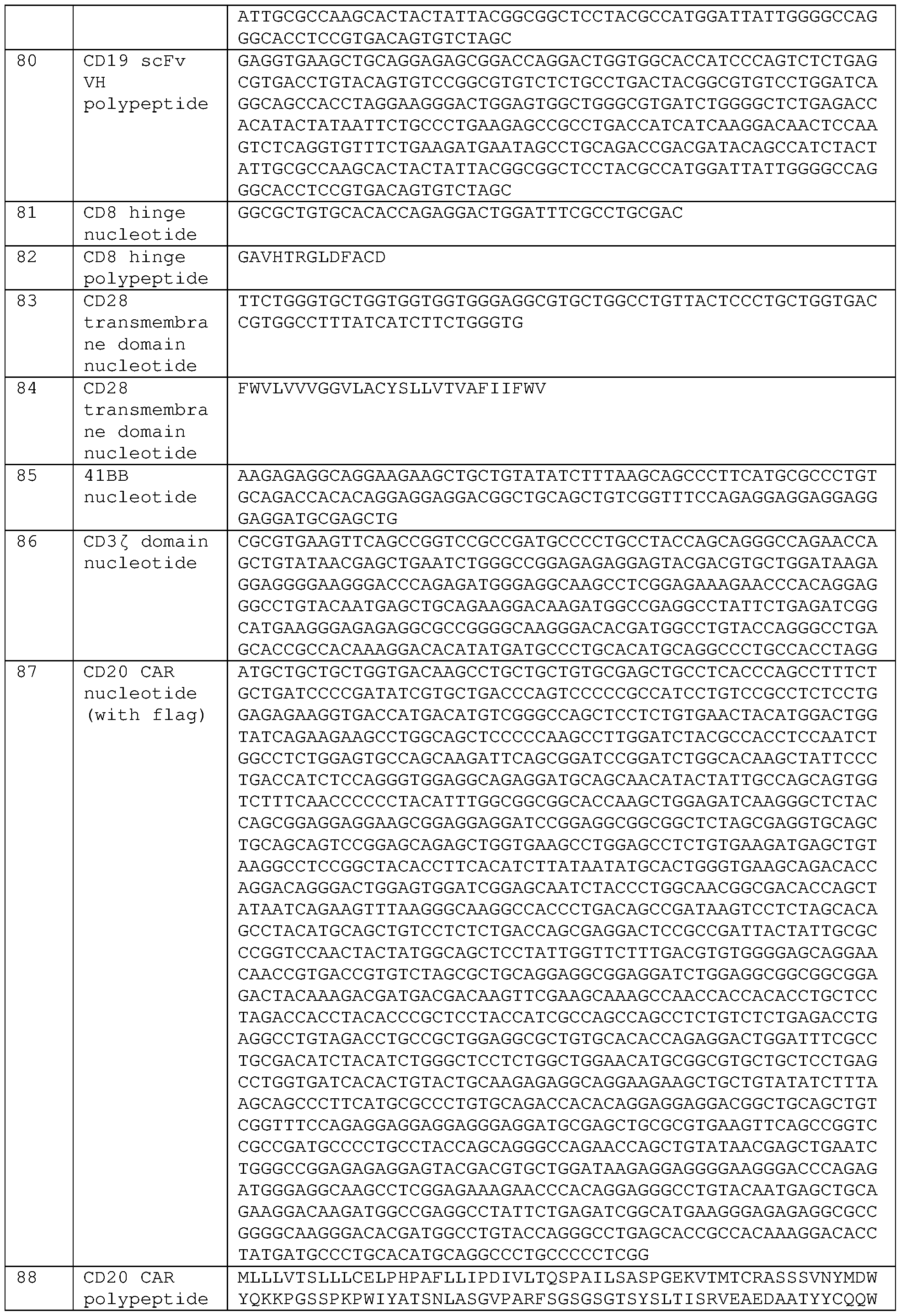


Claims
1. A method for preparing a lentivirus formulation, comprising
(i) filtering a suspension mixture comprising a population of host cells and lentiviral particles to remove contaminants, wherein filtering comprises:
(a) filtering the mixture with a first filter, wherein the first filter is a depth filter, resulting in a first filtrate,
(b) filtering the first filtrate with a second filter, wherein the second filter has a retention threshold smaller than the first filter, resulting in a second filtrate, and
(c) filtering the second filtrate, with a third filter, wherein the third filter has a retention threshold smaller than the second filter, thereby producing a filtered formulation of lentiviral particles; and
(ii) concentrating the filtered formulation of lentiviral particles, wherein concentrating comprises chromatography and ultrafiltration.
2. A method for preparing a lentivirus formulation, comprising
(i) contacting a population of host cells in suspension with at least one plasmid encoding a lentiviral protein;
(ii) culturing the population of host cells of step (i) for a period of time sufficient to produce a suspension mixture comprising a population of host cells and lentiviral particles;
(iii) filtering the suspension mixture to remove contaminants, comprising:
(a) contacting the mixture with an endonuclease,
(b) filtering the mixture with a first filter, wherein the first filter is a depth filter, resulting in a first filtrate
(c) filtering the first filtrate with a second filter, wherein the second filter has a retention threshold smaller than the first filter, resulting in a second filtrate, and
(d) filtering the second filtrate with a third filter, wherein the third filter has a retention threshold smaller than the second filter, thereby producing a filtered formulation of lentiviral particles; and
(iv) concentrating the filtered formulation of lentiviral particles, wherein concentrating comprises chromatography and ultrafiltration.
3. The method of claim 1 or 2, wherein the host cell comprises a human cell.
4. The method of claim 3, wherein the human cell comprises a HEK293 cell, a HEK293T cell, a HEK293F cell, a HEK293FT cell, a Te671 cell, a HT1080 cell, or a CEM cell.
5. The method of claim 3 or 4, wherein the cell comprises a HEK293 cell.
6. The method of claim 3 or 4, wherein the cell comprises a HEK293T cell.
7. The method of any one of claims 1-6, wherein the first filter has a retention threshold of 1-60 μm.
8. The method of claim 7, wherein the first filter has a retention threshold of 60 μm.
9. The method of any one of claims 1-8, wherein the second filter has a retention threshold of 0.4-4 μm.
10. The method of claim 9, wherein the second filter has a retention threshold of 0.45 μm.
11. The method of any of claims 1-10, wherein the third filter has a retention threshold of 0.45 μm ± 0.2 μm.
12. The method of any one of claims 1-10, wherein the third filter has a retention threshold of 0.2-0.3 μm.
13. The method of claim 12, wherein the third filter has a retention threshold of 0.2 μm.
14. The method of any one of claims 1-10, 12 and 13, wherein the first filter has a retention threshold of 60 μm, the second filter has a retention threshold of 0.45 μm, and the third filter has a retention threshold of 0.2 μm.
15. The method of any one of claims 1 and 3-14, wherein an endonuclease is present through steps (i)(a)-(i)(c).
16. The method of any one of claims 2-15, wherein the endonuclease is present through steps (iii)(b)-(iii)(d).
17. The method of any one of claims 1-16, wherein the second filter and the third filter are two layers within a dual-layer filter component.
18. The method of any one of claims 1-16, wherein the third filter is a dual-layer filter comprising a first layer filter and a second layer filter, wherein the second layer filter has a retention threshold smaller than the first layer filter.
19. The method of claim 18, wherein the retention threshold of the first filter is 60 μm, the retention threshold of the second filter is 0.45 μm, the retention threshold of the first layer filter is 0.45 μm, and the retention threshold of the second layer filter is 0.2 μm.
20. The method of any one of claims 1-19, wherein the chromatography is anion exchange chromatography (AEX).
21. The method of claim 20, wherein the AEX chromatography comprises eluting the lentiviral particles with a salt buffer.
22. The method of claim 21, wherein the salt buffer comprises NaC1.
23. The method of claim 22, wherein the NaC1 is at a concentration from about 0.5M to 3M.
24. The method of claim 22, wherein the NaC1 is at a concentration from about 0.5 M to 1 M.
25. The method of any of claims 22-24, wherein the NaC1 is or about 0.75M.
26. The method of claim 22, wherein the NaC1 is at a concentration from about IM to 3 M.
27. The method of claim 22, wherein the NaC1 is at a concentration from about 1.5 M to 2.5 M.
28. The method of any of claims 22, 26 or 27, wherein the NaC1 is about 2M.
29. The method of any one of claims 1-28, wherein the chromatography is performed before ultrafiltration.
30. The method of any one of claims 1-29, wherein the ultrafiltration is ultrafiltration/diafiltration (UF/DF) .
31. The method of claim 30, wherein the UF/DF is by one or more tangential flow filtration (TFF) steps.
32. The method of claim 31, wherein the filter of the one or more TFF is a hollow fiber filter.
33. The method of claim 32, wherein the nominal molecular weight cutoff (NMWC) of the hollow fiber filter is or is about 500 kDa.
34. The method of any of claims 31-33, wherein the TFF comprises a first tangential flow filtration (TFF) step and second tangential flow filtration (TFF) step.
35. The method of claim 33, wherein the first TFF is performed with a first hollow fiber filter and the second TFF is performed with a second hollow fiber filter.
36. The method of claim 35, wherein the first and second hollow fiber filter have the same nominal molecular weight cutoff (NMWC).
37. The method of claim 36, wherein the NMWC is 500 kDa.
38. The method of claim 35, wherein the first hollow fiber filter has a greater nominal molecular weight cutoff (NMWC) than the second hollow fiber filter.
39. The method of claim 38, wherein the NMWC of the first hollow fiber filter is 500 kDa.
40. The method of any of claims 35-39, wherein the first hollow fiber filter has a greater surface area than the second hollow fiber filter.
41. The method of claim 40, wherein the surface area of the first hollow fiber filter is between 790 cm2 to 1600 cm2.
42. The method of any of claims 35-41, wherein the first hollow fiber filter holds a larger volume than the second hollow fiber filter.
43. The method of claim 42, wherein the volume of the first hollow fiber filter is 300 mL.
44. The method of any one of claims 1-43, comprising sterilizing filtration of the filtered formulation after concentration, thereby producing a sterilized formulation.
45. The method of claim 44, wherein sterilizing filtration comprises filtering the filtered formulation with a fourth filter.
46. The method of claim 45, wherein the fourth filter has a retention threshold of 0.2 pm.
47. The method of any one of claims 44-46, comprising formulating the sterilized formulation in a buffer, thereby producing a drug substance.
48. The method of any one of claims 1-47, wherein the method occurs at a pH of 6-
8.
49. The method of any one of claims 1-48, wherein the lentivirus formulation is for in vivo administration to a subject.
50. The method of any one of claims 1-49, wherein the amount of contaminants in the filtered formulation is reduced compared to the amount of contaminants in the second filtrate.
51. The method of any one of claims 44-50, wherein the amount of contaminants in the sterilized formulation is at a level acceptable for in vivo administration to a subject.
52. The method of any one of claims 1-51, wherein the contaminants comprise host cells, host cell DNA (hcDNA), and/or host cell proteins (HCP).
53. The method of claim 52, wherein the amount of hcDNA is less than 10%, less than 9%, less than 8%, less than 7%, less than 6%, less than 5%, less than 4%, less than 3%, less than 2%, less than 1% or less than 0.5% of the sterilized formulation.
54. The method of claim 52, wherein the amount of hcDNA is reduced greater than 90%, greater than 91%, greater than 92%, greater than 93%, greater than 94%, greater than 95%, greater than 96%, greater than 97%, greater than 98%, or greater than 99% in the sterilized formulation.
55. The method of claim 52, wherein the amount of HCP is less than 10%, less than 9%, less than 8%, less than 7%, less than 6%, less than 5%, less than 4%, less than 3%, less than 2%, less than 1% or less than 0.5% of the sterilized formulation.
56. The method of claim 52, wherein the amount of HCP is reduced greater than 90%, greater than 91%, greater than 92%, greater than 93%, greater than 94%, greater than 95%, greater than 96%, greater than 97%, greater than 98%, or greater than 99% in the sterilized formulation.
57. The method of any of claims 52-56, wherein the hcDNA amount in the filtered formulation is less than about 2500 ng/lE9 TU.
58. The method of any one of claims 52-57, wherein the hcDNA amount in the filtered formulation is at least about 80-fold lower compared to the hcDNA amount in the suspension mixture.
59. The method of any one of claims 52-58, wherein the hcDNA amount in the filtered formulation is at least about 5-fold lower compared to the hcDNA amount in the second filtrate.
60. The method of any one of claims 52-59, wherein the HCP amount after chromatography is less than about 3000 μg/lE9 TU.
61. The method of any one of claims 52-60, wherein the HCP amount after chromatography is at least about 40-fold lower compared to the HCP amount before chromatography .
62. The method of any one of claims 52-61, wherein the HCP amount after chromatography is at least about 99% lower compared to the HCP amount before chromatography .
63. The method of any one of claims 52-62, wherein the HCP amount is less than about 1500 μg/lE9 TU after a first UF/DF step.
64. The method of any one of claims 52-63, wherein the HCP amount is not detectable after a second UF/DF step.
65. The method of any one of claims 1-64, wherein the suspension mixture comprises a media, wherein the media does not contain serum and/or animal by-products.
66. The method of any one of claims 2-65, wherein the culturing of step (ii) is for
40-48 hours.
67. The method of any one of claims 1-66, wherein the filtering and concentrating occurs over a time period of 5-8 hours.
68. The method of any one of claims 1-67, wherein the suspension mixture has a volume of 3-50 liters.
69. The method of any of claims 1-67, wherein the suspension mixture has a volume of 5 L to 200 L.
70. The method of any of claims 1-67, wherein the suspension mixture has a volume of 100 L to 200 L.
71. The method of any of claims 1-67, wherein the suspension mixture has a volume of at or about 180 L to at or about 200 L.
72. The method of any one of claims 1-71, wherein the lentiviral particle comprises at least one payload.
73. The method of claim 72, wherein the at least one payload comprises a non- coding nucleic acid, optionally wherein the non-coding nucleic acid is an siRNA, a miRNA, or a shRNA.
74. The method of claim 72, wherein the at least payload is a polynucleotide encoding a polypeptide of interest.
75. The method of any one of claims 2-74, wherein the at least one plasmid is a polynucleotide encoding a polypeptide of interest.
76. The method of claim 74 or 75, wherein the polypeptide of interest is a chimeric antigen receptor (CAR).
77. The method of claim 76, wherein the CAR is specific for a tumor-associated antigen.
78. The method of claim 77, wherein the tumor-associated antigen is CD 19,
BCMA, GPRC5D, R0R1, FcRL5, alpha-fetoprotein, or Her2.
79. The method of claim 76, wherein the CAR is a universal CAR.
80. The method of claim 79, wherein the universal CAR comprises an extracellular domain comprising a tag binding domain.
81. The method of claim 80, wherein the tag is a fluorescein.
82. The method of claim 76, wherein the CAR comprises an extracellular domain comprising a hapten binding domain.
83. The method of any one of claims 1-82, wherein the lentiviral particle comprises a surface engineered fusion protein exposed on the surface of the lentiviral particle, optionally wherein the surface engineered protein is embedded in the lipid bilayer.
84. The method of claim 83, wherein the surface engineered protein is composed of a single binding domain protein that binds to a target molecule on a target cell.
85. The method of claim 84, wherein the surface engineered protein is composed of a multiple binding domain protein, wherein each binding domain binds to a target molecule on a target cell, optionally wherein each binding domain binds to a different target molecule.
86. The method of claim 84 or claim 85 wherein the single binding domain protein or the multiple binding domain protein is a transduction enhancer protein.
87. The method of claim 83-86, wherein the surface engineered protein is a fusion protein comprising a transduction enhancer and a viral envelope protein.
88. The method of any one of claims 1-87, wherein the lentiviral particle comprises a viral envelope comprising a transduction enhancer protein and a viral envelope protein.
89. The method of any one of claims 2-88, wherein the at least one plasmid is a plasmid encoding a fusion protein comprising a transduction enhancer protein and a viral envelope protein, optionally wherein the transduction enhancer comprises an immune-cell activating protein and/or costimulatory molecule.
90. The method of any one of claims 2-89, wherein the at least one plasmid is a plasmid encoding a fusion protein comprising an immune-cell activating protein and a viral envelope protein.
91. The method of claim 89 or claim 90, wherein the immune-cell activating protein comprises at least one binding domain that specifically binds CD2, CD3, CD28H, LFA-1, DNAM-1, CD27, ICOS, LIGHT, GITR, CD30, SLAM, Ly-9, CD84, Lyl08, NKG2D, NKp46, NKp44, NKp30, CD244, TCR α chain, TCR β chain, TCR ζ chain, TCR γ chain, TCR δ chain, CD3 ε TCR subunit, CD3 y TCR subunit, CD3 δ TCR subunit, or NKp80, or combinations thereof.
92. The method of any one of claims 2-89, wherein the at least one plasmid is a plasmid encoding a fusion protein comprising at least one costimulatory molecule and a viral envelope protein.
93. The method of claim 89 and claim 92, wherein the costimulatory molecule is CD45, CD2, CD4, CD5, CD8, CD9, CD16, CD22, CD33, CD28, CD37, CD64, CD80, CD86, CD134, CD137, CD154, 0X40, 4-1BB, CD40L, or any combination thereof.
94. The method of any one of claims 86-90, wherein the transduction enhancer comprises at least one binding domain that binds a target molecule selected from the group consisting of an immune cell activating receptor, a T cell costimulatory receptor or an adhesion molecule.
95. The method of any one of claims 86-94, wherein:
the transduction enhancer comprises a single binding domain that binds to one target molecule selected from the group consisting of a immune cell- activating receptor, a T cell costimulatory receptor or an adhesion molecule; the transduction enhancer comprises multiple binding domains that bind to two or more target molecules selected from the group consisting of an immune cell activating receptor, a T cell costimulatory receptor and an adhesion molecule; or the transduction enhancer comprises multiple binding domains that each bind to a different target molecule that is an immune cell activating receptor, a T cell costimulatory receptor and an adhesion molecule.
96. The method of any of claims 94-95, wherein: the immune cell activating receptor is CD3; the costimulatory molecule is CD28, CD137 or CD134; and/or the adhesion molecule is CD58 or CD2.
97. The method of any of claims 84-96, wherein each of the at least one binding domain is independently selected from an antibody or antigen-binding fragment or an ectodomain of a native ligand of the target molecule.
98. The method of any one of claims 87-97, wherein the viral envelope protein is a VSV-G envelope protein, a measles virus envelope protein, a nipah virus envelope protein, or a cocal virus G protein.
99. The method of any one of claims 2-98, wherein the at least one plasmid is a plasmid encoding a helper viral protein.
100. The method of claim 99, wherein the helper viral protein is rev and/or gagpol.
101. The method of any one of claims 2-100, wherein the population of host cells is contacted with a mixture of plasmids comprising (i) a plasmid encoding a gene of interest; (ii) a plasmid encoding a rev viral protein; (iii) a plasmid encoding a gagpol viral protein; and (iv) a plasmid encoding a viral envelope protein.
102. The method of claim 101, wherein the mixture of plasmids comprises (v) a plasmid encoding an immune cell-activating protein, (vi) a plasmid encoding a co- stimulatory molecule, or (vii) any combination of (v)-(vi).
103. A lentiviral formulation produced by the method of any one of claims 1-102.
104. The lentiviral formulation of claim 103, wherein the formulation has an infectious titer of 2.0 TU/mL to 6 x 10⁸ TU/mL.
105. The lentiviral formulation of claim 103 or claim 104, wherein the formulation has an infectious titer of 2.5 TU/mL to 4.7 x 10⁸ TU/mL.
106. The lentiviral formulation of claim 104 or claim 105, wherein the total number of infectious units in the formulation is 4 x 10¹⁰ TU to 8 x 10¹⁰ TU.
107. The lentiviral formulation of any of claims 104-106, wherein the total number of infectious units in the formulation is 5 x 10¹⁰ TU to 7 x 10¹⁰ TU, optionally at or about 6 x 10¹⁰ TU.
108. The lentiviral formulation of any of claims 104-107, wherein the formulation comprises less than 10%, less than 9%, less than 8%, less than 7%, less than 6%, less than 5%, less than 4%, less than 3%, less than 2%, less than 1% or less than 0.5% of HCP.
109. The lentiviral formulation of any of claims 104-108, wherein the formulation comprises less than 10%, less than 9%, less than 8%, less than 7%, less than 6%, less than 5%, less than 4%, less than 3%, less than 2%, less than 1% or less than 0.5% of hcDNA.
110. The lentiviral formulation of any of claims 104-109, wherein the formulation comprises greater than 90%, greater than 91%, greater than 92%, greater than 93%, greater than 94%, greater than 95%, greater than 96%, greater than 97%, greater than 98%, or greater than 99% reduction of HCP.
111. The lentiviral formulation of any of claims 104-110, wherein the formulation comprises greater than 90%, greater than 91%, greater than 92%, greater than 93%, greater than 94%, greater than 95%, greater than 96%, greater than 97%, greater than 98%, or greater than 99% reduction of hcDNA, optionally reduced compared to the filtered formulation prior to the concentrating.
112. The lentiviral formulation of any of claims 104-111, wherein the formulation comprises greater than 99% reduction of hcDNA and greater than 99% reduction in HCP, optionally reduced compared to the filtered formulation prior to the concentrating.
113. The lentiviral formulation of any of claims 104-112, wherein the formulation comprises less than 1% hcDNA and less than 1% HCP.
114. A lentiviral formulation comprising a lentiviral vector at a titer of 2.5 to 4.7 x 10⁸ TU/mL, wherein the formulation comprises less than 1% hcDNA and less than 1% HCP.
115. The lentiviral formulation of any of claims 103-114, wherein the volume of the formulation is 1 mL to 500 mL, optionally 10 mL to 100 mL.
Priority Applications (1)
| Application Number | Priority Date | Filing Date | Title |
|---|---|---|---|
| AU2023272490A AU2023272490A1 (en) | 2022-05-17 | 2023-05-17 | Manufacturing viral particles |
Applications Claiming Priority (8)
| Application Number | Priority Date | Filing Date | Title |
|---|---|---|---|
| US202263342975P | 2022-05-17 | 2022-05-17 | |
| US63/342,975 | 2022-05-17 | ||
| US202263371756P | 2022-08-17 | 2022-08-17 | |
| US63/371,756 | 2022-08-17 | ||
| US202263371864P | 2022-08-18 | 2022-08-18 | |
| US63/371,864 | 2022-08-18 | ||
| US202363440093P | 2023-01-19 | 2023-01-19 | |
| US63/440,093 | 2023-01-19 |
Publications (1)
| Publication Number | Publication Date |
|---|---|
| WO2023225569A1 true WO2023225569A1 (en) | 2023-11-23 |
Family
ID=86692905
Family Applications (1)
| Application Number | Title | Priority Date | Filing Date |
|---|---|---|---|
| PCT/US2023/067136 WO2023225569A1 (en) | 2022-05-17 | 2023-05-17 | Manufacturing viral particles |
Country Status (2)
| Country | Link |
|---|---|
| AU (1) | AU2023272490A1 (en) |
| WO (1) | WO2023225569A1 (en) |
Citations (41)
| Publication number | Priority date | Publication date | Assignee | Title |
|---|---|---|---|---|
| US5399363A (en) | 1991-01-25 | 1995-03-21 | Eastman Kodak Company | Surface modified anticancer nanoparticles |
| US5466468A (en) | 1990-04-03 | 1995-11-14 | Ciba-Geigy Corporation | Parenterally administrable liposome formulation comprising synthetic lipids |
| US5543158A (en) | 1993-07-23 | 1996-08-06 | Massachusetts Institute Of Technology | Biodegradable injectable nanoparticles |
| US5641515A (en) | 1995-04-04 | 1997-06-24 | Elan Corporation, Plc | Controlled release biodegradable nanoparticles containing insulin |
| US5994136A (en) | 1997-12-12 | 1999-11-30 | Cell Genesys, Inc. | Method and means for producing high titer, safe, recombinant lentivirus vectors |
| US6013516A (en) | 1995-10-06 | 2000-01-11 | The Salk Institute For Biological Studies | Vector and method of use for nucleic acid delivery to non-dividing cells |
| US7446190B2 (en) | 2002-05-28 | 2008-11-04 | Sloan-Kettering Institute For Cancer Research | Nucleic acids encoding chimeric T cell receptors |
| US8093042B2 (en) | 1999-10-12 | 2012-01-10 | Institut Pasteur | Lentiviral triplex DNA, and vectors and recombinant cells containing lentiviral triplex DNA |
| US8399964B2 (en) | 2006-06-02 | 2013-03-19 | Honeywell International Inc. | Multilayer structures for magnetic shielding |
| WO2015057852A1 (en) | 2013-10-15 | 2015-04-23 | The California Institute For Biomedical Research | Chimeric antigen receptor t cell switches and uses thereof |
| US20160122782A1 (en) | 2014-11-05 | 2016-05-05 | Juno Therapeutics, Inc. | Methods for transduction and cell processing |
| US20160152723A1 (en) | 2014-08-28 | 2016-06-02 | Juno Therapeutics, Inc. | Antibodies and chimeric antigen receptors specific for cd19 |
| WO2016139463A1 (en) | 2015-03-02 | 2016-09-09 | Ucl Business Plc | Retroviral and lentiviral vectors |
| US20160272718A1 (en) | 2015-03-17 | 2016-09-22 | Chimera Bioengineering, Inc. | Smart CAR Devices, DE CAR Polypeptides, Side CARs and Uses Thereof |
| US20170037369A1 (en) | 2014-04-23 | 2017-02-09 | Juno Therapeutics, Inc. | Methods for isolating, culturing, and genetically engineering immune cell populations for adoptive therapy |
| US9624276B2 (en) | 2013-10-15 | 2017-04-18 | The California Institute For Biomedical Research | Peptidic chimeric antigen receptor T cell switches and uses thereof |
| US20170240612A1 (en) | 2014-08-29 | 2017-08-24 | Gemoab Monoclonals Gmbh | Universal chimeric antigen expressing immune cells for targeting of diverse multiple antigens and method of manufacturing the same and use of the same for treatment of cancer, infections and autoimmune disorders |
| WO2017156311A2 (en) * | 2016-03-09 | 2017-09-14 | American Gene Technologies International Inc. | Combination vectors and methods for treating cancer |
| US20170275362A1 (en) | 2014-12-05 | 2017-09-28 | Memorial Sloan-Kettering Cancer Center | Chimeric antigen receptors targeting fc receptor-like 5 and uses thereof |
| US20170290900A1 (en) | 2012-12-20 | 2017-10-12 | Purdue Research Foundation | Chimeric antigen receptor-expressing t cells as anti-cancer therapeutics |
| US9856322B2 (en) | 2003-11-05 | 2018-01-02 | St Jude Children's Research Hospital, Inc. | Chimeric receptors with 4-1BB stimulatory signaling domain |
| US20180118803A1 (en) | 2014-12-05 | 2018-05-03 | Memorial Sloan Kettering Cancer Center | Chimeric antigen receptors targeting g-protein coupled receptor and uses thereof |
| WO2019057124A1 (en) | 2017-09-22 | 2019-03-28 | Wuxi Biologics (Shanghai) Co., Ltd. | Novel bispecific cd3/cd19 polypeptide complexes |
| US20190091308A1 (en) | 2016-04-08 | 2019-03-28 | Purdue Research Foundation | Methods and compositions for car t cell therapy |
| WO2019070674A2 (en) * | 2017-10-02 | 2019-04-11 | American Gene Technologies International Inc. | Vectors with promoter and enhancer combinations for treating phenylketonuria |
| US20190161553A1 (en) | 2017-11-01 | 2019-05-30 | Juno Therapeutics, Inc. | Chimeric antigen receptors specific for b-cell maturation antigen and encoding polynucleotides |
| US10357514B2 (en) | 2014-04-07 | 2019-07-23 | The Trustees Of The University Of Pennsylvania | Treatment of cancer using anti-CD19 Chimeric Antigen Receptor |
| US20190269727A1 (en) | 2015-12-28 | 2019-09-05 | Novartis Ag | Methods of making chimeric antigen receptor-expressing cells |
| US20200023009A1 (en) | 2017-02-28 | 2020-01-23 | Endocyte, Inc. | Compositions and methods for car t cell therapy |
| US20200123224A1 (en) | 2016-12-13 | 2020-04-23 | Seattle Children's Hospital (dba Seattle Children's Research Institute) | Methods of exogenous drug activation of chemical-induced signaling complexes expressed in engineered cells in vitro and in vivo |
| WO2020106992A1 (en) | 2018-11-21 | 2020-05-28 | Umoja Biopharma, Inc. | Multicistronic vector for surface engineering lentiviral particles |
| US20200216502A1 (en) | 2017-09-22 | 2020-07-09 | Centre National De La Recherche Scientifique (Cnrs) | Mutated Glycoprotein of Vesicular Stomatitis Virus |
| US20200246381A1 (en) | 2018-02-01 | 2020-08-06 | Nanjing Iaso Biotherapeutics Co., Ltd. | Chimeric antigen receptor (car) binding to bcma, and uses thereof |
| US10736918B2 (en) | 2012-08-20 | 2020-08-11 | Fred Hutchinson Cancer Research Center | Method and compositions for cellular immunotherapy |
| US10918665B2 (en) | 2014-12-05 | 2021-02-16 | Memorial Sloan Kettering Cancer Center | Chimeric antigen receptors targeting B-cell maturation antigen and uses thereof |
| WO2021076788A2 (en) | 2019-10-16 | 2021-04-22 | Umoja Biopharma, Inc. | Retroviral vector for univeral receptor therapy |
| US20210163893A1 (en) | 2018-08-09 | 2021-06-03 | Juno Therapeutics, Inc. | Processes for generating engineered cells and compositions thereof |
| US20210244871A1 (en) | 2018-05-11 | 2021-08-12 | Lupagen, Inc. | Systems and methods for closed loop, real-time modifications of patient cells |
| US20210393689A1 (en) | 2018-11-01 | 2021-12-23 | Juno Therapeutics, Inc. | Chimeric antigen receptors specific for g protein-coupled receptor class c group 5 member d (gprc5d) |
| US20220096651A1 (en) | 2019-01-29 | 2022-03-31 | Juno Therapeutics, Inc. | Antibodies and chimeric antigen receptors specific for receptor tyrosine kinase like orphan receptor 1 (ror1) |
| WO2022072885A1 (en) | 2020-10-02 | 2022-04-07 | Lupagen, Inc. | Closed loop, bedside cell purification systems and methods |
-
2023
- 2023-05-17 AU AU2023272490A patent/AU2023272490A1/en active Pending
- 2023-05-17 WO PCT/US2023/067136 patent/WO2023225569A1/en unknown
Patent Citations (41)
| Publication number | Priority date | Publication date | Assignee | Title |
|---|---|---|---|---|
| US5466468A (en) | 1990-04-03 | 1995-11-14 | Ciba-Geigy Corporation | Parenterally administrable liposome formulation comprising synthetic lipids |
| US5399363A (en) | 1991-01-25 | 1995-03-21 | Eastman Kodak Company | Surface modified anticancer nanoparticles |
| US5543158A (en) | 1993-07-23 | 1996-08-06 | Massachusetts Institute Of Technology | Biodegradable injectable nanoparticles |
| US5641515A (en) | 1995-04-04 | 1997-06-24 | Elan Corporation, Plc | Controlled release biodegradable nanoparticles containing insulin |
| US6013516A (en) | 1995-10-06 | 2000-01-11 | The Salk Institute For Biological Studies | Vector and method of use for nucleic acid delivery to non-dividing cells |
| US5994136A (en) | 1997-12-12 | 1999-11-30 | Cell Genesys, Inc. | Method and means for producing high titer, safe, recombinant lentivirus vectors |
| US8093042B2 (en) | 1999-10-12 | 2012-01-10 | Institut Pasteur | Lentiviral triplex DNA, and vectors and recombinant cells containing lentiviral triplex DNA |
| US7446190B2 (en) | 2002-05-28 | 2008-11-04 | Sloan-Kettering Institute For Cancer Research | Nucleic acids encoding chimeric T cell receptors |
| US9856322B2 (en) | 2003-11-05 | 2018-01-02 | St Jude Children's Research Hospital, Inc. | Chimeric receptors with 4-1BB stimulatory signaling domain |
| US8399964B2 (en) | 2006-06-02 | 2013-03-19 | Honeywell International Inc. | Multilayer structures for magnetic shielding |
| US10736918B2 (en) | 2012-08-20 | 2020-08-11 | Fred Hutchinson Cancer Research Center | Method and compositions for cellular immunotherapy |
| US20170290900A1 (en) | 2012-12-20 | 2017-10-12 | Purdue Research Foundation | Chimeric antigen receptor-expressing t cells as anti-cancer therapeutics |
| WO2015057852A1 (en) | 2013-10-15 | 2015-04-23 | The California Institute For Biomedical Research | Chimeric antigen receptor t cell switches and uses thereof |
| US9624276B2 (en) | 2013-10-15 | 2017-04-18 | The California Institute For Biomedical Research | Peptidic chimeric antigen receptor T cell switches and uses thereof |
| US10357514B2 (en) | 2014-04-07 | 2019-07-23 | The Trustees Of The University Of Pennsylvania | Treatment of cancer using anti-CD19 Chimeric Antigen Receptor |
| US20170037369A1 (en) | 2014-04-23 | 2017-02-09 | Juno Therapeutics, Inc. | Methods for isolating, culturing, and genetically engineering immune cell populations for adoptive therapy |
| US20160152723A1 (en) | 2014-08-28 | 2016-06-02 | Juno Therapeutics, Inc. | Antibodies and chimeric antigen receptors specific for cd19 |
| US20170240612A1 (en) | 2014-08-29 | 2017-08-24 | Gemoab Monoclonals Gmbh | Universal chimeric antigen expressing immune cells for targeting of diverse multiple antigens and method of manufacturing the same and use of the same for treatment of cancer, infections and autoimmune disorders |
| US20160122782A1 (en) | 2014-11-05 | 2016-05-05 | Juno Therapeutics, Inc. | Methods for transduction and cell processing |
| US20180118803A1 (en) | 2014-12-05 | 2018-05-03 | Memorial Sloan Kettering Cancer Center | Chimeric antigen receptors targeting g-protein coupled receptor and uses thereof |
| US10918665B2 (en) | 2014-12-05 | 2021-02-16 | Memorial Sloan Kettering Cancer Center | Chimeric antigen receptors targeting B-cell maturation antigen and uses thereof |
| US20170275362A1 (en) | 2014-12-05 | 2017-09-28 | Memorial Sloan-Kettering Cancer Center | Chimeric antigen receptors targeting fc receptor-like 5 and uses thereof |
| WO2016139463A1 (en) | 2015-03-02 | 2016-09-09 | Ucl Business Plc | Retroviral and lentiviral vectors |
| US20160272718A1 (en) | 2015-03-17 | 2016-09-22 | Chimera Bioengineering, Inc. | Smart CAR Devices, DE CAR Polypeptides, Side CARs and Uses Thereof |
| US20190269727A1 (en) | 2015-12-28 | 2019-09-05 | Novartis Ag | Methods of making chimeric antigen receptor-expressing cells |
| WO2017156311A2 (en) * | 2016-03-09 | 2017-09-14 | American Gene Technologies International Inc. | Combination vectors and methods for treating cancer |
| US20190091308A1 (en) | 2016-04-08 | 2019-03-28 | Purdue Research Foundation | Methods and compositions for car t cell therapy |
| US20200123224A1 (en) | 2016-12-13 | 2020-04-23 | Seattle Children's Hospital (dba Seattle Children's Research Institute) | Methods of exogenous drug activation of chemical-induced signaling complexes expressed in engineered cells in vitro and in vivo |
| US20200023009A1 (en) | 2017-02-28 | 2020-01-23 | Endocyte, Inc. | Compositions and methods for car t cell therapy |
| WO2019057124A1 (en) | 2017-09-22 | 2019-03-28 | Wuxi Biologics (Shanghai) Co., Ltd. | Novel bispecific cd3/cd19 polypeptide complexes |
| US20200216502A1 (en) | 2017-09-22 | 2020-07-09 | Centre National De La Recherche Scientifique (Cnrs) | Mutated Glycoprotein of Vesicular Stomatitis Virus |
| WO2019070674A2 (en) * | 2017-10-02 | 2019-04-11 | American Gene Technologies International Inc. | Vectors with promoter and enhancer combinations for treating phenylketonuria |
| US20190161553A1 (en) | 2017-11-01 | 2019-05-30 | Juno Therapeutics, Inc. | Chimeric antigen receptors specific for b-cell maturation antigen and encoding polynucleotides |
| US20200246381A1 (en) | 2018-02-01 | 2020-08-06 | Nanjing Iaso Biotherapeutics Co., Ltd. | Chimeric antigen receptor (car) binding to bcma, and uses thereof |
| US20210244871A1 (en) | 2018-05-11 | 2021-08-12 | Lupagen, Inc. | Systems and methods for closed loop, real-time modifications of patient cells |
| US20210163893A1 (en) | 2018-08-09 | 2021-06-03 | Juno Therapeutics, Inc. | Processes for generating engineered cells and compositions thereof |
| US20210393689A1 (en) | 2018-11-01 | 2021-12-23 | Juno Therapeutics, Inc. | Chimeric antigen receptors specific for g protein-coupled receptor class c group 5 member d (gprc5d) |
| WO2020106992A1 (en) | 2018-11-21 | 2020-05-28 | Umoja Biopharma, Inc. | Multicistronic vector for surface engineering lentiviral particles |
| US20220096651A1 (en) | 2019-01-29 | 2022-03-31 | Juno Therapeutics, Inc. | Antibodies and chimeric antigen receptors specific for receptor tyrosine kinase like orphan receptor 1 (ror1) |
| WO2021076788A2 (en) | 2019-10-16 | 2021-04-22 | Umoja Biopharma, Inc. | Retroviral vector for univeral receptor therapy |
| WO2022072885A1 (en) | 2020-10-02 | 2022-04-07 | Lupagen, Inc. | Closed loop, bedside cell purification systems and methods |
Non-Patent Citations (44)
| Title |
|---|
| "Oligonucleotide Synthesis: A Practical Approach", 1984, IRL PRESS |
| "Protein Purification Techniques", 2001, OXFORD UNIVERSITY PRESS |
| "Remington: The Science and Practice of Pharmacy", 2005, LIPPINCOTT WILLIAMS & WILKINS |
| "Remington's Pharmaceutical Sciences", 1980 |
| "Tissue Culture", 1973, ACADEMIC PRESS |
| ABOU-EL-ENEIN, M. ET AL., BLOOD CANCER DISCOV, vol. 2, no. 5, 2021, pages 408 - 422 |
| ALTVATER BLANDMEIER SPSCHERER S ET AL., CLIN CANCER RES., vol. 15, 2009, pages 4857 - 4866 |
| ALVAREZ-VALLINA, L. ET AL., EUR J IMMUNOL., vol. 26, no. 10, 1996, pages 2304 - 9 |
| ARCANGELI ET AL., TRANSL CANCER RES, vol. 5, no. 2, 2016, pages S174 - S177 |
| ARCANGELI, S. ET AL., FRONT. IMMUNOL, vol. 11, no. 1217, 19 June 2020 (2020-06-19), pages 1 - 13 |
| AUSUBEL, F. M. ET AL.: "Pharmaceutical Process Filtration Catalogue", vol. 9, 1995, JOHN WILEY & SONS, pages: 177 - 202 |
| CARTELLIERI ET AL., BLOOD CANCER JOURNAL, vol. 6, 2016, pages e458 |
| CHU JDENG YBENSON DM ET AL., LEUKEMIA, vol. 28, 2014, pages 917 - 927 |
| D. M. J. LILLEYJ. E. DAHLBERG: "Methods of Enzymology: DNA Structure Part A: Synthesis and Physical Analysis of DNA ;Methods in Enzymology", 1992, ACADEMIC PRESS |
| GAERERTS ET AL.: "Comparison of retroviral vector titration methods", BMC BIOTECHNOL, vol. 6, 2006, pages 34, XP021017061, DOI: 10.1186/1472-6750-6-34 |
| GHASSEMI, S. ET AL., NAT BIOMED ENG, vol. 6, no. 2, February 2022 (2022-02-01), pages 118 - 128 |
| HAN JCHU JKEUNG CW ET AL., SCI REP., vol. 5, 2015, pages 11483 |
| HENNIG ET AL., INTERNATIONAL JOURNAL OF CANCER, vol. 61, pages 786 - 792 |
| HUMBERT ET AL.: "Development of third-generation Cocal Envelope Producer Cell Lines for Robust Retroviral Gene Transfer into Hematopoietic Stem Cells and T-cells", MOLECULAR THERAPY, vol. 24, 2016, pages 1237 - 1246, XP055659688, DOI: 10.1038/mt.2016.70 |
| IMAI, C. ET AL., LEUKEMIA, vol. 18, 2004, pages 676 - 84 |
| J. M. POLAKJAMES O'D. MCGEE: "In Situ Hybridization: Principles and Practice", 1990, OXFORD UNIVERSITY PRESS |
| J. SAMBROOKE. F. FRITSCHT. MANIATIS: "Molecular Cloning: A Laboratory Manual", 1989, COLD SPRING HARBOR LABORATORY PRESS |
| JOGLEKAR ET AL., HUMAN GENE THERAPY METHODS, vol. 28, 2017, pages 291 - 301 |
| KLOSS SOBERSCHMIDT OMORGAN M ET AL., HUM GENE THER., vol. 28, 2017, pages 897 - 913 |
| KOLMAR H. ET AL., THE FEBS JOURNAL, vol. 275, no. 11, 2008, pages 26684 - 90 |
| KRUSCHINSKI AMOOSMANN APOSCHKE I ET AL., PROC NATL ACAD SCI U S A., vol. 105, 2008, pages 17481 - 17486 |
| KULARATNE, S.A. ET AL., MOL PHARM., vol. 6, no. 3, 2009, pages 780 - 9 |
| LATZA, U. ET AL., EUR. J. IMMUNOL., vol. 24, 1994, pages 677 |
| LI YHERMANSON DLMORIARITY BSKAUFMAN DS, CELL STEM CELL, vol. 23, 2018, pages 181 - 192 |
| LIU ETONG YDOTTI G ET AL., LEUKEMIA, vol. 32, 2018, pages 520 - 531 |
| MOREIRA ANA SOFIA ET AL: "Advances in Lentivirus Purification", BIOTECHNOLOGY JOURNAL, vol. 16, no. 1, 1 January 2021 (2021-01-01), DE, pages 2000019, XP093051358, ISSN: 1860-6768, DOI: 10.1002/biot.202000019 * |
| MULLER NMICHEN STIETZE S ET AL., J IMMUNOTHER., vol. 38, 2015, pages 197 - 210 |
| MUNIR CHERYAN: "Ultrafiltration Handbook", 1986, TECHNOMIC PUBLISHING |
| NALDINI ET AL., SCIENCE, vol. 272, 1996, pages 263 - 7 |
| OTTO-WILHELM MERTEN ET AL: "Large-Scale Manufacture and Characterization of a Lentiviral Vector Produced for Clinical Ex Vivo Gene Therapy Application", HUMAN GENE THERAPY, vol. 22, no. 3, 1 March 2011 (2011-03-01), pages 343 - 356, XP055023182, ISSN: 1043-0342, DOI: 10.1089/hum.2010.060 * |
| R.I. FRESHNEY: "Culture of animal cells: A manual of basic technique", 2000, OXFORD UNIVERSITY PRESS |
| REICHERT, J.M., MABS, vol. 1, no. 3, 2009, pages 190 - 209 |
| SEGA, E.I. ET AL., CANCER METASTASIS REV., vol. 27, no. 4, 2008, pages 655 - 64 |
| SOFER, GAILHAGEL, LARS: "Handbook of Process Chromatography: A Guide to Optimization, Scale-up, and Validation", 1997, COLD SPRING HARBOR LABORATORY PRESS, pages: 758 - 763 |
| VALKAMA ANNIINA J. ET AL: "Development of Large-Scale Downstream Processing for Lentiviral Vectors", MOLECULAR THERAPY- METHODS & CLINICAL DEVELOPMENT, vol. 17, 1 June 2020 (2020-06-01), GB, pages 717 - 730, XP093051352, ISSN: 2329-0501, DOI: 10.1016/j.omtm.2020.03.025 * |
| VORMITTAG, P. ET AL., CURR OPIN BIOTECHNOL, vol. 54, October 2018 (2018-10-01), pages 164 - 181 |
| WAYUA. C. ET AL., MOLECULAR PHARMACEUTICS, 2013 |
| XU YLIU QZHONG M ET AL., J HEMATOL ONCOL., vol. 12, 2019, pages 49 |
| ZUFFEREY ET AL., J. VIROL., vol. 72, 1998, pages 8150 - 8471 |
Also Published As
| Publication number | Publication date |
|---|---|
| AU2023272490A1 (en) | 2024-12-12 |
Similar Documents
| Publication | Publication Date | Title |
|---|---|---|
| JP7171871B2 (en) | Costimulatory Chimeric Antigen Receptor T Cells Targeting IL13Rα2 | |
| JP7252379B2 (en) | CS1-targeted chimeric antigen receptor-modified T cells | |
| JP2024086948A (en) | Improved methods for manufacturing adoptive cell therapeutics | |
| AU2016206868B2 (en) | Modified hepatitis post-transcriptional regulatory elements | |
| US20230348617A1 (en) | Chimeric antigen receptors targeted to psca | |
| US20230348624A1 (en) | Bispecific transduction enhancer | |
| JP2023551220A (en) | Vector systems and their use for delivering multiple polynucleotides | |
| CA3035829A1 (en) | Perfusion bioreactor bag assemblies | |
| AU2023265993A1 (en) | Viral particle with surface stimulating molecules | |
| WO2019241334A1 (en) | Chimeric antigen receptor tumor infiltrating lymphocytes | |
| WO2023225569A1 (en) | Manufacturing viral particles | |
| JP2024515793A (en) | Viral Vector Production System | |
| US20240228959A9 (en) | Large scale car-t immune cell manufacturing method utilizing lentiviral vector transfection | |
| US20230323394A1 (en) | Improved generation of lentiviral vectors for t cell transduction using cocal envelope | |
| WO2024089639A1 (en) | Lentiviral formulations | |
| TW202434735A (en) | Particles displaying adhesion-molecule fusions | |
| WO2024097992A2 (en) | Particles displaying adhesion-molecule fusions | |
| WO2024098038A2 (en) | Polynucleotide construct and related viral vectors and methods |
Legal Events
| Date | Code | Title | Description |
|---|---|---|---|
| 121 | Ep: the epo has been informed by wipo that ep was designated in this application |
Ref document number: 23729004 Country of ref document: EP Kind code of ref document: A1 |