WO2018221969A1 - A novel anti-c-met antibody and use thereof - Google Patents
A novel anti-c-met antibody and use thereof Download PDFInfo
- Publication number
- WO2018221969A1 WO2018221969A1 PCT/KR2018/006182 KR2018006182W WO2018221969A1 WO 2018221969 A1 WO2018221969 A1 WO 2018221969A1 KR 2018006182 W KR2018006182 W KR 2018006182W WO 2018221969 A1 WO2018221969 A1 WO 2018221969A1
- Authority
- WO
- WIPO (PCT)
- Prior art keywords
- seq
- antibody
- substituted
- represented
- variable region
- Prior art date
Links
Images


Classifications
-
- C—CHEMISTRY; METALLURGY
- C07—ORGANIC CHEMISTRY
- C07K—PEPTIDES
- C07K16/00—Immunoglobulins [IGs], e.g. monoclonal or polyclonal antibodies
- C07K16/18—Immunoglobulins [IGs], e.g. monoclonal or polyclonal antibodies against material from animals or humans
- C07K16/28—Immunoglobulins [IGs], e.g. monoclonal or polyclonal antibodies against material from animals or humans against receptors, cell surface antigens or cell surface determinants
- C07K16/2863—Immunoglobulins [IGs], e.g. monoclonal or polyclonal antibodies against material from animals or humans against receptors, cell surface antigens or cell surface determinants against receptors for growth factors, growth regulators
-
- G—PHYSICS
- G01—MEASURING; TESTING
- G01N—INVESTIGATING OR ANALYSING MATERIALS BY DETERMINING THEIR CHEMICAL OR PHYSICAL PROPERTIES
- G01N33/00—Investigating or analysing materials by specific methods not covered by groups G01N1/00 - G01N31/00
- G01N33/48—Biological material, e.g. blood, urine; Haemocytometers
- G01N33/50—Chemical analysis of biological material, e.g. blood, urine; Testing involving biospecific ligand binding methods; Immunological testing
- G01N33/53—Immunoassay; Biospecific binding assay; Materials therefor
- G01N33/574—Immunoassay; Biospecific binding assay; Materials therefor for cancer
- G01N33/5748—Immunoassay; Biospecific binding assay; Materials therefor for cancer involving oncogenic proteins
-
- A—HUMAN NECESSITIES
- A61—MEDICAL OR VETERINARY SCIENCE; HYGIENE
- A61K—PREPARATIONS FOR MEDICAL, DENTAL OR TOILETRY PURPOSES
- A61K39/00—Medicinal preparations containing antigens or antibodies
- A61K2039/505—Medicinal preparations containing antigens or antibodies comprising antibodies
-
- A—HUMAN NECESSITIES
- A61—MEDICAL OR VETERINARY SCIENCE; HYGIENE
- A61K—PREPARATIONS FOR MEDICAL, DENTAL OR TOILETRY PURPOSES
- A61K39/00—Medicinal preparations containing antigens or antibodies
- A61K2039/505—Medicinal preparations containing antigens or antibodies comprising antibodies
- A61K2039/507—Comprising a combination of two or more separate antibodies
-
- C—CHEMISTRY; METALLURGY
- C07—ORGANIC CHEMISTRY
- C07K—PEPTIDES
- C07K2317/00—Immunoglobulins specific features
- C07K2317/20—Immunoglobulins specific features characterized by taxonomic origin
- C07K2317/24—Immunoglobulins specific features characterized by taxonomic origin containing regions, domains or residues from different species, e.g. chimeric, humanized or veneered
-
- C—CHEMISTRY; METALLURGY
- C07—ORGANIC CHEMISTRY
- C07K—PEPTIDES
- C07K2317/00—Immunoglobulins specific features
- C07K2317/30—Immunoglobulins specific features characterized by aspects of specificity or valency
- C07K2317/31—Immunoglobulins specific features characterized by aspects of specificity or valency multispecific
-
- C—CHEMISTRY; METALLURGY
- C07—ORGANIC CHEMISTRY
- C07K—PEPTIDES
- C07K2317/00—Immunoglobulins specific features
- C07K2317/30—Immunoglobulins specific features characterized by aspects of specificity or valency
- C07K2317/33—Crossreactivity, e.g. for species or epitope, or lack of said crossreactivity
-
- C—CHEMISTRY; METALLURGY
- C07—ORGANIC CHEMISTRY
- C07K—PEPTIDES
- C07K2317/00—Immunoglobulins specific features
- C07K2317/30—Immunoglobulins specific features characterized by aspects of specificity or valency
- C07K2317/34—Identification of a linear epitope shorter than 20 amino acid residues or of a conformational epitope defined by amino acid residues
-
- C—CHEMISTRY; METALLURGY
- C07—ORGANIC CHEMISTRY
- C07K—PEPTIDES
- C07K2317/00—Immunoglobulins specific features
- C07K2317/50—Immunoglobulins specific features characterized by immunoglobulin fragments
- C07K2317/52—Constant or Fc region; Isotype
- C07K2317/53—Hinge
-
- C—CHEMISTRY; METALLURGY
- C07—ORGANIC CHEMISTRY
- C07K—PEPTIDES
- C07K2317/00—Immunoglobulins specific features
- C07K2317/50—Immunoglobulins specific features characterized by immunoglobulin fragments
- C07K2317/56—Immunoglobulins specific features characterized by immunoglobulin fragments variable (Fv) region, i.e. VH and/or VL
- C07K2317/565—Complementarity determining region [CDR]
-
- C—CHEMISTRY; METALLURGY
- C07—ORGANIC CHEMISTRY
- C07K—PEPTIDES
- C07K2317/00—Immunoglobulins specific features
- C07K2317/60—Immunoglobulins specific features characterized by non-natural combinations of immunoglobulin fragments
- C07K2317/62—Immunoglobulins specific features characterized by non-natural combinations of immunoglobulin fragments comprising only variable region components
- C07K2317/622—Single chain antibody (scFv)
-
- C—CHEMISTRY; METALLURGY
- C07—ORGANIC CHEMISTRY
- C07K—PEPTIDES
- C07K2317/00—Immunoglobulins specific features
- C07K2317/70—Immunoglobulins specific features characterized by effect upon binding to a cell or to an antigen
- C07K2317/73—Inducing cell death, e.g. apoptosis, necrosis or inhibition of cell proliferation
-
- C—CHEMISTRY; METALLURGY
- C07—ORGANIC CHEMISTRY
- C07K—PEPTIDES
- C07K2317/00—Immunoglobulins specific features
- C07K2317/70—Immunoglobulins specific features characterized by effect upon binding to a cell or to an antigen
- C07K2317/77—Internalization into the cell
-
- C—CHEMISTRY; METALLURGY
- C07—ORGANIC CHEMISTRY
- C07K—PEPTIDES
- C07K2317/00—Immunoglobulins specific features
- C07K2317/90—Immunoglobulins specific features characterized by (pharmaco)kinetic aspects or by stability of the immunoglobulin
- C07K2317/92—Affinity (KD), association rate (Ka), dissociation rate (Kd) or EC50 value
-
- G—PHYSICS
- G01—MEASURING; TESTING
- G01N—INVESTIGATING OR ANALYSING MATERIALS BY DETERMINING THEIR CHEMICAL OR PHYSICAL PROPERTIES
- G01N2333/00—Assays involving biological materials from specific organisms or of a specific nature
- G01N2333/435—Assays involving biological materials from specific organisms or of a specific nature from animals; from humans
- G01N2333/705—Assays involving receptors, cell surface antigens or cell surface determinants
- G01N2333/71—Assays involving receptors, cell surface antigens or cell surface determinants for growth factors; for growth regulators
-
- G—PHYSICS
- G01—MEASURING; TESTING
- G01N—INVESTIGATING OR ANALYSING MATERIALS BY DETERMINING THEIR CHEMICAL OR PHYSICAL PROPERTIES
- G01N2333/00—Assays involving biological materials from specific organisms or of a specific nature
- G01N2333/90—Enzymes; Proenzymes
- G01N2333/91—Transferases (2.)
- G01N2333/912—Transferases (2.) transferring phosphorus containing groups, e.g. kinases (2.7)
Definitions
- the present invention relates to an antibody or an antigen binding fragment thereof, specifically binding to a human hepatocyte growth factor receptor (c-Met), and a composition for preventing or treating cancer comprising the same.
- c-Met human hepatocyte growth factor receptor
- RTK Receptor tyrosine kinases
- HGFR hepatocyte growth factor receptor
- HGF/SF scatter factor
- An abnormal c-Met activation by HGF which is one of the representative oncogenic mechanisms, is known to be associated with tumor proliferation, apoptosis inhibition, neovascularization, invasion, metastasis and the like (Bottaro DP et al., Science 251: 802-804 (1991), Day RM et al., Oncogene 18: 3399-3406 (1999)).
- c-Met has drawn much attention as a target antigen for treating such various cancers and various approaches have been made to inhibit the expression and activity of c-Met.
- a c-Met-specific small molecule tyrosine kinase inhibitor which has been known so far, there are Tivantinib (ArQule), INC280 (Novatis), AMG337 (Amgen), etc.
- Rilotumumab Amgen
- Ficlatuzumab AVEP Pharmaceuticals
- HuL2G7 Huaxy Biotech
- Onartuzumab (WO 2006/015371) in clinical phase III of development by Genentech, Emibetuzumab (WO 2010/059654) in clinical phase II by Lilly, SAIT-301 (US 2014154251) in clinical phase I of development, ABT-700 (Wang J et al., BMC Cancer. 16: 105-118(2016)), etc.
- Onartuzumab is a monovalent antagonistic antibody derived from a bivalent monoclonal antibody (5D5), which acts on c-Met as an agent (Mark Merchant, et al., Proc Natl Acad Sci U S A. 110(32): E2987-E299 (2013)).
- c-Met is associated with the occurrence and progression of various cancers as described above, thus it is constantly driving a continuous demand for developing a new therapeutic agent capable of treating cancer by targeting c-Met.
- the present inventors have developed a novel anti-c-Met antibody binding to c-Met with a high affinity and have also identified that such anti-c-Met antibody, a chimera thereof and humanized and affinity-optimized antibodies remarkably inhibit a proliferation of tumor cells and have an excellent anticancer effect, thus having completed the present invention.
- One objective of the present invention is to provide an antibody or an antigen binding fragment thereof that specifically binds to a hepatocyte growth factor receptor (c-Met).
- c-Met hepatocyte growth factor receptor
- Another objective of the present invention is to provide a nucleic acid molecule encoding the antibody or the antigen binding fragment thereof, an expression vector comprising the nucleic acid molecule, a host cell having the expression vector introduced therein, a method for producing an antibody or an antigen binding fragment thereof using the host cell.
- Yet another objective of the present invention is to provide a composition for detecting c-Met comprising the antibody or the antigen binding fragment thereof, a kit for detection comprising the same, and a method for detecting a c-Met antigen using the same.
- Still yet another objective of the present invention is to provide a composition for preventing or treating cancer comprising the antibody or the antigen binding fragment thereof.
- the antibody or the antigen binding fragment thereof of the present invention that specifically binds to a hepatocyte growth factor receptor (c-Met), has a novel sequence, and shows an excellent cancer cell proliferation inhibitory activity and a remarkably excellent anticancer activity even by a little amount thereof, thus effectively preventing or treating the disease such as cancer.
- c-Met hepatocyte growth factor receptor
- FIG. 1 shows results of an in vitro test on tumor cell proliferation inhibitory activity of hybridoma c-Met antibody of the present invention.
- FIG. 2 shows a schematic diagram of a vector for expressing a separate transcriptome for scFv display.
- FIG. 3 shows results of analyzing a tumor cell proliferation inhibitory activity by hu8C4 affinity-optimized antibody of the present invention.
- FIG. 4 shows results of analyzing a tumor cell proliferation inhibitory activity by a bispecific antibody of the present invention.
- FIG. 5 shows results of analyzing a tumor cell proliferation inhibitory activity by a bispecific antibody of the present invention.
- FIG. 6 shows results of comparing a tumor cell proliferation inhibitory activity between the bispecific antibody of the present invention and a combined therapy in U-87 MG (glioblatoma), NCI-H292 (NSCLC), NCI-H1648 (NSCLC) and NCI-H596 (NSCLC) cell lines.
- FIG. 7 shows results of comparing a tumor cell proliferation inhibitory activity between the bispecific antibody of the present invention and a combined therapy in LS174T (colon), BT20 (TNBC) and KP4 (pancreatic) cell lines.
- FIG. 8 shows results of comparing a tumor cell proliferation inhibitory activity between the bispecific antibody of the present invention and a combined therapy in HCC827 (NSCLC) and NCI-H596 (NSCLC) cell lines.
- FIG. 9 shows results of measuring a binding capacity of the anti-c-Met antibody and the bispecific antibody of the present invention with regard to various kinds of c-Met and EGFR antigens by an ELISA method.
- FIG. 10 shows results of measuring an effect of decreasing a receptor level by the bispecific antibody of the present invention in an NCI-H820 (NSCLC) cell line.
- FIG. 11 shows results of measuring an inhibition of c-Met and EGFR phosphorylation by the anti-c-Met antibody and the bispecific antibody of the present invention in an NCI-H820 (NSCLC) cell line.
- FIG. 12 shows results of measuring an anticancer effect of the bispecific antibody of the present invention in a U-87 MG (glioblastoma) cell xenograft model.
- FIG. 13 shows results of measuring an anticancer effect of the bispecific antibody of the present invention in an NCI-H820 (NSCLC) cell xenograft model.
- FIG. 14 shows results of analyzing a tumor cell proliferation inhibitory activity by treating the anti-c-Met antibody of the present invention and the anti-HER2 antibody by a combined therapy in an NCI-H2170 (NSCLC) cell line.
- FIG. 15 shows results of measuring an anticancer effect of a combined therapy with the anti-c-Met antibody of the present invention and the anti-HER2 antibody in an NCI-H2170 (NSCLC) cell xenograft model.
- FIG. 16 shows results of measuring an anticancer effect of the bispecific antibody of the present invention in an NCI-H596 (NSCLC) cell xenograft model.
- FIG. 17 shows results of measuring an anticancer effect of the bispecific antibody of the present invention in an EBC-1 (NSCLC) cell xenograft model.
- FIG. 18 shows results of indicating an amount of c-Met on the surface of cells, measured after treating an HCC827 cell line with a bispecific antibody (hu8C4 x Vectibix scFv), etc.
- FIG. 19 shows results of indicating an amount of EGFR on the surface of cells, measured after treating an HCC827 cell line with a bispecific antibody (hu8C4 x Vectibix scFv), etc.
- FIG. 20 shows results of indicating an epitope of a bispecific antibody, analyzed by a hydrogen-deuterium exchange mass spectrometry (HDX-MS), in a tertiary structure.
- HDX-MS hydrogen-deuterium exchange mass spectrometry
- one aspect of the present invention provides an antibody or an antigen binding fragment thereof that specifically binds to a hepatocyte growth factor receptor (c-Met).
- c-Met hepatocyte growth factor receptor
- the antibody or the antigen binding fragment thereof of the present invention specifically binding to c-Met, binds to c-Met with a high affinity to inhibit an expression or activity thereof, thus showing an excellent tumor cell proliferation inhibitory activity, such that the antibody alone or with conventional pharmaceutically acceptable carriers, other anticancer drugs, anticancer adjuvants, etc. may be valuably used as an anticancer composition for preventing or treating cancer.
- the term "antibody” means a protein molecule serving as a receptor for specifically recognizing an antigen, comprising an immunoglobulin molecule immunologically having reactivity with a certain antigen, wherein examples thereof may comprise a monoclonal antibody, a polyclonal antibody, a full-length antibody and antibody fragments all. Also, the term may comprise a bivalent or bispecific molecule (e.g., a bispecific antibody), a diabody, a triabody or a tetrabody.
- the term “monoclonal antibody” refers to an antibody molecule of a single molecule composition obtained from substantially the same antibody population, wherein such monoclonal antibody shows a single binding specificity and affinity for a certain epitope.
- the term “full-length antibody” has a structure with two full-length light chains and two full-length heavy chains, wherein each of light chains is linked to a heavy chain by a disulfide bond.
- a constant region of the heavy chain has gamma ( ⁇ ), mu ( ⁇ ), alpha ( ⁇ ), delta ( ⁇ ) and epsilon ( ⁇ ) types, and also has gamma1 ( ⁇ 1), gamma2 ( ⁇ 2), gamma3 ( ⁇ 3), gamma4 ( ⁇ 4), alpha1 ( ⁇ 1) and alpha2 ( ⁇ 2) as a subclass.
- a constant region of the light chain has kappa ( ⁇ ) and lambda ( ⁇ ) types.
- IgG comprises IgG1, IgG2, IgG3 and IgG4 as a subtype.
- fragment refers to any fragments of the antibody of the present invention having an antigen binding function of the antibody, wherein such terms are used interchangeably with each other.
- exemplary antigen binding fragments comprise Fab, Fab', F(ab') 2 , Fv and the like, but not limited thereto.
- the Fab has a structure with a variable region of light and heavy chains, a constant region of light chain and a first constant region of heavy chain (CH1 domain), and also has one antigen binding site.
- An antigen binding fragment of an antibody molecule or an antibody fragment means a fragment having an antigen binding function, and Fab' is different from Fab in that the former has a hinge region having one or more cysteine residue in C terminus of a heavy chain CH1 domain.
- F(ab') 2 antibody is created in such a way that a cysteine residue of a hinge region of Fab' forms a disulfide bond.
- Fv is a minimal antibody fragment having only a heavy chain variable region and a light chain variable region, wherein a recombinant technology for creating Fv fragments is disclosed in PCT International Patent Publication Applications WO 88/10649, WO 88/106630, WO 88/07085, WO 88/07086, WO 88/09344 and the like.
- Two-chain Fv is formed in such a way that a heavy chain variable region and a light chain variable region are linked to each other by a non-covalent bond
- single-chain Fv is formed in such a way that a heavy chain variable region and a single chain variable region are generally linked with each other either by a covalent bond through a peptide linker or directly linked in C-terminus, thus forming a structure like a dimer as shown in the two-chain Fv.
- Such antibody fragment may be obtained by using a protein hydrolase (for example, Fab may be obtained by performing a restriction digestion of a whole antibody by papain and F(ab') 2 fragment may be obtained by performing a digestion of the same by pepsin) or may be produced by a gene recombination technology, but not limited thereto.
- a protein hydrolase for example, Fab may be obtained by performing a restriction digestion of a whole antibody by papain and F(ab') 2 fragment may be obtained by performing a digestion of the same by pepsin
- F(ab') 2 fragment may be obtained by performing a digestion of the same by pepsin
- the antibody specifically binding to c-Met is:
- an antibody comprising a light chain variable region comprising a light chain CDR1 represented by SEQ ID NO: 1; a light chain CDR2 represented by SEQ ID NO: 2; a light chain CDR3 represented by SEQ ID NO: 3, and a heavy chain variable region comprising a heavy chain CDR1 represented by SEQ ID NO: 7; a heavy chain CDR2 represented by SEQ ID NO: 8; and a heavy chain CDR3 represented by SEQ ID NO: 9;
- an antibody comprising a light chain variable region comprising a light chain CDR1 represented by SEQ ID NO: 4; a light chain CDR2 represented by SEQ ID NO: 5; a light chain CDR3 represented by SEQ ID NO: 6, and a heavy chain variable region comprising a heavy chain CDR1 represented by SEQ ID NO: 10; a heavy chain CDR2 represented by SEQ ID NO: 11; and a heavy chain CDR3 represented by SEQ ID NO: 12; or
- the term “heavy chain” may comprise both a full-length heavy chain and a fragment thereof comprising a variable region domain VH with an amino acid sequence having a variable region sequence enough to give specificity to an antigen, as well as three constant region domains CH1, CH2 and CH3.
- the term “light chain” may comprise both a full-length light chain and a fragment thereof comprising a variable region domain VL with an amino acid sequence having a variable region sequence enough to give specificity to an antigen, as well as a constant region domain CL.
- the antibody may comprise both a mouse antibody produced from a mouse, and a mutant thereof, wherein a part of an amino acid sequence of a parent antibody is substituted, added and/or deleted to improve the affinity, immunity, etc., of the antibody.
- the mutant may comprise a chimeric antibody, a humanized antibody, an affinity-optimized antibody, etc., as an example, but not limited thereto.
- the mutant comprehensively refers to an antibody, wherein a part of a CDR amino acid sequence of a parent antibody is mutated (substituted, added or deleted) on condition of having the same CDR as that of the parent antibody or targeting the same epitope as that of the parent antibody.
- Such mutant may be appropriately adjusted by those skilled in the art to improve the affinity, immunity and the like of an antibody within the scope of maintaining a binding capacity for the same epitope.
- the antibody or the antigen binding fragment thereof of the present invention may comprise a sequence of anti-c-Met antibody described herein as well as biological equivalents thereof, within the scope of specifically recognizing c-Met.
- an additional change may be made in an amino acid sequence of the antibody, in order to further improve the binding affinity and/or other biological characteristics of the antibody.
- Such change comprises, for example, the deletion, insertion and/or substitution of an amino acid sequence residue of the antibody.
- Such amino acid mutation is made based on relative similarity of amino acid side chain substituent, e.g., hydrophobicity, hydrophilicity, charge, size, etc.
- arginine, lysine and histidine are all positive charge residues; alanine, glycine and serine have a similar size; and phenylalanine, tryptophan and tyrosine have a similar shape.
- arginine, lysine and histidine; alanine, glycine and serine; and phenylalanine, tryptophan and tyrosine are biologically functional equivalents.
- chimeric antibody is an antibody formed in such a way that a variable region of a mouse antibody is recombined with a constant region of a human antibody, which results in a greatly improved immune reaction in comparison with a mouse antibody.
- the term "humanized antibody” means an antibody formed in such a way that a protein sequence of an antibody derived from other species than human is modified to be similar to that of an antibody mutant naturally produced from human.
- the humanized antibody may be prepared by preparing a humanized variable region through a recombination of CDR derived from a mouse with FR derived from a human antibody and then by recombining the same with a constant region of a preferred human antibody.
- a simple CDR grafting only results in a low affinity of the humanized antibody, so several key FR amino acid residues, which are considered to possibly influence a three-dimensional structure of CDR, may develop an affinity with those of mouse antibody, thus reaching the same level as the affinity of an original mouse antibody.
- affinity-optimized antibody which is a mutant formed in such a way that a part of CDR sequence of a certain antibody is substituted, added or deleted, means an antibody with a better binding affinity to an antigen while binding to the same antigen epitope as that of the certain antibody.
- the affinity-optimized antibody of the present invention refers to a mutant antibody binds to the same epitope as that of: (a) an antibody comprising a light chain variable region comprising a light chain CDR1 represented by SEQ ID NO: 1; a light chain CDR2 represented by SEQ ID NO: 2; a light chain CDR3 represented by SEQ ID NO: 3, and a heavy chain variable region comprising a heavy chain CDR1 represented by SEQ ID NO: 7; a heavy chain CDR2 represented by SEQ ID NO: 8; a heavy chain CDR3 represented by SEQ ID NO: 9; or (b) an antibody comprising a light chain variable region comprising a light chain CDR1 represented by SEQ ID NO: 4; a light chain CDR2 represented by SEQ ID NO: 5; a light chain CDR3 represented by SEQ ID NO: 6, and a heavy chain variable region comprising a heavy chain CDR1 represented by SEQ ID NO: 10; a heavy chain CDR2 represented by SEQ ID NO:
- the affinity-optimized antibody of the present invention may be prepared through a phage display.
- the term "phage display” refers to a technology, which displays a mutant polypeptide as a fusion protein with at least a part of coat protein on a phage, for example, on the surface of fibrous phage particles.
- the usefulness of the phage display lies in the fact that it targets a large library of randomized protein mutants, thus promptly and efficiently classifying sequences binding to a target antigen with a high affinity. Displaying a library of peptides and proteins on the phage has been used for screening millions of polypeptides in order to see a polypeptide with a specific binding characteristic.
- the antibody is an antibody comprising: (a) a light chain variable region represented by SEQ ID NO: 13 and a heavy chain variable region represented by SEQ ID NO: 15; or (b) a light chain variable region represented by SEQ ID NO: 14 and a heavy chain variable region represented by SEQ ID NO: 16.
- the antibody is an antibody comprising: (a) a light chain variable region coded by a nucleotide represented by SEQ ID NO: 17 and a heavy chain variable region coded by a nucleotide represented by SEQ ID NO: 19; or (b) a light chain variable region coded by a nucleotide represented by SEQ ID NO: 18 and a heavy chain variable region coded by a nucleotide represented by SEQ ID NO: 20, but not limited thereto.
- a hybridoma cell group was obtained from a mouse, wherein a human c-Met Sema domain/Fc fusion protein is an antigen, from which anti-c-Met antibody specifically binding to c-Met was selected by screening with an ELISA analysis method using c-Met/His fusion protein as an antigen.
- the selected antibody and the chimeric antibody thereof have a tumor cell proliferation inhibitory activity, which is equal to or more excellent than even commercially available known LY2875358 and OA-5D5 (Table 3 and FIG. 1), thus being very valuably used in prevention or treatment of cancer.
- the antibody comprises:
- the antibody is an antibody comprising: (a) a light chain variable region coded by a nucleotide represented by SEQ ID NO: 25 and a heavy chain variable region coded by a nucleotide represented by SEQ ID NO: 27; (b) a light chain variable region coded by a nucleotide represented by SEQ ID NO: 26 and a heavy chain variable region coded by a nucleotide represented by SEQ ID NO: 28; (c) a light chain variable region coded by a nucleotide represented by SEQ ID NO: 33 and a heavy chain variable region coded by a nucleotide represented by SEQ ID NO: 35; or (d) a light chain variable region coded by a nucleotide represented by SEQ ID NO: 34 and a heavy chain variable region coded by a nucleotide represented by SEQ ID NO: 36, but not limited thereto. Also, it may be provided that the antibody comprises a hinge region represented by one of SEQ ID NO: 25 and a
- a humanized antibody comprising CDR of the antibody obtained through a phage display selection was prepared, and it was identified that such antibody showed an anticancer activity, which was similar to that of the chimera antibody of the present invention (Examples 2 and 3).
- a tumor cell proliferation inhibitory activity of the antibody was evaluated according to a hinge region sequence, and it was identified that a proliferation of most tumor cells was effectively inhibited, even with a somewhat difference in the activity depending on the difference of hinge sequence (Table 7).
- an affinity-optimized antibody for the humanized antibody is an antibody, wherein one or more amino acid sequence is substituted from an antibody comprising: a light chain variable region comprising a light chain CDR1 represented by SEQ ID NO: 1; a light chain CDR2 represented by SEQ ID NO: 2; a light chain CDR3 represented by SEQ ID NO: 3, and a heavy chain variable region comprising a heavy chain CDR1 represented by SEQ ID NO: 7; a heavy chain CDR2 represented by SEQ ID NO: 8; a heavy chain CDR3 represented by SEQ ID NO: 9, and wherein, (i) G in a 1st position of the light chain CDR1 is substituted with A, E, K, L, N, R, S, V or W; A in a 2nd position thereof is substituted with C, G, I, P, S, T or V; S in a 3rd position thereof is substituted with G, M, N,
- the light chain CDR1 comprises 0 to 5 substitutions
- the light chain CDR2 comprises 0 to 1 substitution
- the light chain CDR3 comprises 0 to 7 substitutions
- the heavy chain CDR1 comprises 0 to 1 substitution
- the heavy chain CDR2 comprises 0 to 11 substitutions
- the heavy chain CDR3 comprises 0 to 6 substitutions.
- the affinity-optimized antibody comprises a light chain variable region comprising a light chain CDR1 represented by any one of SEQ ID NO: 1 and SEQ ID NO: 229 to SEQ ID NO: 268; a light chain CDR2 represented by any one of SEQ ID NO: 2, SEQ ID NO: 182 to SEQ ID NO: 190, SEQ ID NO: 227 and SEQ ID NO: 228; a light chain CDR3 represented by any one of SEQ ID NO: 3, SEQ ID NO: 142 to SEQ ID NO: 181, SEQ ID NO: 191 to SEQ ID NO: 226 and SEQ ID NO: 269 to SEQ ID NO: 301; and a heavy chain variable region comprising a heavy chain CDR1 represented by any one of SEQ ID NO: 7 and SEQ ID NO: 108 to SEQ ID NO: 112; a heavy chain CDR2 represented by any one of SEQ ID NO: 8, SEQ ID NO: 54 to
- a competitive selection method was used to select an antibody with a more improved affinity than the humanized antibody, thus obtaining a number of affinity-optimized antibodies (Tables 8 to 10 and 12).
- the affinity-optimized antibody has a tumor cell proliferation inhibitory effect that is 4.3 to 28.5 times more excellent than the humanized body (Table 11, 13 and FIG. 3).
- the antibody is an antibody or an antigen binding fragment thereof specifically further binding to an epidermal growth factor receptor (EGFR) in addition to specifically binding to c-Met.
- EGFR epidermal growth factor receptor
- EGFR ErbB tyrosine kinases
- Gefitinib (Iressa), elotinib (Tarceva) and osimertinib (Tagrisso), which are EGFR tyrosine kinase inhibitors, are used as a representative lung cancer therapeutic agent; and cetuximab (Erbitux) and panitumumab (Vectibix), which are EGFR target antibodies, are used as a colon cancer therapeutic agent (Yewale C et al., Biomaterials. 2013 34(34):8690-707 (2013), Deric L. Wheeler et al., Nature Reviews Clinical Oncology 7, 493-507 (2010)).
- Such EGFR target therapeutic agents cause resistance one year before and after treatment, wherein c-Met amplification, mutation and HGF-induced activation are known as a key mechanism of resistance (Simona Corso Cancer Discovery 3:978-992 (2013), Curtis R Chong et al., Nature Medicine 19, 1389-1400 (2013)). Also, it is reported that EGFR and c-Met are simultaneously expressed in various tumor cells, wherein, upon inhibiting EGFR, c-Met becomes activated, thus promptly developing the resistance of EGFR TKI (Engelman, J.A., et al., Science, 316:1039-43 (2007)).
- a single treatment with a c-Met target drug alone and a combined treatment with an EGFR target drug have been now in a clinical trial, but their efficacy has not been verified yet as a therapeutic agent and there is a need for developing a therapeutic agent for c-Met-related cancerous tumors, known as a key cause of resistance.
- the present inventors have prepared c-Met/EGFR bispecific antibody based on the antibody described above.
- the bispecific antibody not only effectively inhibits a proliferation of tumor cells, which are resistant to existing EGFR therapeutic agents, but also shows an excellent proliferation inhibitory activity against tumor cells, thus being valuably used in treatment of diseases such as c-Met-mediated cancers through various mechanisms.
- the bispecific antibody is formed in such a way that an antibody or an antigen binding fragment thereof specifically binding to EGFR is linked to one light chain or heavy chain terminus of c-Met specific antibody, for example, being linked to a heavy chain C-terminus, but not limited thereto.
- binding fragment specifically binding to EGFR is Fab, Fab', F(ab') 2 or Fv.
- the Fv is a scFv fragment, wherein the scFv fragment is linked by a connector capable of linking the scFv fragment to one light chain or heavy chain terminus of c-Met antibody.
- an antibody specifically binding to EGFR is further prepared by linking with a connector represented by SEQ ID NO: 312.
- the EGFR scFv fragment is an EGFR scFv capable of specifically binding to EGFR, known in the art, wherein, for example, there are Erbitux, Vectibix, Portrazza, TheraCIM or the like, but not limited thereto.
- the EGFR scFv is an Erbitux or Vectibix scFv fragment, particularly the EGFR scFv comprises an amino acid sequence represented by SEQ ID NO: 313 or SEQ ID NO: 314, wherein the Vectibix scFv comprises an amino acid sequence represented by SEQ ID NO: 315, but not limited thereto.
- an anticancer effect of the antibody of the present invention is not particularly limited by an abnormality of c-Met expression or a presence or absence of c-Met mutation, etc.
- the bispecific antibody of the present invention had a more excellent tumor cell proliferation inhibitory capacity than a combined therapy of two antibodies (Tables 18 to 21 and FIGS. 6 to 8). Also, as a result of identifying an effect of the bispecific antibody of the present invention on the activity of antigens and signal transduction materials, it was identified that the bispecific antibody of the present invention had a more excellent signal transduction inhibitory efficacy than an antibody alone (FIG. 11).
- the antibody or the antigen binding fragment thereof of the present invention binds to an epitope region represented by an amino acid sequence selected from the group represented by SEQ ID NO: 331, SEQ ID NO: 332, SEQ ID NO: 333 and/or SEQ ID NO: 334.
- An affinity-optimized antibody prepared based on a certain antibody is characterized by having a high homology with the light chain and heavy chain CDR sequences of a variable region with regard to the reference antibody, thus binding to the same epitope region as the reference antibody, such that such affinity-optimized antibody can share all the biological characteristics such as a pharmaceutical mechanism and a pharmaceutical efficacy caused by a binding site, specificity and antibody and exhibit a more excellent effect on binding affinity than the reference antibody.
- the epitope region respectively means, for example, YVSKPGAQL (SEQ ID NO: 331) in 321th to 329th positions, IGASLNDDI (SEQ ID NO: 332) in 333th to 341th positions, PIKYVND (SEQ ID NO: 333) in 366th to 372th positions, and QVVVSRSGPST (SEQ ID NO: 334) in 464th to 474th positions from N-terminus of a reference c-Met antigen (SEQ ID NO: 335), wherein c-Met antigen sequence with the antibody or the antigen binding fragment thereof of the present invention binding thereto comprises a partial mutation (substitution, addition or deletion) or a binding antigen exists in a form of a c-Met fragment, precursor or subtype, thus its binding sites or sequences may somewhat vary accordingly. Nevertheless, a person of ordinary skill in the art may clearly specify a position and a sequence, to which the antigen or the antigen binding fragment thereof of the present invention binds based
- the bispecific antibody hu8C4 x Vectibix scFv of the present invention binds to 4 epitope regions of Y321 - L329 (SEQ ID NO: 331), I333 - I341 (SEQ ID NO: 332), P366 - D372 (SEQ ID NO: 333), and Q464 - S474 (SEQ ID NO: 334) of a human c-Met sema domain ⁇ chain (Table 28).
- the "antibody or antigen binding fragment thereof specifically binding to c-Met" of the present invention means the one binding to a human c-Met by K D 1 ⁇ 10 -7 M or less. It may be provided that the antibody or the antigen binding fragment thereof binds to human c-Met, for example, by K D 5 ⁇ 10 -8 M or less, K D 1 ⁇ 10 -8 M or less, K D 5 ⁇ 10 -9 M or less, or K D 1 ⁇ 10 -9 M or less, but not limited thereto.
- the antibody or the antigen binding fragments thereof of the present invention had a high binding affinity to c-Met antigen by identifying a binding affinity of hu8C4, hu8C4 AH71 and hu8C4 x Vectibix scFv to c-Met ECD, thus identifying K D values of 3.173 ⁇ 10 -10 , 9.993 ⁇ 10 -11 and 2.78 ⁇ 10 -10 , respectively (Table 22).
- the antibody or the antigen binding fragment thereof of the present invention had a cross-reactivity to a c-Met antigen of a cynomolgus monkey, which is an ape (Table 22), but did not bind to other animal-derived antigens (e.g., rodents) (FIG. 9). Also, it was identified that the antibody or the antigen binding fragment thereof of the present invention did not bind to other receptors on the surface of cells than c-Met (Table 24). Thus, it can be seen from the results above that the antibody or the antigen binding fragment thereof of the present invention showed a binding specificity to c-Met antigen of humans and monkeys.
- binding constant (K on ) means a binding ratio of a certain antibody-antigen interaction
- dissociation constant (K off ) means a dissociation ratio of a certain antibody-antigen interaction
- affinity to antigen (K D ) is the one that a ratio of K off : K on (i.e., K off / K on ) is indicated as a molar concentration (M). It may be provided that a K D value for an antibody is measured by using a method widely established in the art. For example, as a method for measuring a K D value of an antibody, it may be provided by a surface plasmon resonance analysis using a Biocore TM system, but not limited thereto.
- Another aspect of the present invention provides a method for producing a nucleic acid molecule for coding the antibody or the antigen binding fragment thereof, an expression vector comprising the nucleic acid molecule, a host cell having the expression vector introduced therein, an antibody using the host cell or an antigen binding fragment thereof.
- the antibody and the antigen binding fragment thereof are such as that described above.
- nucleic acid molecule has a meaning that comprehensively comprises DNA and RNA molecules, wherein a nucleotide, a basic constituent unit in the nucleic acid molecule, comprises not only a natural nucleotide, but also an analogue, in which a sugar or base portion is modified (Scheit, Nucleotide Analogs, John Wiley, New York (1980); Uhlman and Peyman, Chemical Reviews, (1990) 90:543-584).
- a sequence of a nucleic acid molecule for coding the heavy chain and light chain variable regions of the present invention may be modified, wherein the modification comprises an addition, deletion, or non-conservative or conservative substitution of nucleotide.
- the nucleic acid molecule of the present invention also comprises a nucleotide sequence representing a substantial identity with the aforementioned nucleotide sequence.
- the substantial identity means a nucleotide sequence that represents a minimal 80% homology, particularly a minimal 90% homology, more particularly a minimal 95% homology.
- the term "vector,” which is a means for expressing a target gene in a host cell, comprises a plasmid vector; a cosmid vector; and virus vector such as a bacteriophage vector, an adenovirus vector, a retrovirus vector and an adeno-related virus, particularly a plasmid vector, but not limited thereto.
- nucleic acid molecule for coding a light chain variable region and a nucleic acid molecule for coding a heavy chain variable region are operatively linked with a promoter.
- operatively linked means a functional binding between a nucleic acid expression regulatory sequence (e.g., a promoter, a signal sequence, or an array in a transcriptional regulatory factor binding site) and other nucleic acid sequence, thus the regulatory sequence controls a transcription and/or decoding of the other nucleic acid sequence.
- a nucleic acid expression regulatory sequence e.g., a promoter, a signal sequence, or an array in a transcriptional regulatory factor binding site
- the recombinant vector system of the present invention may be built through various methods known in the art. For example, such detailed methods are disclosed in Sambrook et al., Molecular Cloning, A Laboratory Manual, Cold Spring Harbor Laboratory Press (2001), the documents of which are hereby incorporated by reference.
- the vector of the present invention may be typically built as a vector for cloning or a vector for expression. Also, the vector of the present invention may be built in such a way that a prokaryotic cell or an eukaryotic cell is a host.
- the vector of the present invention is an expression vector and the prokaryotic cell is a host
- it is general to comprise powerful promotors capable of carrying out transcription (e.g., tac promotor, lac promotor, lacUV5 promotor, lpp promotor, pL ⁇ promotor, pR ⁇ promotor, rac5 promotor, amp promotor, recA promotor, SP6 promotor, trp promotor, T7 promotor and the like), a ribosome binding site for starting decoding and transcription/decoding termination sequence.
- E. coli e.g., HB101, BL21, DH5 ⁇ , etc.
- promotor and operator portions of E are used as a host cell, promotor and operator portions of E.
- Bacillus sp . is used as a host cell, a promotor of toxin protein gene of Bacillus thuringiensis (Appl. Environ. Microbiol. (1998) 64:3932-3938; Mol. Gen. Genet. (1996) 250:734-741) or any promotors expressible in Bacillus sp . may be used as a regulatory portion.
- the recombinant vector of the present invention may be prepared by manipulating plasmid (e.g., pCL, pSC101, pGV1106, pACYC177, ColE1, pKT230, pME290, pBR322, pUC8/9, pUC6, pBD9, pHC79, pIJ61, pLAFR1, pHV14, pGEX series, pET series, pUC19 and the like), phage (e.g., ⁇ gt4 ⁇ B, ⁇ -Charon, ⁇ z1, M13 and the like) or virus (e.g., SV40, etc.) often used in the art.
- plasmid e.g., pCL, pSC101, pGV1106, pACYC177, ColE1, pKT230, pME290, pBR322, pUC8/9, pUC6, pBD9, pHC79, pIJ61, pLA
- the vector of the present invention is an expression vector and an eukaryotic cell is a host
- promotors derived from a genome of mammal cells e.g., metallothionein promotor, ⁇ -actin promotor, human hemoglobin promotor and human muscle creatin promotor
- promotors derived from mammal virus e.g., adenoviral late promotor, vaccinia virus 7.5K promotor, SV40 promotor, cytomegalovirus (CMV) promotor, tk promotor of HSV, mouse breast tumor virus (MMTV) promotor, LTR promotor of HIV, promotor of Moloney virus, promotor of Epstein-barr virus (EBV) and promotor of Rous sarcoma virus (RSV)
- CMV promotor e.g., adenoviral late promotor, vaccinia virus 7.5K promotor, SV40 promotor, cytomegalovirus (CMV) promotor, tk promotor of HSV, mouse
- the recombinant vector of the present invention may be fused with other sequences in order to facilitate refining of an antibody expressed therefrom.
- fused sequences there are glutathione S-transferase (Pharmacia, USA), maltose binding protein (NEB, USA), FLAG (IBI, USA), 6x His (hexahistidine; Quiagen, USA) and the like.
- a protein expressed by the vector of the present invention is an antibody, thus the expressed antibody may be easily purified through a protein A column, etc., without an additional sequence for refining.
- the recombinant vector of the present invention comprises an antibiotic resistance gene conventionally used in the art as a selected marker, wherein it may comprise, for example, resistance genes to ampicillin, gentamicin, carbenicillin, chloramphenicol, streptomycin, kanamycin, geneticin, neomycin and tetracycline.
- a vector for expressing the antibody of the present invention there may be both a vector system, in which a light chain and a heavy chain are simultaneously expressed in one vector, and a system, in which a light chain and a heavy chain are respectively expressed in a separate vector.
- two vectors may be introduced into a host cell, for example, through co-transformation or targeted transformation.
- the co-transformation is a method for selecting cells that express both light and heavy chains after simultaneously introducing each vector DNA for coding light and heavy chains into a host cell.
- the targeted transformation is a method for selecting a cell transformed with a vector comprising a light (or heavy) chain and transforming a selected cell again with a vector comprising a heavy (or light) chain to finally select a cell that expresses both light and heavy chains.
- any host cells known in the art may be used, wherein such host cells may comprise Bacillus sp . strains such as Escherichia coli , Bacillus subtilis and Bacillus thuringiensis and prokaryotic host cells such as Streptomyces , Pseudomonas (e.g., Pseudomonas putida ), Proteus mirabilis or Staphylococcus (e.g., Staphylococcus carnosus ), but not limited thereto.
- Bacillus sp strains such as Escherichia coli , Bacillus subtilis and Bacillus thuringiensis and prokaryotic host cells such as Streptomyces , Pseudomonas (e.g., Pseudomonas putida ), Proteus mirabilis or Staphylococcus (e.g., Staphylococcus carnosus ), but not limited thereto.
- eukaryotic host cells of the vector there may be mycetes such as Aspergillus species , yeasts such as Pichia pastoris , Saccharomyces cerevisiae , Schizosaccharomyces and Neurospora crassa , other lower eukaryotic cells, cells of higher eukaryotes such as insect-derived cells, and cells derived from plants or mammals.
- mycetes such as Aspergillus species , yeasts such as Pichia pastoris , Saccharomyces cerevisiae , Schizosaccharomyces and Neurospora crassa , other lower eukaryotic cells, cells of higher eukaryotes such as insect-derived cells, and cells derived from plants or mammals.
- host cells may be COS7 cells (monkey kidney cells), NSO cells, SP2/0, Chinese hamster ovary (CHO) cells, W138, baby hamster kidney (BHK) cells, MDCK, myeloma cell lines, HuT 78 cells or 293 cells, more particularly CHO cells, but not limited thereto.
- COS7 cells normal kidney cells
- NSO cells normal kidney cells
- SP2/0 Chinese hamster ovary (CHO) cells
- W138 W138
- baby hamster kidney (BHK) cells baby hamster kidney (BHK) cells
- MDCK myeloma cell lines
- HuT 78 cells or 293 cells more particularly CHO cells, but not limited thereto.
- transformation and/or “transfection” into host cells may be performed by selecting a suitable standard technology according to host cells as known in the art, comprising any methods for introducing nucleic acid into organisms, cells, tissues or organs.
- the methods comprise electroporation, plasmogamy, calcium phosphate (CaPO 4 ) precipitation, calcium chloride (CaCl 2 ) precipitation, agitation using silicon carbide fiber, agrobacteria-mediated transformation, PEG, dextran sulfate, lipofectamine, drying/suppression-mediated transformation and the like, but not limited thereto.
- the method for producing an antibody or an antigen binding fragment thereof using a host cell may particularly comprise steps of: (a) culturing a host cell transformed with a recombinant vector of the present invention; and (b) expressing an anti-c-Met antibody or an antigen binding fragment thereof in the host cell.
- culturing of a transformed host cell may be performed in an appropriate medium and under culturing conditions known in the art. Such culturing process may be easily adjusted according to a selected strain by those skilled in the art.
- Such culturing method is disclosed in various documents (e.g., James M. Lee, Biochemical Engineering, Prentice-Hall International Editions, 138-176).
- Cell culture is divided into suspension culture and attachment culture according to a cell growth type, and batch culture, fed-batch culture and continuous culture according to a culture method.
- a medium used in culture has to appropriately satisfy requirements of a certain strain.
- the medium comprises various carbon sources, nitrogen sources and microelement ingredients.
- usable carbon sources may comprise carbohydrates such as glucose, sucrose, lactose, fructose, maltose, starch and cellulose; fats such as soybean oil, sunflower oil, castor oil and coconut oil; fat acids such as palmitic acid, stearic acid and linoleic acid; alcohols such as glycerol and ethanol; and organic acids such as acetic acid, wherein such carbon sources may be used alone or in combination.
- Nitrogen sources may comprise, for example, organic nitrogen sources such as peptone, yeast extract, meat juice, malt extract, corn steep liquor (CSL) and soybean-wheat, and inorganic nitrogen sources such as urea, ammonium sulfate, ammonium chloride, ammonium phosphate, ammonium carbonate and ammonium nitrate, wherein such nitrogen sources may be used alone or in combination.
- organic nitrogen sources such as peptone, yeast extract, meat juice, malt extract, corn steep liquor (CSL) and soybean-wheat
- inorganic nitrogen sources such as urea, ammonium sulfate, ammonium chloride, ammonium phosphate, ammonium carbonate and ammonium nitrate, wherein such nitrogen sources may be used alone or in combination.
- the medium may comprise potassium dihydrogen phosphate, dipotassium hydrogen phosphate and sodium-containing salt corresponding thereto.
- the medium may comprise metallic salts such as magnesium sulphate or iron sulfate
- a culture product During culture, compounds such as ammonium hydroxide, potassium hydroxide, ammonia, phosphoric acid and sulfuric acid are added to a culture product in an appropriate way to adjust a pH of the culture product. Also, during culture, bubble formation may be suppressed by using a defoaming agent such as fatty acid polyglycol ester. Also, oxygen or oxygen-containing gas (e.g., air) is injected into a culture product in order to maintain an aerobic state of the culture product.
- a temperature of the culture product is normally 20°C to 45°C, preferably 25°C to 40°C.
- the production method may further comprise a step of: (c) collecting an anti-c-Met antibody or an antigen binding fragment thereof expressed in the host cell.
- An antibody obtained by culturing the transformed host cell may be used in a non-purified state, or further used in a purified state with high purity by using various conventional methods, for example, dialysis, salt precipitation, chromatography and the like.
- a method for using chromatography is most often used, wherein a type and order of column may be selected from ion-exchange chromatography, size exclusion chromatography, affinity chromatography, etc., according to antibody characteristics, culture method, etc.
- Another aspect of the present invention provides a composition for detecting c-Met, comprising the antibody or the antigen binding fragment thereof, a kit for detection comprising the same, and a method for detecting c-Met antibody using the same.
- composition for detecting c-Met and the kit comprising the same form an antigen-antibody complex in such a way that an antibody specifically binding to c-Met or an antigen binding fragment thereof comes into contact with a specimen sample, thus effectively detecting c-Met.
- the term "antigen-antibody complex” means a conjugate between c-Met and an antibody for recognizing the same, in order to identify a tumor or a cancer cell of expressing c-Met in a sample.
- a method for quantifying c-Met antigen using a composition for detecting c-Met and using a kit comprising the same may be performed by identifying a formation of an antigen-antibody complex, wherein identifying of the formation of an antigen-antibody complex may be performed by enzyme immunoassay (ELISA), western blotting, immunofluorescence, immunohistochemistry staining, flow cytometry, immunocytochemistry, radioimmunoassay (RIA), immunoprecipitation assay, immunodiffusion assay, complement fixation assay, a protein chip, etc., but not limited thereto.
- ELISA enzyme immunoassay
- western blotting immunofluorescence
- immunohistochemistry staining flow cytometry
- immunocytochemistry immunocytochemistry
- RIA radioimmunoassay
- immunoprecipitation assay immunodiffusion assay
- complement fixation assay a protein chip, etc., but not limited thereto.
- the ELISA comprises various ELISA methods such as a direct ELISA using a labeled antibody for recognizing an antigen attached to a solid support; an indirect ELISA using a labeled secondary antibody for recognizing a capture antibody in a complex of an antibody for recognizing an antigen attached to a solid support; a direct sandwich ELISA using another labeled antibody for recognizing an antigen in a complex of an antibody and an antigen attached to a solid support; an indirect sandwich ELISA using a labeled secondary antibody for reacting with another antibody for recognizing an antigen in a complex of an antibody and an antigen attached to a solid support and then recognizing such antibody, etc.
- an enzyme As a label for qualitatively or quantitatively making a formation of an antigen-antibody complex measurable, there are an enzyme, a fluorescent material, a ligand, a luminous material, a microparticle, a redox molecule, radio isotope and the like, but not necessarily limited thereto.
- ⁇ -glucuronidase As the enzymes, there are ⁇ -glucuronidase, ⁇ -D-glucosidase, ⁇ -D-galactosidase, urease, peroxidase, alkaline phosphatase, acetylcholinesterase, glucose oxidase, hexokinase and GDPase, RNase, glucose oxidase and luciferase, phosphofructokinase, phosphoenolpyruvate carboxylase, aspartate aminotransferase, phosphoenolpyruvate decarboxylase, ⁇ -lactamase, etc., but not limited thereto.
- Another aspect of the present invention provides a composition for preventing or treating cancer comprising the antibody or the antigen binding fragment thereof of the present invention.
- Yet another aspect of the present invention provides a method for preventing or treating cancer, comprising a step of administering a composition comprising the antibody or the antigen binding fragment thereof of the present invention to an individual being in danger of developing cancer or having the same.
- Still yet another aspect of the present invention provides a use of cancer treatment and a use of preparing an anticancer drug, with regard to a composition comprising the antibody or the antigen binding fragment thereof of the present invention.
- the antibody and the antigen binding fragment thereof are such as that described above.
- the antibody or the antigen binding fragment thereof of the present invention is capable of binding to c-Met alone or a combination of c-Met and EGFR with high affinity to inhibit a growth of cancer cells, such that the antibody alone or in combination with conventional pharmaceutically acceptable carriers can be used in treatment, prevention and diagnosis of hyperproliferative diseases such as cancer.
- prevention means all the acts, which prevent or delay diseases such as cancer, etc., from occurrence or recurrence by an administration of the composition of the present invention
- treatment means an inhibition of development of diseases such as cancer, reduction of cancer, or removal of cancer.
- cancer a disease applied to the composition of the present invention, is particularly lung cancer, stomach cancer, colon cancer, rectal cancer, triple negative breast cancer (TNBC), glioblastoma, pancreatic cancer, head and neck cancer, breast cancer, ovarian cancer, renal cancer, bladder cancer, prostate cancer, solenoma, salivary gland tumor or thyroid cancer, more particularly lung cancer, stomach cancer, colon cancer, rectal cancer, triple negative breast cancer (TNBC), glioblastoma, pancreatic cancer, head and neck cancer, breast cancer, and much more particularly lung cancer, stomach cancer, colon cancer, rectal cancer, triple negative breast cancer (TNBC), glioblastoma, pancreatic cancer, head and neck cancer, but not limited thereto.
- TNBC triple negative breast cancer
- TNBC triple negative breast cancer
- cancer is the one caused by, in particular, c-Met overexpression, amplification, mutation or activation, but not limited thereto.
- a composition comprising the antibody or the binding fragment thereof of the present invention has an inhibitory effect on proliferation of all the cancerous tumors irrespective of abnormal expression or mutation of c-Met, such that a pharmaceutical use of the present invention is not limited by an expression aspect or presence or absence of mutation of c-Met.
- the composition may be a form of a pharmaceutical composition, a quasi-drug composition and a composition for health food.
- composition of the present invention for preventing or treating cancer may further comprise a pharmaceutically acceptable carrier.
- the pharmaceutically acceptable carrier is the one conventionally used in preparing a formulation, comprising lactose, dextrose, sucrose, sorbitol, mannitol, starch, acacia rubber, calcium phosphate, alginate, gelatin, calcium silicate, microcrystalline cellulose, polyvinylpyrrolidone, cellulose, water, syrup, methylcellulose, methyl hydroxybenzoate, propyl hydroxybenzoate, talc, magnesium stearate, mineral oil and the like, but not limited thereto.
- composition of the present invention for preventing or treating cancer may further comprise lubricant, humectant, sweetening agent, flavoring agent, emulsifier, suspending agent, preservative, etc.
- lubricant preferably glycerol, glycerol, glycerol, glycerol, glycerol, glycerol, glycerol, glycerol, glycerol, glycerin, glycerin, glycerin, glycerin, glycerin, glycerin, glycerin, glycerin, glycerin, glycerin, glycerol, glycerol, glycerol, glycerol, glycerol, glycerol, glycerol, glycerol, glycerol, glycerol, glycerol, glycerol, glycerol, glyce
- composition of the present invention may be administered orally or parenterally wherein a parenteral administration may be performed by intravenous infusion, subcutaneous infusion, intramuscular injection, intraperitoneal injection, endothelial administration, local administration, intranasal administration, intrapulmonary administration, rectal administration and the like.
- a parenteral administration may be performed by intravenous infusion, subcutaneous infusion, intramuscular injection, intraperitoneal injection, endothelial administration, local administration, intranasal administration, intrapulmonary administration, rectal administration and the like.
- protein or peptide is digested, so an oral composition may be formulated in such a way that its active drug is coated or protected from decomposition in stomach.
- a composition of the present invention may be administered by a predetermined device through which an active substance may be moved into a target cell.
- a suitable dosage of the composition of the present invention for preventing or treating cancer varies depending on such factors as a formulation method, an administration type, a patient' age, weight, gender, morbid condition, food, administration time, administration path, excretion speed and response sensitivity, wherein an ordinary skilled doctor may easily determine and prescribe an effective dose for a desired treatment or prevention.
- a daily dose of the pharmaceutical composition of the present invention may amount to 0.001-100 mg/kg or more.
- the term "pharmaceutical effective dose” means an amount enough to treat, prevent and diagnose diseases such as cancer.
- composition of the present invention for preventing or treating cancer may be formulated into a preparation by using pharmaceutically acceptable carriers and/or expedients according to a method, which may be easily performed by those skilled in the art, to which the present invention pertains, such that such composition can be prepared in a mono-dose form or prepared by being inserted into a multi-dose container.
- a dosage form may be in a form of solution in oil or aqueous medium, suspension or emulsion, or in a form of extract, powder, suppository, powdered drug, granule, tablet or capsule, and may further comprise a dispersing agent or a stabilizer.
- composition of the present invention may be administered as an individual therapeutic agent or administered in combination with other therapeutic agents, and may be administered sequentially or simultaneously with conventional therapeutic agents.
- the antibody or the antigen binding fragment thereof of the present invention may be used in treatment of cancer in such a way that it is injected in vivo in a form of an antibody-therapeutic agent (functional molecule) and a bispecific antibody-therapeutic agent (functional molecule) conjugate, which are such as that described above.
- an antibody-therapeutic agent functional molecule
- a bispecific antibody-therapeutic agent functional molecule conjugate
- c-Met targeted by an antibody or an antigen binding fragment thereof included in the composition of the present invention is a molecule expressed on the surface of cancer cells, thus it may be used in the prevention, treatment and diagnosis of c-Met related cancer in such a way that a functional molecule further is bound to the antibody of the present invention or is administered in combination therewith.
- the functional molecule may comprise a chemical substance, radioactive nuclide, immunotherapeutic agent, cytokine, chemokine, toxin, biotic agent, enzyme inhibitor and the like.
- the functional molecule capable of coupling with the antibody or the fragment thereof of the present invention results in antibody drug-conjugates may be a chemical substance, cytokine or chemokine, but not limited thereto.
- the chemical substance may be, for example, an anticancer drug, particularly, acivicin, aclarubicin, acodazole, acronycine, adozelesin, alanosine, aldesleukin, allopurinol sodium, altretamine, aminoglutethimide, amonafide, ampligen, amsacrine, androgens, anguidine, aphidicolin glycinate, asaley, asparaginase, 5-azacytidine, azathioprine, bacillus calmette-guerin (BCG), Baker's antifol, beta-2-dioxythioguanosine, bisantrene HCl, bleomycin sulfate, bulsufan, but
- Example 1 Preparation of hybridoma cell for producing c-Met specific antibody and identification of tumor cell proliferation inhibitory activity thereof
- a human c-Met Sema domain/Fc fusion protein (self-produced) was intraperitoneally injected as an antigen into a mouse, in order to obtain an immunized mouse needed for developing a hybridoma cell line through animal immunization. Screening was performed through an ELISA analysis method using a human c-Met/His fusion protein as an antigen, in order to select a hybridoma cell specifically responding to c-Met protein only out of a hybridoma cell group.
- a tumor cell proliferation inhibitory activity was tested in a human glioblastoma cell line U-87 MG and a human stomach cancer cell line MKN45.
- the U-87 MG cells (ATCC, #HTB14) were diluted in a culture medium EMEM (ATCC, #30-2003) containing 10% (v/v) FBS, 100 U / 500 ml penicillin and 100 ⁇ g / 500 ml streptomycin (Invitrogen, #15140-122), after which resulting cells were added by 100 ⁇ l into each well of a 96-well plate at a concentration of 2.5 ⁇ 10 3 cells, such that the plate was cultured under 37°C, 95% RH and 5% (v/v) CO 2 conditions for 18 - 24 hours.
- EMEM ATCC, #30-2003
- the cell culture medium was removed from each well, after which an EMEM medium containing 2% (v/v) FBS was added by 100 ⁇ l into each well, and an antibody prepared at 2X of a final concentration (100 nM) was continuously diluted at a ratio of 1/10, such that resulting cells were added by 100 ⁇ l into each well at six concentrations (i.e., 200 nM, 20 nM, 2 nM, 200 pM, 20 pM and 2 pM) for each antibody.
- concentrations i.e., 200 nM, 20 nM, 2 nM, 200 pM, 20 pM and 2 pM
- the plate was cultured for 5 days under 37°C, 95% RH and 5% (v/v) CO 2 conditions, after which resulting cells were fixed with 10% TCA (Trichloroacetic acid; Sigma, #T0699) solution on a final day.
- TCA Terichloroacetic acid
- the resulting fixed cells were dyed for 25 minutes in such a way that 80 ⁇ l of 0.4% SRB (sulforhodamine B) solution was added into each well, after which resulting cells were washed 5 times with 1% acetic acid solution.
- 150 ⁇ l of 10 mM Tris solution was inserted into each well of a dried plate to dissolve SRB dye, after which its optical density was measured at a wavelength of 540nm by using a microplate reader.
- MKN45 (#JCRB0254) cell lines were diluted in an RPMI-1640 medium (Gibco, #A10491) containing 10% (v/v) FBS, after which the resulting cell lines were divided by 2.5 ⁇ 10 3 into each well of a 96-well plate, such that the resulting plate was cultured overnight under 37°C, 5% CO 2 conditions.
- the medium of each well of the plate was replaced with 100 ⁇ l of an RPMI-1640 medium containing 1% (v/v) FBS, after which a test antibody was sequentially diluted at a ratio of 1/10 (i.e., 100 nM, 10 nM, 1 nM, 100 pM, 10 pM and 1 pM) to reach 1 pM at a final concentration of 100 nM, such that the resulting antibody was added by 100 ⁇ l into each well.
- the plate was cultured for 5 days under 37°C, 5% CO 2 conditions, after which the medium was removed therefrom, such that a TCA solution was inserted by 200 ⁇ l into each well to fix cells.
- the cells of the plate were dyed according to a conventional SRB colorimetric assay method, after which an optical density of each well was measured at a wavelength of 540 nm by using a microplate reader. Results of the U87 MG and MKN45 cell lines are shown in Table 3 and FIG. 1.
- the anti-c-Met 8C4, 5G3 antibodies and chimera antibodies thereof of the present invention all have a tumor cell proliferation inhibitory activity, which is equal to or more excellent than the known c-Met antibodies LY2875358 and OA-5D5 (control group).
- the 8C4, 5G3 antibodies and mutants thereof such as chimera antibodies, humanized antibodies and affinity-optimized antibodies to antigen of the present invention may be very valuably used in preventing or treating c-Met related cancer.
- the mouse antibody 8C4 was humanized and an in vitro tumor cell proliferation inhibitory activity thereof was identified, in order to further identify an effect of an antibody prepared in the present invention.
- a human germline gene having a high homology with a gene in a heavy chain variable region of a mouse antibody 8C4 was analyzed first through Ig Blast (http://www.ncbi.nlm.nih.gov/igblast/). In result, it was identified that IGHV3-23 had 48% homology with the 8C4 antibody in an amino acid level, and also identified that IGHV3-11 had 46% homology with the 8C4 antibody in an amino acid level.
- the CDR-H1, CDR-H2 and CDR-H3 of the mouse antibody 8C4 was defined by Kabat numbering, and hu8C4-1 was prepared in such a way that the CDR portion of the mouse antibody 8C4 was represented by be introduced into a framework of IGHV3-23.
- no. 48 (V ⁇ I), no. 49 (S ⁇ G), no. 71 (R ⁇ A), no. 73 (N ⁇ K), no. 78 (L ⁇ A) and no. 94 (K ⁇ G) amino acids were back-mutated into an original amino acid sequence of the mouse antibody 8C4 to finally build a heavy chain of hu8C4-1.
- the CDR portion of the mouse antibody 8C4 was represented by be introduced into a framework of IGHV3-11, and no. 48 (V ⁇ I), no. 49 (S ⁇ G), no. 71 (R ⁇ A), no. 73 (N ⁇ K), no. 78 (L ⁇ A) and no. 94 (R ⁇ G) amino acids were back-mutated into an original amino acid sequence of the mouse antibody 8C4 to finally build a heavy chain of hu8C4-2.
- the CDR-L1, CDR-L2 and CDR-L3 of the mouse antibody 8C4 were defined by Kabat numbering, and the CRD portion of the mouse antibody 8C4 was represented by be introduced into a framework of IGKV1-33 and a framework of IGKV1-27, thus preparing hu8C4-1 and hu8C4-2 respectively.
- amino acid no. 69 (T ⁇ R) of both and hu8C4-2 were back-mutated into an original amino acid sequence of the mouse antibody 8C4.
- the 8C4 humanized antibody was expressed in a 293T cell by using a pCLS05 vector (Korea Patent Registration No. 10-1420274). With regard to such obtained humanized antibodies in a form of IgG1, it was identified whether or not they had a tumor cell proliferation inhibitory activity in U-87 MG, a human glioblastoma cell line, by the same method as shown in Example 1 above.
- mouse antibody 5G3 of the present invention was humanized to identify an in vitro tumor cell proliferation inhibitory activity thereof.
- a human germline gene having a highest homology with a gene in a heavy chain variable region of the mouse antibody 5G3 was analyzed first through Ig Blast (http://www.ncbi.nlm.nih.gov/igblast/). In result, it was identified that IGHV1-46 had 67.3% homology with the 5G3 antibody in an amino acid level.
- the CDR-H1, CDR-H2 and CDR-H3 of the mouse antibody 5G3 were defined by Kabat numbering, and the CRD portion of the mouse antibody 5G3 was represented by be introduced into a framework of IGHV1-46. At this time, amino acid no. 48 (M ⁇ I), no.
- CDR-grafting was performed in IGKV3-20 gene having 63.5% homology with the 5G3 antibody, and amino acid no. 43 (A ⁇ S), no. 60 (D ⁇ A) and no. 71 (F ⁇ N) were back-mutated to build a light chain of hu5G3-1.
- the 5G3 humanized antibody was expressed in a 293T cell by using a pCLS05 vector (Korea Patent Registration No. 10-1420274). With regard to such obtained humanized antibodies in a form of IgG2, it was identified whether or not they had a tumor cell proliferation inhibitory activity in MKN45, a human stomach cancer cell line, by the same method as shown in Example 1 above.
- a hinge of the human IgG1 heavy chain constant region had an amino acid sequence of "EPKSCDKTHTCPPCP (SEQ ID NO: 37)," which was substituted to obtain a hinge region mutant having an amino acid sequence of SEQ ID NO: 38 to SEQ ID NO: 44.
- the resulting mutants were respectively cloned into a vector comprising the heavy chain variable region of hu8C4-1, hu8C4-2 humanized antibodies prepared in Example 2 above.
- An in vitro tumor cell proliferation inhibitory activity according to a hinge sequence was identified in U-87 MG by the same method as shown in Example 1 above.
- NCI-H1993 non-small cell lung cancer cell line NCI-H1993 (ATCC, #CRL-5909).
- the NCI-H1993 cell lines were diluted in an RPMI-1640 medium (Gibco, #A10491) containing 10% (v/v) FBS, after which resulting cell lines were divided by 3.0 ⁇ 10 3 into each well of a 96-well plate, such that the resulting plate was cultured overnight under 37°C, 5% CO 2 conditions.
- the medium of each well of the plate was replaced with 100 ⁇ l of an RPMI-1640 medium containing 2% (v/v) FBS, after which a test antibody was sequentially diluted at a ratio of 1/10 (i.e., 100 nM, 10 nM, 1 nM, 100 pM, 10 pM and 1 pM) to reach 0.001 nM at a final concentration of 100 nM, such that the resulting antibody was added by 100 ⁇ l into each well.
- the plate was cultured for 5 days under 37°C, 5% CO 2 conditions, after which the medium was removed therefrom, such that a TCA solution (Sigma, #T0699) was inserted by 200 ⁇ l into each well to fix the cells.
- the cells of the plate were dyed according to a conventional SRB colorimetric assay method, after which an optical density of each well was measured at a wavelength of 540 nm by using a microplate reader.
- hu8C4 an IgG1 humanized antibody representatively having a hinge region of SEQ ID NO: 38 in hu8C4-1 was named as hu8C4, and an affinity-optimized antibody thereto was prepared to identify an effect thereof.
- a phage-displayed scFv library was first prepared by using a phagemid vector displayed in a combined form of scFv and pIII, wherein a schematic structure of the vector is illustrated in FIG. 2.
- the phagemid vector comprises a scFv fragment of an antibody under a control of an IPTG-inductive lac promotor, wherein a linker sequence used was GGGGS GGGGS GGGGS (SEQ. No. 53).
- a mutation-inducing oligonucleotide having an NNK codon was used to introduce variety into the heavy chain and light chain CDR domain of hu8C4. Accordingly, a hu8C4 scFv library with a fusion of His, HA and pIII was prepared, after which a human c-Met specific antibody was selected from the prepared antibody library.
- a competitive selection method was used to select an antibody with an improved affinity.
- a human c-Met antigen was bound according to the manufacturer guidelines in Dynabeads® M-280 (Thermo Fisher Scientific, 11205D).
- a bead with an antigen binding thereto was blocked for 2 hours by a superblock Tris buffered saline (TBS, Pierce).
- TBS Tris buffered saline
- recombinant phage grew overnight at 37°C, and then recombinant phage was centrifuged and a phage of its supernatant was blocked with superblock TBS, 0.05% Tween 20 for 2 hours. Then, the bead was washed with PBS containing 0.05% Twin 20.
- a blocked phage solution was added into the washed bead, after which the resulting bead was incubated in a rotator for 2 hours for phage binding, such that the resulting bead was washed with PBS containing 0.05% Twin 20. Then, a human c-Met antigen was added into PBS 1 ml containing 0.05% Twin 20, after which the resulting antigen was incubated in a rotator for 24 hours (Rouet R et al. (2012) Nat Protoc . 7:364-373).
- the phage binding to the bead was eluted with 100 mM triethanolamine for 5 minutes, after which an eluent was neutralized with 0.5 M Tris/Cl (pH 7.2). An eluted phage neutralization liquid was infected with E. coli TG1.
- hu8C4 as well as 10 kinds of key antibody with a combination of light chain and heavy chain variable regions of an affinity-optimized antibody thereof showed a tumor cell proliferation inhibitory activity, too.
- IC 50 of the 10 kinds of antibody amounted to 1.7 - 5.3 nM and it was identified that they had a tumor cell proliferation inhibitory effect, which was 9.2 - 28.5 times more excellent than the parent antibody hu8C4.
- a tumor cell proliferation inhibitory activity was evaluated by using NCI-H1993, NCI-H292 and NCI-H820 lung cancer cell lines.
- NCI-H1993 ATCC, #CRL-5909
- NCI-H292 ATCC, #CRL-1848
- NCI-H820 ATCC, #HTB-181 with threonine (T) mutated into methionine (M) in EGFR amino acid no. 790
- T threonine
- M methionine
- Each cell line was diluted in an RPMI-1640 medium (Gibco, #A10491) containing 10% (v/v) FBS, after which the resulting cell lines were divided by 2.0 ⁇ 10 3 into each well of a 96-well plate, such that the resulting plate was cultured overnight under 37°C, 5% CO 2 conditions. Then, each well of the plate was replaced with 100 ⁇ l of a serum-free medium, after which the resulting plate was cultured under 37°C, 5% CO 2 conditions for 18 hours.
- the medium was replaced with 100 ⁇ l of the RPMI-1640 medium containing 2% (v/v) FBS or HGF 50 ng/ml, after which a test antibody was sequentially diluted at a ratio of 1/10 (i.e., 100 nM, 10 nM, 1 nM, 100 pM, 10 pM and 1 pM) to reach 0.001 nM at a final concentration of 100 nM, such that the resulting antibody was added by 100 ⁇ l into each well.
- the plate was cultured for 5 days under 37°C, 5% CO 2 conditions, after which the medium was removed therefrom, such that a TCA solution was inserted by 200 ⁇ l into each well to fix cells.
- the cells of the plate were dyed according to a conventional SRB colorimetric assay method, after which an optical density of each well was measured at a wavelength of 540 nm by using a microplate reader.
- the antibody of the present invention has a proliferation inhibitory effect on all the cancer types regardless of an overexpression or mutation of c-Met and EGFR, thus may be effectively used in these cancer types.
- Example 7 Comparative evaluation of in vitro tumor cell proliferation inhibitory activity of bispecific antibody compared to combined therapy
- Eight types of cancer were used to compare a tumor cell proliferation inhibitory activity between a combined therapy of each antibody targeting c-Met and EGFR respectively and the bispecific antibody of the present invention.
- a tumor cell proliferation inhibitory activity was evaluated in a lung cancer cell line NCI-H292 (ATCC, #CRL-1848), an HGF-autocrinal glioblastoma cell line U-87 MG (ATCC, #HTB-14), lung cancer cell lines NCI-H1648 (ATCC #CRL-5882) and NCI-H596 (ATCC #HTB-178), HCC827 (ATCC, #CRL2868), a colon cancer cell line LS174T (ATCC, #CL-188), a triple negative breast cancer (TNBC) cell line BT20 (ATCC, #HTB-19) and a pancreatic cancer cell line KP4 (JCRB, #RCB1005).
- NCI-H292 ATCC, #CRL-1848
- an HGF-autocrinal glioblastoma cell line U-87 MG ATCC, #HTB-14
- lung cancer cell lines NCI-H1648 ATCC #CRL-5882
- NCI-H596 AT
- the NCI-H1648 cell line is characterized by a normal expression of EGFR and c-Met
- the NCI-H596 cell line is characterized by a deletion of some sequence of exon no. 14 of MET gene
- the HCC827 cell line is characterized by a deletion of some sequence of exon no. 19 of EGFR gene.
- the LS174T cell line has a KRAS mutation and the KP4 is characterized by autocrining HGF.
- the U-87 MG cell line was evaluated by a method of Example 1 and the NCI-H292 cell line was evaluated by a method of Example 6. Also, the NCI-H1648, NCI-H596 and HCC827 cell lines were diluted in an RPMI-1640 medium (Gibco, #A10491) containing 10% (v/v) FBS, after which the resulting cell lines were divided by 2.0 ⁇ 10 3 in each well of a 96-well plate. The LS174T cell line was diluted in a DMEM medium (Gibco, #11995-065) containing 10% (v/v) FBS, after which the resulting cell lines were divided by 2.0 ⁇ 10 3 .
- RPMI-1640 medium Gibco, #A10491
- the LS174T cell line was diluted in a DMEM medium (Gibco, #11995-065) containing 10% (v/v) FBS, after which the resulting cell lines were divided by 2.0 ⁇ 10 3 .
- the BT20 cell line was diluted in an EMEM medium (ATCC, #30-2003) containing 10% (v/v) FBS, after which the resulting cell lines were divided by 3.0 ⁇ 10 3 .
- the KP4 cell line was diluted in an RPMI-1640 medium (Gibco, #A10491) containing 10% (v/v) FBS, after which the resulting cell lines were divided by 1.5 ⁇ 10 3 , such that the resulting plate was cultured overnight under 37°C, 5% CO 2 conditions. Then, each well of the plate was replaced with 100 ⁇ l of a serum-free medium, after which the resulting plate was cultured under 37°C, 5% CO 2 conditions for 18 hours.
- the medium was replaced with 100 ⁇ l of the RPMI-1640 medium containing 2% (v/v) FBS or HGF 50 ng/ml, after which a test antibody was sequentially diluted at a ratio of 1/10 (i.e., 100 nM, 10 nM, 1 nM, 100 pM, 10 pM and 1 pM) to reach 1 pM at a final concentration of 100 nM, such that the resulting antibody was added by 100 ⁇ l into each well. Then, the plate was incubated for 5 days under 37°C, 5% CO 2 conditions, after which the medium was removed therefrom, such that a TCA solution was inserted by 200 ⁇ l into each well to fix cells. Also, the cells of the plate were dyed according to a conventional SRB colorimetric assay method, after which an optical density of each well was measured at a wavelength of 540 nm by using a microplate reader.
- a tumor cell proliferation inhibitory capacity of the bispecific antibody of the present invention was more excellent than that of hu8C4, Vectibix or a combined therapy of two antibodies in the 8 kinds of tumor cell line all. Also, it was identified that it had a remarkably excellent tumor cell proliferation inhibitory capacity in U-87MG, NCI-H292, BT20 and KP4 cell lines when compared to EM1-MAb (Janssen) used as a control bispecific antibody.
- hu8C4 and hu8C4 x Vectibix scFv had an excellent tumor cell proliferation inhibitory capacity compared to a control antibody, when compared to LA480 (Lilly), OA-5D5 (Genentech) and AbF46 (Samsung), which were c-Met target antibodies in U-87MG cell lines.
- Tarceva an EGFR tyrosine kinase inhibitor in HCC827 cell line, showed resistance under HGF processing conditions, but it was identified that it showed an excellent tumor cell proliferation inhibitory capacity when being processed in combination with Tarceva, hu8C4, hu8C4 x Vectibix scFv or c-Met inhibitors under such conditions.
- binding capacity of the c-Met antibody of the present invention to an extracellular domain (ECD) binding of c-Met antibody and bispecific antibody to c-Met ECD and EGFR ECD was measured between human and cynomolgus monkey by using BIAcore.
- a human c-Met ECD ACROBiosystems, MET-H5227
- a cynomolgus monkey c-Met ECD SiNo. Biological, 90304-C08H
- a human EGFR ECD strep ACROBiosystems, EGR-H5285
- a cynomolgus monkey EGFR ECD SiNo. Biological, 90285-C08B
- an Fc-specific anti-human IgG antibody (SouthernBiotech, 2047-01) was fixed to a CM5 sensor chip in the level of 10000 RU.
- the antibodies were diluted in HBS-EP buffer (0.01 M HEPES pH 7.4, 0.15 M NaCl, 3 mM EDTA and 0.005% (v/v) Surfactant P20) at a concentration of 1 - 2 ⁇ g/ml, after which the resulting antibodies were injected into a CM5 chip with an anti-human Ig Fc fixed thereto at a flow rate of 30 ⁇ l/min for 10 - 120 seconds, and then captured in a range of 150 - 200 RU.
- Each antigen was used after being diluted at 10, 5, 2.5, 1.25, 0.625, 0.3125 and 0.15625 nM, after which the resulting antigens were sequentially injected from a lower concentration. Then, the resulting antigens were injected at a flow rate of 30 ⁇ l/min for 5 minutes to carry out binding, after which a running buffer was injected thereinto for 10 - 15 minutes to carry out a dissociation. 15 ⁇ l of 10 mM Glycine-HCl (pH 1.5) was used to revive the chip. A binding and dissociation speed for each cycle was evaluated by using a "1:1 Langmuir binding" model in BIAevaluation software version 4.1, and biacore data are summarized in Tables 22 and 23.
- Example 9 Measurement of c-Met antibody binding capacity to c-Met ECD , EGFR ECD between various animal species (ELISA)
- Binding of c-Met antibody and bispecific antibody to c-Met ECD and EGFR ECD between mouse, cynomolgus monkey and human was measured by using ELISA.
- mouse c-Met (SiNo. Biological Inc, 50622-M08H), cynomolgus monkey c-Met (SiNo. Biological Inc, 90304-C08H), human c-Met (R&D Systems, 358-MT), mouse EGFR (SiNo. Biological Inc, 51091-M08H), cynomolgus monkey EGFR (SiNo. Biological, 90285-C08B) and human EGFR (Abcam, 155639) antigens were all divided into a 96-well plate at a concentration of 2 ⁇ g/ml, after which the resulting plate was reacted at 4°C overnight.
- hu8C4 x Vectibix scFv bispecific antibody was sequentially diluted at a ratio of 1/5 from 100 nM to measure its binding capacity in 7 concentration sections (i.e., 100 nM, 20 nM, 4 nM, 800 pM, 160 pM, 32 pM and 6.4 pM).
- hu8C4 monospecific antibody and hu8C4 x Vectibix scFv bispecific antibody did not bind to a mouse c-Met and a mouse EGFR, but bind to monkey and human c-Mets and EGFRs.
- a human IgG antibody, used as a negative control group did not bind at all.
- the results above suggest that the c-Met antibody of the present invention is specific only to human and monkey c-Mets and EGFRs.
- human c-Met Fc chimera (R&D systems, 358-MT_CF), human FGF R3 (IIIc) Fc chimera (R&D systems, 766-FR), human IGF-I R (R&D systems, 391-GR-050), human PDGF R ⁇ Fc chimera (R&D systems, 385-PR_CF), human VEGF R2 Fc chimera (R&D systems, 357-KD_CF) and human MSP R/Ron (R&D systems, 1947-MS-050) were used as an antigen.
- Each antigen was diluted in 0.05 M carbonate-bicarbonate (Sigma, C3041) buffer at a concentration of 1 ⁇ g/ml, after which the resulting antigen was added into each well of a 96-well plate (Corning, #2592), such that the resulting plate was coated at 4°C overnight.
- the plate was washed once with TBS-T, after which TBS-T containing 4% - skim milk was added by 200 ⁇ l into each well of the resulting plate in order to inhibit a non-specific binding, such that the resulting plate was reacted at 37°C for 1 hour.
- the plate was washed once with TBS-T buffer, after which a primary antibody was sequentially diluted in TBS-T buffer containing 2% - skim milk from a highest concentration of 30 nM to 0.002 nM, such that the resulting antibody was added by 100 ⁇ l into each well, thus being reacted at 37°C for 2 hours.
- an anti-human kappa light chains-peroxidase (Sigma, A7164) was diluted at a ratio of 1 : 5000 as a secondary antibody, after which the resulting antibody was added by 100 ⁇ l into each well, thus being reacted at 37°C for 1 hour.
- TMB solution (Sigma, T4444) was added by 100 ⁇ l into each well to carry out an color developing reaction, after which 2 N ammonium sulfate solution was added by 50 ⁇ l into each well to stop the reaction.
- An optical density was measured based on a value at a wavelength of 450 nm by using a microplate reader and a reference wavelength of 570 nm was used.
- a degree of binding of an anti-c-Met antibody to each antigen was proportionate to an optical density signal value, wherein results thereof are shown in Table 24.
- the hu8C4 antibody of the present invention preferentially binds to c-Met, and it was identified that it did hardly bind to other antigens of FGF R3, VEGFR R2, IGF IR, PDGF R and RON.
- Example 11 In vitro internalization activity of c-Met antibody and c-Met level inhibitory activity of bispecific antibody
- c-Met antibody of the present invention had an in vitro internalization activity in tumor cells as well as an effect on reducing a receptor level by a bispecific antibody capable of simultaneously binding to c-Met and EGFR.
- an antibody internalization occurs by a physiological activity of a normal receptor, wherein, when binding to a specific ligand, the receptor normally expressed outside cells becomes activated through a homo- or hetero- dimerization and causes a receptor-mediated endocytosis.
- An antibody specific to a receptor of a cell has a capacity to induce such phenomenon and is internalized into the cell by causing the endocytosis, thus inducing a decomposition of the receptor, reducing a degree of expression thereof, and possibly inhibiting a signal transduction by a certain receptor.
- An amount of antibodies bound outside cells may be detected by using a fluorescence-activated cell sorting (FACS) device, thus finding an amount of antibodies internalized inside the cells.
- FACS fluorescence-activated cell sorting
- MKN45 cell line (#JCRB0254), which was a stomach cancer cell line, was used to identify an in vitro internalization activity of c-Met antibody inside tumor cells.
- MKN45 expresses a c-Met receptor at a high level by amplification of MET gene, such that a phosphorylation of the c-Met receptor is induced in an HGF-nondependent way.
- a test was performed as follows to see if a c-Met receptor is internalized into a cell by an anti-c-Met antibody hu8C4, thus reducing a level of expression.
- MKN45 stomach cancer cell lines were divided by 5.0 x 10 5 into each well of a 6-well plate containing an RPMI-1640 medium (2 ml) containing 10% (v/v) FBS, after which the plate was cultured under 37°C, RH 95% and 5% CO 2 conditions for 24 hours.
- An anti-c-Met antibody to be analyzed as well as an anti-IgG antibody (control group) were diluted to reach a final concentration of 100 nM, after which the resulting antibodies were reacted overnight.
- As a plate to be used as a non-internalized control group was treated as an anti-c-Met antibody and a human IgG antibody (control group), after which the resulting plate was reacted at 4°C for 1 hour.
- the NCI-H820 cell line is a cell line suitable for measuring an effect of reducing a receptor level by an anti-c-Met x EGFR bispecific antibody, because a c-Met receptor was expressed in a level of about 83,000 SABC (specific antibody-binding capacity) and an EGFR receptor is expressed in a level of about 74,000 SABC.
- NCI-H820 cell lines were divided by 1.0 x 10 5 into each well of a 6-well plate with an RPMI-1640 medium (2 ml) containing 10% (v/v) FBS, after which the resulting plate was cultured overnight under 37°C, RH 95% and 5% CO 2 conditions for 24 hours. Then, it was replaced with a serum-free medium, after which the resulting plate was cultured overnight under 37°C, RH 95% and 5% CO 2 conditions for 24 hours.
- an anti-c-Met antibody, an anti-c-Met x EGFR bispecific antibody, an anti-EGFR antibody and a human IgG antibody as a control group were diluted and treated in a medium containing 2% - FBS to reach a final concentration of 10 nM, after which the resulting antibodies were cultured for 5 days. After that, cells of each well were collected with 1 ml of an enzyme-free cell dissociation buffer, after which the collected cells were washed twice with a cold PBS.
- goat F(ab ⁇ ) 2 anti-mouse IgG-CSF (R&D Systems Cat.#F0103B) was added by 10 ⁇ l into each well as a secondary antibody, thus being reacted at 4°C for 1 hour.
- the cells were washed twice with PBS, after which the resulting cells were fixed with 100 ⁇ l of BD Cytofix (BD, #554655) and washed once with PBS, such that an FITC geo-mean (MFI) value, a degree of fluorescent staining, was measured by using a BD FACS Canto II parenchymatous cell analyzer.
- MFI FITC geo-mean
- an EGFR receptor was hardly decreased, but a c-Met receptor was remarkably decreased to a level of 2% (FIG. 10).
- an anti-EGFR antibody Vectibix reduced the EGFR receptor to a level of about 83%, but a c-Met receptor was hardly decreased.
- the hu8C4 x Vectibix bispecific antibody of the present invention simultaneously binding to c-Met and EGFR receptors, it was identified that the EGFR receptor was decreased to a level of about 21% and the c-Met receptor was decreased to a level of about 4%, respectively.
- the hu8C4 x Vectibix bispecific antibody of the present invention remarkably reduced a level of expression of c-Met and EGFR receptors simultaneously.
- NCI-H820 cell lines were divided into a 6-well plate at a concentration of 5 x 10 5 cells per well, after which the resulting plate was cultured overnight under 37°C, 5% CO 2 conditions, such that it was replaced with a serum-free medium and cultured overnight again.
- An antibody was diluted and treated in a serum-free medium at a concentration of 100 nM, after which the resulting antibody was reacted for 24 hours, such that HGF (Gibco, PHG0254) and EGF (R&D Systems, 236-EG-200) were treated at a concentration of 50 ng/ml and 10 ng/ml respectively 15 minutes before collecting cells.
- the cells were dissolved in a dissolution buffer to carry out a collection of cells, after which a protein concentration was quantified by using a Lowry assay method.
- 20 ⁇ g of protein was loaded onto each well and run in SDS-PAGE, after which blotting was performed in a nitrocellulose membrane.
- all the primary antibodies were diluted and reacted at a ratio of 1 : 1,000, after which HRP-binding anti-rabbit antibody was diluted at a ratio of 1 : 5,000 and reacted as secondary cells.
- the antibodies absorbed onto the membrane were reacted with enhanced chemiluminescence (ECL), after which the resulting antibodies were measured by using an LC-3000 device.
- ECL enhanced chemiluminescence
- the hu8C4 x Vectibix scFv bispecific antibody of the present invention may reduce an activity of receptor such as EGFR, Erk, Akt, etc., and downstream signal transduction substances in NCI-H820 cell line.
- receptor such as EGFR, Erk, Akt, etc.
- the antibody of the present invention shows an efficacy through a signal transduction inhibition.
- human glioblastoma U-87 MG cell lines were cultured under 37°C, 5% CO 2 conditions by using an EMEM (ATCC® 30-2003 TM ) medium containing L-glutamine (300 mg/l), 25 mM HEPES, 25 mM NaHCO 3 , 10 % heat inactivated FBS and the like. Then, U-87 MG cells were subcutaneously inoculated by 200 ⁇ l into a flank of a 6 to 8 week-old male athymic nude mouse (Harlan) at a concentration of 1 x 10 7 per mouse.
- EMEM ATCC® 30-2003 TM
- L-glutamine 300 mg/l
- 25 mM HEPES 25 mM NaHCO 3
- 10 % heat inactivated FBS 10 % heat inactivated FBS
- a grouping was performed, after which a test material was intraperitoneally administered once a week for 4 weeks (total 5 times: 0, 7, 14, 21 and 28 days). The test material was administered 5 mg/kg, and a tumor volume and a mouse weight were measured twice a week.
- a comparison between an excipient control group and a test material-administered group was generally verified by using Student t-test, and a statistical method used was Origin Pro 8.5 program.
- "Maximum inhibition %" indicates an inhibition % of tumor growth compared to a solvent-treated control group.
- a group administered with 3.5 mg/kg and 6.8 mg/kg of hu8C4 IgG2 x Vectibix scFv had a maximum inhibition 96% for a tumor volume compared to a solvent control group, and a group administered with 1.5 mg/kg thereof had a maximum inhibition 80%, thus reducing a tumor volume to a significant level from a 7th day after administration until the final day of the test (p ⁇ 0.01) (FIG. 12).
- the bispecific antibody of the present invention reduced a tumor growth to a significant level (p ⁇ 0.01).
- the bispecific antibody of the present invention remarkably reduced a tumor growth, thus having an excellent antitumor efficacy.
- Example 14 Identification of tumor cell proliferation inhibitory activity in NCI-H820 xeno graft mouse model
- NCI-H820 cell line which is a cell line with threonine (T) of EGFR amino acid no. 790 mutated into methionine (M) and with a MET gene amplified, is known as a resistant cell line of AZD9291 (osimertinib, tagrisso), which is a third generation EGFR TKI ( Spotify A. E. Cross, et al., Cancer Discov. 4(9): 1046-1061 (2014)).
- a mouse used in this Example was a 6-week-old male mouse (Jackson Laboratory, STOCK Hgftm1.1 (HGF) Aveo Prkdcscid/J), wherein a mouse HGF gene was removed therefrom and transformed to express a human HGF gene.
- the NCI-H820 (ATCC, #HTB-181) cell line was inserted into a flask for cell culture along with an RPMI1640 medium containing 10% FBS, after which the resulting flask was cultured under 37°C, 5% CO 2 conditions. Then, the resulting cells were washed with PBS and 2.5% trypsin-EDTA (Gibco, 15090) was diluted 10 times, after which it was added thereinto to separate the cells.
- a centrifugation 1,000 rpm, 5 min. was performed to get rid of supernatant and obtain a cell suspension in a new medium.
- a cell viability was identified by a microscope, after which the resulting cells were diluted in a serum-free medium at a concentration of 5.0 x 10 7 cells/ml, thus preparing cell lines.
- the cell lines prepared were subcutaneously administered into a mouse by an amount of 0.1 ml/head. After administration, when a tumor size in a region with cell lines transplanted thereinto reached about 100 - 150 mm 3 , cell lines were distributed so that a tumor size of each group can be evenly dispersed according to a ranked tumor size.
- oncogenesis was identified twice a week from a 7th day after starting cell administration until 28th day after a day of grouping (day of starting an administration of test material) and after closing an administration of test material, after which a tumor's major axis and minor axis were measured by a calipers, thus calculating a tumor size (ab 2 /2 (a: a length of major axis, b: a length of minor axis)).
- Statistical analysis was performed by Prism 5.03 (GraphPad Software Inc., San Diego, CA, USA). If a p value is less than 0.05, it was judged as statistically significant.
- Example 15 Identification of in vitro tumor cell proliferation inhibitory activity by a combined administration of 5G3 c-Met antibody and HER2 antibody
- NCI-H2170 cell line An in vitro test on cell proliferation inhibitory activity was performed by NCI-H2170 cell line, in order to evaluate a tumor cell proliferation inhibitory activity according to a combination of the anti-c-Met antibody 5G3 of the present invention and anti-HER2 antibody.
- NCI-H2170 cell line (ATCC #CRL-5928) is a non-small cell lung cancer (NSCLC) tumor cell line, wherein, as a result of measuring its receptor level, EGFR was expressed in the level of about 2,700 specific antibody-binding capacity (SABC), while c-Met was expressed in the level of about 11,000 SABC.
- SABC specific antibody-binding capacity
- NCI-H2170 cells were diluted in an RPMI-1640 culture medium containing 10% (v/v) FBS, after which the resulting cells were added by 100 ⁇ l into a plate at a concentration of 3.0 ⁇ 10 3 cells per well, such that the resulting plate was cultured under 37°C, 95% RH and 5% (v/v) CO 2 conditions for 18 - 24 hours. Then, the cell culture medium of each well was removed therefrom, after which an RPMI-1640 medium containing 2% (v/v) FBS was added by 100 ⁇ l into each well.
- antibodies prepared at 2X of a final concentration (100 nM) were continuously diluted at a ratio of 1/10, such that the resulting antibodies were added by 100 ⁇ l into each well at six concentrations (i.e., 200 nM, 20 nM, 2 nM, 200 pM, 20 pM and 2 pM) for each antibody.
- the plate was cultured for 5 days under 37°C, 95% RH and 5% (v/v) CO 2 conditions, after which 20 ⁇ l of WST-8 solution (CCK-8, Dojindo) was added into each well on the final day to carry out color development for 1 - 2 hours, such that an optical density was measured at a wavelength of 450 nm by a microplate reader.
- Example 16 Identification of in vivo tumor cell proliferation inhibitory activity by a combined administration of 5G3 c-Met antibody and HER2 antibody in an NCI-H2170 xeno graft mouse model as a human lung cancer cell line
- An anticancer activity experiment was performed on an NCI-H2170 xenograft mouse model as a lung cancer cell line, in order to see a combined efficacy of HER2 antibody and c-Met antibody.
- Example 15 a tumor size of a mouse was measured by the same method as shown in Example 14 by using the same mouse as shown in Example 13 above. Results of evaluating an antitumor efficacy by a combination of A091 and 5G3 in an NCI-H2170 xenograft mouse model as a lung tumor cell are shown in FIG. 15.
- a tumor volume was decreased to a significant level compared to a solvent control group from a 14th day after administration (p ⁇ 0.05). Also, a group administered with a combination of A091 and 5G3 showed a significant decrease in a tumor volume compared to a group administered with A091 alone or a group administered with BsAB02 (US2010/0254988 A1) as a control bispecific antibody (p ⁇ 0.01).
- Example 17 Identification of tumor cell proliferation inhibitory activity in NCI-H596 xeno graft mouse model
- NCI-H596 cell line was a lung cancer cell line with a mutation in exon14 of c-Met
- an evaluation was made on an NCI-H596 xenograft mouse model, in order to identify an anticancer effect of hu8C4 x Vectibix scFv.
- Example 14 a tumor size of a mouse was measured by using the same mouse and the same method as shown in Example 14 above.
- Results of evaluating an anticancer efficacy after administering hu8C4 x Vectibix scFv once or twice a week for total 4 weeks in an NCI-H596 xenograft model as a lung tumor cell are shown in FIG. 16.
- a level of tumor size in a group administered with hu8C4 x Vectibix scFv 10 mg/kg twice a week showed a statistically significant difference compared to a control group from an 11th day after starting an administration of test material until the end of an experiment, and levels of tumor sizes in a group administered with hu8C4 x Vectibix scFv 5 mg/kg twice a week and a group administered with hu8C4 x Vectibix scFv 10 mg/kg once a week were also significantly lower compared to a control group from an 18th day after starting an administration of test material.
- a level of tumor size in a group administered with test material had a tendency of change in a dose-correlated way according to a test material dose, and a tumor size of a test group was lower compared to a control group even after a final day of administering a test material (Day 28).
- Example 18 Identification of tumor cell proliferation inhibitory activity in EBC -1 xeno graft mouse model
- EBC-1 was a lung cancer cell line with an amplification of c-Met gene
- an evaluation was made on an EBC-1 xenograft mouse model, in order to identify an anticancer effect of hu8C4 x Vectibix scFv.
- a mouse used in this Example was a six-week-old female athymic nude mouse (Harlan).
- EBC-1 (JCRB, #JCRB0820) cell lines were inserted into a flask for cell culture together with an EMEM medium containing 10% FBS, after which the resulting cell lines were cultured under 37°C, 5% CO 2 conditions.
- Cell lines were prepared in such a way that the resulting cell lines were diluted in a serum-free medium at a concentration of 5.0 x 10 7 cells/ml, after which the cell lines were subcutaneously administered into a mouse by an amount of 0.1 ml/head.
- hu8C4 x Vectibix scFv was administered once or twice a week for total 4 weeks, after which a tumor size of the mouse was measured by the same method as shown in Example 14.
- a level of tumor size in a group administered with hu8C4 x Vectibix scFv 10 mg/kg twice a week showed a statistically significant difference compared to a control group from a 7th day after starting an administration of test material until a 56th day after starting an administration of test material.
- a group administered with hu8C4 x Vectibix scFv 5 mg/kg twice a week and a group administered with the same once a week showed a significant low level compared to a control group from an 18th day after starting an administration of test material.
- a level of tumor size in a group administered with test material had a tendency of change in a dose-correlated way according to a test material dose
- a level of tumor size in a group administered with hu8C4 x Vectibix scFv 10 mg/kg twice a week during an observation period after a final day (Day 28) of administering a test material was significantly low compared to a control group until a 56th day after starting an administration of test material.
- Example 19 Effect of reducing c-Met and EGFR on the surface of cancer cells by bispecific antibody
- a receptor generally located on a cell membrane was internalized into a cell when binding to an antibody, thus an amount thereof located on the cell membrane was decreased.
- a decrease in the receptor on such cell membrane causes an inhibition of receptor activation and a decrease in a downstream signal thereof by a ligand binding.
- a lung adenocarcinoma cell line HCC827 was used to observe a decrease in c-Met and EGFR on a cell membrane.
- HCC827 has an EGFR E746-A750 deletion mutation and overexpresses c-Met.
- HCC827 was treated with the bispecific antibody (hu8C4 x Vectibix scFv) of the present invention and other antibodies, after which immunofluorescence staining was performed by an antibody specific to c-Met and EGFR, such that the resulting cell line was analyzed with a fluorescence activated cell sorter, thus measuring an amount of c-Met and EGFR on the surface of cells.
- a detailed method is as follows.
- HCC827 cells (ATCC® CRL-2868 TM ) were divided by 3.0 x 10 5 into each well of a 6-well plate containing an RPMI-1640 medium (2 ml) containing 10% (v/v) FBS, after which the plate was cultured under 37°C, RH 95% and 5% CO 2 conditions for 24 hours.
- the bispecific antibody (hu8C4 x Vectibix scFv) of the present invention, the c-Met antibody (hu8C4) of the present invention, vectibix, a mixture of the c-Met antibody (hu8C4) of the present invention and vectibix, C-EM1 and LA480 were diluted to reach a final concentration of 100 nM, after which the resulting antibodies were treated and reacted for 18 hours.
- a human IgG antibody was treated and reacted for 18 hours.
- a goat-derived c-Met antibody (R&D systems, AF276), a goat-derived EGFR antibody (R&D systems, AF231) or a non-specific goat-derived antibody for measuring an amount of staining were mixed by 2 ⁇ g respectively with 200 ⁇ l of a cold PBS containing 2% (v/v) FBS, after which the resulting antibodies were treated into each well, such that the resulting plate was reacted at 4°C for 1 hour. Then, the resulting plate was washed twice with a cold PBS containing 2% (v/v) FBS.
- ALEXA488 was bound as a secondary antibody, after which 1 ⁇ l of a donkey-derived antibody (Thermo Fisher, A-11055) binding to a goat antibody was diluted with 200 ⁇ l of a cold PBS containing 2% (v/v) FBS, such that the resulting antibody was used. After being reacted with the secondary antibody at 4°C for 1 hour, the resulting cells were washed twice with a cold PBS containing 2% (v/v) FBS, after which the resulting cells were fixed by using 200 ⁇ l of BD Cytofix(BD, #554655).
- MFI ALEXA488 Geo-mean
- c-Met or EGFR surface amount geo MFI [experimental group] - geo MFI [non-specific goat-derived antibody]
- c-Met sema domain consists of two ⁇ / ⁇ chains, thus identifying each coverage for the two chains. Due to a presence of a number of disulfide bonds in a sample, a peptide coverage was optimized by adjusting a quench holding time, a TCEP concentration, a pepsin concentration, etc. Finally, an experiment was performed under quench buffer conditions with 100 mM K.Phosphate, 125 mM TCEP, 0.5 M Guanidine-HCl and pH 2.66.
- Antigens and antibodies were prepared at a concentration of 3.3 mg/ml and 65 mg/ml respectively, and 37 pmol of cMET antigens and 36 pmol of antibodies were bound 3 hours before the experiment.
- a deuterium labeling buffer was reacted for 0, 0.33, 10, 60 and 240 minutes. Labeling was stopped with a quench buffer in accordance with each labeling time and vortexing was performed, after which they were immediately frozen in liquid nitrogen, thus being stored at -80°C before the analysis.
- the resulting antigens and antibodies were loaded onto a pepsin column and analyzed with a mass spectrometer (MS).
- the bispecific antibody (hu8C4 x Vectibix scFv) of the present invention binds to a 3-dimensional form of epitopes in 4 regions of Y321 - L329 (SEQ. No. 331), I333 - I341 (SEQ. No. 332), P366 - D372 (SEQ. No. 333), and Q464 - S474 (SEQ. No. 334) of a human c-Met sema domain ⁇ chain (Table 28).
- a labeling was performed on a tertiary structure of a human c-Met antigen (PDB No. 4K3J) by using a PyMOL program, wherein results thereof are shown in FIG. 20.
- the mouse antibody, humanized antibody, affinity-optimized antibody or antigen binding fragments thereof of the present invention selectively act on c-Met, wherein they show an excellent cancer cell proliferation inhibitory activity as well as a remarkably excellent anticancer activity even by a little amount thereof, thus effectively preventing or treating cancer.
Landscapes
- Health & Medical Sciences (AREA)
- Chemical & Material Sciences (AREA)
- Immunology (AREA)
- Life Sciences & Earth Sciences (AREA)
- Molecular Biology (AREA)
- Engineering & Computer Science (AREA)
- Urology & Nephrology (AREA)
- Medicinal Chemistry (AREA)
- Organic Chemistry (AREA)
- Hematology (AREA)
- General Health & Medical Sciences (AREA)
- Biomedical Technology (AREA)
- Biochemistry (AREA)
- Oncology (AREA)
- Analytical Chemistry (AREA)
- Food Science & Technology (AREA)
- Physics & Mathematics (AREA)
- Biotechnology (AREA)
- Hospice & Palliative Care (AREA)
- Microbiology (AREA)
- General Physics & Mathematics (AREA)
- Pathology (AREA)
- Biophysics (AREA)
- Genetics & Genomics (AREA)
- Proteomics, Peptides & Aminoacids (AREA)
- Cell Biology (AREA)
- Peptides Or Proteins (AREA)
Abstract
Description


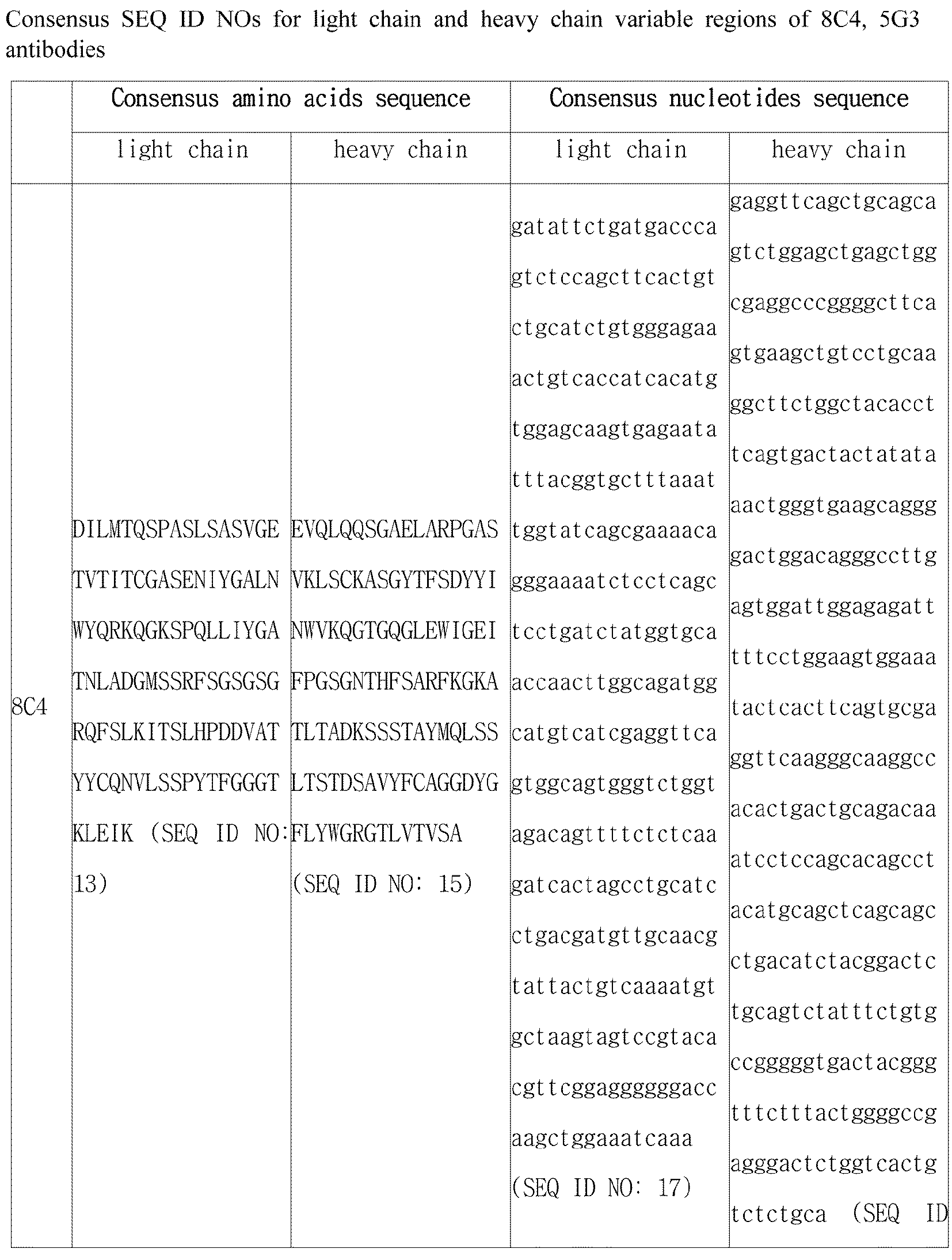


Claims (31)
- An antibody or an antigen binding fragment thereof that specifically binds to a hepatocyte growth factor receptor (c-Met).
- The antibody or the antigen binding fragment thereof according to claim 1, wherein the antibody binds to one or more epitope region represented by an amino acid sequence selected from the group consisting of SEQ ID NO: 331, SEQ ID NO: 332, SEQ ID NO: 333 and SEQ ID NO: 334.
- The antibody or the antigen binding fragment thereof according to claim 2, wherein the antibody or the antigen binding fragment thereof binds to a human c-Met by KD of 1 Х 10-7 M or less, wherein the KD is measured by surface plasmon resonance (Biacore) analysis.
- The antibody or the antigen binding fragment thereof according to claim 1, wherein the antibody is:(a) an antibody comprising a light chain variable region comprising a light chain CDR1 represented by SEQ ID NO: 1; a light chain CDR2 represented by SEQ ID NO: 2; a light chain CDR3 represented by SEQ ID NO: 3, and a heavy chain variable region comprising a heavy chain CDR1 represented by SEQ ID NO: 7; a heavy chain CDR2 represented by SEQ ID NO: 8; and a heavy chain CDR3 represented by SEQ ID NO: 9;(b) an antibody comprising a light chain variable region comprising a light chain CDR1 represented by SEQ ID NO: 4; a light chain CDR2 represented by SEQ ID NO: 5; a light chain CDR3 represented by SEQ ID NO: 6, and a heavy chain variable region comprising a heavy chain CDR1 represented by SEQ ID NO: 10; a heavy chain CDR2 represented by SEQ ID NO: 11; a heavy chain CDR3 represented by SEQ ID NO: 12; or(c) an affinity-optimized antibodies thereof.
- The antibody or the antigen binding fragment thereof according to claim 4, wherein the antibody comprises: (a) a light chain variable region represented by SEQ ID NO: 13 and a heavy chain variable region represented by SEQ ID NO: 15; or (b) a light chain variable region represented by SEQ ID NO: 14 and a heavy chain variable region represented by SEQ ID NO: 16.
- The antibody or the antigen binding fragment thereof according to claim 4, wherein the antibody comprises:(a) a light chain variable region represented by SEQ ID NO: 21 and a heavy chain variable region represented by SEQ ID NO: 23;(b) a light chain variable region represented by SEQ ID NO: 22 and a heavy chain variable region represented by SEQ ID NO: 24;(c) a light chain variable region represented by SEQ ID NO: 29 and a heavy chain variable region represented by SEQ ID NO: 31; or(d) a light chain variable region represented by SEQ ID NO: 30 and a heavy chain variable region represented by SEQ ID NO: 32.
- The antibody or the antigen binding fragment thereof according to claim 4, wherein the antibody comprises a hinge region represented by any one of SEQ ID NO: 37 to SEQ ID NO: 44.
- The antibody or the antigen binding fragment thereof according to claim 4, wherein the affinity-optimized antibody is an antibody, in which at least one amino acid sequence is substituted from an antibody, comprising:a light chain variable region comprising a light chain CDR1 represented by SEQ ID No: 1; a light chain CDR2 represented by SEQ ID No: 2; a light chain CDR3 represented by SEQ ID No: 3, and a heavy chain variable region comprising a heavy chain CDR1 represented by SEQ ID No: 7; a heavy chain CDR2 represented by SEQ ID No: 8; and a heavy chain CDR3 represented by SEQ ID No: 9; and wherein(i) G in a 1st position of the light chain CDR1 is substituted with A, E, K, L, N, R, S, V or W; A in a 2nd position thereof is substituted with C, G, I, P, S, T or V; S in a 3rd position thereof is substituted with G, M, N, P, Q, R, S or T; E in a 4th position thereof is substituted with A, D, F, G, H, K, M, Q, R, S, T or V; N in a 5th position thereof is substituted with A, D, E, G, K, L, P, Q, R, S, T or V; I in a 6th position thereof is substituted with A, F, L, M, Q, R, S, T or V; Y in a 7th position thereof is substituted with F, H, R or V; or G in a 8th position thereof is substituted with D, F, H, M, N, R, S, T or V;(ii) G in a 1st position of the light chain CDR2 is substituted with D, F, H, K, P, Q, S, V or Y; T in a 3rd position thereof is substituted with Q; or N in a 4th position thereof is substituted with G;(iii) Q in a 1st position of the light chain CDR3 is substituted with E, G, I, M or N; N in a 2nd position thereof is substituted with A, D, E, H, L, Q, S or T; V in a 3rd position thereof is substituted with I, L, M, N, Q, S or T; L in a 4th position thereof is substituted with F, H, I, M, R, S, V, W or Y; S in a 5th position thereof is substituted with C, D, E, F, G, H, K, L, N, Q, R, T, V or Y; S in a 6th position thereof is substituted with D, E, F, G, H, I, L, M, N, P, Q, R, T, V or Y; P in a 7th position thereof is substituted with A, D, E, G, N, Q, S or V; Y in an 8th position thereof is substituted with E, F, L, M or Q; or T in a 9th position thereof is substituted with D, F, G, I, L, N, S, V, W or Y;(iv) D in a 1st position of the heavy chain CDR1 is substituted with G or Q; Y in a 2nd position thereof is substituted with Q; or I in a 4th position thereof is substituted with A or Q;(v) F in a 3rd position of the heavy chain CDR2 is substituted with D, E, W or Y; G in a 5th position thereof is substituted with D, H or Y; S in a 6th position thereof is substituted with F, P, W or Y; G in a 7th position thereof is substituted with A, F, L, N or T; N in an 8th position thereof is substituted with F, P, S, T or Y; T in a 9th position thereof is substituted with A, D, E, F, G, H, L, P, S or V; H in a 10th position thereof is substituted with A, D, F, M, R, S, T, V, W or Y; F in an 11th position thereof is substituted with G, H, I, L, M, N, P, Q, V or Y; S in a 12th position thereof is substituted with A, D, G, H, I, L, P, T or V; A in a 13th position thereof is substituted with D, E, F, G, H, I, K, L, M, P, R, S, T, V or Y; R in a 14th position thereof is substituted with A, E, G, H, L, N, P, Q, S, W or Y; F in a 15th position thereof is substituted with D, E, G, L, M, P, R, S, V or W; K in a 16th position thereof is substituted with A, E, F, G, H, L, R, S, T, V or Y; or G in a 17th position thereof is substituted with E, F, H, L, M, N, P, Q, R, S, T, V or W; or(vi) G in a 1st position of the heavy chain CDR3 is substituted with E, F, H, N, Q, V or W; D in a 2nd position thereof is substituted with E; Y in a 3rd position thereof is substituted with L, Q, T or V; G in a 4th position thereof is substituted with W; F in a 5th position thereof is substituted with L or Y; L in a 6th position thereof is substituted with Q, S or Y; or Y in a 7th position thereof is substituted with C, L, M, N or Q,wherein the light chain CDR1 comprises 0 to 5 substitutions, the light chain CDR2 comprises 0 to 1 substitution, the light chain CDR3 comprises 0 to 7 substitutions, the heavy chain CDR1 comprises 0 to 1 substitution, the heavy chain CDR2 comprises 0 to 11 substitutions, and the heavy chain CDR3 comprises 0 to 6 substitutions.
- The antibody or the antigen binding fragment thereof according to claim 4, wherein the affinity-optimized antibody comprises:a light chain variable region comprising a light chain CDR1 represented by any one of SEQ ID NO: 1 and SEQ ID NO: 229 to SEQ ID NO: 268; a light chain CDR2 represented by any one of SEQ ID NO: 2, SEQ ID NO: 182 to SEQ ID NO: 190, SEQ ID NO: 227 and SEQ ID NO: 228; a light chain CDR3 represented by any one of SEQ ID NO: 3, SEQ ID NO: 142 to SEQ ID NO: 181, SEQ ID NO: 191 to SEQ ID NO: 226 and SEQ ID NO: 269 to SEQ ID NO: 301; anda heavy chain variable region comprising a heavy chain CDR1 represented by any one of SEQ ID NO: 7 and SEQ ID NO: 108 to SEQ ID NO: 112; a heavy chain CDR2 represented by any one of SEQ ID NO: 8, SEQ ID NO: 54 to SEQ ID NO: 63, SEQ ID NO: 72 to SEQ ID NO: 107 and SEQ ID NO: 118 to SEQ ID NO: 141; a heavy chain CDR3 represented by any one of SEQ ID NO: 9, SEQ ID NO: 64 to SEQ ID NO: 71 and SEQ ID NO: 113 to SEQ ID NO: 117.
- The antibody or the antigen binding fragment thereof according to claim 9, wherein the affinity-optimized antibody comprises a light chain variable region represented by any one of SEQ ID NO: 21 and SEQ ID NO: 306 to SEQ ID NO: 311, and a heavy chain variable region represented by any one of SEQ ID NO: 23 and SEQ ID NO: 302 to SEQ ID NO: 305.
- The antibody or the antigen binding fragment thereof according to claim 10, wherein the affinity-optimized antibody comprises:(a) a light chain variable region represented by SEQ ID NO: 21 and a heavy chain variable region represented by SEQ ID NO: 302;(b) a light chain variable region represented by SEQ ID NO: 21 and a heavy chain variable region represented by SEQ ID NO: 305;(c) a light chain variable region represented by SEQ ID NO: 310 and a heavy chain variable region represented by SEQ ID NO: 23;(d) a light chain variable region represented by SEQ ID NO: 308 and a heavy chain variable region represented by SEQ ID NO: 305;(e) a light chain variable region represented by SEQ ID NO: 306 and a heavy chain variable region represented by SEQ ID NO: 303;(f) a light chain variable region represented by SEQ ID NO: 307 and a heavy chain variable region represented by SEQ ID NO: 304;(g) a light chain variable region represented by SEQ ID NO: 308 and a heavy chain variable region represented by SEQ ID NO: 304;(h) a light chain variable region represented by SEQ ID NO: 309 and a heavy chain variable region represented by SEQ ID NO: 304;(i) a light chain variable region represented by SEQ ID NO: 311 and a heavy chain variable region represented by SEQ ID NO: 304; or(j) a light chain variable region represented by SEQ ID NO: 306 and a heavy chain variable region represented by SEQ ID NO: 302.
- The antibody or the antigen binding fragment thereof according to claim 4, wherein the antibody further specifically binds to an epidermal growth factor receptor (EGFR).
- The antibody or the antigen binding fragment thereof according to claim 12, wherein the antibody is that an antibody or an antigen binding fragment thereof binding to EGFR is linked to one light chain or heavy chain terminus of c-Met specific antibody.
- The antibody or the antigen binding fragment thereof according to claim 12, wherein the antigen binding fragment binding to the EGFR is Fab, Fab', F(ab')2 or Fv.
- The antibody or the antigen binding fragment thereof according to claim 12, wherein the antigen binding fragment binding to the EGFR is Fab, Fab', F(ab')2 or Fv.
- The antibody or the antigen binding fragment thereof according to claim 15, wherein the Erbitux scFv comprises an amino acid sequence represented by SEQ ID NO: 313 or SEQ ID NO: 314.
- The antibody or the antigen binding fragment thereof according to claim 15, wherein the Vectibix scFv comprises an amino acid sequence represented by SEQ ID NO: 315.
- The antibody or the antigen binding fragment thereof according to claim 13, wherein the antibody or the antigen binding fragment thereof is linked by a connector represented by SEQ ID NO: 312.
- The antibody or the antigen binding fragment thereof according to claim 4, wherein the antigen binding fragment is Fab, Fab', F(ab')2 or Fv.
- A nucleic acid molecule encoding the antibody or the antigen binding fragment thereof of any one of claims 1 to 19.
- An expression vector comprising the nucleic acid molecule of claim 20.
- A host cell having the expression vector introduced therein of claim 21.
- A method for producing an antibody or an antigen binding fragment thereof, using the host cell of claim 22.
- A composition for detecting c-Met, comprising the antibody or the antigen binding fragment thereof of any one of claims 1 to 19.
- A kit for detecting c-Met, comprising the composition for detecting c-Met of claim 24.
- A method for detecting a c-Met antigen using the antibody or the antigen binding fragment thereof of any one of claims 1 to 19.
- A composition for preventing or treating cancer, comprising the antibody or the antigen binding fragment of any one of claims 1 to 19.
- The composition for preventing or treating cancer according to claims 27, wherein the antibody or the antigen binding fragment thereof binds to c-Met to inhibit a receptor activity.
- The composition for preventing or treating cancer according to claim 28, wherein the antibody or the antigen binding fragment thereof further binds to EGFR to inhibit the receptor activity.
- The composition for preventing or treating cancer according to claim 27, wherein the cancer is caused by c-Met overexpression, amplification, mutation or activation.
- The composition for preventing or treating cancer according to claim 27, wherein the cancer is selected from the group consisting of lung cancer, stomach cancer, colon cancer, rectal cancer, triple negative breast cancer (TNBC), glioblastoma, pancreatic cancer, head and neck cancer, breast cancer, ovarian cancer, liver cancer, renal cancer, bladder cancer, prostate cancer, brain cancer, uterine cancer, solenoma, thyroid cancer, acute myeloid leukemia, chronic myeloid leukemia, myeloma, multiple myeloma, melanoma, lymphoma and adrenal cortex cancer.
Priority Applications (12)
| Application Number | Priority Date | Filing Date | Title |
|---|---|---|---|
| AU2018278730A AU2018278730B2 (en) | 2017-05-30 | 2018-05-30 | A novel anti-c-Met antibody and use thereof |
| MX2019014316A MX2019014316A (en) | 2017-05-30 | 2018-05-30 | A novel anti-c-met antibody and use thereof. |
| US16/616,345 US11479612B2 (en) | 2017-05-30 | 2018-05-30 | Anti-c-Met antibody and use thereof |
| CN201880036099.7A CN110770254B (en) | 2017-05-30 | 2018-05-30 | Novel anti-c-MET antibodies and uses thereof |
| RU2019143101A RU2751720C2 (en) | 2017-05-30 | 2018-05-30 | New anti-c-met-antibody and application thereof |
| JP2019566586A JP7325339B2 (en) | 2017-05-30 | 2018-05-30 | Novel anti-c-Met antibody and use thereof |
| CA3061704A CA3061704C (en) | 2017-05-30 | 2018-05-30 | Anti-c-met antibody and use thereof |
| NZ758605A NZ758605A (en) | 2017-05-30 | 2018-05-30 | A novel anti-c-met antibody and use thereof |
| MYPI2019006353A MY192630A (en) | 2017-05-30 | 2018-05-30 | A novel anti-c-met antibody and use thereof |
| EP18809820.6A EP3630844A4 (en) | 2017-05-30 | 2018-05-30 | A novel anti-c-met antibody and use thereof |
| BR112019025070-0A BR112019025070A2 (en) | 2017-05-30 | 2018-05-30 | innovative anti-c-met antibody and its use |
| PH12019550233A PH12019550233A1 (en) | 2017-05-30 | 2019-11-12 | A novel anti-c-met antibody and use thereof |
Applications Claiming Priority (4)
| Application Number | Priority Date | Filing Date | Title |
|---|---|---|---|
| KR10-2017-0067106 | 2017-05-30 | ||
| KR20170067106 | 2017-05-30 | ||
| KR1020180061888A KR102078292B1 (en) | 2017-05-30 | 2018-05-30 | A novel anti c-Met antibody and use thereof |
| KR10-2018-0061888 | 2018-05-30 |
Publications (1)
| Publication Number | Publication Date |
|---|---|
| WO2018221969A1 true WO2018221969A1 (en) | 2018-12-06 |
Family
ID=64455558
Family Applications (1)
| Application Number | Title | Priority Date | Filing Date |
|---|---|---|---|
| PCT/KR2018/006182 WO2018221969A1 (en) | 2017-05-30 | 2018-05-30 | A novel anti-c-met antibody and use thereof |
Country Status (1)
| Country | Link |
|---|---|
| WO (1) | WO2018221969A1 (en) |
Cited By (3)
| Publication number | Priority date | Publication date | Assignee | Title |
|---|---|---|---|---|
| CN113195536A (en) * | 2018-12-07 | 2021-07-30 | 首尔大学校产学协力团 | anti-c-Met agonist antibodies and uses thereof |
| US11142578B2 (en) | 2016-11-16 | 2021-10-12 | Regeneron Pharmaceuticals, Inc. | Anti-MET antibodies, bispecific antigen binding molecules that bind MET, and methods of use thereof |
| US11896682B2 (en) | 2019-09-16 | 2024-02-13 | Regeneron Pharmaceuticals, Inc. | Radiolabeled MET binding proteins for immuno-PET imaging and methods of use thereof |
Citations (4)
| Publication number | Priority date | Publication date | Assignee | Title |
|---|---|---|---|---|
| US20140141000A1 (en) * | 2012-11-21 | 2014-05-22 | Janssen Biotech, Inc. | Bispecific EGFR/C-Met Antibodies |
| US20150118238A1 (en) * | 2012-05-09 | 2015-04-30 | Eli Lilly And Company | Anti-c-Met Antibodies |
| KR20160061199A (en) * | 2014-11-21 | 2016-05-31 | 삼성전자주식회사 | Pharmaceutical composition for combination therapy containing dual inhibitor of c-Met and EGFR and IGF-1R inhibitor |
| US9631027B2 (en) * | 2010-11-03 | 2017-04-25 | Argen-X N.V. | Anti c-Met antibodies |
-
2018
- 2018-05-30 WO PCT/KR2018/006182 patent/WO2018221969A1/en active Application Filing
Patent Citations (4)
| Publication number | Priority date | Publication date | Assignee | Title |
|---|---|---|---|---|
| US9631027B2 (en) * | 2010-11-03 | 2017-04-25 | Argen-X N.V. | Anti c-Met antibodies |
| US20150118238A1 (en) * | 2012-05-09 | 2015-04-30 | Eli Lilly And Company | Anti-c-Met Antibodies |
| US20140141000A1 (en) * | 2012-11-21 | 2014-05-22 | Janssen Biotech, Inc. | Bispecific EGFR/C-Met Antibodies |
| KR20160061199A (en) * | 2014-11-21 | 2016-05-31 | 삼성전자주식회사 | Pharmaceutical composition for combination therapy containing dual inhibitor of c-Met and EGFR and IGF-1R inhibitor |
Non-Patent Citations (2)
| Title |
|---|
| MOORES, SHERI L. ET AL.: "A novel bispecific antibody targeting EGFR and cMet is effective against EGFR inhibitor-resistant lung tumors", CANCER RESEARCH, vol. 76, no. 13, 2016, pages 3942 - 3953, XP055431654 * |
| See also references of EP3630844A4 * |
Cited By (3)
| Publication number | Priority date | Publication date | Assignee | Title |
|---|---|---|---|---|
| US11142578B2 (en) | 2016-11-16 | 2021-10-12 | Regeneron Pharmaceuticals, Inc. | Anti-MET antibodies, bispecific antigen binding molecules that bind MET, and methods of use thereof |
| CN113195536A (en) * | 2018-12-07 | 2021-07-30 | 首尔大学校产学协力团 | anti-c-Met agonist antibodies and uses thereof |
| US11896682B2 (en) | 2019-09-16 | 2024-02-13 | Regeneron Pharmaceuticals, Inc. | Radiolabeled MET binding proteins for immuno-PET imaging and methods of use thereof |
Similar Documents
| Publication | Publication Date | Title |
|---|---|---|
| AU2018278730B2 (en) | A novel anti-c-Met antibody and use thereof | |
| WO2019098682A1 (en) | Anti-her2 antibody or antigen-binding fragment thereof, and chimeric antigen receptor comprising same | |
| WO2018194381A1 (en) | Binding molecule specific for lrig-1 protein and use thereof | |
| WO2018221969A1 (en) | A novel anti-c-met antibody and use thereof | |
| WO2014090053A1 (en) | Monoclonal antibody for antagonizing and inhibiting binding of vascular endothelial cell growth factor and its receptor, and coding sequence and use thereof | |
| WO2022039490A1 (en) | Anti-b7-h4/anti-4-1bb bispecific antibodies and use thereof | |
| WO2016137108A1 (en) | Novel antibody binding to tfpi and composition comprising the same | |
| WO2018174544A2 (en) | Antibody binding specifically to muc1 and use thereof | |
| WO2016200189A1 (en) | Rabies virus g protein epitope, and rabies virus neutralising binding molecule that binds specifically thereto | |
| WO2019132533A1 (en) | Anti-pd-l1 antibody and use thereof | |
| WO2022177394A1 (en) | Bispecific single domain antibody to pd-l1 and cd47 and use thereof | |
| WO2019078699A2 (en) | Anti-vista antibody and use thereof | |
| WO2018034434A1 (en) | Vaccine composition containing cyclic peptide, and antibody for cyclic peptide or anti-cancer composition containing same | |
| WO2018026248A1 (en) | Novel antibody against programmed cell death protein (pd-1), and use thereof | |
| WO2021020846A1 (en) | Anti-her2/anti-4-1bb bispecific antibody and use thereof | |
| WO2022149837A1 (en) | Anti-fgfr3 antibody and use thereof | |
| WO2022103245A1 (en) | Single domain antibody against sars-cov-2, and use thereof | |
| WO2018026249A1 (en) | Antibody against programmed death-ligand 1 (pd-l1), and use thereof | |
| WO2022085905A1 (en) | Antibody binding specifically to sars-cov-2 spike protein and use thereof | |
| WO2021020845A1 (en) | Anti-egfr/anti-4-1bb bispecific antibody and use thereof | |
| WO2021101346A1 (en) | Anti-ror1/anti-4-1bb bispecific antibodies and uses thereof | |
| WO2021006714A1 (en) | Composition for preventing, ameliorating or treating immune checkpoint inhibitor-resistant cancer | |
| WO2022240159A1 (en) | Anti-tigit antibodies and use thereof | |
| WO2020209645A1 (en) | Antibody to programmed cell death protein ligand-1 (pd-l1), and use thereof | |
| WO2022177392A1 (en) | Single domain antibody against cd47 and use thereof |
Legal Events
| Date | Code | Title | Description |
|---|---|---|---|
| 121 | Ep: the epo has been informed by wipo that ep was designated in this application |
Ref document number: 18809820 Country of ref document: EP Kind code of ref document: A1 |
|
| ENP | Entry into the national phase |
Ref document number: 3061704 Country of ref document: CA |
|
| ENP | Entry into the national phase |
Ref document number: 2018278730 Country of ref document: AU Date of ref document: 20180530 Kind code of ref document: A |
|
| ENP | Entry into the national phase |
Ref document number: 2019566586 Country of ref document: JP Kind code of ref document: A |
|
| NENP | Non-entry into the national phase |
Ref country code: DE |
|
| REG | Reference to national code |
Ref country code: BR Ref legal event code: B01A Ref document number: 112019025070 Country of ref document: BR |
|
| WWE | Wipo information: entry into national phase |
Ref document number: 2018809820 Country of ref document: EP |
|
| ENP | Entry into the national phase |
Ref document number: 2018809820 Country of ref document: EP Effective date: 20200102 |
|
| ENP | Entry into the national phase |
Ref document number: 112019025070 Country of ref document: BR Kind code of ref document: A2 Effective date: 20191127 |