US20100274270A1 - Guidewire support catheter - Google Patents
Guidewire support catheter Download PDFInfo
- Publication number
- US20100274270A1 US20100274270A1 US12/689,748 US68974810A US2010274270A1 US 20100274270 A1 US20100274270 A1 US 20100274270A1 US 68974810 A US68974810 A US 68974810A US 2010274270 A1 US2010274270 A1 US 2010274270A1
- Authority
- US
- United States
- Prior art keywords
- distal tip
- guidewire
- catheter
- wedge
- elongate
- Prior art date
- Legal status (The legal status is an assumption and is not a legal conclusion. Google has not performed a legal analysis and makes no representation as to the accuracy of the status listed.)
- Granted
Links
- 238000005520 cutting process Methods 0.000 claims abstract description 42
- 238000000034 method Methods 0.000 claims abstract description 32
- 230000001681 protective effect Effects 0.000 claims description 30
- 210000004204 blood vessel Anatomy 0.000 claims description 12
- 230000002441 reversible effect Effects 0.000 claims description 11
- 230000001684 chronic effect Effects 0.000 abstract description 5
- 210000001519 tissue Anatomy 0.000 description 20
- 238000005452 bending Methods 0.000 description 12
- 239000000463 material Substances 0.000 description 12
- FAPWRFPIFSIZLT-UHFFFAOYSA-M Sodium chloride Chemical compound [Na+].[Cl-] FAPWRFPIFSIZLT-UHFFFAOYSA-M 0.000 description 10
- 230000033001 locomotion Effects 0.000 description 10
- 239000011780 sodium chloride Substances 0.000 description 10
- 210000001367 artery Anatomy 0.000 description 9
- 206010053648 Vascular occlusion Diseases 0.000 description 6
- 210000002808 connective tissue Anatomy 0.000 description 6
- 239000010935 stainless steel Substances 0.000 description 6
- 229910001220 stainless steel Inorganic materials 0.000 description 6
- 230000002146 bilateral effect Effects 0.000 description 5
- 239000012530 fluid Substances 0.000 description 5
- 238000003780 insertion Methods 0.000 description 5
- 230000037431 insertion Effects 0.000 description 5
- 230000003902 lesion Effects 0.000 description 5
- 230000002093 peripheral effect Effects 0.000 description 4
- 210000005166 vasculature Anatomy 0.000 description 4
- 238000002399 angioplasty Methods 0.000 description 3
- 230000008901 benefit Effects 0.000 description 3
- 208000029078 coronary artery disease Diseases 0.000 description 3
- 238000002224 dissection Methods 0.000 description 3
- 238000001531 micro-dissection Methods 0.000 description 3
- 238000007789 sealing Methods 0.000 description 3
- 230000002792 vascular Effects 0.000 description 3
- 206010003210 Arteriosclerosis Diseases 0.000 description 2
- 208000037260 Atherosclerotic Plaque Diseases 0.000 description 2
- 208000018262 Peripheral vascular disease Diseases 0.000 description 2
- 238000005553 drilling Methods 0.000 description 2
- 238000002594 fluoroscopy Methods 0.000 description 2
- 238000011010 flushing procedure Methods 0.000 description 2
- 238000003384 imaging method Methods 0.000 description 2
- 230000007246 mechanism Effects 0.000 description 2
- 238000002324 minimally invasive surgery Methods 0.000 description 2
- 230000036961 partial effect Effects 0.000 description 2
- 208000030613 peripheral artery disease Diseases 0.000 description 2
- 229920000642 polymer Polymers 0.000 description 2
- 210000002435 tendon Anatomy 0.000 description 2
- 208000019553 vascular disease Diseases 0.000 description 2
- 208000004434 Calcinosis Diseases 0.000 description 1
- 241000282327 Felis silvestris Species 0.000 description 1
- 206010017711 Gangrene Diseases 0.000 description 1
- ATJFFYVFTNAWJD-UHFFFAOYSA-N Tin Chemical compound [Sn] ATJFFYVFTNAWJD-UHFFFAOYSA-N 0.000 description 1
- 239000003929 acidic solution Substances 0.000 description 1
- 230000009471 action Effects 0.000 description 1
- 230000004913 activation Effects 0.000 description 1
- 230000006978 adaptation Effects 0.000 description 1
- 238000002266 amputation Methods 0.000 description 1
- 230000003143 atherosclerotic effect Effects 0.000 description 1
- 230000015572 biosynthetic process Effects 0.000 description 1
- 239000008280 blood Substances 0.000 description 1
- 210000004369 blood Anatomy 0.000 description 1
- 230000017531 blood circulation Effects 0.000 description 1
- 230000000295 complement effect Effects 0.000 description 1
- 239000000994 contrast dye Substances 0.000 description 1
- 239000002872 contrast media Substances 0.000 description 1
- 230000001276 controlling effect Effects 0.000 description 1
- 230000008878 coupling Effects 0.000 description 1
- 238000010168 coupling process Methods 0.000 description 1
- 238000005859 coupling reaction Methods 0.000 description 1
- 235000021438 curry Nutrition 0.000 description 1
- 208000037265 diseases, disorders, signs and symptoms Diseases 0.000 description 1
- 208000035475 disorder Diseases 0.000 description 1
- 238000009826 distribution Methods 0.000 description 1
- 239000000975 dye Substances 0.000 description 1
- 229920001971 elastomer Polymers 0.000 description 1
- 239000000806 elastomer Substances 0.000 description 1
- 238000005516 engineering process Methods 0.000 description 1
- 210000003414 extremity Anatomy 0.000 description 1
- 210000003195 fascia Anatomy 0.000 description 1
- 239000002657 fibrous material Substances 0.000 description 1
- -1 for example Substances 0.000 description 1
- 210000005224 forefinger Anatomy 0.000 description 1
- 230000006870 function Effects 0.000 description 1
- 210000004283 incisor Anatomy 0.000 description 1
- 238000010348 incorporation Methods 0.000 description 1
- 239000007924 injection Substances 0.000 description 1
- 238000002347 injection Methods 0.000 description 1
- 230000000670 limiting effect Effects 0.000 description 1
- 229910052751 metal Inorganic materials 0.000 description 1
- 239000002184 metal Substances 0.000 description 1
- 229910001000 nickel titanium Inorganic materials 0.000 description 1
- HLXZNVUGXRDIFK-UHFFFAOYSA-N nickel titanium Chemical compound [Ti].[Ti].[Ti].[Ti].[Ti].[Ti].[Ti].[Ti].[Ti].[Ti].[Ti].[Ni].[Ni].[Ni].[Ni].[Ni].[Ni].[Ni].[Ni].[Ni].[Ni].[Ni].[Ni].[Ni].[Ni] HLXZNVUGXRDIFK-UHFFFAOYSA-N 0.000 description 1
- 230000037361 pathway Effects 0.000 description 1
- 229920003023 plastic Polymers 0.000 description 1
- 239000004033 plastic Substances 0.000 description 1
- 238000002360 preparation method Methods 0.000 description 1
- 230000009467 reduction Effects 0.000 description 1
- 230000001105 regulatory effect Effects 0.000 description 1
- 229910001285 shape-memory alloy Inorganic materials 0.000 description 1
- 239000007787 solid Substances 0.000 description 1
- 238000009987 spinning Methods 0.000 description 1
- 238000010561 standard procedure Methods 0.000 description 1
- 230000003068 static effect Effects 0.000 description 1
- 239000000126 substance Substances 0.000 description 1
- 238000006467 substitution reaction Methods 0.000 description 1
- 238000010408 sweeping Methods 0.000 description 1
- 230000001225 therapeutic effect Effects 0.000 description 1
- 210000003813 thumb Anatomy 0.000 description 1
- 238000012546 transfer Methods 0.000 description 1
- 210000004231 tunica media Anatomy 0.000 description 1
- 208000021331 vascular occlusion disease Diseases 0.000 description 1
- 238000003466 welding Methods 0.000 description 1
Images


Classifications
-
- A—HUMAN NECESSITIES
- A61—MEDICAL OR VETERINARY SCIENCE; HYGIENE
- A61B—DIAGNOSIS; SURGERY; IDENTIFICATION
- A61B5/00—Measuring for diagnostic purposes; Identification of persons
- A61B5/06—Devices, other than using radiation, for detecting or locating foreign bodies ; Determining position of diagnostic devices within or on the body of the patient
- A61B5/065—Determining position of the probe employing exclusively positioning means located on or in the probe, e.g. using position sensors arranged on the probe
- A61B5/066—Superposing sensor position on an image of the patient, e.g. obtained by ultrasound or x-ray imaging
-
- A—HUMAN NECESSITIES
- A61—MEDICAL OR VETERINARY SCIENCE; HYGIENE
- A61M—DEVICES FOR INTRODUCING MEDIA INTO, OR ONTO, THE BODY; DEVICES FOR TRANSDUCING BODY MEDIA OR FOR TAKING MEDIA FROM THE BODY; DEVICES FOR PRODUCING OR ENDING SLEEP OR STUPOR
- A61M25/00—Catheters; Hollow probes
- A61M25/01—Introducing, guiding, advancing, emplacing or holding catheters
-
- A—HUMAN NECESSITIES
- A61—MEDICAL OR VETERINARY SCIENCE; HYGIENE
- A61B—DIAGNOSIS; SURGERY; IDENTIFICATION
- A61B17/00—Surgical instruments, devices or methods
- A61B17/32—Surgical cutting instruments
- A61B17/3205—Excision instruments
- A61B17/3207—Atherectomy devices working by cutting or abrading; Similar devices specially adapted for non-vascular obstructions
- A61B17/320758—Atherectomy devices working by cutting or abrading; Similar devices specially adapted for non-vascular obstructions with a rotating cutting instrument, e.g. motor driven
-
- A—HUMAN NECESSITIES
- A61—MEDICAL OR VETERINARY SCIENCE; HYGIENE
- A61M—DEVICES FOR INTRODUCING MEDIA INTO, OR ONTO, THE BODY; DEVICES FOR TRANSDUCING BODY MEDIA OR FOR TAKING MEDIA FROM THE BODY; DEVICES FOR PRODUCING OR ENDING SLEEP OR STUPOR
- A61M25/00—Catheters; Hollow probes
- A61M25/01—Introducing, guiding, advancing, emplacing or holding catheters
- A61M25/0105—Steering means as part of the catheter or advancing means; Markers for positioning
- A61M25/0133—Tip steering devices
- A61M25/0138—Tip steering devices having flexible regions as a result of weakened outer material, e.g. slots, slits, cuts, joints or coils
-
- A—HUMAN NECESSITIES
- A61—MEDICAL OR VETERINARY SCIENCE; HYGIENE
- A61B—DIAGNOSIS; SURGERY; IDENTIFICATION
- A61B17/00—Surgical instruments, devices or methods
- A61B17/00234—Surgical instruments, devices or methods for minimally invasive surgery
- A61B2017/00292—Surgical instruments, devices or methods for minimally invasive surgery mounted on or guided by flexible, e.g. catheter-like, means
- A61B2017/003—Steerable
- A61B2017/00305—Constructional details of the flexible means
- A61B2017/00309—Cut-outs or slits
-
- A—HUMAN NECESSITIES
- A61—MEDICAL OR VETERINARY SCIENCE; HYGIENE
- A61B—DIAGNOSIS; SURGERY; IDENTIFICATION
- A61B17/00—Surgical instruments, devices or methods
- A61B17/22—Implements for squeezing-off ulcers or the like on inner organs of the body; Implements for scraping-out cavities of body organs, e.g. bones; for invasive removal or destruction of calculus using mechanical vibrations; for removing obstructions in blood vessels, not otherwise provided for
- A61B2017/22038—Implements for squeezing-off ulcers or the like on inner organs of the body; Implements for scraping-out cavities of body organs, e.g. bones; for invasive removal or destruction of calculus using mechanical vibrations; for removing obstructions in blood vessels, not otherwise provided for with a guide wire
-
- A—HUMAN NECESSITIES
- A61—MEDICAL OR VETERINARY SCIENCE; HYGIENE
- A61B—DIAGNOSIS; SURGERY; IDENTIFICATION
- A61B17/00—Surgical instruments, devices or methods
- A61B17/32—Surgical cutting instruments
- A61B17/3205—Excision instruments
- A61B17/3207—Atherectomy devices working by cutting or abrading; Similar devices specially adapted for non-vascular obstructions
- A61B17/320758—Atherectomy devices working by cutting or abrading; Similar devices specially adapted for non-vascular obstructions with a rotating cutting instrument, e.g. motor driven
- A61B2017/320775—Morcellators, impeller or propeller like means
-
- A—HUMAN NECESSITIES
- A61—MEDICAL OR VETERINARY SCIENCE; HYGIENE
- A61M—DEVICES FOR INTRODUCING MEDIA INTO, OR ONTO, THE BODY; DEVICES FOR TRANSDUCING BODY MEDIA OR FOR TAKING MEDIA FROM THE BODY; DEVICES FOR PRODUCING OR ENDING SLEEP OR STUPOR
- A61M25/00—Catheters; Hollow probes
- A61M25/01—Introducing, guiding, advancing, emplacing or holding catheters
- A61M25/0105—Steering means as part of the catheter or advancing means; Markers for positioning
- A61M25/0133—Tip steering devices
- A61M25/0147—Tip steering devices with movable mechanical means, e.g. pull wires
-
- A—HUMAN NECESSITIES
- A61—MEDICAL OR VETERINARY SCIENCE; HYGIENE
- A61M—DEVICES FOR INTRODUCING MEDIA INTO, OR ONTO, THE BODY; DEVICES FOR TRANSDUCING BODY MEDIA OR FOR TAKING MEDIA FROM THE BODY; DEVICES FOR PRODUCING OR ENDING SLEEP OR STUPOR
- A61M25/00—Catheters; Hollow probes
- A61M25/01—Introducing, guiding, advancing, emplacing or holding catheters
- A61M25/0105—Steering means as part of the catheter or advancing means; Markers for positioning
- A61M25/0133—Tip steering devices
- A61M25/0155—Tip steering devices with hydraulic or pneumatic means, e.g. balloons or inflatable compartments
-
- A—HUMAN NECESSITIES
- A61—MEDICAL OR VETERINARY SCIENCE; HYGIENE
- A61M—DEVICES FOR INTRODUCING MEDIA INTO, OR ONTO, THE BODY; DEVICES FOR TRANSDUCING BODY MEDIA OR FOR TAKING MEDIA FROM THE BODY; DEVICES FOR PRODUCING OR ENDING SLEEP OR STUPOR
- A61M25/00—Catheters; Hollow probes
- A61M25/01—Introducing, guiding, advancing, emplacing or holding catheters
- A61M25/0172—Exchanging a guidewire while keeping the catheter in place
Definitions
- Described herein are guidewire placement and support catheters that may be used to place a guidewire within an occluded lumen.
- guidewire placement and support catheters having a rotating, cutting and/or bending distal end region that may be used to position a guidewire through an occluded lumen of a vessel, including for treatment of chronic total occlusions.
- vascular diseases such as coronary artery disease and peripheral vascular disease
- CAD coronary artery disease
- PAD peripheral artery disease
- CTO Chronic Total Occlusion
- treatment of such blockages may include angioplasty or atherectomy procedures in which a guidewire, often via a catheter, is inserted into the diseased artery and threaded to the blocked region. There the blockage may be either squeezed into a more open position, for example, by pressure from an inflated catheter balloon (e.g., balloon angioplasty), and/or the blocked region may be held open by a stent. Alternatively a physician may use a catheter to surgically remove the plaque from the inside of the artery (e.g., an atherectomy).
- a guidewire often via a catheter
- the blocked region may be held open by a stent.
- a physician may use a catheter to surgically remove the plaque from the inside of the artery (e.g., an atherectomy).
- a guidewire must typically be forced across or through the occlusion so that the treatment catheter may be positioned to treat the occluding plaque.
- the guidewire must be positioned across the plaque because the active (e.g., plaque removing or displacing) portions of most currently available catheters are usually located on the sides of the catheters. For example, stents, balloons, atherectomy cutting tools, and other active portions are usually mounted on the sides of the catheter.
- An obstruction or plaque may be composed of relatively tough fibrous material, often including hard calcium deposits. Forcing a guidewire or catheter past such obstructions may cause the guidewire to exit the lumen of the vessel (e.g., artery), including causing it to enter the layers forming the artery, further damaging the tissue.
- Control of this device is mostly limited to extending/retracting the blades, and the cutting edges may damage the tissue, particularly if the blades contact the edges of the vessel; potentially exiting the lumen of the vessel.
- U.S. Pat. No. 6,152,938 describes a catheter drilling device for opening a wide variety of different blocked (occluded) tubes. The device anchors the tip of the drill head against a face of the occlusion, and partially rotates the drill head using a rein attached to the drill head so that the drill head faces at an angle. The blades of the drill are only controlled in rotation and may cut even non-target tissue, also leading the tip to exit and/or damage the vessel.
- 6,730,063 teaches a catheter device that can chemically treat calcified vascular occlusions by delivering acidic solutions and other fluids to calcified plaque to chemically dissolving the calcified material. As in the other known methods, this chemical treatment may be difficult to control, and in particular may be difficult to prevent damage to the vessel.
- Commercially available devices for treating occluded vessels include those marketed by Cordis Corporation, FlowCardia Technology, Kensey Nash Corporation, and other companies. These devices suffer from the same inadequacies as described above, and have also proven to have only limited success. The best reported success rates of overcoming CTOs with prior art devices range from 56% to 75%.
- the device may be guided by a guidewire, and may include a vacuum source to remove cut material.
- these devices are not adapted to self-center.
- these blades are typically side-cutting, meaning that the cutting typically occurs at the sides, where the rotating blade may interact with a (typically fixed) complementary surface, cutting the tissue between the surfaces.
- the distal rotating cutting head is smaller than the diameter of the rest of the elongate distal portion of the device.
- Described herein are guidewire positioning and support devices, method for using them, method of treating a subject using them, and systems including such guidewire positioning and support devices.
- These devices typically include an active distal tip region that is rotatable and may include one or more wedges that may be extended from the rotatable distal tip.
- the distal end of the device may be steerable, and may be steered while still rotating the distal end of the device.
- the wedges may be fully or partially retracted into distal housing. Both the distal housing and the wedges may rotate.
- the wedges (which may be sharp blades or may be blunt) can be continuously extended from the distal housing and locked in any position (extended, partially extended or retracted) and rotated clockwise and/or counterclockwise while locked in a retracted, extended or partially extended position.
- the distal region of the device may also be controllable steered from the proximal end of the device.
- the distal region of the device may be deflected.
- the bending distal region may be located immediately proximal to the rotatable distal tip.
- the device is typically elongate, and includes at least one lumen extending along the length (e.g., a central lumen) through which a guidewire may be passed.
- the lumen (or a separate lumen) may also be used to pass a material such as a contrast dye, saline, an imaging device, etc.
- the proximal end of the device typically includes a handle region that may be used to control the distal end.
- the device may include a rotation control, a wedge extension control and/or a steering control.
- controls may be combined into one or more controls or these functions may be distributed or divided between different controls. Any appropriate control may be used, including slides, knobs, dials, buttons, levers, switches, etc.
- the controls may be automated or computer-controlled.
- a driver e.g., motor, mechanical driver, etc.
- rotation of the distal tip region may be driven by a motor, and may be geared or otherwise controlled. The rotation may be manually or automatically controlled.
- the rotatable distal tip may a single integrated wedge having a protective surface that prevents cutting of tissue when rotated in a reverse direction, while allowing cutting of tissue when rotating in the reverse direction.
- the wedges at the distal end may be referred to as a blade or blades, even though they may be substantially blunt.
- the wedges are configured so that they are forward-cutting, but not side-cutting. This means that they may include a forward-facing cutting edge, and the more lateral edges may be blunted or less sharp.
- the rotating distal tip includes only a single wedge, rather than multiple wedges.
- the wedge (blade) may be helically arranged at the distal tip.
- the rotating distal tip including the wedge(s) had a diameter that is equal to (or greater than) the diameter of the rest of the elongate body (including the regions more proximal to the distal end of the device.
- the rotating distal end comprises three or more wedges that are radially separated around the tip region (e.g., spaced equally apart radially). It may be advantageous to have three or more wedges spaced around the tip, which may improve centering of the device, as described herein.
- the devices described herein are self-centering within the lumen of the vessel.
- the self-centering property of the device arises from the configuration of the distal tip as well as the rotational properties of the device.
- the wedges may be blunt, or laterally blunt (having only a forward-cutting face).
- both the wedges at the rotating distal portion and the housing into which the wedges may be extended or retracted may rotate.
- the diameter of the rotating distal end may be at least equal to the diameter of the more proximal regions of the elongate body of the device which may also allow self-centering within the lumen of a vessel.
- guidewire positioning and support catheter devices described herein may be referred to as “guidewire support catheters,” “guidewire positioning catheters,” “guidewire steering catheters,” “steerable rotating wedge” catheters, or the like.
- these guidewire positioning and support catheters may be referred to as merely “the devices” or “the catheter devices”.
- the guidewire positioning and support devices described herein may be configured as single-use, over-the-wire device, devices.
- the device may be compatible other guidewires of standard sizes used in minimally invasive procedures.
- the outer diameter of the elongate device (including the distal end region) may fit within a 7F sheath.
- the devices may be used with any appropriate guidewire, including steerable guidewires.
- the guidewire may be a 0.035′′ guidewire. These devices may generally be described as “steering” a guidewire, although they may be used with a guidewire within the catheter, or the guidewire may be positioned within the device after the catheter has been positioned.
- these devices provide support and guidance for positioning a guidewire across an occlusion.
- the devices may support probing and exchange of an assortment of guidewires, by traversing an occluding in a vessel.
- the devices described herein may be used to deliver contrast.
- the internal lumen which may be used by the guidewire may also be used for local dye injections without removing the device from the vessel.
- the steerable (deflecting) distal end and the rotating distal tip allow the device to be passed through an occlusion such as a plaque occluding a blood vessel (or artery) without requiring removal of the plaque.
- the device may be used to bluntly form a pathway through a plaque using the retractable/extendable rotating wedges at the distal tip, or simply using the rotating distal tip alone.
- the exposable wedge(s) at the distal tip may also allow for helical AND blunt microdissection.
- the wedges at the distal tip are sharp (e.g., cutting or knife-edged), while in other variations the wedges are substantially blunt.
- the wedges are typically curved around or along the longitudinal axis of the distal tip. For example, a wedge may extend helically around the distal tip end of the device (when extended).
- the extension of the wedge from the distal housing may be limited.
- the wedges may be prevented from extending fully out of the distal housing, thereby preventing material (such as a plaque or tissue) from getting caught between the wedges and the housing.
- the steerable distal end region may be steerable in any appropriate manner.
- the distal end region may be steerable by defecting in one direction (e.g., ‘down’) or in more than one direction (e.g., down/up, right/left, etc.).
- the degree of deflection may also be controlled.
- the tip may be deflected a maximum of 10 degrees, 15 degrees, 20 degrees, 25 degrees, 30 degrees, 45 degrees, 60 degrees, 90 degrees, or any angle there between.
- the distal end region of the device is pre-biased to be straight. Therefore the straight configuration of the distal tip may be restored once the deflection (controlled at the proximal end) is restored.
- the distal end of the device is flexible.
- the end region may include the tip (e.g., the rotating tip) or it may include the region immediately proximal to the tip.
- the “tip region” refers to the rotatable tip region at the distal end.
- the deflectable/steerable distal end region (which may be referred to as the “steerable tip”) may allow the catheter to re-enter the true lumen of the vessel if it becomes subintimal (e.g., if it extends into the fascia or region around the vessel).
- the device may include a distal end that has exposable bilateral wedges within a bullet-shaped housing, a catheter shaft, and a proximal handle that allows manipulation of the distal end of the device (e.g., rotation, steering, etc.) and may also provide a channel for flushing of the catheter lumen.
- both the distal tip and bilateral wedges are visible through fluoroscopy, and the catheter can be delivered over-the-wire through a 7F introducer sheath.
- the device also facilitates the delivery and exchange of guidewires and other therapeutic devices, such as stents, angioplasty balloons, or atherectomy catheters. It may also be used to deliver saline or contrast.
- the proximal handle may act as an interface with the operator, and allows a physician to expose or retract the wedges as needed.
- This part of the device may include four main components: a rotator, a pin, a slider, and a luer.
- the operator can use the slider and rotator to manipulate the pin, which in turn controls whether the wedges are exposed or retracted.
- the pin acts as a locking mechanism to control the deployment of the wedges.
- the luer allows for flushing of the catheter with saline prior to use and serves as a portal to deliver contrast or saline to a desired area.
- the catheter shaft provides pushability and torque to the bilateral wedges, and serves to center the device towards the middle of a CTO.
- the distal tip contains the bilateral wedges within a bullet-shaped housing ( FIG. 4 ).
- the housing with the wedges either retracted or exposed and rotating in a counter-clockwise direction can deliver blunt microdissection with more force than a guidewire.
- the spinning action creates helical microdissection planes to pull the catheter through the CTO lesion.
- any of the devices describe herein may include one or more of the following features, including: a steerable distal end region, a relatively short length, an elongate torque (drive) shaft) for rotating the distal tip, and reversible and retractable wedges having limited extension.
- the steerable distal end region is designed for and allows luminal re-entry.
- the may be adjustable up to a 45° angle and can help orient the device.
- the devices described herein may be any appropriate length, but relatively short length catheters may be particularly useful.
- the device may be approximately 100 cm, allowing the device to reach any occlusion down to the popliteal and ease physician handling outside the body.
- the device is between about 50 and 150 cm, between about 70 and 120 cm, etc.
- the distal tip may be driven by an elongate drive short (or “torque shaft”) that extends the length of the device.
- This drive shaft is typically robust, allowing increased torque precision and manipulation during the operation. Examples of this torque shaft are provided herein.
- the torque shaft may be made of a braided stainless steel material having a hollow lumen (forming the central lumen of the device). Rotation of the drive shaft typically drives the distal tip rotation.
- the drive shaft is typically flexible and connects to the rotational control at the proximal end (e.g., handle).
- the extendable/retractable wedges may include one or more features which act as safety features.
- the (typically helical) wedges may be configured to disengage more readily if needed during operation of the device.
- the distal tip, including the wedges and housing may rotate both clockwise and counterclockwise.
- the wedges may be extended or retracted independently of the rotation.
- the distal end may be rotated with the wedges locked in any desired extended or retracted position.
- the extension of the wedges may be limited to maximum extension that prevents the formation of a gap or space between the housing and the wedges between which material may be trapped.
- the wedges may be prevented from further extension by a lock or pin that prevents the wedges from fully extending out of the housing.
- the handle region at the proximal end of the device may also be adapted for handheld use.
- the controls at the handle region may be adapted so that the device may be manipulated by a single hand.
- the handle region may also include one or more indicators or displays for displaying the status of the distal end of the device.
- the handle may include an indicator indicating the extent to which the wedges are extended from the distal tip.
- the indicator may be a dial, slider, or the like.
- the indicator may be marked or unmarked.
- the wedge(s) may be extended to any degree (or to a maximum extension), or retracted completely.
- the handle, or the controller for the extension/retraction on the handle may include a lock locking the wedges at a desired extension.
- the same, or a different, lock may also be included on the handle for locking the configuration/position of the distal end region that is steerable or deflectable.
- a lock may be used to lock the distal end at a 45 degree angle.
- the proximal handle may be otherwise configured to be handheld.
- the controls may be clustered or located at one end of the handle with a gripper region at the other end.
- the controls may be clustered at the proximal portion of the handle (allowing the distal portion of the handle to be gripped in the palm of the hand, and manipulated by the thumb and forefinger of the right.
- the elongate outer sheath of the catheter is flexible, and may be coated so that it can be readily inserted into a lumen of a sheath, catheter, or directly into a body lumen.
- the elongate outer sheath of the catheter may be formed of a braided stainless steel, a polymer, or the like.
- the elongate outer sheath is typically tubular, and may be coated (either or both inner and outer diameter) with a protective cover.
- the elongate outer sheath may be referred to as a shaft (e.g., catheter shaft), and may be coated with a lubricious material or may be formed of a smooth and/or lubricious material.
- Described herein are devices, including at least one specific example of a device, including some examples in which dimensions are provided. It is to be understood that these dimensions may be varied while staying within the scope of the invention as generally described. Unless otherwise indicated, these dimensions are intended as merely illustrative, and not limiting.
- the forward cutting blades may prevent cutting on the sides/walls of the lumen.
- This configuration may also help with self-centering, as mentioned.
- the device may be configured so that the diameter of the blade (e.g., wedge) region is the same as the diameter of the rest of the catheter.
- the diameter of the distal end having the rotatable wedge is maxed out, so that the blades are as big across as the rest of the catheter, which may allow for maximum traction in the lumen of the vessel.
- the diameter of the blades (the distal region including the blade or blades) may therefore be equal to the diameter of the catheter. This in turn may allow the blades to engage better with the tissue of the vessel because there is more region of the vessel (e.g., width) available for the blade to engage with.
- the distal end region may be rotated manually.
- the tip may be spun or rotated by rotating a control or portion of the proximal handle.
- the device may be used as a screwdriver or drill element that is manually turning at a slow, more controlled (e.g., continuously and manually variable) rate. This may also provide tactile feedback to the operator, who can gauge how much force is applied by the feel of the device. For example, a clinician may determine how much force to apply both to advance and to spin the blades. Compared to true “drilling” elements and augering devices, the devices described herein may be worked into the media of the vessel more readily.
- Rotation of the tip may be clockwise or counterclockwise. In some variations the direction of rotation determines whether the wedge(s) at the distal tip are cutting or non-cutting. The user may alter the direction of cutting at any time during the procedure, and may manually or automatically control the rate and/or direction.
- the devices may advantageously be advanced into the vessel, and may self-centered. These variations may also prevent damaging the lumen of the vessel.
- the same control that allows extension (and/or rotation) of the distal end to extended/retract the wedges from the catheter/protective housing may be used to steer (e.g., bend) the distal end.
- the controls are simplified, allowing the device to be controlled more easily with a single activation scheme.
- these devices realize previously unexpected advantages because the entire distal end, including (in some variations) the housing into which the wedges/blades may retract, rotates.
- the rotation of the distal protective housing may permit blunt tip movement/dissection and allows powered blunt dissection.
- the device may exhibit a substantial mechanical advantage, apparently allowing the device to overcome the static friction at the tip and advance across an occlusion.
- the rotating housing also helps with self-centering and steering of the device.
- the ability to rotate the device in both the clockwise and counterclockwise (e.g., forward and backward) directions manually is also particularly helpful for blunt dissection.
- Turning the catheter tip (with blades retracted) in one direction (i.e., clockwise) allows the helical slots to engage the tissue more as the edges are rotated in the direction of the helix, rather than the opposite direction (i.e., counterclockwise) that moves it along the direction of the helix, thereby preventing or allowing less motion forward.
- the devices described herein can be locked in the open/closed position while still being rotatable.
- the devices can be locked with the wedges/blades partially extended, completely extended, and/or completely retracted.
- the handle includes a lock or locking control for securing the blades/wedges in a particular extension, while still allowing the entire distal end (including the blades/wedges and housing) to rotate.
- elongate guidewire support catheter devices for positioning a guidewire across an occluded portion of a vessel including: an elongate catheter body having an outer diameter; a rotatable distal tip configured to be rotated in a forward direction to cut tissue and a reverse direction for bluntly contacting tissue without cutting; a proximal handle region including a control for rotating the distal tip in either the forward or reverse directions; and a central lumen extending through the elongate guidewire support catheter configured to pass a guidewire.
- the outer diameter of the distal tip may be approximately the same as the outer diameter of the elongate catheter body.
- the device also includes a steerable distal end region configured to controllably deflect the rotatable distal tip.
- the rotatable tip may comprise a helical blade edge.
- the rotatable distal tip in some variations has a substantially smooth, curved outer surface that presents an atraumatic tissue-contacting surface when rotated in the reverse direction and that presents a sharp, tissue-cutting surface when rotated in the forward direction.
- Also described herein are methods of placing a guidewire across an occlusion of the lumen of a blood vessel including the steps of: advancing a catheter having an elongate catheter body and a rotatable distal tip towards the occlusion in the lumen of the blood vessel (wherein the distal tip comprises a forward-cutting wedge having atraumatic lateral edges and a protective housing); rotating and extending the wedge of the distal tip out of the protective housing while rotating both the wedge and the protective housing (wherein the wedge and protective housing have an outer diameter at least as large as the outer diameter of the elongate catheter body); advancing the distal tip across the occlusion; and passing a guidewire through a lumen of the catheter once it has crossed at least partially through the occlusion.
- the step of rotating the distal tip of the catheter may include rotating the distal tip both clockwise and counterclockwise.
- the method may also include steering the distal tip.
- the step of steering may include manipulating a control on the handle of the catheter to bend the distal end of the device.
- Also described herein are methods of placing a guidewire across an occlusion of the lumen of a blood vessel including: advancing a catheter having an elongate catheter body and a rotatable distal tip towards the occlusion in the lumen of the blood vessel; rotating the distal tip relative to the elongate body in a forward direction to cut tissue rotating the distal tip relative to the elongate body in a reverse direction to bluntly contacting tissue without cutting; advancing the distal tip across the occlusion; and passing a guidewire through a lumen of the catheter once it has crossed at least partially through the occlusion.
- the step of rotating the distal tip may comprise manually rotating the distal tip.
- the method may also include steering the distal tip.
- the step of steering may include manipulating a control on the handle of the catheter to bend the distal end of the device.
- FIG. 1 is a perspective view of one variation of a guidewire positioning and support catheter devices as described herein.
- FIGS. 2A-2D illustrate operation of one variation of a guidewire positioning and support catheter device.
- FIG. 3 illustrates steering of the distal end region of a guidewire positioning and support catheter device.
- FIG. 4A-4C illustrate sequential extension of the wedge portion(s) of a guidewire positioning and support catheter device and deflection of the distal end of the device.
- FIG. 5 is a side perspective view of another variation of a guidewire positioning and support catheter device.
- FIG. 6 is a side perspective view of the proximal handle region of the guidewire positioning and support catheter device shown in FIG. 5 .
- FIG. 7 is an exploded view of the proximal handle region shown in FIG. 6 .
- FIG. 8 is a side perspective view of the distal end region of one variation of a guidewire positioning and support catheter device.
- FIGS. 9A and 9B are exploded views of the distal end region shown in FIG. 8 .
- FIGS. 10A-10C show side perspective, side and end views, respectively, of the luer fitting at the proximal end region of one variation of a guidewire positioning and support catheter device.
- FIGS. 10D and 10E show alternative side perspective views of the luer fitting shown in FIG. 10A .
- FIGS. 11A-11D show side perspective, side, end and front views, respectively, of one variation of a slider component of a handle of a guidewire positioning and support catheter device.
- FIG. 11E shows another side perspective view.
- FIGS. 12A-12C show side perspective, end and side views, respectively, of a lock (shown as a rotator cap in this example) which may be used to lock the slider of FIG. 11A .
- a lock shown as a rotator cap in this example
- FIGS. 13A-13C show side perspective, end and side views, respectively, of a slider seal which may be used as part of a guidewire positioning and support catheter device.
- FIGS. 14A-14C show side perspective, end and side views, respectively, of a fluid sealing tube portion of a handle for a guidewire positioning and support catheter device.
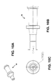
- FIGS. 15A-15C show side perspective, side and end views, respectively, of a set screw portion of a handle for a guidewire positioning and support catheter device.
- FIGS. 16A-16C show side perspective, side and end views, respectively, of a rotator portion of a handle for a guidewire positioning and support catheter device.
- FIG. 16D is another side perspective view of the rotator portion shown in FIG. 16A .
- FIGS. 17A-17D show side perspective, side, cross-sectional, and end views, respectively, of a handle body portion of a handle for a guidewire positioning and support catheter device.
- FIGS. 18A-18C show side perspective, side and end views, respectively, of a cap portion of a handle body for a handle of a guidewire positioning and support catheter device.
- FIGS. 19A-19C show side perspective, front and side views, respectively, of a sealing O-ring portion of one variations of a handle for a guidewire positioning and support catheter device.
- FIGS. 20A-20D show side perspective, front and side views, respectively, of an o-ring cap portion of a handle for a guidewire positioning and support catheter device.
- FIG. 21 is a side perspective view of an outer elongate shaft for a guidewire positioning and support catheter device.
- FIG. 22 is a side perspective view of a drive shaft (e.g., torque shaft) for a guidewire positioning and support catheter device.
- a drive shaft e.g., torque shaft
- FIG. 23A-23C show first and second side perspective views, and an end view, respectively, of a steerable distal end portion of a guidewire positioning and support catheter device as described in one variation herein.
- FIG. 23D shows the steerable (or flexible) distal end of FIGS. 23A-23C projected flat (e.g., “unrolled”) so that the cut pattern is apparent.
- FIGS. 24A-24C show side perspective, side and end views, respectively, of an attachment ring for a steerable distal end portion of a guidewire positioning and support catheter device as described in one variation herein.
- FIGS. 25A-25C show side perspective, side and end views, respectively, of a slip collar for a rotatable distal tip portion of a guidewire positioning and support catheter device as described in one variation herein.
- FIGS. 26A-26C show side perspective, side and end views, respectively, of a drive coupler element for a rotatable distal tip portion of a guidewire positioning and support catheter device as described in one variation herein.
- FIG. 26D shows an end perspective view of the drive coupler shown in FIGS. 26A-26C .
- FIGS. 27A-27C show side perspective, side and end views, respectively, of a distal housing for a rotatable distal tip portion of a guidewire positioning and support catheter device as described in one variation herein.
- FIG. 27D is another side perspective view of the distal housing shown in FIG. 27A .
- FIGS. 28A-28C show side perspective, side and end views, respectively, of a wedge (blade) element for a rotatable distal tip portion of a guidewire positioning and support catheter device as described in one variation herein.
- FIGS. 28D and 28E show alternative perspective views of the wedge element shown in FIG. 28A .
- FIG. 29 is a fluoroscopic image thorough a region of a subject's body having an occluded vessel.
- FIG. 30A-30C illustrate operation of a guidewire positioning and support catheter such as the one illustrated above to traverse the occluded vessel shown in FIG. 29 .
- FIG. 31A shows a cross-section through a region of a vessel including a guidewire positioning device as described herein.
- FIG. 31B is a partial longitudinal view of the same region of FIG. 31A .
- FIG. 32A shows an alternative view of a cross-section through a region of a vessel including a guidewire positioning device.
- FIG. 32B is a partial longitudinal view of the same region of FIG. 32A .
- FIG. 33 is another variation of a guidewire positioning and support device.
- FIG. 34 shows an enlarged view of the distal end of the device of FIG. 33 .
- FIG. 35 shows another view of the distal tip region of the device of FIG. 33 .
- FIG. 36 is an exploded view of the distal tip region of the device of FIG. 33 .
- FIGS. 37A and 37B show increasingly enlarged views of the handle region of the device of FIG. 33 .
- FIG. 38 is an exploded view of the handle shown for the device of FIG. 33 .
- FIG. 39A shows one variation of a guidewire positioning and support device having three wedges in the retracted position.
- FIG. 39B shows the same device in which the wedges are extended.
- FIG. 40A illustrates one variation of a guidewire positioning device having a rotatable distal tip with a plurality (two in this example) of extendable wedges retractable into a rotating housing.
- FIG. 40B shows another variation having an integrated rotatable distal tip with a pair of channeled flutes extending around from the distal end of the rotating tip that are configured to present a sharp cutting edge when the tip is rotated counter-clockwise, and to present a non-cutting (e.g., atraumatic) surface when the tip is rotated clockwise.
- FIGS. 41A-41D show the rotatable distal tip of FIG. 40B in various views.
- FIGS. 41A and 41D show side perspective views.
- FIG. 41B shows an end view.
- FIG. 41C shows a side view.
- FIG. 1 illustrates one variation of a guidewire positioning and support catheter having a rotatable distal end with extendable/retractable wedges.
- This device may be used to position (e.g., providing a channel for) a guidewire or for traversing an occlusion in a vessel, particularly for traversing complete occlusions.
- This device may be used to support steerable guidewires (and may be used to guide them) in accessing discrete regions of the peripheral vasculature. It may be used to facilitate placement and exchange of guidewires and other interventional devices. It may also be used to deliver saline or contrast.
- these devices include a rotatable distal tip.
- the distal tip may be rotated manually (by operation of a control on the proximal handle).
- both counterclockwise and clockwise rotation is possible.
- the devices may include one or more extendable wedges that may be extended from or retracted into a distal housing that is atraumatic (e.g., smooth, non-sharp).
- the wedges may be blades, or they may include one or more sharp regions, or they may be dull.
- the wedges may be extended continuously, so that the user can operate a control on the proximal handle to extend and/or retract the wedges.
- the wedge or wedges may extend laterally from the region along the length of the tip.
- the wedges may extend helically at least partially around the distal tip.
- a single wedge (which may be a single helical wedge) extends from the tip of the distal end. Multiple wedges may extend from the distal tip.
- two bilateral wedges may extend from the distal tip.
- the rotation of the wedges may be independent of the extension of the distal tip. For example, the distal tip may be rotated even with the wedges retracted or with the wedges all or completely extended.
- the devices described herein may also include a steerable distal end region.
- the steerable distal end region may be steerable in one or more directions.
- the steerable distal tip may be displaced in one direction (e.g., bent in one direction).
- the steerable distal end region is coordinated with the extension of the wedge or wedges from the distal tip.
- the steerable distal end region may be configured to bend in one or more direction using the same actuator that extends the wedge from the rotatable distal tip. This configuration may reduce or eliminate the need for an additional bending/steering mechanism (e.g., tendon wires or the like) to traverse the elongate body of the catheter.
- an additional bending/steering mechanism e.g., tendon wires or the like
- the steering of the distal end region may be independent of the extension of the wedge from the distal tip.
- one or more control wires e.g., tendons
- hydraulic or pressure lines may be use to control steering (bending) of the distal end region.
- FIG. 1 shows one variation of a guidewire support catheter.
- This example includes a 6F catheter (outer) sheath 103 and is a 0.035′′ guidewire compatible over-the-wire device.
- An exemplary guidewire 115 is shown inserted in the proximal end of the device handle 120 .
- a system may include a guidewire, although a guidewire does not have to be included as part of the device.
- the guidewire support catheters described herein may include a catheter shaft 103 with handle assembly 120 at the proximal end and an atraumatic distal tip 101 .
- a locking luer connecter 117 at the proximal end provides entry to a lumen that supports and facilitates movement of a guidewire 115 .
- the catheter may be sterilized.
- the blunt distal tip 101 houses one or more deployable spiral wedges (not visible in FIG. 1 ) and allows atraumatic advancement of the catheter through the vasculature.
- manipulation of a control e.g., slider 111
- Slider 111 controls the deployment and retraction of the wedges and deflection of the flexible distal tip 101 .
- Advancing the slider 111 in the distal direction deploys the wedges from the distal tip, and pulling the slider to its furthest proximal position retracts the wedges inside the distal tip 101 .
- a tip indicator 109 moves in conjunction with longitudinal movement of the slider and provides information about wedge deployment and the amount of deflection at the distal tip (described in more detail below for FIGS. 2A-2D ).
- the wedges are in the retracted position. If the tip indicator 109 resides in its mid-point location, the wedges are fully deployed. As the slider 111 is advanced distally from this mid-point and the tip indicator 109 moves towards its furthest proximal location, the distal tip 101 deflects at an increasing angle (see FIGS. 2C and 2D ). The flexing of the distal tip 101 is visible under fluoroscopic guidance.
- the handle assembly 120 typically consists of a handle body 105 , a rotator 107 , a tip indicator 109 , a slider 111 , a slider lock 113 , and a luer 117 .
- the handle body 120 controls rotational orientation of the catheter, including the flexible distal tip.
- the rotator 107 controls the rotational movement to the distal tip independent of the catheter orientation. Rotational movement of the distal tip 101 can be achieved either with wedges deployed or retracted.
- the slider 111 can move through the rotator in a longitudinal direction to control wedge deployment/retraction and the amount of tip deflection.
- the slider lock 113 restricts the slider movement.
- the slider lock 113 can be engaged by turning it in the clockwise direction until tight, and can be disengaged by counterclockwise rotation.
- the Luer is provided to facilitate fluid flush and the entry/exit of the guidewire. This port also acts as an entry port for the guidewire.
- FIGS. 2A-2D illustrate extension/retraction of the wedge (e.g., “blade”) elements from the distal tip of the device.
- moving the slider 111 portion of the handle distally (as indicated by the movement of the tip indicator 109 ), extends the wedges 49 from the retracted position shown in FIG. 2A , to a deployed condition, shown in FIG. 2B .
- the wedges are fully deployed. In this example, the wedges do not extend out of the protective housing at the distal tip 50 .
- Moving the slider further distally acts to deflect (steer or bend) the distal end region of the device.
- the distal end region includes a laser-cut tubular region (scaffold 46 ) that is pre-biased in a straight configuration, but may be bent or flexed to deflect at an angle from the straight configuration, as shown in FIGS. 2C and 2D .
- the inner shaft (a torque shaft or drive shaft) in this example is connected to the slider on the handle, and is slideable within the catheter relative to the distal housing 50 .
- the drive shaft (not visible in this figure) is connected to the wedges 49 at the distal tip, and sliding the slider causes the drive shaft to advance the distal tip until it is fully deployed, as shown in FIG. 2B . Further advancing of the slider, as illustrated in FIGS.
- the cuts in the tubular region allow the region 46 to flex or bend in a predictable region.
- One side of the tubular region 46 is not cut, and acts as a living hinge region that may bend as the spacing between the cuts of the cut region expands.
- these cuts may be spaced and angled to limit the ability of the tubular region 46 to bend beyond a certain point (e.g., to form an angle of less than some predetermined amount, such as 30 degrees, 40 degrees, 45 degrees, 50 degrees, 60 degrees, etc.).
- some predetermined amount such as 30 degrees, 40 degrees, 45 degrees, 50 degrees, 60 degrees, etc.
- the distal end region is shown with the wedges deployed at a minimal tip deflection (also indicated by the slider tip indicator 109 slightly proximal from the distal end of the indicator window).
- the slider is fully deployed distally and the distal end of the device is bent to the maximum tip deflection, with the wedges extended.
- the deflecting/bending tubular region 46 which may be referred to as the deflecting, bending or hinged region, is configured to bend by the application of a longitudinal pushing force that is applied from the drive shaft in this example. Because this deflecting region is ‘hinged’ on one side in this example, a symmetric (or even asymmetric) force applied across the region causes the region to bend against the hinge.
- the tubular region 46 in this example may be made of any appropriate material, for example, metal (stainless steel, shape memory alloys such as Nitinol, or the like), polymers (plastics, elastomers, etc.) or the like. It may be helpful if the region is configured to include a restoring force and restores itself to the original (straight) shape when the distal force is removed.
- FIGS. 3 and 4 A- 4 C illustrate steering of the distal end of a catheter as just discussed.
- the hinge in this example is simply a narrow band that is solid, while the rest of the circumference of the region includes a plurality of approximately transverse (i.e., transverse to the longitudinal length of the catheter) cuts.
- the cuts in this case include regions that double back on themselves to form a zig-zag pattern.
- This pattern shown in detail in FIGS. 23A-23D ) effectively limits the bending of the region since, for any given spacing between the cuts, the opposite walls of the cuts in the doubled-back region will contact each other, preventing further bending.
- the hinged region shown in FIGS. 3-4C is formed by laser-cut stainless steel, however other materials, and method of cutting them, may be used, as mentioned.
- FIG. 5 shows a side-perspective view of one example of a catheter 40 as described herein.
- the catheter includes a distal end region 101 having a flexible or deflection region 46 and a rotatable tip region consisting of a protective housing 49 and an extendable/retractable wedged region 50 .
- the distal end region 101 is connected to the proximal handle region 120 by an elongate catheter 45 .
- This catheter includes an outer flexible catheter body 45 , and one or more inner members, such as an inner drive member (not visible in FIG. 5 ) that can drive rotation of the distal tip.
- This catheter also includes a central passageway (not visible) that opens at the proximal and distal ends.
- the handle region 210 includes a handle body 41 that is connected to a handle body cap 44 which interfaces with a rotator portion 56 .
- the rotator 56 partially covers a slider 54 .
- the rotator portion 56 may be rotated; this rotation is translated (e.g., via a drive shaft) into rotation of the distal tip.
- the slider may move distally or proximally within the handle 120 .
- a window in the slider shows a position indicator (set screw 55 ) that indicates the longitudinal position of the slider, and therefore indicates the extension/retraction state of the wedges at the distal tip. Sliding the slider distally extends the wedges from the housing and may also deflect or steer the device, as just described.
- the proximal end of the device includes a luer fitting region which may be used to connect to the central lumen of the device (e.g., for applying contrast, saline, etc.).
- the luer region may include a threaded or lockable region.
- the luer region may also include a flanged or funnel-shaped opening to help assist in threading a guidewire into the catheter.
- FIG. 6 shows a close-up perspective view of just the proximal handle region 120 of the device shown in FIG. 5 .
- FIG. 7 is an exploded view of the handle region 120 of FIGS. 5 and 6 , indicating all of the component parts, including internal elements that may be included.
- the device may include an inner drive catheter 52 connected to both the rotator 56 and the slider 54 .
- the inner drive catheter is a braided stainless steel catheter that is connected to a more rigid (e.g., stainless steel) tube 53 at the proximal end.
- the rigid tube 53 may help provide a fluid seal against one or more sealing elements, such as O-rings 42 and slider seals 58 . These seals may help isolate the components of the device.
- fluid e.g., blood, etc.
- the inner lumen may be continuous with the body lumen, but can be regulated by attachment of saline, plasma, or other material to the proximal end of the device, to help regulate the pressure differential between the lumen and the environment outside of the patient.
- Multiple seals may be used between the inner and outer catheters, including redundant seals.
- a locking member 57 configured as a screw-lock, may be included on at least the slider portion of the device.
- This lock may be used (e.g., locking by screwing distally/unscrewing proximally to unlock) to secure the slider in a desired position.
- the position of the wedges (extended/retracted position) and/or the deflection of the distal end may be locked in a desired position.
- Other locks e.g., preventing or allowing rotation
- FIG. 8 is an enlarged view of the distal end of the device shown in FIG. 5 .
- the distal end includes a rotatable tip region including a protective housing 49 and extendable/retractable wedge portion 50 .
- a steering or steerable bending region 46 is also shown, as previously described.
- the flexible bending region 46 connects to the outer catheter 45 .
- FIGS. 9A and 9B show exploded views of the distal end region of the catheter shown in FIGS. 5 and 8 .
- the inner drive shaft catheter 52 is shown within the outer catheter 45 . Both the inner 52 and outer 45 catheters are flexible, so that the entire elongate length of the device is flexible.
- the inner drive (or torque) shaft catheter 52 connects to a rotatable mount, drive coupler 51 , that connects to the wedge element 50 .
- the rotatable mount is also engaged with the protective housing 49 , and both of these elements are rotatably connected to the slip collar 48 .
- rotation of the drive shaft catheter 52 rotates the wedge element and the protective housing at the distal tip, causing the entire distal tip region to rotate.
- the drive shaft 52 may be extended and/or retracted to extend or retract the wedge element, as previously described, and apply longitudinal force to bend the flexible region 46 .
- the flexible region 46 is rigidly connected (e.g., welded) to an attachment ring 47 that connects to the slip ring 48 to allow a rotatable coupling between the distal tip region (e.g., protective housing 49 ) and the rest of the catheter.
- the proximal end of the flexible region 46 is connected (e.g., by welding) to the outer catheter 45 .
- FIGS. 7 and 9 A- 9 B are illustrated in greater detail in FIGS. 10A-28E in various views.
- Many of these figures include exemplary dimensions and tolerances. These dimensions are intended only as examples, and other dimensions may be used, or ranges of dimensions (e.g., +/ ⁇ 5% of the value shown, +/ ⁇ 10% of the value shown, etc.). For simplicity, the numbering of each component is maintained throughout the figures.
- any of the devices described herein may be used to treat an occlusion.
- these devices may be used to position a guidewire across an occlusion such as a chronic total occlusion of the peripheral vascular system.
- treatment may include insertion of the catheter device into a vessel (e.g., blood vessel or artery) that is occluded. Fluoroscopy or other imaging technique may be used to visualize both the device and the vessel. Thus, appropriate contrast material may be used, and may be applied through the device or prior to insertion of the device.
- the device may be inserted using known minimally invasive (e.g., percutaneous) techniques.
- the device may be inserted with the wedge element retracted, so that the distal tip region is essentially atraumatic, to prevent damage to tissue or miss-guidance of the device.
- the device may be positioned against the occlusion, and the distal tip region may be rotated with or without the wedge element deployed.
- the deploying the wedge element, in combination with the (manual) rotation may allow the device to penetrate the occlusion.
- the device may thus be advanced by rotation of the distal tip, either with or without the wedge element extended.
- the catheter may be gently driven (e.g., by the medical professional) distally to advance the device.
- the device may be used prior to insertion of a guidewire through the device, or it may be used in conjunction with a guidewire.
- a guidewire may be extended from the distal end as it is advanced to also help penetrate the occlusion or steer the device.
- the distal region of the device may also be steered by controlling the bending of the flexible distal region, as illustrated and described above. If the device should penetrate or exit the lumen of the vessel, it may be guided back into the vessel (re-entry) using the steerable distal end.
- a catheter in operation, may be removed from a sterile package and transfer to a sterile field where it can be first flushed with heparanized saline through the proximal port located on the handle assembly. The catheter may then be lightly wiped with heparanized saline in preparation for insertion. Before use, the catheter may be tested to confirm that the wedges extend and retract and the distal end region of the device bends for steering.
- the rotator may be turned clockwise and counterclockwise to rotate the distal tip region.
- the slider may be longitudinally slide distally to extend the wedges from the tip region (from out of the housing) and further distally to bend the steerable distal end region.
- the slider may then be slid proximally to ‘unbend’ (restore to straight) the distal end, and to retract the wedge region.
- the slider lock may be rotated clockwise to engage (lock) the slider in position (and therefore the extension of the wedge(s) and the bend of the distal end region, and the slider lock may be rotated counterclockwise to disengage and allow longitudinal slider movement.
- the device may be inserted into the vessel by first retracting the wedges into the housing at the distal tip.
- a guidewire may be inserted up to (or adjacent to) the occlusion using standard techniques.
- the catheter may then be advanced over the guidewire under fluoroscopic guidance through a sheath to the target lesion (occlusion).
- the guidewire support catheter is used with a guidewire within the central lumen, which may provide additional stiffness and support to the catheter.
- the slider lock When the device is positioned, manually torque and advance the catheter through a region of the peripheral vasculature while maintaining catheter position at the lesion site. If additional support is necessary to advance beyond the region, deploy the wedges by pushing on the slider the slider lock may be engaged if desired.
- the rotator may be manipulated as needed to advance the catheter. To ensure the distal tip of the device remains in the vessel's true lumen, the Slider may be pushed until the tip is positioned properly and engage the slider lock.
- the wedges may be fully retracted under fluoroscopic guidance, and the catheter may be retracted, leaving the guidewire in place.
- FIG. 29 shows a fluoroscopic image thorough a region of a subject's body having an occluded vessel.
- the occluded vessel extends from the top to the bottom of the figure.
- a dark column extending from the top of the figure shows the un-occluded portion of the vessel filled with contrast.
- FIGS. 30A-30C illustrate the use of the devices as described herein to traverse this occluded region carrying a guidewire that may be left in position (spanning the occlusion) after the catheter is removed.
- the catheter does not remove any substantial amount of material from the occlusion, but the rotation of the wedges at the distal tip allows it to advance around and through the occlusion within the vessel, forming a passage for the guidewire.
- the catheter may be removed, leaving the guidewire in position.
- the guidewire may then be used in conjunction with other devices, including stents or expanding balloon catheters that may be used to expand or remove the plaque or otherwise enlarge the vessel.
- a catheter holding a balloon and/or stent may be delivered over the guidewire after it has been positioned.
- FIGS. 31A and 31B illustrate the insertion of a device into a blood vessel.
- the vessel includes the media (e.g., tunica media) surrounded by adventitia (e.g., tunica adventitia).
- adventitia e.g., tunica adventitia
- the devices described herein may be self-centering within the “true” lumen of the vessel, rather than a “false” lumen region between the adventitia and media, as illustrated in FIGS. 32A and 32B .
- any of the catheters described herein may be used to treat peripheral vascular disease.
- the manually rotatable distal end (including the wedges) of the catheter may have been found to guide the catheter in the true lumen, especially when the reaching the distal cap of a lesion. Centering (self-centering) of the catheter appears to result from the rotation of the blades, coupled with the dimension of the device, including the diameter of the wedges and distal tip (which are typically the same as the diameter of the rest of the catheter). This rotating distal tip helps the device to stay in true lumen of the blood vessel.
- the blades are configured to engage and dissect their way through tissue like the atheroma.
- the rotating blades may create a sweeping path that is not only self-centering, but also is much bigger than the vessel wall (media). If a blade does engage with the media, it returns in the true lumen because the configuration of the device may help keep it within that space.
- the wedges may include a forward-cutting face (e.g., a blade or sharp edge that faces distally, away from the lateral sides, and therefore away from the walls of the lumen). The lateral portions of the wedge may be atraumatic, so that they don't cut or tear the lumen tissue.
- An atraumatic wedge portion may be rounded or otherwise dulled and configured so as not to cut the lumen.
- more than one wedge e.g., two or more wedges
- the other blade continues making its way through the atheroma.
- the catheter device may reside between the media on one side and the adventitia on the other.
- the elastic nature of Adventitia typically prevents the wedge (and particularly atraumatic surfaces of the wedge) from engaging the tissue.
- the rotating wedge(s) may selectively dissect their way through the media and not through adventitia. Once the device dissects its way through the medial, it ends up back in the true lumen.
- FIG. 33 is another variation of a guidewire positioning and support catheter having a rotatable distal end with one or more extendable/retractable wedges.
- This device may also be used to position (e.g., providing a channel for) a guidewire or for traversing an occlusion in a vessel, particularly for traversing complete occlusions.
- This device may be used to support steerable guidewires (and may be used to guide them) in accessing discrete regions of the peripheral vasculature. It may be used to facilitate placement and exchange of guidewires and other interventional devices. It may also be used to deliver saline or contrast.
- the variation shown in FIGS. 33-38 does not include a steerable distal end, as illustrated above.
- the catheter includes an elongate catheter body having an outer diameter, and a rotatable distal tip.
- the rotatable distal tip includes a rotatable, extendable, wedge, and a rotatable protective housing.
- the extendable wedge is configured to be extended from the protective housing.
- both the extendable wedge and protective housing have an outer diameter at least as wide as the diameter of the catheter (e.g., the distal half of the catheter).
- the wedge is a forward-cutting wedge having atraumatic lateral edges.
- the wedge region of the distal tip shown in FIGS. 33-38 are substantially similar to the wedge regions shown above (e.g., FIGS. 28A-28E ).
- FIG. 34 shows an enlarged view of the distal end of the catheter of FIG. 33 .
- FIG. 35 shows an alternative view in which the more proximal portion of the catheter has been removed.
- FIG. 36 shows an exploded view of the distal end of the device of FIGS. 33-35 .
- the rotatable wedge portion 50 ′ fits within the housing 49 ′, and both the housing and the wedge portion are rotatable, as already described.
- FIGS. 37A and 37B illustrate progressively enlarged views of the proximal (handle) region of the device.
- advancing the slider 111 ′ in the distal direction will extend the wedge(s) from the protective housing.
- the device may be configured so that the wedge portion cannot be fully extended from the housing.
- the device may also include an indicator.
- FIG. 38 shows an exploded view of the catheter handle.
- any of the variations described herein may include one, two, three, or more wedges in the distal tip. It may be advantageous for the device to include three or more wedges that extend from the distal end. Having three or more wedges positioned around the distal end of the device may permit more equal distribution of the forces around the center of the tip. In addition, having three or more wedge may enhance the centering capability of the device. Devices having two wedges may engage the target tissue with only a single wedge at a time (e.g., in a lumen that is larger in diameter then the diameter of the device), which may cause the device to tend to spin around the engaged wedge or blade rather than the center of the device (e.g., the true center point of the catheter).
- FIGS. 39A and 39B illustrate one variation of a device having three symmetrically positioned wedges shown in the retracted ( FIG. 39A ) and extended ( FIG. 39B ) positions.
- FIGS. 40B to 41D illustrate another variation of a guidewire placement device.
- This variation may include many of the features described above, but includes an integrated (or “unified”) rotatable distal tip.
- FIG. 40A is another illustration of the two-part rotatable distal tip including both an extendable/retractable rotatable wedge portion and a rotatable distal housing.
- the variations shown in FIG. 40B includes a rotatable distal tip in which the wedge and housing are integrated into a rotation-direction sensitive embodiment.
- the distal tip is similar tin configuration to the distal housing, however the one or more spiral slots from which the wedges may extend are replaced with spiral flutes that act as spiral wedge members.
- the outer surface is smooth and flat when traveling in the counter-clockwise direction (e.g., see FIGS. 41A-41D ), while traversing the tip in the clockwise direction encounters multiple sharp edges.
- the tissue is passively engaged by the rotating atraumatic (non-cutting) surface.
- Rotation of the tip in the opposite (counterclockwise) direction presents multiple cutting edges.
- the variation shown in FIGS. 40B-41D may be operated similar to those described above, however the wedges do not need to be extended/retracted as described. Instead, the cutting/non-cutting surfaces (corresponding to the extended/retracted configuration of other variations) may by operated depending on the direction of rotation of the distal tip. As mentioned above, the distal tip may be manually rotated, allowing both control of the speed and/or direction of rotation (e.g., by twisting a control to rotate the distal tip, etc.).
Landscapes
- Health & Medical Sciences (AREA)
- Life Sciences & Earth Sciences (AREA)
- Engineering & Computer Science (AREA)
- Veterinary Medicine (AREA)
- Biomedical Technology (AREA)
- Heart & Thoracic Surgery (AREA)
- Animal Behavior & Ethology (AREA)
- General Health & Medical Sciences (AREA)
- Public Health (AREA)
- Surgery (AREA)
- Biophysics (AREA)
- Pulmonology (AREA)
- Anesthesiology (AREA)
- Hematology (AREA)
- Nuclear Medicine, Radiotherapy & Molecular Imaging (AREA)
- Medical Informatics (AREA)
- Molecular Biology (AREA)
- Vascular Medicine (AREA)
- Gynecology & Obstetrics (AREA)
- Radiology & Medical Imaging (AREA)
- Human Computer Interaction (AREA)
- Physics & Mathematics (AREA)
- Pathology (AREA)
- Media Introduction/Drainage Providing Device (AREA)
- Surgical Instruments (AREA)
Abstract
Description
- This patent application claims priority to U.S. Provisional Patent Applications: 61/173,542 (filed Apr. 28, 2009), titled “Guidewire Support Catheter;” and 61/233,093 (filed Aug. 11, 2009), also titled “Guidewire Support Catheter.” The disclosures of these applications are incorporated herein by reference in their entirety.
- This application may be related to U.S. patent application Ser. No. 12/108,433, titled “Catheter System and Method for Boring through Blocked Vascular Passages,” filed Apr. 23, 2008, and U.S. patent application Ser. No. 12/272,697, titled “Dual-tip Catheter System for Boring through Blocked Vascular Passages,” and filed Nov. 17, 2008. Both of these applications are herein incorporated by reference in their entirety.
- All publications and patent applications mentioned in this specification are herein incorporated by reference in their entirety to the same extent as if each individual publication or patent application was specifically and individually indicated to be incorporated by reference.
- Described herein are guidewire placement and support catheters that may be used to place a guidewire within an occluded lumen. In particular, described herein are guidewire placement and support catheters having a rotating, cutting and/or bending distal end region that may be used to position a guidewire through an occluded lumen of a vessel, including for treatment of chronic total occlusions.
- Guidewire placement is a critical step in many medical procedures, particularly in minimally invasive procedures and treatment of vascular diseases. For example, a number of vascular diseases, such as coronary artery disease and peripheral vascular disease, are caused by the build-up of fatty atherosclerotic deposits (plaque) in the arteries, which limit blood flow to the tissues that are supplied by that particular artery. Disorders caused by occluded body vessels, including coronary artery disease (CAD) and peripheral artery disease (PAD) may be debilitating and life-threatening. Chronic Total Occlusion (CTO) can result in limb gangrene, requiring amputation, and may lead to other complications and eventually death. Increasingly, treatment of such blockages may include angioplasty or atherectomy procedures in which a guidewire, often via a catheter, is inserted into the diseased artery and threaded to the blocked region. There the blockage may be either squeezed into a more open position, for example, by pressure from an inflated catheter balloon (e.g., balloon angioplasty), and/or the blocked region may be held open by a stent. Alternatively a physician may use a catheter to surgically remove the plaque from the inside of the artery (e.g., an atherectomy).
- Thus, placement of a guidewire completely or partially across the occlusion is often a critical, though difficult step. Devices and method for guidewire placement are expensive, complex, and all too often ineffective. A guidewire must typically be forced across or through the occlusion so that the treatment catheter may be positioned to treat the occluding plaque. The guidewire must be positioned across the plaque because the active (e.g., plaque removing or displacing) portions of most currently available catheters are usually located on the sides of the catheters. For example, stents, balloons, atherectomy cutting tools, and other active portions are usually mounted on the sides of the catheter.
- However, when the artery is totally blocked by plaque, it is extremely difficult, and potentially dangerous to force the guidewire through the occlusion. An obstruction or plaque may be composed of relatively tough fibrous material, often including hard calcium deposits. Forcing a guidewire or catheter past such obstructions may cause the guidewire to exit the lumen of the vessel (e.g., artery), including causing it to enter the layers forming the artery, further damaging the tissue.
- Currently available devices and methods for crossing occlusions are inadequate. For example, patents such as U.S. Pat. No. 5,556,405 to Lary, U.S. Pat. No. 6,152,938 to Curry, and U.S. Pat. No. 6,730,063 to Delaney et. al. describe devices and methods for passing an occlusion that do not adequately allow placement of a guidewire across a plaque. U.S. Pat. No. 5,556,405 teaches an incisor catheter having blades that can protrude from the catheter to push outward and form linear cuts in the plaque. Control of this device is mostly limited to extending/retracting the blades, and the cutting edges may damage the tissue, particularly if the blades contact the edges of the vessel; potentially exiting the lumen of the vessel. U.S. Pat. No. 6,152,938 describes a catheter drilling device for opening a wide variety of different blocked (occluded) tubes. The device anchors the tip of the drill head against a face of the occlusion, and partially rotates the drill head using a rein attached to the drill head so that the drill head faces at an angle. The blades of the drill are only controlled in rotation and may cut even non-target tissue, also leading the tip to exit and/or damage the vessel. As one alternative, U.S. Pat. No. 6,730,063 teaches a catheter device that can chemically treat calcified vascular occlusions by delivering acidic solutions and other fluids to calcified plaque to chemically dissolving the calcified material. As in the other known methods, this chemical treatment may be difficult to control, and in particular may be difficult to prevent damage to the vessel. Commercially available devices for treating occluded vessels include those marketed by Cordis Corporation, FlowCardia Technology, Kensey Nash Corporation, and other companies. These devices suffer from the same inadequacies as described above, and have also proven to have only limited success. The best reported success rates of overcoming CTOs with prior art devices range from 56% to 75%.
- In addition, many of the devices and methods for crossing an occlusion described above may still require manually forcing the guidewire through the occlusion with only minimal (or no) assistance from the devices. Finally, many of these devices may inadvertently exit the vessel, but few, if any of them, are readily re-positionable to reenter the vessel.
- A large family of patents and pending application including U.S. Pat. No. 6,001,112, U.S. Pat. No. 6,454,779, U.S. Pat. No. 6,206,989, U.S. Pat. No. 7,479,147, U.S. Pat. No. 6,451,036, U.S. Pat. No. 6,482,217, U.S. Pat. No. 7,172,610, U.S. Pat. No. 7,235,088 and U.S. Pat. No. 6,666,874 (“Taylor patents”) describe a rotatable cutter that is configured for atherectomy cutting. These devices typically include an inner rotating member (that may extend from a fixed housing at the distal end of the device). The device may be guided by a guidewire, and may include a vacuum source to remove cut material. However, these devices are not adapted to self-center. In addition, these blades are typically side-cutting, meaning that the cutting typically occurs at the sides, where the rotating blade may interact with a (typically fixed) complementary surface, cutting the tissue between the surfaces. Further, the distal rotating cutting head is smaller than the diameter of the rest of the elongate distal portion of the device.
- Thus, there remains a need for guidewire positioning devices that can effectively traverse occluded vessels, and particularly chronically occluded vessels. Such devices would enable positioning of a guidewire and therefore enable positioning of stents and other devices to be more successfully used in high occlusion situations, leading to improved patient outcomes and a reduction in patient morbidity and mortality.
- Described herein are guidewire positioning and support devices, method for using them, method of treating a subject using them, and systems including such guidewire positioning and support devices.
- These devices typically include an active distal tip region that is rotatable and may include one or more wedges that may be extended from the rotatable distal tip. In some variations the distal end of the device may be steerable, and may be steered while still rotating the distal end of the device. The wedges may be fully or partially retracted into distal housing. Both the distal housing and the wedges may rotate. The wedges (which may be sharp blades or may be blunt) can be continuously extended from the distal housing and locked in any position (extended, partially extended or retracted) and rotated clockwise and/or counterclockwise while locked in a retracted, extended or partially extended position. The distal region of the device may also be controllable steered from the proximal end of the device. For example, the distal region of the device may be deflected. The bending distal region may be located immediately proximal to the rotatable distal tip. The device is typically elongate, and includes at least one lumen extending along the length (e.g., a central lumen) through which a guidewire may be passed. The lumen (or a separate lumen) may also be used to pass a material such as a contrast dye, saline, an imaging device, etc. The proximal end of the device typically includes a handle region that may be used to control the distal end. For example, the device may include a rotation control, a wedge extension control and/or a steering control. In some variations these controls may be combined into one or more controls or these functions may be distributed or divided between different controls. Any appropriate control may be used, including slides, knobs, dials, buttons, levers, switches, etc. In some variations the controls may be automated or computer-controlled. In some variations a driver (e.g., motor, mechanical driver, etc.) may be used to drive the controls. For example, rotation of the distal tip region may be driven by a motor, and may be geared or otherwise controlled. The rotation may be manually or automatically controlled.
- In some variations, described in greater detail below, rather than an extendable wedge (or wedges) and a housing, the rotatable distal tip may a single integrated wedge having a protective surface that prevents cutting of tissue when rotated in a reverse direction, while allowing cutting of tissue when rotating in the reverse direction.
- The wedges at the distal end may be referred to as a blade or blades, even though they may be substantially blunt. In some variations the wedges are configured so that they are forward-cutting, but not side-cutting. This means that they may include a forward-facing cutting edge, and the more lateral edges may be blunted or less sharp. In some variations the rotating distal tip includes only a single wedge, rather than multiple wedges. The wedge (blade) may be helically arranged at the distal tip. In many variations (including those illustrated below), the rotating distal tip including the wedge(s) had a diameter that is equal to (or greater than) the diameter of the rest of the elongate body (including the regions more proximal to the distal end of the device.
- In some variations, the rotating distal end comprises three or more wedges that are radially separated around the tip region (e.g., spaced equally apart radially). It may be advantageous to have three or more wedges spaced around the tip, which may improve centering of the device, as described herein.
- In operation, many of the devices described herein are self-centering within the lumen of the vessel. The self-centering property of the device arises from the configuration of the distal tip as well as the rotational properties of the device. In particular, as mentioned, the wedges may be blunt, or laterally blunt (having only a forward-cutting face). Further, both the wedges at the rotating distal portion and the housing into which the wedges may be extended or retracted may rotate. Finally, the diameter of the rotating distal end (including the wedges) may be at least equal to the diameter of the more proximal regions of the elongate body of the device which may also allow self-centering within the lumen of a vessel.
- In general, the guidewire positioning and support catheter devices described herein may be referred to as “guidewire support catheters,” “guidewire positioning catheters,” “guidewire steering catheters,” “steerable rotating wedge” catheters, or the like. For simplicity, these guidewire positioning and support catheters may be referred to as merely “the devices” or “the catheter devices”.
- The guidewire positioning and support devices described herein may be configured as single-use, over-the-wire device, devices. For example, the device may be compatible other guidewires of standard sizes used in minimally invasive procedures. The outer diameter of the elongate device (including the distal end region) may fit within a 7F sheath. The devices may be used with any appropriate guidewire, including steerable guidewires. For example, the guidewire may be a 0.035″ guidewire. These devices may generally be described as “steering” a guidewire, although they may be used with a guidewire within the catheter, or the guidewire may be positioned within the device after the catheter has been positioned.
- In general, these devices provide support and guidance for positioning a guidewire across an occlusion. As described herein, the devices may support probing and exchange of an assortment of guidewires, by traversing an occluding in a vessel. In addition, the devices described herein may be used to deliver contrast. The internal lumen which may be used by the guidewire may also be used for local dye injections without removing the device from the vessel.
- As described in greater detail below, in operation the steerable (deflecting) distal end and the rotating distal tip allow the device to be passed through an occlusion such as a plaque occluding a blood vessel (or artery) without requiring removal of the plaque. Thus, the device may be used to bluntly form a pathway through a plaque using the retractable/extendable rotating wedges at the distal tip, or simply using the rotating distal tip alone. The exposable wedge(s) at the distal tip may also allow for helical AND blunt microdissection. In some variations, the wedges at the distal tip are sharp (e.g., cutting or knife-edged), while in other variations the wedges are substantially blunt. The wedges are typically curved around or along the longitudinal axis of the distal tip. For example, a wedge may extend helically around the distal tip end of the device (when extended).
- In variations in which the rotatable distal tip includes one or more separate wedge and housing, the extension of the wedge from the distal housing may be limited. For example, the wedges may be prevented from extending fully out of the distal housing, thereby preventing material (such as a plaque or tissue) from getting caught between the wedges and the housing.
- The steerable distal end region may be steerable in any appropriate manner. In particular, the distal end region may be steerable by defecting in one direction (e.g., ‘down’) or in more than one direction (e.g., down/up, right/left, etc.). The degree of deflection may also be controlled. In some variations, the tip may be deflected a maximum of 10 degrees, 15 degrees, 20 degrees, 25 degrees, 30 degrees, 45 degrees, 60 degrees, 90 degrees, or any angle there between. In some variations the distal end region of the device is pre-biased to be straight. Therefore the straight configuration of the distal tip may be restored once the deflection (controlled at the proximal end) is restored. In addition to be deflectable, in some variations the distal end of the device is flexible. The end region may include the tip (e.g., the rotating tip) or it may include the region immediately proximal to the tip. In general the “tip region” refers to the rotatable tip region at the distal end.
- Deflection or steering of the distal end may help with re-entry of the device. For example, the deflectable/steerable distal end region (which may be referred to as the “steerable tip”) may allow the catheter to re-enter the true lumen of the vessel if it becomes subintimal (e.g., if it extends into the fascia or region around the vessel).
- In some variations, the device may include a distal end that has exposable bilateral wedges within a bullet-shaped housing, a catheter shaft, and a proximal handle that allows manipulation of the distal end of the device (e.g., rotation, steering, etc.) and may also provide a channel for flushing of the catheter lumen. In this variation (and other variations) both the distal tip and bilateral wedges are visible through fluoroscopy, and the catheter can be delivered over-the-wire through a 7F introducer sheath. The device also facilitates the delivery and exchange of guidewires and other therapeutic devices, such as stents, angioplasty balloons, or atherectomy catheters. It may also be used to deliver saline or contrast.
- The proximal handle may act as an interface with the operator, and allows a physician to expose or retract the wedges as needed. This part of the device may include four main components: a rotator, a pin, a slider, and a luer. The operator can use the slider and rotator to manipulate the pin, which in turn controls whether the wedges are exposed or retracted. The pin acts as a locking mechanism to control the deployment of the wedges. At the end of the handle, the luer allows for flushing of the catheter with saline prior to use and serves as a portal to deliver contrast or saline to a desired area. The catheter shaft provides pushability and torque to the bilateral wedges, and serves to center the device towards the middle of a CTO. The distal tip contains the bilateral wedges within a bullet-shaped housing (
FIG. 4 ). The housing with the wedges either retracted or exposed and rotating in a counter-clockwise direction can deliver blunt microdissection with more force than a guidewire. With the wedges deployed, and rotating in a clockwise motion, the spinning action creates helical microdissection planes to pull the catheter through the CTO lesion. - Any of the devices describe herein may include one or more of the following features, including: a steerable distal end region, a relatively short length, an elongate torque (drive) shaft) for rotating the distal tip, and reversible and retractable wedges having limited extension. The steerable distal end region is designed for and allows luminal re-entry. For example, the may be adjustable up to a 45° angle and can help orient the device.
- The devices described herein may be any appropriate length, but relatively short length catheters may be particularly useful. For example, the device may be approximately 100 cm, allowing the device to reach any occlusion down to the popliteal and ease physician handling outside the body. In other variations, the device is between about 50 and 150 cm, between about 70 and 120 cm, etc.
- The distal tip may be driven by an elongate drive short (or “torque shaft”) that extends the length of the device. This drive shaft is typically robust, allowing increased torque precision and manipulation during the operation. Examples of this torque shaft are provided herein. For example, the torque shaft may be made of a braided stainless steel material having a hollow lumen (forming the central lumen of the device). Rotation of the drive shaft typically drives the distal tip rotation. The drive shaft is typically flexible and connects to the rotational control at the proximal end (e.g., handle).
- The extendable/retractable wedges (including the wedge element as well as the housing into which the wedges may be retracted) may include one or more features which act as safety features. For example, the (typically helical) wedges may be configured to disengage more readily if needed during operation of the device. The distal tip, including the wedges and housing, may rotate both clockwise and counterclockwise. In addition the wedges may be extended or retracted independently of the rotation. Thus, the distal end may be rotated with the wedges locked in any desired extended or retracted position. In addition the extension of the wedges may be limited to maximum extension that prevents the formation of a gap or space between the housing and the wedges between which material may be trapped. For example, the wedges may be prevented from further extension by a lock or pin that prevents the wedges from fully extending out of the housing.
- The handle region at the proximal end of the device may also be adapted for handheld use. In particular, the controls at the handle region may be adapted so that the device may be manipulated by a single hand. In some variations the handle region may also include one or more indicators or displays for displaying the status of the distal end of the device. For example, the handle may include an indicator indicating the extent to which the wedges are extended from the distal tip. The indicator may be a dial, slider, or the like. The indicator may be marked or unmarked.
- As mentioned, in some variations the wedge(s) may be extended to any degree (or to a maximum extension), or retracted completely. The handle, or the controller for the extension/retraction on the handle, may include a lock locking the wedges at a desired extension. The same, or a different, lock may also be included on the handle for locking the configuration/position of the distal end region that is steerable or deflectable. For example, a lock may be used to lock the distal end at a 45 degree angle.
- The proximal handle may be otherwise configured to be handheld. For example, the controls may be clustered or located at one end of the handle with a gripper region at the other end. For example, the controls may be clustered at the proximal portion of the handle (allowing the distal portion of the handle to be gripped in the palm of the hand, and manipulated by the thumb and forefinger of the right.
- In general, the elongate outer sheath of the catheter is flexible, and may be coated so that it can be readily inserted into a lumen of a sheath, catheter, or directly into a body lumen. For example, the elongate outer sheath of the catheter may be formed of a braided stainless steel, a polymer, or the like. The elongate outer sheath is typically tubular, and may be coated (either or both inner and outer diameter) with a protective cover. The elongate outer sheath may be referred to as a shaft (e.g., catheter shaft), and may be coated with a lubricious material or may be formed of a smooth and/or lubricious material.
- Described herein are devices, including at least one specific example of a device, including some examples in which dimensions are provided. It is to be understood that these dimensions may be varied while staying within the scope of the invention as generally described. Unless otherwise indicated, these dimensions are intended as merely illustrative, and not limiting.
- As mentioned briefly above, various aspects of the devices described herein provide substantially benefits compared to other device which may be used in occluded vessels, including rotating devices. For example, the forward cutting blades may prevent cutting on the sides/walls of the lumen. This configuration may also help with self-centering, as mentioned. In addition, the device may be configured so that the diameter of the blade (e.g., wedge) region is the same as the diameter of the rest of the catheter. Thus, the diameter of the distal end having the rotatable wedge is maxed out, so that the blades are as big across as the rest of the catheter, which may allow for maximum traction in the lumen of the vessel. The diameter of the blades (the distal region including the blade or blades) may therefore be equal to the diameter of the catheter. This in turn may allow the blades to engage better with the tissue of the vessel because there is more region of the vessel (e.g., width) available for the blade to engage with.
- As mentioned, the distal end region may be rotated manually. For example, the tip may be spun or rotated by rotating a control or portion of the proximal handle. Thus, the device may be used as a screwdriver or drill element that is manually turning at a slow, more controlled (e.g., continuously and manually variable) rate. This may also provide tactile feedback to the operator, who can gauge how much force is applied by the feel of the device. For example, a clinician may determine how much force to apply both to advance and to spin the blades. Compared to true “drilling” elements and augering devices, the devices described herein may be worked into the media of the vessel more readily. Rotation of the tip may be clockwise or counterclockwise. In some variations the direction of rotation determines whether the wedge(s) at the distal tip are cutting or non-cutting. The user may alter the direction of cutting at any time during the procedure, and may manually or automatically control the rate and/or direction.
- In variations having blades (wedges) that are substantially blunt on the longitudinal side (e.g. the side facing the wall of the vessel), the devices may advantageously be advanced into the vessel, and may self-centered. These variations may also prevent damaging the lumen of the vessel.
- In variations of the devices having a steerable distal end, the same control that allows extension (and/or rotation) of the distal end to extended/retract the wedges from the catheter/protective housing may be used to steer (e.g., bend) the distal end. By combining the steering with blade extension, the controls are simplified, allowing the device to be controlled more easily with a single activation scheme.
- In addition, these devices realize previously unexpected advantages because the entire distal end, including (in some variations) the housing into which the wedges/blades may retract, rotates. The rotation of the distal protective housing may permit blunt tip movement/dissection and allows powered blunt dissection. Compared to simply advancing a blunt (non-rotating tip) through the vessel, rotating the blunt or blunted distal end (e.g., when the blades are retracted into the housing), the device may exhibit a substantial mechanical advantage, apparently allowing the device to overcome the static friction at the tip and advance across an occlusion. As mentioned above, the rotating housing also helps with self-centering and steering of the device. The ability to rotate the device in both the clockwise and counterclockwise (e.g., forward and backward) directions manually is also particularly helpful for blunt dissection. Turning the catheter tip (with blades retracted) in one direction (i.e., clockwise) allows the helical slots to engage the tissue more as the edges are rotated in the direction of the helix, rather than the opposite direction (i.e., counterclockwise) that moves it along the direction of the helix, thereby preventing or allowing less motion forward.
- Finally, the devices described herein can be locked in the open/closed position while still being rotatable. For example, the devices can be locked with the wedges/blades partially extended, completely extended, and/or completely retracted. In many variations the handle includes a lock or locking control for securing the blades/wedges in a particular extension, while still allowing the entire distal end (including the blades/wedges and housing) to rotate.
- Also described herein are elongate guidewire support catheter devices for positioning a guidewire across an occluded portion of a vessel including: an elongate catheter body having an outer diameter; a rotatable distal tip configured to be rotated in a forward direction to cut tissue and a reverse direction for bluntly contacting tissue without cutting; a proximal handle region including a control for rotating the distal tip in either the forward or reverse directions; and a central lumen extending through the elongate guidewire support catheter configured to pass a guidewire.
- The outer diameter of the distal tip may be approximately the same as the outer diameter of the elongate catheter body.
- In some variations, the device also includes a steerable distal end region configured to controllably deflect the rotatable distal tip. The rotatable tip may comprise a helical blade edge. The rotatable distal tip in some variations has a substantially smooth, curved outer surface that presents an atraumatic tissue-contacting surface when rotated in the reverse direction and that presents a sharp, tissue-cutting surface when rotated in the forward direction.
- Also described herein are methods of placing a guidewire across an occlusion of the lumen of a blood vessel including the steps of: advancing a catheter having an elongate catheter body and a rotatable distal tip towards the occlusion in the lumen of the blood vessel (wherein the distal tip comprises a forward-cutting wedge having atraumatic lateral edges and a protective housing); rotating and extending the wedge of the distal tip out of the protective housing while rotating both the wedge and the protective housing (wherein the wedge and protective housing have an outer diameter at least as large as the outer diameter of the elongate catheter body); advancing the distal tip across the occlusion; and passing a guidewire through a lumen of the catheter once it has crossed at least partially through the occlusion.
- The step of rotating the distal tip of the catheter may include rotating the distal tip both clockwise and counterclockwise.
- The method may also include steering the distal tip. The step of steering may include manipulating a control on the handle of the catheter to bend the distal end of the device.
- Also described herein are methods of placing a guidewire across an occlusion of the lumen of a blood vessel including: advancing a catheter having an elongate catheter body and a rotatable distal tip towards the occlusion in the lumen of the blood vessel; rotating the distal tip relative to the elongate body in a forward direction to cut tissue rotating the distal tip relative to the elongate body in a reverse direction to bluntly contacting tissue without cutting; advancing the distal tip across the occlusion; and passing a guidewire through a lumen of the catheter once it has crossed at least partially through the occlusion.
- The step of rotating the distal tip may comprise manually rotating the distal tip. The method may also include steering the distal tip. As mentioned, the step of steering may include manipulating a control on the handle of the catheter to bend the distal end of the device.
-
FIG. 1 is a perspective view of one variation of a guidewire positioning and support catheter devices as described herein. -
FIGS. 2A-2D illustrate operation of one variation of a guidewire positioning and support catheter device. -
FIG. 3 illustrates steering of the distal end region of a guidewire positioning and support catheter device. -
FIG. 4A-4C illustrate sequential extension of the wedge portion(s) of a guidewire positioning and support catheter device and deflection of the distal end of the device. -
FIG. 5 is a side perspective view of another variation of a guidewire positioning and support catheter device. -
FIG. 6 is a side perspective view of the proximal handle region of the guidewire positioning and support catheter device shown inFIG. 5 . -
FIG. 7 is an exploded view of the proximal handle region shown inFIG. 6 . -
FIG. 8 is a side perspective view of the distal end region of one variation of a guidewire positioning and support catheter device. -
FIGS. 9A and 9B are exploded views of the distal end region shown inFIG. 8 . -
FIGS. 10A-10C show side perspective, side and end views, respectively, of the luer fitting at the proximal end region of one variation of a guidewire positioning and support catheter device.FIGS. 10D and 10E show alternative side perspective views of the luer fitting shown inFIG. 10A . -
FIGS. 11A-11D show side perspective, side, end and front views, respectively, of one variation of a slider component of a handle of a guidewire positioning and support catheter device.FIG. 11E shows another side perspective view. -
FIGS. 12A-12C show side perspective, end and side views, respectively, of a lock (shown as a rotator cap in this example) which may be used to lock the slider ofFIG. 11A . -
FIGS. 13A-13C show side perspective, end and side views, respectively, of a slider seal which may be used as part of a guidewire positioning and support catheter device. -
FIGS. 14A-14C show side perspective, end and side views, respectively, of a fluid sealing tube portion of a handle for a guidewire positioning and support catheter device. -
FIGS. 15A-15C show side perspective, side and end views, respectively, of a set screw portion of a handle for a guidewire positioning and support catheter device. -
FIGS. 16A-16C show side perspective, side and end views, respectively, of a rotator portion of a handle for a guidewire positioning and support catheter device.FIG. 16D is another side perspective view of the rotator portion shown inFIG. 16A . -
FIGS. 17A-17D show side perspective, side, cross-sectional, and end views, respectively, of a handle body portion of a handle for a guidewire positioning and support catheter device. -
FIGS. 18A-18C show side perspective, side and end views, respectively, of a cap portion of a handle body for a handle of a guidewire positioning and support catheter device. -
FIGS. 19A-19C show side perspective, front and side views, respectively, of a sealing O-ring portion of one variations of a handle for a guidewire positioning and support catheter device. -
FIGS. 20A-20D show side perspective, front and side views, respectively, of an o-ring cap portion of a handle for a guidewire positioning and support catheter device. -
FIG. 21 is a side perspective view of an outer elongate shaft for a guidewire positioning and support catheter device. -
FIG. 22 is a side perspective view of a drive shaft (e.g., torque shaft) for a guidewire positioning and support catheter device. -
FIG. 23A-23C show first and second side perspective views, and an end view, respectively, of a steerable distal end portion of a guidewire positioning and support catheter device as described in one variation herein.FIG. 23D shows the steerable (or flexible) distal end ofFIGS. 23A-23C projected flat (e.g., “unrolled”) so that the cut pattern is apparent. -
FIGS. 24A-24C show side perspective, side and end views, respectively, of an attachment ring for a steerable distal end portion of a guidewire positioning and support catheter device as described in one variation herein. -
FIGS. 25A-25C show side perspective, side and end views, respectively, of a slip collar for a rotatable distal tip portion of a guidewire positioning and support catheter device as described in one variation herein. -
FIGS. 26A-26C show side perspective, side and end views, respectively, of a drive coupler element for a rotatable distal tip portion of a guidewire positioning and support catheter device as described in one variation herein.FIG. 26D shows an end perspective view of the drive coupler shown inFIGS. 26A-26C . -
FIGS. 27A-27C show side perspective, side and end views, respectively, of a distal housing for a rotatable distal tip portion of a guidewire positioning and support catheter device as described in one variation herein.FIG. 27D is another side perspective view of the distal housing shown inFIG. 27A . -
FIGS. 28A-28C show side perspective, side and end views, respectively, of a wedge (blade) element for a rotatable distal tip portion of a guidewire positioning and support catheter device as described in one variation herein.FIGS. 28D and 28E show alternative perspective views of the wedge element shown inFIG. 28A . -
FIG. 29 is a fluoroscopic image thorough a region of a subject's body having an occluded vessel. -
FIG. 30A-30C illustrate operation of a guidewire positioning and support catheter such as the one illustrated above to traverse the occluded vessel shown inFIG. 29 . -
FIG. 31A shows a cross-section through a region of a vessel including a guidewire positioning device as described herein.FIG. 31B is a partial longitudinal view of the same region ofFIG. 31A . -
FIG. 32A shows an alternative view of a cross-section through a region of a vessel including a guidewire positioning device.FIG. 32B is a partial longitudinal view of the same region ofFIG. 32A . -
FIG. 33 is another variation of a guidewire positioning and support device. -
FIG. 34 shows an enlarged view of the distal end of the device ofFIG. 33 . -
FIG. 35 shows another view of the distal tip region of the device ofFIG. 33 . -
FIG. 36 is an exploded view of the distal tip region of the device ofFIG. 33 . -
FIGS. 37A and 37B show increasingly enlarged views of the handle region of the device ofFIG. 33 . -
FIG. 38 is an exploded view of the handle shown for the device ofFIG. 33 . -
FIG. 39A shows one variation of a guidewire positioning and support device having three wedges in the retracted position.FIG. 39B shows the same device in which the wedges are extended. -
FIG. 40A illustrates one variation of a guidewire positioning device having a rotatable distal tip with a plurality (two in this example) of extendable wedges retractable into a rotating housing.FIG. 40B shows another variation having an integrated rotatable distal tip with a pair of channeled flutes extending around from the distal end of the rotating tip that are configured to present a sharp cutting edge when the tip is rotated counter-clockwise, and to present a non-cutting (e.g., atraumatic) surface when the tip is rotated clockwise. -
FIGS. 41A-41D show the rotatable distal tip ofFIG. 40B in various views.FIGS. 41A and 41D show side perspective views.FIG. 41B shows an end view.FIG. 41C shows a side view. -
FIG. 1 illustrates one variation of a guidewire positioning and support catheter having a rotatable distal end with extendable/retractable wedges. This device may be used to position (e.g., providing a channel for) a guidewire or for traversing an occlusion in a vessel, particularly for traversing complete occlusions. This device may be used to support steerable guidewires (and may be used to guide them) in accessing discrete regions of the peripheral vasculature. It may be used to facilitate placement and exchange of guidewires and other interventional devices. It may also be used to deliver saline or contrast. - One example of such a guidewire support catheter is provided below, although other variations are contemplated. In general, these devices include a rotatable distal tip. The distal tip may be rotated manually (by operation of a control on the proximal handle). In some variations both counterclockwise and clockwise rotation is possible. The devices may include one or more extendable wedges that may be extended from or retracted into a distal housing that is atraumatic (e.g., smooth, non-sharp). The wedges may be blades, or they may include one or more sharp regions, or they may be dull. The wedges may be extended continuously, so that the user can operate a control on the proximal handle to extend and/or retract the wedges. The wedge or wedges may extend laterally from the region along the length of the tip. For example, the wedges may extend helically at least partially around the distal tip. In some variations a single wedge (which may be a single helical wedge) extends from the tip of the distal end. Multiple wedges may extend from the distal tip. In some variations two bilateral wedges may extend from the distal tip. The rotation of the wedges may be independent of the extension of the distal tip. For example, the distal tip may be rotated even with the wedges retracted or with the wedges all or completely extended.
- The devices described herein may also include a steerable distal end region. The steerable distal end region may be steerable in one or more directions. For example, the steerable distal tip may be displaced in one direction (e.g., bent in one direction). In some variations, the steerable distal end region is coordinated with the extension of the wedge or wedges from the distal tip. For example the steerable distal end region may be configured to bend in one or more direction using the same actuator that extends the wedge from the rotatable distal tip. This configuration may reduce or eliminate the need for an additional bending/steering mechanism (e.g., tendon wires or the like) to traverse the elongate body of the catheter. In other variations, including those allow for steering in multiple directions the steering of the distal end region may be independent of the extension of the wedge from the distal tip. For example, one or more control wires (e.g., tendons), hydraulic or pressure lines may be use to control steering (bending) of the distal end region.
-
FIG. 1 shows one variation of a guidewire support catheter. This example includes a 6F catheter (outer)sheath 103 and is a 0.035″ guidewire compatible over-the-wire device. Anexemplary guidewire 115 is shown inserted in the proximal end of thedevice handle 120. Thus, a system may include a guidewire, although a guidewire does not have to be included as part of the device. The guidewire support catheters described herein may include acatheter shaft 103 withhandle assembly 120 at the proximal end and an atraumaticdistal tip 101. A lockingluer connecter 117 at the proximal end provides entry to a lumen that supports and facilitates movement of aguidewire 115. The catheter may be sterilized. - The blunt
distal tip 101 houses one or more deployable spiral wedges (not visible inFIG. 1 ) and allows atraumatic advancement of the catheter through the vasculature. At the proximal end of the catheter, manipulation of a control (e.g., slider 111) controls the deployment and retraction of the wedges and deflection of the flexibledistal tip 101. Advancing theslider 111 in the distal direction deploys the wedges from the distal tip, and pulling the slider to its furthest proximal position retracts the wedges inside thedistal tip 101. Atip indicator 109 moves in conjunction with longitudinal movement of the slider and provides information about wedge deployment and the amount of deflection at the distal tip (described in more detail below forFIGS. 2A-2D ). In the variation illustrated inFIGS. 1-2D , at the tip indicator's 109 most proximal location, the wedges are in the retracted position. If thetip indicator 109 resides in its mid-point location, the wedges are fully deployed. As theslider 111 is advanced distally from this mid-point and thetip indicator 109 moves towards its furthest proximal location, thedistal tip 101 deflects at an increasing angle (seeFIGS. 2C and 2D ). The flexing of thedistal tip 101 is visible under fluoroscopic guidance. - The
handle assembly 120 typically consists of ahandle body 105, arotator 107, atip indicator 109, aslider 111, aslider lock 113, and aluer 117. Thehandle body 120 controls rotational orientation of the catheter, including the flexible distal tip. Therotator 107 controls the rotational movement to the distal tip independent of the catheter orientation. Rotational movement of thedistal tip 101 can be achieved either with wedges deployed or retracted. As referenced above, theslider 111 can move through the rotator in a longitudinal direction to control wedge deployment/retraction and the amount of tip deflection. Theslider lock 113 restricts the slider movement. Theslider lock 113 can be engaged by turning it in the clockwise direction until tight, and can be disengaged by counterclockwise rotation. The Luer is provided to facilitate fluid flush and the entry/exit of the guidewire. This port also acts as an entry port for the guidewire. -
FIGS. 2A-2D illustrate extension/retraction of the wedge (e.g., “blade”) elements from the distal tip of the device. As shown inFIGS. 2A and 2B , moving theslider 111 portion of the handle distally (as indicated by the movement of the tip indicator 109), extends thewedges 49 from the retracted position shown inFIG. 2A , to a deployed condition, shown inFIG. 2B . When the slider is about halfway along the indicator, the wedges are fully deployed. In this example, the wedges do not extend out of the protective housing at thedistal tip 50. Moving the slider further distally (as shown inFIGS. 2C and 2D ) acts to deflect (steer or bend) the distal end region of the device. - In the variation shown in
FIGS. 1-2D , the distal end region includes a laser-cut tubular region (scaffold 46) that is pre-biased in a straight configuration, but may be bent or flexed to deflect at an angle from the straight configuration, as shown inFIGS. 2C and 2D . The inner shaft (a torque shaft or drive shaft) in this example is connected to the slider on the handle, and is slideable within the catheter relative to thedistal housing 50. Thus, the drive shaft (not visible in this figure) is connected to thewedges 49 at the distal tip, and sliding the slider causes the drive shaft to advance the distal tip until it is fully deployed, as shown inFIG. 2B . Further advancing of the slider, as illustrated inFIGS. 2C and 2D , applies a force at the distal end of the device that result in bending of thetubular region 46. In this example, the cuts in the tubular region allow theregion 46 to flex or bend in a predictable region. One side of thetubular region 46 is not cut, and acts as a living hinge region that may bend as the spacing between the cuts of the cut region expands. As illustrate below, these cuts may be spaced and angled to limit the ability of thetubular region 46 to bend beyond a certain point (e.g., to form an angle of less than some predetermined amount, such as 30 degrees, 40 degrees, 45 degrees, 50 degrees, 60 degrees, etc.). Thus, inFIG. 2C the distal end region is shown with the wedges deployed at a minimal tip deflection (also indicated by theslider tip indicator 109 slightly proximal from the distal end of the indicator window). InFIG. 2D the slider is fully deployed distally and the distal end of the device is bent to the maximum tip deflection, with the wedges extended. - Although the control of the extension/retraction of the wedges and the deflection or steering of the distal tip are conjoined in this example, which may simplify the one-handed control of the catheter and allow the control of extension/retraction and deflection using a single drive shaft, in other variations these controls may be separated, and separate drive shafts may be used. In addition, the deflecting/bending
tubular region 46, which may be referred to as the deflecting, bending or hinged region, is configured to bend by the application of a longitudinal pushing force that is applied from the drive shaft in this example. Because this deflecting region is ‘hinged’ on one side in this example, a symmetric (or even asymmetric) force applied across the region causes the region to bend against the hinge. Thetubular region 46 in this example may be made of any appropriate material, for example, metal (stainless steel, shape memory alloys such as Nitinol, or the like), polymers (plastics, elastomers, etc.) or the like. It may be helpful if the region is configured to include a restoring force and restores itself to the original (straight) shape when the distal force is removed. - FIGS. 3 and 4A-4C illustrate steering of the distal end of a catheter as just discussed. For example, in
FIG. 3 the ‘hinge’ portion of the tubular region extends laterally along the top of the device shown. The hinge in this example is simply a narrow band that is solid, while the rest of the circumference of the region includes a plurality of approximately transverse (i.e., transverse to the longitudinal length of the catheter) cuts. The cuts in this case include regions that double back on themselves to form a zig-zag pattern. This pattern (shown in detail inFIGS. 23A-23D ) effectively limits the bending of the region since, for any given spacing between the cuts, the opposite walls of the cuts in the doubled-back region will contact each other, preventing further bending. The hinged region shown inFIGS. 3-4C is formed by laser-cut stainless steel, however other materials, and method of cutting them, may be used, as mentioned. -
FIG. 5 shows a side-perspective view of one example of acatheter 40 as described herein. In this variation, the catheter includes adistal end region 101 having a flexible ordeflection region 46 and a rotatable tip region consisting of aprotective housing 49 and an extendable/retractable wedgedregion 50. Thedistal end region 101 is connected to theproximal handle region 120 by anelongate catheter 45. This catheter includes an outerflexible catheter body 45, and one or more inner members, such as an inner drive member (not visible inFIG. 5 ) that can drive rotation of the distal tip. This catheter also includes a central passageway (not visible) that opens at the proximal and distal ends. - The
handle region 210 includes ahandle body 41 that is connected to ahandle body cap 44 which interfaces with arotator portion 56. Therotator 56 partially covers aslider 54. Therotator portion 56 may be rotated; this rotation is translated (e.g., via a drive shaft) into rotation of the distal tip. The slider may move distally or proximally within thehandle 120. A window in the slider shows a position indicator (set screw 55) that indicates the longitudinal position of the slider, and therefore indicates the extension/retraction state of the wedges at the distal tip. Sliding the slider distally extends the wedges from the housing and may also deflect or steer the device, as just described. The proximal end of the device includes a luer fitting region which may be used to connect to the central lumen of the device (e.g., for applying contrast, saline, etc.). The luer region may include a threaded or lockable region. The luer region may also include a flanged or funnel-shaped opening to help assist in threading a guidewire into the catheter. -
FIG. 6 shows a close-up perspective view of just theproximal handle region 120 of the device shown inFIG. 5 . -
FIG. 7 is an exploded view of thehandle region 120 ofFIGS. 5 and 6 , indicating all of the component parts, including internal elements that may be included. For example, the device may include aninner drive catheter 52 connected to both therotator 56 and theslider 54. In this example, the inner drive catheter is a braided stainless steel catheter that is connected to a more rigid (e.g., stainless steel)tube 53 at the proximal end. Therigid tube 53 may help provide a fluid seal against one or more sealing elements, such as O-rings 42 and slider seals 58. These seals may help isolate the components of the device. Without seals between thedrive shaft 52 and theouter catheter 45, fluid (e.g., blood, etc.) may leak from the body vessel through the device; in operation, the inner lumen may be continuous with the body lumen, but can be regulated by attachment of saline, plasma, or other material to the proximal end of the device, to help regulate the pressure differential between the lumen and the environment outside of the patient. Multiple seals may be used between the inner and outer catheters, including redundant seals. - In addition to the seals, a locking
member 57 configured as a screw-lock, may be included on at least the slider portion of the device. This lock may be used (e.g., locking by screwing distally/unscrewing proximally to unlock) to secure the slider in a desired position. Thus, the position of the wedges (extended/retracted position) and/or the deflection of the distal end may be locked in a desired position. Other locks (e.g., preventing or allowing rotation) may also be included. -
FIG. 8 is an enlarged view of the distal end of the device shown inFIG. 5 . In this example, the distal end includes a rotatable tip region including aprotective housing 49 and extendable/retractable wedge portion 50. A steering orsteerable bending region 46 is also shown, as previously described. Theflexible bending region 46 connects to theouter catheter 45. -
FIGS. 9A and 9B show exploded views of the distal end region of the catheter shown inFIGS. 5 and 8 . In the exploded views shown, the innerdrive shaft catheter 52 is shown within theouter catheter 45. Both the inner 52 and outer 45 catheters are flexible, so that the entire elongate length of the device is flexible. The inner drive (or torque)shaft catheter 52 connects to a rotatable mount, drivecoupler 51, that connects to thewedge element 50. The rotatable mount is also engaged with theprotective housing 49, and both of these elements are rotatably connected to theslip collar 48. Thus, rotation of thedrive shaft catheter 52 rotates the wedge element and the protective housing at the distal tip, causing the entire distal tip region to rotate. In addition, thedrive shaft 52 may be extended and/or retracted to extend or retract the wedge element, as previously described, and apply longitudinal force to bend theflexible region 46. Theflexible region 46 is rigidly connected (e.g., welded) to anattachment ring 47 that connects to theslip ring 48 to allow a rotatable coupling between the distal tip region (e.g., protective housing 49) and the rest of the catheter. The proximal end of theflexible region 46 is connected (e.g., by welding) to theouter catheter 45. - Each of the various components shown in the exploded views of FIGS. 7 and 9A-9B are illustrated in greater detail in
FIGS. 10A-28E in various views. Many of these figures include exemplary dimensions and tolerances. These dimensions are intended only as examples, and other dimensions may be used, or ranges of dimensions (e.g., +/−5% of the value shown, +/−10% of the value shown, etc.). For simplicity, the numbering of each component is maintained throughout the figures. - In operation, any of the devices described herein may be used to treat an occlusion. In particular, these devices may be used to position a guidewire across an occlusion such as a chronic total occlusion of the peripheral vascular system. In general, treatment may include insertion of the catheter device into a vessel (e.g., blood vessel or artery) that is occluded. Fluoroscopy or other imaging technique may be used to visualize both the device and the vessel. Thus, appropriate contrast material may be used, and may be applied through the device or prior to insertion of the device. The device may be inserted using known minimally invasive (e.g., percutaneous) techniques. The device may be inserted with the wedge element retracted, so that the distal tip region is essentially atraumatic, to prevent damage to tissue or miss-guidance of the device.
- The device may be positioned against the occlusion, and the distal tip region may be rotated with or without the wedge element deployed. The deploying the wedge element, in combination with the (manual) rotation may allow the device to penetrate the occlusion. The device may thus be advanced by rotation of the distal tip, either with or without the wedge element extended. The catheter may be gently driven (e.g., by the medical professional) distally to advance the device. The device may be used prior to insertion of a guidewire through the device, or it may be used in conjunction with a guidewire. For example, a guidewire may be extended from the distal end as it is advanced to also help penetrate the occlusion or steer the device.
- The distal region of the device may also be steered by controlling the bending of the flexible distal region, as illustrated and described above. If the device should penetrate or exit the lumen of the vessel, it may be guided back into the vessel (re-entry) using the steerable distal end.
- For example, in operation, a catheter may be removed from a sterile package and transfer to a sterile field where it can be first flushed with heparanized saline through the proximal port located on the handle assembly. The catheter may then be lightly wiped with heparanized saline in preparation for insertion. Before use, the catheter may be tested to confirm that the wedges extend and retract and the distal end region of the device bends for steering. In general, to control the device, the rotator may be turned clockwise and counterclockwise to rotate the distal tip region. The slider may be longitudinally slide distally to extend the wedges from the tip region (from out of the housing) and further distally to bend the steerable distal end region. The slider may then be slid proximally to ‘unbend’ (restore to straight) the distal end, and to retract the wedge region. The slider lock may be rotated clockwise to engage (lock) the slider in position (and therefore the extension of the wedge(s) and the bend of the distal end region, and the slider lock may be rotated counterclockwise to disengage and allow longitudinal slider movement.
- The device may be inserted into the vessel by first retracting the wedges into the housing at the distal tip. Prior to inserting the catheters describe herein, a guidewire may be inserted up to (or adjacent to) the occlusion using standard techniques. The catheter may then be advanced over the guidewire under fluoroscopic guidance through a sheath to the target lesion (occlusion). In operation, the guidewire support catheter is used with a guidewire within the central lumen, which may provide additional stiffness and support to the catheter.
- When the device is positioned, manually torque and advance the catheter through a region of the peripheral vasculature while maintaining catheter position at the lesion site. If additional support is necessary to advance beyond the region, deploy the wedges by pushing on the slider the slider lock may be engaged if desired. The rotator may be manipulated as needed to advance the catheter. To ensure the distal tip of the device remains in the vessel's true lumen, the Slider may be pushed until the tip is positioned properly and engage the slider lock.
- After advancing beyond the lesion, the wedges may be fully retracted under fluoroscopic guidance, and the catheter may be retracted, leaving the guidewire in place.
-
FIG. 29 shows a fluoroscopic image thorough a region of a subject's body having an occluded vessel. The occluded vessel extends from the top to the bottom of the figure. A dark column extending from the top of the figure shows the un-occluded portion of the vessel filled with contrast.FIGS. 30A-30C illustrate the use of the devices as described herein to traverse this occluded region carrying a guidewire that may be left in position (spanning the occlusion) after the catheter is removed. As illustrated, the catheter does not remove any substantial amount of material from the occlusion, but the rotation of the wedges at the distal tip allows it to advance around and through the occlusion within the vessel, forming a passage for the guidewire. Thereafter, the catheter may be removed, leaving the guidewire in position. The guidewire may then be used in conjunction with other devices, including stents or expanding balloon catheters that may be used to expand or remove the plaque or otherwise enlarge the vessel. For example, a catheter holding a balloon and/or stent may be delivered over the guidewire after it has been positioned. -
FIGS. 31A and 31B illustrate the insertion of a device into a blood vessel. The vessel includes the media (e.g., tunica media) surrounded by adventitia (e.g., tunica adventitia). During operation, the devices described herein may be self-centering within the “true” lumen of the vessel, rather than a “false” lumen region between the adventitia and media, as illustrated inFIGS. 32A and 32B . - For example, any of the catheters described herein (referred to for convenience as a “Wildcat” device) may be used to treat peripheral vascular disease. When used to treat chronic total occlusions, the manually rotatable distal end (including the wedges) of the catheter may have been found to guide the catheter in the true lumen, especially when the reaching the distal cap of a lesion. Centering (self-centering) of the catheter appears to result from the rotation of the blades, coupled with the dimension of the device, including the diameter of the wedges and distal tip (which are typically the same as the diameter of the rest of the catheter). This rotating distal tip helps the device to stay in true lumen of the blood vessel. The blades (wedges) are configured to engage and dissect their way through tissue like the atheroma. For example, the rotating blades may create a sweeping path that is not only self-centering, but also is much bigger than the vessel wall (media). If a blade does engage with the media, it returns in the true lumen because the configuration of the device may help keep it within that space. As mentioned, the wedges may include a forward-cutting face (e.g., a blade or sharp edge that faces distally, away from the lateral sides, and therefore away from the walls of the lumen). The lateral portions of the wedge may be atraumatic, so that they don't cut or tear the lumen tissue. An atraumatic wedge portion may be rounded or otherwise dulled and configured so as not to cut the lumen. In some variations more than one wedge (e.g., two or more wedges) may be used. Thus, when one blade (wedge) is engaged with the vessel wall, the other blade continues making its way through the atheroma.
- If the catheter device does end up in the sub-intimal plane (e.g., false lumen), as illustrated in
FIGS. 32A and 32B , the device may reside between the media on one side and the adventitia on the other. The elastic nature of Adventitia typically prevents the wedge (and particularly atraumatic surfaces of the wedge) from engaging the tissue. At the same time it is easy for the rotating wedge(s) to engage with the media if it is presented in the front of the device, which may include the tissue-cutting surface(s). Thus, the rotating wedges may selectively dissect their way through the media and not through adventitia. Once the device dissects its way through the medial, it ends up back in the true lumen. -
FIG. 33 is another variation of a guidewire positioning and support catheter having a rotatable distal end with one or more extendable/retractable wedges. This device may also be used to position (e.g., providing a channel for) a guidewire or for traversing an occlusion in a vessel, particularly for traversing complete occlusions. This device may be used to support steerable guidewires (and may be used to guide them) in accessing discrete regions of the peripheral vasculature. It may be used to facilitate placement and exchange of guidewires and other interventional devices. It may also be used to deliver saline or contrast. - The variation shown in
FIGS. 33-38 does not include a steerable distal end, as illustrated above. In this variation, the catheter includes an elongate catheter body having an outer diameter, and a rotatable distal tip. The rotatable distal tip includes a rotatable, extendable, wedge, and a rotatable protective housing. The extendable wedge is configured to be extended from the protective housing. In this variation, both the extendable wedge and protective housing have an outer diameter at least as wide as the diameter of the catheter (e.g., the distal half of the catheter). In some variations the wedge is a forward-cutting wedge having atraumatic lateral edges. The wedge region of the distal tip shown inFIGS. 33-38 are substantially similar to the wedge regions shown above (e.g.,FIGS. 28A-28E ). -
FIG. 34 shows an enlarged view of the distal end of the catheter ofFIG. 33 .FIG. 35 shows an alternative view in which the more proximal portion of the catheter has been removed.FIG. 36 shows an exploded view of the distal end of the device ofFIGS. 33-35 . InFIG. 36 , therotatable wedge portion 50′ fits within thehousing 49′, and both the housing and the wedge portion are rotatable, as already described. -
FIGS. 37A and 37B illustrate progressively enlarged views of the proximal (handle) region of the device. In this variation, advancing theslider 111′ in the distal direction will extend the wedge(s) from the protective housing. As mentioned above, the device may be configured so that the wedge portion cannot be fully extended from the housing. The device may also include an indicator. Finally,FIG. 38 shows an exploded view of the catheter handle. - Any of the variations described herein may include one, two, three, or more wedges in the distal tip. It may be advantageous for the device to include three or more wedges that extend from the distal end. Having three or more wedges positioned around the distal end of the device may permit more equal distribution of the forces around the center of the tip. In addition, having three or more wedge may enhance the centering capability of the device. Devices having two wedges may engage the target tissue with only a single wedge at a time (e.g., in a lumen that is larger in diameter then the diameter of the device), which may cause the device to tend to spin around the engaged wedge or blade rather than the center of the device (e.g., the true center point of the catheter). For example,
FIGS. 39A and 39B illustrate one variation of a device having three symmetrically positioned wedges shown in the retracted (FIG. 39A ) and extended (FIG. 39B ) positions. -
FIGS. 40B to 41D illustrate another variation of a guidewire placement device. This variation may include many of the features described above, but includes an integrated (or “unified”) rotatable distal tip.FIG. 40A is another illustration of the two-part rotatable distal tip including both an extendable/retractable rotatable wedge portion and a rotatable distal housing. The variations shown inFIG. 40B includes a rotatable distal tip in which the wedge and housing are integrated into a rotation-direction sensitive embodiment. In this variation, the distal tip is similar tin configuration to the distal housing, however the one or more spiral slots from which the wedges may extend are replaced with spiral flutes that act as spiral wedge members. Around the surface of the tip, the outer surface is smooth and flat when traveling in the counter-clockwise direction (e.g., seeFIGS. 41A-41D ), while traversing the tip in the clockwise direction encounters multiple sharp edges. Thus, as the tip is rotated clockwise, the tissue is passively engaged by the rotating atraumatic (non-cutting) surface. Rotation of the tip in the opposite (counterclockwise) direction presents multiple cutting edges. - In operation, the variation shown in
FIGS. 40B-41D may be operated similar to those described above, however the wedges do not need to be extended/retracted as described. Instead, the cutting/non-cutting surfaces (corresponding to the extended/retracted configuration of other variations) may by operated depending on the direction of rotation of the distal tip. As mentioned above, the distal tip may be manually rotated, allowing both control of the speed and/or direction of rotation (e.g., by twisting a control to rotate the distal tip, etc.). - The examples and illustrations included herein show, by way of illustration and not of limitation, specific embodiments in which the subject matter may be practiced. Other embodiments may be utilized and derived there from, such that structural and logical substitutions and changes may be made without departing from the scope of this disclosure. Such embodiments of the inventive subject matter may be referred to herein individually or collectively by the term “invention” merely for convenience and without intending to voluntarily limit the scope of this application to any single invention or inventive concept, if more than one is in fact disclosed. Thus, although specific embodiments have been illustrated and described herein, any arrangement calculated to achieve the same purpose may be substituted for the specific embodiments shown. This disclosure is intended to cover any and all adaptations or variations of various embodiments. Combinations of the above embodiments, and other embodiments not specifically described herein, will be apparent to those of skill in the art upon reviewing the above description.
Claims (27)
Priority Applications (5)
| Application Number | Priority Date | Filing Date | Title |
|---|---|---|---|
| US12/689,748 US8696695B2 (en) | 2009-04-28 | 2010-01-19 | Guidewire positioning catheter |
| US13/929,579 US9642646B2 (en) | 2009-04-28 | 2013-06-27 | Guidewire positioning catheter |
| US14/171,583 US9572492B2 (en) | 2008-04-23 | 2014-02-03 | Occlusion-crossing devices, imaging, and atherectomy devices |
| US15/480,238 US11076773B2 (en) | 2009-04-28 | 2017-04-05 | Guidewire positioning catheter |
| US17/443,398 US11998311B2 (en) | 2009-04-28 | 2021-07-26 | Guidewire positioning catheter |
Applications Claiming Priority (3)
| Application Number | Priority Date | Filing Date | Title |
|---|---|---|---|
| US17354209P | 2009-04-28 | 2009-04-28 | |
| US23309309P | 2009-08-11 | 2009-08-11 | |
| US12/689,748 US8696695B2 (en) | 2009-04-28 | 2010-01-19 | Guidewire positioning catheter |
Related Child Applications (1)
| Application Number | Title | Priority Date | Filing Date |
|---|---|---|---|
| US13/929,579 Continuation US9642646B2 (en) | 2009-04-28 | 2013-06-27 | Guidewire positioning catheter |
Publications (2)
| Publication Number | Publication Date |
|---|---|
| US20100274270A1 true US20100274270A1 (en) | 2010-10-28 |
| US8696695B2 US8696695B2 (en) | 2014-04-15 |
Family
ID=42992780
Family Applications (4)
| Application Number | Title | Priority Date | Filing Date |
|---|---|---|---|
| US12/689,748 Active 2030-12-05 US8696695B2 (en) | 2008-04-23 | 2010-01-19 | Guidewire positioning catheter |
| US13/929,579 Active US9642646B2 (en) | 2009-04-28 | 2013-06-27 | Guidewire positioning catheter |
| US15/480,238 Active US11076773B2 (en) | 2009-04-28 | 2017-04-05 | Guidewire positioning catheter |
| US17/443,398 Active US11998311B2 (en) | 2009-04-28 | 2021-07-26 | Guidewire positioning catheter |
Family Applications After (3)
| Application Number | Title | Priority Date | Filing Date |
|---|---|---|---|
| US13/929,579 Active US9642646B2 (en) | 2009-04-28 | 2013-06-27 | Guidewire positioning catheter |
| US15/480,238 Active US11076773B2 (en) | 2009-04-28 | 2017-04-05 | Guidewire positioning catheter |
| US17/443,398 Active US11998311B2 (en) | 2009-04-28 | 2021-07-26 | Guidewire positioning catheter |
Country Status (3)
| Country | Link |
|---|---|
| US (4) | US8696695B2 (en) |
| EP (1) | EP2424608B1 (en) |
| WO (1) | WO2010129075A1 (en) |
Cited By (61)
| Publication number | Priority date | Publication date | Assignee | Title |
|---|---|---|---|---|
| US20120226341A1 (en) * | 2011-03-01 | 2012-09-06 | Endologix, Inc. | Catheter system and methods of using same |
| US20120253186A1 (en) * | 2011-03-28 | 2012-10-04 | Simpson John B | Occlusion-crossing devices, imaging, and atherectomy devices |
| US20120288929A1 (en) * | 2011-05-13 | 2012-11-15 | Victor Font Castell | Inoculating apparatus for biological samples in agar surface of petri capsules |
| US8361097B2 (en) | 2008-04-23 | 2013-01-29 | Avinger, Inc. | Catheter system and method for boring through blocked vascular passages |
| WO2013071299A2 (en) | 2011-11-11 | 2013-05-16 | Avinger, Inc. | Occlusion-crossing devices, atherectomy devices, and imaging |
| US8548571B2 (en) | 2009-12-08 | 2013-10-01 | Avinger, Inc. | Devices and methods for predicting and preventing restenosis |
| US8579958B2 (en) | 2002-03-12 | 2013-11-12 | Covidien Lp | Everting stent and stent delivery system |
| US8591566B2 (en) | 2012-02-23 | 2013-11-26 | Covidien Lp | Methods and apparatus for luminal stenting |
| WO2014143064A1 (en) | 2013-03-15 | 2014-09-18 | Avinger, Inc. | Chronic total occlusion crossing devices with imaging |
| WO2015031525A1 (en) * | 2013-08-27 | 2015-03-05 | Reflow Medical, Inc. | Shapeable re-entry devices and associated systems and methods |
| WO2015042190A3 (en) * | 2013-09-18 | 2015-06-18 | Xablecath Inc. | Systems and methods for crossing and treating an occlusion |
| WO2015095475A1 (en) * | 2013-12-19 | 2015-06-25 | Bendit Technologies Ltd. | Steering tool |
| US9072624B2 (en) | 2012-02-23 | 2015-07-07 | Covidien Lp | Luminal stenting |
| US9078659B2 (en) | 2012-04-23 | 2015-07-14 | Covidien Lp | Delivery system with hooks for resheathability |
| EP2812047A4 (en) * | 2012-02-09 | 2015-10-21 | Therix Medical Dev Ltd | Occlusion access system |
| US9204893B2 (en) | 2009-09-02 | 2015-12-08 | Reflow Medical, Inc. | Systems, methods and devices for ablation, crossing, and cutting of occlusions |
| CN105142709A (en) * | 2013-04-26 | 2015-12-09 | 泰尔茂株式会社 | Medical elongated body |
| US9345406B2 (en) | 2011-11-11 | 2016-05-24 | Avinger, Inc. | Occlusion-crossing devices, atherectomy devices, and imaging |
| US9345510B2 (en) | 2010-07-01 | 2016-05-24 | Avinger, Inc. | Atherectomy catheters with longitudinally displaceable drive shafts |
| US9474639B2 (en) | 2013-08-27 | 2016-10-25 | Covidien Lp | Delivery of medical devices |
| WO2016183178A1 (en) | 2015-05-11 | 2016-11-17 | Dfine, Inc. | System for use in treatment of vertebral fractures |
| US9642646B2 (en) | 2009-04-28 | 2017-05-09 | Avinger, Inc. | Guidewire positioning catheter |
| CN106659518A (en) * | 2014-07-03 | 2017-05-10 | 塔雅格医疗有限公司 | Improved atherectomy device |
| US9700701B2 (en) | 2008-07-01 | 2017-07-11 | Endologix, Inc. | Catheter system and methods of using same |
| US9724222B2 (en) | 2012-07-20 | 2017-08-08 | Covidien Lp | Resheathable stent delivery system |
| CN107106190A (en) * | 2014-07-08 | 2017-08-29 | 阿维格公司 | Cross device in the chronic fully closed piston part of high speed |
| US9782186B2 (en) | 2013-08-27 | 2017-10-10 | Covidien Lp | Vascular intervention system |
| US10130500B2 (en) | 2013-07-25 | 2018-11-20 | Covidien Lp | Methods and apparatus for luminal stenting |
| US10363062B2 (en) | 2011-10-17 | 2019-07-30 | Avinger, Inc. | Atherectomy catheters and non-contact actuation mechanism for catheters |
| US10376396B2 (en) | 2017-01-19 | 2019-08-13 | Covidien Lp | Coupling units for medical device delivery systems |
| US10463380B2 (en) | 2016-12-09 | 2019-11-05 | Dfine, Inc. | Medical devices for treating hard tissues and related methods |
| US10478241B2 (en) | 2016-10-27 | 2019-11-19 | Merit Medical Systems, Inc. | Articulating osteotome with cement delivery channel |
| CN110575604A (en) * | 2019-10-14 | 2019-12-17 | 苏州法兰克曼医疗器械有限公司 | medical instrument with angle adjusting device |
| US10624652B2 (en) | 2010-04-29 | 2020-04-21 | Dfine, Inc. | System for use in treatment of vertebral fractures |
| US10660656B2 (en) | 2017-01-06 | 2020-05-26 | Dfine, Inc. | Osteotome with a distal portion for simultaneous advancement and articulation |
| CN111417427A (en) * | 2017-11-30 | 2020-07-14 | 巴德股份有限公司 | Intravascular device |
| US10729326B2 (en) | 2009-07-01 | 2020-08-04 | Avinger, Inc. | Catheter-based off-axis optical coherence tomography imaging system |
| US10786377B2 (en) | 2018-04-12 | 2020-09-29 | Covidien Lp | Medical device delivery |
| CN112292079A (en) * | 2018-05-01 | 2021-01-29 | 贝克顿·迪金森公司 | Squeeze-activated blood collection set |
| US11026744B2 (en) | 2016-11-28 | 2021-06-08 | Dfine, Inc. | Tumor ablation devices and related methods |
| CN113143413A (en) * | 2017-05-03 | 2021-07-23 | 美敦力瓦斯科尔勒公司 | Tissue removal catheter with guidewire isolation bushing |
| US11071637B2 (en) | 2018-04-12 | 2021-07-27 | Covidien Lp | Medical device delivery |
| JP2021517840A (en) * | 2018-04-10 | 2021-07-29 | ボストン サイエンティフィック リミテッド | Rotating medical device |
| JP2021521916A (en) * | 2018-04-19 | 2021-08-30 | アビンガー・インコーポレイテッドAvinger, Inc. | Blockage crossing device |
| US11123209B2 (en) | 2018-04-12 | 2021-09-21 | Covidien Lp | Medical device delivery |
| US11129737B2 (en) | 2015-06-30 | 2021-09-28 | Endologix Llc | Locking assembly for coupling guidewire to delivery system |
| US11154317B2 (en) * | 2013-12-20 | 2021-10-26 | Stryker Corporation | Rongeur with cutting implement that is selectively driven by a motor so the cutting implement performs either power assisted or manual cutting of tissue |
| CN113710304A (en) * | 2019-04-24 | 2021-11-26 | 洛桑联邦理工学院 | Enhanced torque steerable guidewire |
| US11197681B2 (en) | 2009-05-20 | 2021-12-14 | Merit Medical Systems, Inc. | Steerable curvable vertebroplasty drill |
| US20220134060A1 (en) * | 2015-08-12 | 2022-05-05 | Vesatek, Llc | System and method for manipulating an elongate medical device |
| US11344222B2 (en) | 2016-12-13 | 2022-05-31 | Koninklijke Philips N.V. | Systems and methods for determining the position of a non-shape-sensed guidewire with a shape-sensed catheter and for visualizing the guidewire |
| CN114760941A (en) * | 2019-10-18 | 2022-07-15 | 阿维格公司 | Atherectomy catheter with shapeable distal tip |
| US11413176B2 (en) | 2018-04-12 | 2022-08-16 | Covidien Lp | Medical device delivery |
| US11413174B2 (en) | 2019-06-26 | 2022-08-16 | Covidien Lp | Core assembly for medical device delivery systems |
| WO2022192102A1 (en) * | 2021-03-12 | 2022-09-15 | Avinger, Inc. | Occlusion-crossing devices |
| US11510723B2 (en) | 2018-11-08 | 2022-11-29 | Dfine, Inc. | Tumor ablation device and related systems and methods |
| US11684350B2 (en) * | 2018-05-25 | 2023-06-27 | Pacesetter, Inc. | Deflectable lead delivery catheter having slittable pull ring |
| US11944558B2 (en) | 2021-08-05 | 2024-04-02 | Covidien Lp | Medical device delivery devices, systems, and methods |
| US11986229B2 (en) | 2019-09-18 | 2024-05-21 | Merit Medical Systems, Inc. | Osteotome with inflatable portion and multiwire articulation |
| US12042413B2 (en) | 2021-04-07 | 2024-07-23 | Covidien Lp | Delivery of medical devices |
| US12109137B2 (en) | 2021-07-30 | 2024-10-08 | Covidien Lp | Medical device delivery |
Families Citing this family (39)
| Publication number | Priority date | Publication date | Assignee | Title |
|---|---|---|---|---|
| US9498600B2 (en) | 2009-07-01 | 2016-11-22 | Avinger, Inc. | Atherectomy catheter with laterally-displaceable tip |
| JP6101078B2 (en) | 2009-05-28 | 2017-03-29 | アビンガー・インコーポレイテッドAvinger, Inc. | Optical coherence tomography for bioimaging |
| US11382653B2 (en) | 2010-07-01 | 2022-07-12 | Avinger, Inc. | Atherectomy catheter |
| US10548478B2 (en) * | 2010-07-01 | 2020-02-04 | Avinger, Inc. | Balloon atherectomy catheters with imaging |
| WO2014039096A1 (en) | 2012-09-06 | 2014-03-13 | Avinger, Inc. | Re-entry stylet for catheter |
| US9730756B2 (en) | 2011-02-24 | 2017-08-15 | Eximo Medical Ltd. | Hybrid catheter for vascular intervention |
| US9949754B2 (en) | 2011-03-28 | 2018-04-24 | Avinger, Inc. | Occlusion-crossing devices |
| EP2849661B1 (en) | 2012-05-14 | 2020-12-09 | Avinger, Inc. | Atherectomy catheters with imaging |
| WO2013172972A1 (en) | 2012-05-14 | 2013-11-21 | Avinger, Inc. | Optical coherence tomography with graded index fiber for biological imaging |
| EP2849660B1 (en) | 2012-05-14 | 2021-08-25 | Avinger, Inc. | Atherectomy catheter drive assemblies |
| US11284916B2 (en) | 2012-09-06 | 2022-03-29 | Avinger, Inc. | Atherectomy catheters and occlusion crossing devices |
| US9498247B2 (en) | 2014-02-06 | 2016-11-22 | Avinger, Inc. | Atherectomy catheters and occlusion crossing devices |
| US10383691B2 (en) | 2013-03-13 | 2019-08-20 | The Spectranetics Corporation | Last catheter with helical internal lumen |
| US9283040B2 (en) | 2013-03-13 | 2016-03-15 | The Spectranetics Corporation | Device and method of ablative cutting with helical tip |
| US9456872B2 (en) | 2013-03-13 | 2016-10-04 | The Spectranetics Corporation | Laser ablation catheter |
| EP2967507B1 (en) | 2013-03-15 | 2018-09-05 | Avinger, Inc. | Tissue collection device for catheter |
| US10932670B2 (en) | 2013-03-15 | 2021-03-02 | Avinger, Inc. | Optical pressure sensor assembly |
| JP6517198B2 (en) | 2013-07-08 | 2019-05-22 | アビンガー・インコーポレイテッドAvinger, Inc. | Identification of elastic layers guiding interventions |
| KR102146558B1 (en) * | 2013-11-06 | 2020-08-20 | 삼성전자주식회사 | Fiber scanning optical probe and medical imaging apparatus including the same |
| EP3102127B1 (en) | 2014-02-06 | 2019-10-09 | Avinger, Inc. | Atherectomy catheter |
| CN111317565B (en) | 2014-05-18 | 2023-05-09 | 爱克斯莫医疗有限公司 | System for tissue ablation using pulsed lasers |
| US10405924B2 (en) | 2014-05-30 | 2019-09-10 | The Spectranetics Corporation | System and method of ablative cutting and vacuum aspiration through primary orifice and auxiliary side port |
| MA40946A (en) | 2014-11-14 | 2017-09-19 | Access Closure Inc | APPARATUS AND METHODS FOR MAKING A VASCULAR PUNCTURE WATERTIGHT |
| US9974559B2 (en) | 2014-12-04 | 2018-05-22 | Cook Medical Technologies Llc | Cutting guide wire and method of use thereof |
| EP3248643B1 (en) * | 2015-01-23 | 2023-08-16 | Terumo Kabushiki Kaisha | Guide wire |
| EP3322338A4 (en) | 2015-07-13 | 2019-03-13 | Avinger, Inc. | MICRO-MOLDED ANAMORPHOSING REFLECTIVE LENS FOR IMAGERY-GUIDED DIAGNOSTIC / THERAPEUTIC CATHETERS |
| USD865166S1 (en) | 2015-11-13 | 2019-10-29 | Access Closure, Inc. | Sheath adapter |
| CN108882857A (en) | 2016-01-25 | 2018-11-23 | 阿维格公司 | With the modified OCT image conduit of lag |
| JP6959255B2 (en) | 2016-04-01 | 2021-11-02 | アビンガー・インコーポレイテッドAvinger, Inc. | Catheter device for porridge resection |
| US11684420B2 (en) | 2016-05-05 | 2023-06-27 | Eximo Medical Ltd. | Apparatus and methods for resecting and/or ablating an undesired tissue |
| EP3463123A4 (en) | 2016-06-03 | 2020-01-08 | Avinger, Inc. | Catheter device with detachable distal end |
| CN109414273B (en) | 2016-06-30 | 2023-02-17 | 阿维格公司 | Atherectomy catheter with shapeable distal tip |
| US11490923B2 (en) | 2018-01-05 | 2022-11-08 | Cook Medical Technologies Llc | Device with sharp and blunt regions for removing occlusions |
| US10918366B2 (en) | 2018-12-28 | 2021-02-16 | DePuy Synthes Products, Inc. | Steerable locking catheter |
| KR102152349B1 (en) * | 2019-08-27 | 2020-09-04 | 한국항공대학교산학협력단 | Linear-rotation motion converting mechanism and endoscope steering apparatus including the same |
| JP2022553223A (en) * | 2019-10-18 | 2022-12-22 | アビンガー・インコーポレイテッド | occlusion crossing device |
| US11304723B1 (en) | 2020-12-17 | 2022-04-19 | Avantec Vascular Corporation | Atherectomy devices that are self-driving with controlled deflection |
| EP4301256A1 (en) * | 2021-03-03 | 2024-01-10 | Covidien LP | Guidewires |
| US12038322B2 (en) | 2022-06-21 | 2024-07-16 | Eximo Medical Ltd. | Devices and methods for testing ablation systems |
Citations (99)
| Publication number | Priority date | Publication date | Assignee | Title |
|---|---|---|---|---|
| US4639091A (en) * | 1983-02-25 | 1987-01-27 | Thomson-Csf | Static deflector device for an infrared beam |
| US4654024A (en) * | 1985-09-04 | 1987-03-31 | C.R. Bard, Inc. | Thermorecanalization catheter and method for use |
| US4926858A (en) * | 1984-05-30 | 1990-05-22 | Devices For Vascular Intervention, Inc. | Atherectomy device for severe occlusions |
| US5000185A (en) * | 1986-02-28 | 1991-03-19 | Cardiovascular Imaging Systems, Inc. | Method for intravascular two-dimensional ultrasonography and recanalization |
| US5085662A (en) * | 1989-11-13 | 1992-02-04 | Scimed Life Systems, Inc. | Atherectomy catheter and related components |
| US5190050A (en) * | 1991-11-08 | 1993-03-02 | Electro-Catheter Corporation | Tip deflectable steerable catheter |
| US5312415A (en) * | 1992-09-22 | 1994-05-17 | Target Therapeutics, Inc. | Assembly for placement of embolic coils using frictional placement |
| US5321501A (en) * | 1991-04-29 | 1994-06-14 | Massachusetts Institute Of Technology | Method and apparatus for optical imaging with means for controlling the longitudinal range of the sample |
| US5383467A (en) * | 1992-11-18 | 1995-01-24 | Spectrascience, Inc. | Guidewire catheter and apparatus for diagnostic imaging |
| US5383460A (en) * | 1992-10-05 | 1995-01-24 | Cardiovascular Imaging Systems, Inc. | Method and apparatus for ultrasound imaging and atherectomy |
| US5632754A (en) * | 1994-12-23 | 1997-05-27 | Devices For Vascular Intervention | Universal catheter with interchangeable work element |
| US5722403A (en) * | 1996-10-28 | 1998-03-03 | Ep Technologies, Inc. | Systems and methods using a porous electrode for ablating and visualizing interior tissue regions |
| US5872879A (en) * | 1996-11-25 | 1999-02-16 | Boston Scientific Corporation | Rotatable connecting optical fibers |
| US5904651A (en) * | 1996-10-28 | 1999-05-18 | Ep Technologies, Inc. | Systems and methods for visualizing tissue during diagnostic or therapeutic procedures |
| US5907425A (en) * | 1995-12-19 | 1999-05-25 | The Board Of Trustees Of The Leland Stanford Junior University | Miniature scanning confocal microscope |
| US6010449A (en) * | 1997-02-28 | 2000-01-04 | Lumend, Inc. | Intravascular catheter system for treating a vascular occlusion |
| US6013072A (en) * | 1997-07-09 | 2000-01-11 | Intraluminal Therapeutics, Inc. | Systems and methods for steering a catheter through body tissue |
| US6048349A (en) * | 1997-07-09 | 2000-04-11 | Intraluminal Therapeutics, Inc. | Systems and methods for guiding a medical instrument through a body |
| US6175669B1 (en) * | 1998-03-30 | 2001-01-16 | The Regents Of The Universtiy Of California | Optical coherence domain reflectometry guidewire |
| US6193676B1 (en) * | 1997-10-03 | 2001-02-27 | Intraluminal Therapeutics, Inc. | Guide wire assembly |
| US6228076B1 (en) * | 1999-01-09 | 2001-05-08 | Intraluminal Therapeutics, Inc. | System and method for controlling tissue ablation |
| US20010004700A1 (en) * | 1998-04-10 | 2001-06-21 | Honeycutt John S. | Rotational atherectomy device |
| US20020007190A1 (en) * | 2000-04-05 | 2002-01-17 | Wulfman Edward I. | Intralumenal material removal systems and methods |
| US6402719B1 (en) * | 1997-09-05 | 2002-06-11 | Cordis Webster, Inc. | Steerable DMR catheter with infusion tube |
| US20020082626A1 (en) * | 1999-11-15 | 2002-06-27 | Donohoe Brendan M. | Integrated anastomosis tool with graft vessel attachment device and cutting device |
| US6503261B1 (en) * | 2001-01-17 | 2003-01-07 | Scimed Life Systems, Inc. | Bi-directional atherectomy burr |
| US20030028100A1 (en) * | 2001-05-01 | 2003-02-06 | Tearney Guillermo J. | Method and apparatus for determination of atherosclerotic plaque type by measurement of tissue optical properties |
| US6517528B1 (en) * | 2000-04-13 | 2003-02-11 | Scimed Life Systems, Inc. | Magnetic catheter drive shaft clutch |
| US20030032880A1 (en) * | 2001-06-13 | 2003-02-13 | Pauliina Moore | Apparatus and method for ultrasonically identifying vulnerable plaque |
| US6542665B2 (en) * | 2001-02-17 | 2003-04-01 | Lucent Technologies Inc. | GRIN fiber lenses |
| US6551302B1 (en) * | 1997-09-24 | 2003-04-22 | Michael J. Rosinko | Steerable catheter with tip alignment and surface contact detector |
| US6564087B1 (en) * | 1991-04-29 | 2003-05-13 | Massachusetts Institute Of Technology | Fiber optic needle probes for optical coherence tomography imaging |
| US6563105B2 (en) * | 1999-06-08 | 2003-05-13 | University Of Washington | Image acquisition with depth enhancement |
| US20030095248A1 (en) * | 2001-11-16 | 2003-05-22 | Didier Frot | Optical-fiber refractometer |
| US6572643B1 (en) * | 2000-07-19 | 2003-06-03 | Vascular Architects, Inc. | Endoprosthesis delivery catheter assembly and method |
| US6579298B1 (en) * | 2000-02-29 | 2003-06-17 | Scimed Life Systems, Inc. | Method and apparatus for treating vein graft lesions |
| US6687010B1 (en) * | 1999-09-09 | 2004-02-03 | Olympus Corporation | Rapid depth scanning optical imaging device |
| US20040039371A1 (en) * | 2002-08-23 | 2004-02-26 | Bruce Tockman | Coronary vein navigator |
| US6728571B1 (en) * | 2001-07-16 | 2004-04-27 | Scimed Life Systems, Inc. | Electronically scanned optical coherence tomography with frequency modulated signals |
| US20040092915A1 (en) * | 1990-08-06 | 2004-05-13 | Levatter Jeffrey I. | Fiber optic laser catheter and method of using it |
| US20040147934A1 (en) * | 2002-10-18 | 2004-07-29 | Kiester P. Douglas | Oscillating, steerable, surgical burring tool and method of using the same |
| US6845190B1 (en) * | 2000-11-27 | 2005-01-18 | University Of Washington | Control of an optical fiber scanner |
| US20050020925A1 (en) * | 2003-05-22 | 2005-01-27 | Martin Kleen | Optical coherence tomography system for the examination of human or animal tissue or of organs |
| US6853457B2 (en) * | 2000-09-04 | 2005-02-08 | Forskningscenter Riso | Optical amplification in coherence reflectometry |
| US6856712B2 (en) * | 2000-11-27 | 2005-02-15 | University Of Washington | Micro-fabricated optical waveguide for use in scanning fiber displays and scanned fiber image acquisition |
| US20050043614A1 (en) * | 2003-08-21 | 2005-02-24 | Huizenga Joel T. | Automated methods and systems for vascular plaque detection and analysis |
| US20050054947A1 (en) * | 2003-08-28 | 2005-03-10 | Goldenberg Alec S. | Rotating soft tissue biopsy needle |
| US6867753B2 (en) * | 2002-10-28 | 2005-03-15 | University Of Washington | Virtual image registration in augmented display field |
| US6879851B2 (en) * | 2001-06-07 | 2005-04-12 | Lightlab Imaging, Llc | Fiber optic endoscopic gastrointestinal probe |
| US20050085721A1 (en) * | 2002-04-19 | 2005-04-21 | University Of Washington | System and method for processing specimens and images for optical tomography |
| US20050085708A1 (en) * | 2002-04-19 | 2005-04-21 | University Of Washington | System and method for preparation of cells for 3D image acquisition |
| US20050141843A1 (en) * | 2003-12-31 | 2005-06-30 | Invitrogen Corporation | Waveguide comprising scattered light detectable particles |
| US20060046235A1 (en) * | 1996-09-04 | 2006-03-02 | David Alexander | Interface device and method for interfacing instruments to medical procedure simulation systems |
| US20060084911A1 (en) * | 1998-10-06 | 2006-04-20 | Boston Scientific Scimed, Inc. | Driveable catheter systems and methods |
| US20060109478A1 (en) * | 2004-11-24 | 2006-05-25 | The General Hospital Corporation | Devices and arrangements for performing coherence range imaging using a common path interferometer |
| US7068878B2 (en) * | 2003-01-24 | 2006-06-27 | University Of Washington | Optical beam scanning system for compact image display or image acquisition |
| US20070015669A1 (en) * | 2005-05-02 | 2007-01-18 | Kewei Zhang | Method for making particulate slurries and particulate slurry compositions |
| US20070015979A1 (en) * | 2005-07-14 | 2007-01-18 | Siemens Aktiengesellschaft | Method and device for generating an image using optical coherence tomography |
| US20070035855A1 (en) * | 2003-03-03 | 2007-02-15 | Dickensheets David L | Miniature confocal optical device, system, and method |
| US20080058629A1 (en) * | 2006-08-21 | 2008-03-06 | University Of Washington | Optical fiber scope with both non-resonant illumination and resonant collection/imaging for multiple modes of operation |
| US20080065205A1 (en) * | 2006-09-11 | 2008-03-13 | Duy Nguyen | Retrievable implant and method for treatment of mitral regurgitation |
| US7366376B2 (en) * | 2004-09-29 | 2008-04-29 | The General Hospital Corporation | System and method for optical coherence imaging |
| US20080103516A1 (en) * | 2006-10-04 | 2008-05-01 | Pathway Medical Technologies, Inc. | Interventional catheters having cutter assemblies and differential cutting surfaces for use in such assemblies |
| US7382949B2 (en) * | 2004-11-02 | 2008-06-03 | The General Hospital Corporation | Fiber-optic rotational device, optical system and method for imaging a sample |
| US20080139897A1 (en) * | 2001-05-31 | 2008-06-12 | Ainsworth Robert D | Catheter with optical fiber sensor |
| US20080147000A1 (en) * | 2006-12-13 | 2008-06-19 | University Of Washington | Catheter tip displacement mechanism |
| US20080154293A1 (en) * | 2006-12-22 | 2008-06-26 | The Spectranetics Corporation | Retractable Separating Systems and Methods |
| US7474407B2 (en) * | 2003-02-20 | 2009-01-06 | Applied Science Innovations | Optical coherence tomography with 3d coherence scanning |
| US20090024191A1 (en) * | 2006-03-03 | 2009-01-22 | University Of Washington | Multi-cladding optical fiber scanner |
| US20090028407A1 (en) * | 2005-11-23 | 2009-01-29 | University Of Washington | Scanning beam with variable sequential framing using interrupted scanning resonance |
| US20090073444A1 (en) * | 2003-06-04 | 2009-03-19 | Tomophase Corporation | Optical measurements of properties in substances using propagation modes of light |
| US20090093764A1 (en) * | 2007-10-08 | 2009-04-09 | Ais Gmbh Aachen Innovative Solutions | Catheter device |
| US7530948B2 (en) * | 2005-02-28 | 2009-05-12 | University Of Washington | Tethered capsule endoscope for Barrett's Esophagus screening |
| US7538886B2 (en) * | 2006-08-22 | 2009-05-26 | Imalux Corporation | Common path time domain optical coherence reflectometry/tomography device |
| US7539362B2 (en) * | 2005-11-21 | 2009-05-26 | Fujifilm Corporation | Optical probe and optical tomography system |
| US7538859B2 (en) * | 2006-02-01 | 2009-05-26 | The General Hospital Corporation | Methods and systems for monitoring and obtaining information of at least one portion of a sample using conformal laser therapy procedures, and providing electromagnetic radiation thereto |
| US20090135280A1 (en) * | 2007-11-27 | 2009-05-28 | University Of Washington | Eliminating illumination crosstalk while imaging using multiple imaging devices |
| US20090137893A1 (en) * | 2007-11-27 | 2009-05-28 | University Of Washington | Adding imaging capability to distal tips of medical tools, catheters, and conduits |
| US7542145B2 (en) * | 2005-09-30 | 2009-06-02 | Fujifilm Corporation | Optical tomography method and optical tomography system |
| US7545504B2 (en) * | 2005-10-07 | 2009-06-09 | Biotigen, Inc. | Imaging systems using unpolarized light and related methods and controllers |
| US7544162B2 (en) * | 2005-09-30 | 2009-06-09 | Fujifilm Corporation | Optical probe |
| US20090152664A1 (en) * | 2007-04-18 | 2009-06-18 | Ethan Jacob Dukenfield Klem | Materials, Systems and Methods for Optoelectronic Devices |
| US7555333B2 (en) * | 2000-06-19 | 2009-06-30 | University Of Washington | Integrated optical scanning image acquisition and display |
| US20100049225A1 (en) * | 2007-10-22 | 2010-02-25 | Atheromed, Inc. | Atherectomy devices and methods |
| US7674253B2 (en) * | 2006-08-18 | 2010-03-09 | Kensey Nash Corporation | Catheter for conducting a procedure within a lumen, duct or organ of a living being |
| US7706863B2 (en) * | 2004-01-21 | 2010-04-27 | University Of Washington | Methods for assessing a physiological state of a mammalian retina |
| US20100130996A1 (en) * | 2008-10-13 | 2010-05-27 | Fox Hollow Technologies, Inc. | Devices and methods for manipulating a catheter shaft |
| US7728985B2 (en) * | 2005-11-14 | 2010-06-01 | Imalux Corporation | Polarization-sensitive common path optical coherence reflectometry/tomography device |
| US7734332B2 (en) * | 2002-10-18 | 2010-06-08 | Ariomedica Ltd. | Atherectomy system with imaging guidewire |
| US7738945B2 (en) * | 2002-04-19 | 2010-06-15 | University Of Washington | Method and apparatus for pseudo-projection formation for optical tomography |
| US20110040238A1 (en) * | 2009-08-12 | 2011-02-17 | Pathway Medical Technologies, Inc. | Interventional catheter assemblies incorporating guide wire brake and management systems |
| US20110106004A1 (en) * | 2009-08-12 | 2011-05-05 | Pathway Medical Technologies, Inc. | Systems and methods for operating interventional catheters using a common operating console and adaptive interface components |
| US7944568B2 (en) * | 2007-12-27 | 2011-05-17 | Fujifilm Corporation | Method and system for producing tomographic image by optical tomography with processing of interference light signals |
| US20110118660A1 (en) * | 2006-10-04 | 2011-05-19 | Pathway Medical Technologies, Inc. | Interventional catheters incorporating an active aspiration system |
| US20110130777A1 (en) * | 2009-12-02 | 2011-06-02 | Fox Hollow Technologies, Inc. | Methods and devices for cutting tissue |
| US20120041307A1 (en) * | 2008-04-23 | 2012-02-16 | Patel Himanshu N | Catheter system and method for boring through blocked vascular passages |
| US20120046679A1 (en) * | 2010-07-01 | 2012-02-23 | Patel Himanshu N | Atherectomy catheters with longitudinally displaceable drive shafts |
| US20130096589A1 (en) * | 2011-10-17 | 2013-04-18 | Maegan K. Spencer | Atherectomy catheters and non-contact actuation mechanism for catheters |
| US20130123615A1 (en) * | 2011-11-11 | 2013-05-16 | Maegan K. Spencer | Occlusion-crossing devices, atherectomy devices, and imaging |
Family Cites Families (439)
| Publication number | Priority date | Publication date | Assignee | Title |
|---|---|---|---|---|
| US3367727A (en) | 1965-10-22 | 1968-02-06 | Abraham W. Ward | Oral surgery tool with interchangeable blades |
| US3908637A (en) | 1974-04-22 | 1975-09-30 | Louis W Doroshow | Rigid urethral instrument |
| US4178935A (en) | 1977-07-21 | 1979-12-18 | Ediny Jury G | Method and apparatus for disintegration of urinary concretions |
| US4527553A (en) | 1980-04-28 | 1985-07-09 | Upsher Michael S | Laryngoscope with improved light source |
| US4578061A (en) | 1980-10-28 | 1986-03-25 | Lemelson Jerome H | Injection catheter and method |
| US5435805A (en) | 1992-08-12 | 1995-07-25 | Vidamed, Inc. | Medical probe device with optical viewing capability |
| US4621353A (en) | 1982-09-09 | 1986-11-04 | Burroughs Corporation | Optical memory system providing improved focusing control and improved beam combining and separating apparatus |
| US4487206A (en) | 1982-10-13 | 1984-12-11 | Honeywell Inc. | Fiber optic pressure sensor with temperature compensation and reference |
| US4611600A (en) | 1983-11-21 | 1986-09-16 | Cordis Corporation | Optical fiber pressure transducer |
| US4598710A (en) | 1984-01-20 | 1986-07-08 | Urban Engineering Company, Inc. | Surgical instrument and method of making same |
| US5178153A (en) | 1984-03-08 | 1993-01-12 | Einzig Robert E | Fluid flow sensing apparatus for in vivo and industrial applications employing novel differential optical fiber pressure sensors |
| US5041082A (en) | 1986-06-16 | 1991-08-20 | Samuel Shiber | Mechanical atherectomy system and method |
| US4781186A (en) | 1984-05-30 | 1988-11-01 | Devices For Vascular Intervention, Inc. | Atherectomy device having a flexible housing |
| US4552554A (en) | 1984-06-25 | 1985-11-12 | Medi-Tech Incorporated | Introducing catheter |
| US4651753A (en) | 1984-10-12 | 1987-03-24 | Jayco Pharmaceuticals | Endoscopic multiple biopsy instrument |
| US4686982A (en) | 1985-06-19 | 1987-08-18 | John Nash | Spiral wire bearing for rotating wire drive catheter |
| US4681106A (en) | 1985-08-12 | 1987-07-21 | Intravascular Surgical Instruments, Inc. | Catheter based surgical methods and apparatus therefor |
| US5182291A (en) | 1986-02-14 | 1993-01-26 | Sanofi | Pyrozala-pyridyl aminoabkoxyphenol compounds |
| US4771774A (en) | 1986-02-28 | 1988-09-20 | Devices For Vascular Intervention, Inc. | Motor drive unit |
| US4691708A (en) | 1986-03-10 | 1987-09-08 | Cordis Corporation | Optical pressure sensor for measuring blood pressure |
| US4842578A (en) | 1986-03-12 | 1989-06-27 | Dyonics, Inc. | Surgical abrading instrument |
| JPH0732758B2 (en) | 1986-05-21 | 1995-04-12 | オリンパス光学工業株式会社 | Endoscope |
| US4729763A (en) | 1986-06-06 | 1988-03-08 | Henrie Rodney A | Catheter for removing occlusive material |
| SE453561B (en) | 1986-06-25 | 1988-02-15 | Radisensor Ab | MINIATURIZED SENSOR FOR PHYSIOLOGICAL PRESSURE SEATS |
| US4841977A (en) | 1987-05-26 | 1989-06-27 | Inter Therapy, Inc. | Ultra-thin acoustic transducer and balloon catheter using same in imaging array subassembly |
| US4850354A (en) | 1987-08-13 | 1989-07-25 | Baxter Travenol Laboratories, Inc. | Surgical cutting instrument |
| US4857046A (en) | 1987-10-21 | 1989-08-15 | Cordis Corporation | Drive catheter having helical pump drive shaft |
| US5529580A (en) | 1987-10-30 | 1996-06-25 | Olympus Optical Co., Ltd. | Surgical resecting tool |
| US5047040A (en) | 1987-11-05 | 1991-09-10 | Devices For Vascular Intervention, Inc. | Atherectomy device and method |
| US4920961A (en) | 1988-06-02 | 1990-05-01 | Circon Corporation | System for disconnetably mounting an endoscope sheath with an endoscope tool |
| DE68925757T2 (en) | 1988-06-13 | 1996-09-05 | Samuel Shiber | Atherectomy system with a guidewire |
| SE460396B (en) | 1988-07-29 | 1989-10-09 | Radisensor Ab | MINIATURIZED SENSOR DEVICE FOR SEATING PHYSIOLOGICAL PRESSURE IN VIVO |
| US5099850A (en) | 1989-01-17 | 1992-03-31 | Olympus Optical Co., Ltd. | Ultrasonic diagnostic apparatus |
| US5431673A (en) | 1989-02-17 | 1995-07-11 | American Biomed, Inc. | Distal atherectomy catheter |
| US4911148A (en) | 1989-03-14 | 1990-03-27 | Intramed Laboratories, Inc. | Deflectable-end endoscope with detachable flexible shaft assembly |
| US5002560A (en) | 1989-09-08 | 1991-03-26 | Advanced Cardiovascular Systems, Inc. | Expandable cage catheter with a rotatable guide |
| US5226909A (en) | 1989-09-12 | 1993-07-13 | Devices For Vascular Intervention, Inc. | Atherectomy device having helical blade and blade guide |
| US5054501A (en) | 1990-05-16 | 1991-10-08 | Brigham & Women's Hospital | Steerable guide wire for cannulation of tubular or vascular organs |
| US5674232A (en) | 1990-06-05 | 1997-10-07 | Halliburton; Alexander George | Catheter and method of use thereof |
| BE1003189A5 (en) | 1990-07-27 | 1992-01-07 | B A Cosurvey Optics S P R L B | PRESSURE SENSOR. |
| US5449372A (en) | 1990-10-09 | 1995-09-12 | Scimed Lifesystems, Inc. | Temporary stent and methods for use and manufacture |
| US5217479A (en) | 1991-02-14 | 1993-06-08 | Linvatec Corporation | Surgical cutting instrument |
| US5142155A (en) | 1991-03-11 | 1992-08-25 | Hewlett-Packard Company | Catheter tip fluorescence-quenching fiber optic pressure sensor |
| US5956355A (en) | 1991-04-29 | 1999-09-21 | Massachusetts Institute Of Technology | Method and apparatus for performing optical measurements using a rapidly frequency-tuned laser |
| US6485413B1 (en) | 1991-04-29 | 2002-11-26 | The General Hospital Corporation | Methods and apparatus for forward-directed optical scanning instruments |
| US5465147A (en) | 1991-04-29 | 1995-11-07 | Massachusetts Institute Of Technology | Method and apparatus for acquiring images using a ccd detector array and no transverse scanner |
| US6501551B1 (en) | 1991-04-29 | 2002-12-31 | Massachusetts Institute Of Technology | Fiber optic imaging endoscope interferometer with at least one faraday rotator |
| US6134003A (en) | 1991-04-29 | 2000-10-17 | Massachusetts Institute Of Technology | Method and apparatus for performing optical measurements using a fiber optic imaging guidewire, catheter or endoscope |
| US7074231B2 (en) | 1991-06-13 | 2006-07-11 | Advanced Cardiovascular Systems, Inc. | Convertible mode vascular catheter system |
| US5192291A (en) | 1992-01-13 | 1993-03-09 | Interventional Technologies, Inc. | Rotationally expandable atherectomy cutter assembly |
| ATE150953T1 (en) | 1992-01-13 | 1997-04-15 | Schneider Usa Inc | SURGICAL CUTTING INSTRUMENT |
| GB9207532D0 (en) | 1992-04-07 | 1992-05-20 | Innovata Biomed Ltd | Medical connection system |
| JPH0627343A (en) | 1992-07-06 | 1994-02-04 | Nippon Telegr & Teleph Corp <Ntt> | Optical fiber juncture for optical fiber amplifier |
| US5425371A (en) | 1992-10-05 | 1995-06-20 | Metatech Corporation | Fiberoptic pressure transducer |
| CA2107741C (en) * | 1992-10-07 | 2000-06-27 | Peter T. Keith | Ablation devices and methods of use |
| US5333142A (en) | 1992-10-26 | 1994-07-26 | The United States Of America As Represented By The Secretary Of The Navy | Technique for intracavity sum frequency generation |
| US5643297A (en) * | 1992-11-09 | 1997-07-01 | Endovascular Instruments, Inc. | Intra-artery obstruction clearing apparatus and methods |
| US5368564A (en) | 1992-12-23 | 1994-11-29 | Angeion Corporation | Steerable catheter |
| US5460168A (en) | 1992-12-25 | 1995-10-24 | Olympus Optical Co., Ltd. | Endoscope cover assembly and cover-system endoscope |
| US5372601A (en) | 1993-03-30 | 1994-12-13 | Lary; Banning G. | Longitudinal reciprocating incisor |
| US5429136A (en) | 1993-04-21 | 1995-07-04 | Devices For Vascular Intervention, Inc. | Imaging atherectomy apparatus |
| DE69412474T2 (en) | 1993-04-28 | 1998-12-17 | Focal, Inc., Lexington, Mass. | DEVICE, PRODUCT AND USE REGARDING INTRALUMINAL PHOTOTHERMO MOLDING |
| US6017359A (en) | 1993-05-25 | 2000-01-25 | Vascular Solutions, Inc. | Vascular sealing apparatus |
| US5383896A (en) | 1993-05-25 | 1995-01-24 | Gershony; Gary | Vascular sealing device |
| US5951583A (en) | 1993-05-25 | 1999-09-14 | Vascular Solutions, Inc. | Thrombin and collagen procoagulant and process for making the same |
| US5868778A (en) | 1995-10-27 | 1999-02-09 | Vascular Solutions, Inc. | Vascular sealing apparatus and method |
| US5579767A (en) | 1993-06-07 | 1996-12-03 | Prince; Martin R. | Method for imaging abdominal aorta and aortic aneurysms |
| US5366464A (en) | 1993-07-22 | 1994-11-22 | Belknap John C | Atherectomy catheter device |
| CH687228A5 (en) | 1993-09-15 | 1996-10-31 | Synthes Ag | Drill head. |
| EP0746374B1 (en) | 1993-10-07 | 2003-12-10 | Boston Scientific Corporation | Dilatation catheter |
| US5507760A (en) | 1993-11-09 | 1996-04-16 | Devices For Vascular Intervention, Inc. | Cutter device |
| JPH07184888A (en) | 1993-12-27 | 1995-07-25 | Toshiba Corp | Ultrasonic diagnostic system |
| US5437284A (en) | 1993-12-30 | 1995-08-01 | Camino Laboratories, Inc. | System and method for in vivo calibration of a sensor |
| US5517998A (en) | 1994-01-24 | 1996-05-21 | Medamicus, Inc. | Closed loop pressure determination system and method for fiber optic pressure transducer system |
| DE69514262T2 (en) | 1994-03-23 | 2001-10-11 | Hamamatsu Photonics K.K., Hamamatsu | Optical fiber catheter |
| JP2869020B2 (en) | 1994-03-23 | 1999-03-10 | 康男 橋本 | Cancer treatment device |
| US5507795A (en) | 1994-04-29 | 1996-04-16 | Devices For Vascular Intervention, Inc. | Catheter with perfusion system |
| US6032673A (en) | 1994-10-13 | 2000-03-07 | Femrx, Inc. | Methods and devices for tissue removal |
| US5836957A (en) | 1994-12-22 | 1998-11-17 | Devices For Vascular Intervention, Inc. | Large volume atherectomy device |
| DE19504261A1 (en) | 1995-02-09 | 1996-09-12 | Krieg Gunther | Angioplasty catheter for dilating and / or opening blood vessels |
| US5613981A (en) | 1995-04-21 | 1997-03-25 | Medtronic, Inc. | Bidirectional dual sinusoidal helix stent |
| US5681336A (en) | 1995-09-07 | 1997-10-28 | Boston Scientific Corporation | Therapeutic device for treating vien graft lesions |
| ATE221338T1 (en) | 1995-09-20 | 2002-08-15 | Texas Heart Inst | YINDICATION OF THERMAL DISCONTINUITY ON VESSEL WALLS |
| US6615071B1 (en) | 1995-09-20 | 2003-09-02 | Board Of Regents, The University Of Texas System | Method and apparatus for detecting vulnerable atherosclerotic plaque |
| US6375615B1 (en) | 1995-10-13 | 2002-04-23 | Transvascular, Inc. | Tissue penetrating catheters having integral imaging transducers and their methods of use |
| US5556405A (en) | 1995-10-13 | 1996-09-17 | Interventional Technologies Inc. | Universal dilator with reciprocal incisor |
| RU2185859C2 (en) | 1995-10-20 | 2002-07-27 | Надим М. Закка | Device for removing stenoses and supporting vascular walls |
| SE9601541D0 (en) | 1995-11-08 | 1996-04-23 | Pacesetter Ab | Guidewire assembly |
| US5843050A (en) | 1995-11-13 | 1998-12-01 | Micro Therapeutics, Inc. | Microcatheter |
| SE9504334D0 (en) | 1995-12-04 | 1995-12-04 | Pacesetter Ab | Guidewire assembly |
| US5676012A (en) | 1995-12-05 | 1997-10-14 | Spectrum Manufacturing, Inc. | Process for forming endoscopic shaver blade from elongate tube |
| US6373573B1 (en) | 2000-03-13 | 2002-04-16 | Lj Laboratories L.L.C. | Apparatus for measuring optical characteristics of a substrate and pigments applied thereto |
| US5733296A (en) | 1996-02-06 | 1998-03-31 | Devices For Vascular Intervention | Composite atherectomy cutter |
| US5935139A (en) | 1996-05-03 | 1999-08-10 | Boston Scientific Corporation | System for immobilizing or manipulating an object in a tract |
| NL1003172C2 (en) | 1996-05-20 | 1997-11-21 | Cordis Europ | Catheter insertion sheath with occlusion balloon. |
| DE69725651T2 (en) | 1996-06-11 | 2004-07-29 | Roke Manor Research Ltd., Romsey | A catheter tracking system |
| US5795295A (en) | 1996-06-25 | 1998-08-18 | Carl Zeiss, Inc. | OCT-assisted surgical microscope with multi-coordinate manipulator |
| US6080170A (en) | 1996-07-26 | 2000-06-27 | Kensey Nash Corporation | System and method of use for revascularizing stenotic bypass grafts and other occluded blood vessels |
| US5779721A (en) | 1996-07-26 | 1998-07-14 | Kensey Nash Corporation | System and method of use for revascularizing stenotic bypass grafts and other blood vessels |
| US6830577B2 (en) | 1996-07-26 | 2004-12-14 | Kensey Nash Corporation | System and method of use for treating occluded vessels and diseased tissue |
| US5830145A (en) | 1996-09-20 | 1998-11-03 | Cardiovascular Imaging Systems, Inc. | Enhanced accuracy of three-dimensional intraluminal ultrasound (ILUS) image reconstruction |
| JPH10161018A (en) | 1996-11-27 | 1998-06-19 | Olympus Optical Co Ltd | Optical system |
| US5899915A (en) | 1996-12-02 | 1999-05-04 | Angiotrax, Inc. | Apparatus and method for intraoperatively performing surgery |
| US6120516A (en) | 1997-02-28 | 2000-09-19 | Lumend, Inc. | Method for treating vascular occlusion |
| US6508825B1 (en) | 1997-02-28 | 2003-01-21 | Lumend, Inc. | Apparatus for treating vascular occlusions |
| US6183487B1 (en) | 1997-03-06 | 2001-02-06 | Scimed Life Systems, Inc. | Ablation device for reducing damage to vessels and/or in-vivo stents |
| US5843103A (en) | 1997-03-06 | 1998-12-01 | Scimed Life Systems, Inc. | Shaped wire rotational atherectomy device |
| US6758854B1 (en) | 1997-05-09 | 2004-07-06 | St. Jude Medical | Splittable occlusion balloon sheath and process of use |
| US5851212A (en) * | 1997-06-11 | 1998-12-22 | Endius Incorporated | Surgical instrument |
| US5987995A (en) | 1997-07-17 | 1999-11-23 | Sentec Corporation | Fiber optic pressure catheter |
| CA2297120A1 (en) | 1997-07-24 | 1999-02-04 | James F. Mcguckin, Jr. | Urinary catheter |
| GB9717580D0 (en) | 1997-08-19 | 1997-10-22 | Curry Paul | Device for opening blocked tubes |
| US5951482A (en) | 1997-10-03 | 1999-09-14 | Intraluminal Therapeutics, Inc. | Assemblies and methods for advancing a guide wire through body tissue |
| JP2001522631A (en) * | 1997-11-07 | 2001-11-20 | プロリフィックス メディカル, インコーポレイテッド | Method and system for treating obstruction in a body lumen |
| US6183432B1 (en) | 1997-11-13 | 2001-02-06 | Lumend, Inc. | Guidewire and catheter with rotating and reciprocating symmetrical or asymmetrical distal tip |
| US5938671A (en) | 1997-11-14 | 1999-08-17 | Reflow, Inc. | Recanalization apparatus and devices for use therein and method |
| US6110164A (en) | 1997-12-05 | 2000-08-29 | Intratherapeutics, Inc. | Guideless catheter segment |
| US6027514A (en) | 1997-12-17 | 2000-02-22 | Fox Hollow Technologies, Inc. | Apparatus and method for removing occluding material from body lumens |
| US20050171478A1 (en) | 1998-01-13 | 2005-08-04 | Selmon Matthew R. | Catheter system for crossing total occlusions in vasculature |
| US6231546B1 (en) | 1998-01-13 | 2001-05-15 | Lumend, Inc. | Methods and apparatus for crossing total occlusions in blood vessels |
| US6416527B1 (en) | 1998-01-28 | 2002-07-09 | St. Jude Medical Cardiovascular Group, Inc. | Vessel cutting device |
| DE19804904C1 (en) | 1998-02-07 | 1999-11-11 | Loh Kg Rittal Werk | Control cabinet with facilities for cooling the interior warm air |
| US6824550B1 (en) | 2000-04-06 | 2004-11-30 | Norbon Medical, Inc. | Guidewire for crossing occlusions or stenosis |
| ES2304807T3 (en) | 1998-03-31 | 2008-10-16 | Medtronic Vascular, Inc. | CATHETERS AND PERCUTANEOUS SYSTEMS OF ARTERIOVENOUS DEVIATION IN SITU. |
| US6482217B1 (en) | 1998-04-10 | 2002-11-19 | Endicor Medical, Inc. | Neuro thrombectomy catheter |
| US6666874B2 (en) * | 1998-04-10 | 2003-12-23 | Endicor Medical, Inc. | Rotational atherectomy system with serrated cutting tip |
| US6290668B1 (en) | 1998-04-30 | 2001-09-18 | Kenton W. Gregory | Light delivery catheter and methods for the use thereof |
| US6285903B1 (en) | 1998-06-30 | 2001-09-04 | Eclipse Surgical Technologies, Inc. | Intracorporeal device with radiopaque marker |
| CA2277409C (en) | 1998-07-10 | 2003-02-11 | Nippon Telegraph And Telephone Corporation | Optical transmission system |
| US6290689B1 (en) | 1999-10-22 | 2001-09-18 | Corazón Technologies, Inc. | Catheter devices and methods for their use in the treatment of calcified vascular occlusions |
| US6440124B1 (en) | 1998-07-22 | 2002-08-27 | Endovasix, Inc. | Flexible flow apparatus and method for the disruption of occlusions |
| US6319227B1 (en) | 1998-08-05 | 2001-11-20 | Scimed Life Systems, Inc. | Automatic/manual longitudinal position translator and rotary drive system for catheters |
| US6106515A (en) | 1998-08-13 | 2000-08-22 | Intraluminal Therapeutics, Inc. | Expandable laser catheter |
| US6241744B1 (en) | 1998-08-14 | 2001-06-05 | Fox Hollow Technologies, Inc. | Apparatus for deploying a guidewire across a complex lesion |
| US6657727B1 (en) | 1998-09-11 | 2003-12-02 | Joseph A. Izatt | Interferometers for optical coherence domain reflectometry and optical coherence tomography using nonreciprocal optical elements |
| US6134002A (en) | 1999-01-14 | 2000-10-17 | Duke University | Apparatus and method for the rapid spectral resolution of confocal images |
| US6445944B1 (en) | 1999-02-01 | 2002-09-03 | Scimed Life Systems | Medical scanning system and related method of scanning |
| US6855123B2 (en) | 2002-08-02 | 2005-02-15 | Flow Cardia, Inc. | Therapeutic ultrasound system |
| WO2000054659A1 (en) | 1999-03-15 | 2000-09-21 | Prolifix Medical, Inc. | Shielded atherectomy device |
| US6911026B1 (en) | 1999-07-12 | 2005-06-28 | Stereotaxis, Inc. | Magnetically guided atherectomy |
| US20040044350A1 (en) | 1999-04-09 | 2004-03-04 | Evalve, Inc. | Steerable access sheath and methods of use |
| US6645217B1 (en) | 1999-05-15 | 2003-11-11 | Advanced Cardiovascular Systems, Inc. | Over-the-wire atherectomy catheter |
| US6294775B1 (en) | 1999-06-08 | 2001-09-25 | University Of Washington | Miniature image acquistion system using a scanning resonant waveguide |
| US6890329B2 (en) | 1999-06-15 | 2005-05-10 | Cryocath Technologies Inc. | Defined deflection structure |
| US6546272B1 (en) | 1999-06-24 | 2003-04-08 | Mackinnon Nicholas B. | Apparatus for in vivo imaging of the respiratory tract and other internal organs |
| US6445939B1 (en) | 1999-08-09 | 2002-09-03 | Lightlab Imaging, Llc | Ultra-small optical probes, imaging optics, and methods for using same |
| US7708749B2 (en) | 2000-12-20 | 2010-05-04 | Fox Hollow Technologies, Inc. | Debulking catheters and methods |
| US6299622B1 (en) | 1999-08-19 | 2001-10-09 | Fox Hollow Technologies, Inc. | Atherectomy catheter with aligned imager |
| US7887556B2 (en) | 2000-12-20 | 2011-02-15 | Fox Hollow Technologies, Inc. | Debulking catheters and methods |
| US6447525B2 (en) | 1999-08-19 | 2002-09-10 | Fox Hollow Technologies, Inc. | Apparatus and methods for removing material from a body lumen |
| US7713279B2 (en) | 2000-12-20 | 2010-05-11 | Fox Hollow Technologies, Inc. | Method and devices for cutting tissue |
| US6638233B2 (en) | 1999-08-19 | 2003-10-28 | Fox Hollow Technologies, Inc. | Apparatus and methods for material capture and removal |
| US20030125757A1 (en) | 2000-12-20 | 2003-07-03 | Fox Hollow Technologies, Inc. | Debulking catheters and methods |
| US8328829B2 (en) | 1999-08-19 | 2012-12-11 | Covidien Lp | High capacity debulking catheter with razor edge cutting window |
| US20030120295A1 (en) | 2000-12-20 | 2003-06-26 | Fox Hollow Technologies, Inc. | Debulking catheters and methods |
| WO2001015609A1 (en) | 1999-08-31 | 2001-03-08 | Fox Hollow Technologies | Atherectomy catheter with a rotating and telescoping cutter |
| US6673042B1 (en) | 1999-11-22 | 2004-01-06 | Wilfred J. Samson | Expandable venous cannula and method of use |
| DK176336B1 (en) | 1999-12-22 | 2007-08-20 | Asahi Optical Co Ltd | Endoscopic tissue collection instrument |
| US6929633B2 (en) | 2000-01-25 | 2005-08-16 | Bacchus Vascular, Inc. | Apparatus and methods for clot dissolution |
| AU2884401A (en) | 2000-01-28 | 2001-08-07 | Asahi Kasei Kabushiki Kaisha | Photothermic transducing spectroscopic analyzer |
| US6629953B1 (en) | 2000-02-18 | 2003-10-07 | Fox Hollow Technologies, Inc. | Methods and devices for removing material from a vascular site |
| US8475484B2 (en) | 2000-04-05 | 2013-07-02 | Medrad, Inc. | Liquid seal assembly for a rotating torque tube |
| US7344546B2 (en) | 2000-04-05 | 2008-03-18 | Pathway Medical Technologies | Intralumenal material removal using a cutting device for differential cutting |
| US10092313B2 (en) | 2000-04-05 | 2018-10-09 | Boston Scientific Limited | Medical sealed tubular structures |
| US6565588B1 (en) | 2000-04-05 | 2003-05-20 | Pathway Medical Technologies, Inc. | Intralumenal material removal using an expandable cutting device |
| US20040243162A1 (en) | 2000-04-05 | 2004-12-02 | Pathway Medical Technologies, Inc. | Interventional catheter assemblies and control systems |
| US6454717B1 (en) | 2000-04-13 | 2002-09-24 | Scimed Life Systems, Inc. | Concentric catheter drive shaft clutch |
| US6975898B2 (en) | 2000-06-19 | 2005-12-13 | University Of Washington | Medical imaging, diagnosis, and therapy using a scanning single optical fiber system |
| US6511474B1 (en) | 2000-07-12 | 2003-01-28 | Corpak, Inc. | Bolus for non-occluding high flow enteral feeding tube |
| US6575995B1 (en) | 2000-07-14 | 2003-06-10 | Advanced Cardiovascular Systems, Inc. | Expandable cage embolic material filter system and method |
| US6451009B1 (en) | 2000-09-12 | 2002-09-17 | The Regents Of The University Of California | OCDR guided laser ablation device |
| US20020072706A1 (en) | 2000-12-11 | 2002-06-13 | Thomas Hiblar | Transluminal drug delivery catheter |
| US20050222519A1 (en) | 2000-12-20 | 2005-10-06 | Fox Hollow Technologies, Inc. | Debulking catheters and methods |
| US20060032508A1 (en) | 2000-12-20 | 2006-02-16 | Fox Hollow Technologies, Inc. | Method of evaluating a treatment for vascular disease |
| US7927784B2 (en) | 2000-12-20 | 2011-04-19 | Ev3 | Vascular lumen debulking catheters and methods |
| US20100121360A9 (en) | 2000-12-20 | 2010-05-13 | Fox Hollow Technologies, Inc | Testing a patient population having a cardiovascular condition for drug efficacy |
| US7699790B2 (en) | 2000-12-20 | 2010-04-20 | Ev3, Inc. | Debulking catheters and methods |
| US20050154407A1 (en) | 2000-12-20 | 2005-07-14 | Fox Hollow Technologies, Inc. | Method of evaluating drug efficacy for treating atherosclerosis |
| US20060235366A1 (en) | 2000-12-20 | 2006-10-19 | Fox Hollow Technologies, Inc. | Method of evaluating a treatment for vascular disease |
| US20040167554A1 (en) | 2000-12-20 | 2004-08-26 | Fox Hollow Technologies, Inc. | Methods and devices for reentering a true lumen from a subintimal space |
| ATE499054T1 (en) | 2000-12-20 | 2011-03-15 | Fox Hollow Technologies Inc | REDUCTION CATHETER |
| IL140780A0 (en) | 2001-01-08 | 2002-02-10 | Gaber Benny | Deflectable guiding apparatus |
| US6497649B2 (en) | 2001-01-21 | 2002-12-24 | University Of Washington | Alleviating motion, simulator, and virtual environmental sickness by presenting visual scene components matched to inner ear vestibular sensations |
| US6760112B2 (en) | 2001-02-17 | 2004-07-06 | Lucent Technologies Inc. | Grin-fiber lens based optical endoscopes |
| US20020115931A1 (en) | 2001-02-21 | 2002-08-22 | Strauss H. William | Localizing intravascular lesions on anatomic images |
| US20020138091A1 (en) | 2001-03-23 | 2002-09-26 | Devonrex, Inc. | Micro-invasive nucleotomy device and method |
| US6616676B2 (en) * | 2001-04-10 | 2003-09-09 | Scimed Life Systems, Inc. | Devices and methods for removing occlusions in vessels |
| US8046057B2 (en) | 2001-04-11 | 2011-10-25 | Clarke Dana S | Tissue structure identification in advance of instrument |
| US6522452B2 (en) | 2001-04-26 | 2003-02-18 | Jds Uniphase Corporation | Latchable microelectromechanical structures using non-newtonian fluids, and methods of operating same |
| US7616986B2 (en) | 2001-05-07 | 2009-11-10 | University Of Washington | Optical fiber scanner for performing multimodal optical imaging |
| US6702744B2 (en) | 2001-06-20 | 2004-03-09 | Advanced Cardiovascular Systems, Inc. | Agents that stimulate therapeutic angiogenesis and techniques and devices that enable their delivery |
| AU2002322520A1 (en) | 2001-07-17 | 2003-03-03 | Kerberos Proximal Solutions | Fluid exchange system for controlled and localized irrigation and aspiration |
| US6599296B1 (en) | 2001-07-27 | 2003-07-29 | Advanced Cardiovascular Systems, Inc. | Ratcheting handle for intraluminal catheter systems |
| US20030045835A1 (en) | 2001-08-30 | 2003-03-06 | Vascular Solutions, Inc. | Method and apparatus for coagulation and closure of pseudoaneurysms |
| US6961123B1 (en) | 2001-09-28 | 2005-11-01 | The Texas A&M University System | Method and apparatus for obtaining information from polarization-sensitive optical coherence tomography |
| US7507245B2 (en) | 2001-10-19 | 2009-03-24 | Cardiovascular Systems, Inc. | Rotational angioplasty device with abrasive crown |
| US6749344B2 (en) | 2001-10-24 | 2004-06-15 | Scimed Life Systems, Inc. | Connection apparatus for optical coherence tomography catheters |
| US20050021075A1 (en) | 2002-12-30 | 2005-01-27 | Bonnette Michael J. | Guidewire having deployable sheathless protective filter |
| FR2832516B1 (en) | 2001-11-19 | 2004-01-23 | Tokendo Sarl | ROTARY ENDOSCOPES WITH A DEVIED DISTAL VIEW |
| US7557929B2 (en) | 2001-12-18 | 2009-07-07 | Massachusetts Institute Of Technology | Systems and methods for phase measurements |
| US6947787B2 (en) | 2001-12-21 | 2005-09-20 | Advanced Cardiovascular Systems, Inc. | System and methods for imaging within a body lumen |
| US7729742B2 (en) | 2001-12-21 | 2010-06-01 | Biosense, Inc. | Wireless position sensor |
| US7493156B2 (en) | 2002-01-07 | 2009-02-17 | Cardiac Pacemakers, Inc. | Steerable guide catheter with pre-shaped rotatable shaft |
| US7428001B2 (en) | 2002-03-15 | 2008-09-23 | University Of Washington | Materials and methods for simulating focal shifts in viewers using large depth of focus displays |
| US6869414B2 (en) | 2002-03-22 | 2005-03-22 | Cardiac Pacemakers, Inc. | Pre-shaped catheter with proximal articulation and pre-formed distal end |
| RU2218191C2 (en) | 2002-04-11 | 2003-12-10 | Научно-исследовательский институт радиоэлектроники и лазерной техники Московского государственного технического университета им. Н.Э.Баумана | Endovasal mini robot |
| US20110201924A1 (en) | 2002-04-30 | 2011-08-18 | The General Hospital Corporation | Method and Apparatus for Improving Image Clarity and Sensitivity in Optical Tomography Using Dynamic Feedback to Control Focal Properties and Coherence Gating |
| US6852109B2 (en) | 2002-06-11 | 2005-02-08 | Intraluminal Therapeutics, Inc. | Radio frequency guide wire assembly with optical coherence reflectometry guidance |
| US6891984B2 (en) | 2002-07-25 | 2005-05-10 | Lightlab Imaging, Llc | Scanning miniature optical probes with optical distortion correction and rotational control |
| US7076135B2 (en) | 2002-09-20 | 2006-07-11 | Nippon Telegraph And Telephone Corporation | Optical module and manufacturing method therefor |
| US7087064B1 (en) | 2002-10-15 | 2006-08-08 | Advanced Cardiovascular Systems, Inc. | Apparatuses and methods for heart valve repair |
| US7493154B2 (en) | 2002-10-23 | 2009-02-17 | Medtronic, Inc. | Methods and apparatus for locating body vessels and occlusions in body vessels |
| US6849064B2 (en) | 2002-10-25 | 2005-02-01 | James S. Hamada | Minimal access lumbar diskectomy instrumentation and method |
| US7715896B2 (en) | 2003-03-21 | 2010-05-11 | Boston Scientific Scimed, Inc. | Systems and methods for internal tissue penetration |
| US20040254599A1 (en) | 2003-03-25 | 2004-12-16 | Lipoma Michael V. | Method and apparatus for pre-lancing stimulation of puncture site |
| US7001369B2 (en) | 2003-03-27 | 2006-02-21 | Scimed Life Systems, Inc. | Medical device |
| US6934444B2 (en) | 2003-04-10 | 2005-08-23 | Sioptical, Inc. | Beam shaping and practical methods of reducing loss associated with mating external sources and optics to thin silicon waveguides |
| US8246640B2 (en) | 2003-04-22 | 2012-08-21 | Tyco Healthcare Group Lp | Methods and devices for cutting tissue at a vascular location |
| US7488340B2 (en) | 2003-06-02 | 2009-02-10 | Vascular Solutions, Inc. | Vascular access closure system |
| USD489973S1 (en) | 2003-06-02 | 2004-05-18 | Vascular Solutions, Inc. | Medical device package |
| US7150749B2 (en) | 2003-06-13 | 2006-12-19 | Sherwood Services Ag | Vessel sealer and divider having elongated knife stroke and safety cutting mechanism |
| US7311723B2 (en) | 2003-07-11 | 2007-12-25 | University Of Washington | Scanning laser device and methods of use |
| US7758625B2 (en) | 2003-09-12 | 2010-07-20 | Abbott Vascular Solutions Inc. | Delivery system for medical devices |
| EP2452648B1 (en) | 2003-09-12 | 2016-02-10 | Vessix Vascular, Inc. | System for selectable eccentric remodeling and/or ablation of atherosclerotic material |
| AU2004279388B2 (en) | 2003-10-03 | 2010-10-14 | Boston Scientific Limited | Systems for a delivering a medical implant to an anatomical location in a patient |
| CA2540470A1 (en) | 2003-10-03 | 2005-04-14 | Acumen Medical, Inc. | Expandable guide sheath and apparatus and methods for making them |
| JP2005114473A (en) | 2003-10-06 | 2005-04-28 | Hitachi Medical Corp | Light detection method and biological light-measuring instrument |
| US20070255252A1 (en) | 2003-10-07 | 2007-11-01 | Mehta Bharat A | Embolectomy Catheter |
| US7776062B2 (en) | 2003-10-27 | 2010-08-17 | Besselink Petrus A | Self-activating endoluminal device |
| EP3009815B1 (en) | 2003-10-27 | 2022-09-07 | The General Hospital Corporation | Method and apparatus for performing optical imaging using frequency-domain interferometry |
| WO2005058137A2 (en) | 2003-12-12 | 2005-06-30 | University Of Washington | Catheterscope 3d guidance and interface system |
| US20050149096A1 (en) | 2003-12-23 | 2005-07-07 | Hilal Said S. | Catheter with conduit traversing tip |
| DE102004001498B4 (en) | 2004-01-09 | 2008-01-10 | Siemens Ag | Catheter for insertion into a vessel |
| US20050159731A1 (en) | 2004-01-16 | 2005-07-21 | Lee Don W. | Intravascular catheter |
| US20050197623A1 (en) | 2004-02-17 | 2005-09-08 | Leeflang Stephen A. | Variable steerable catheters and methods for using them |
| DE102004008370B4 (en) | 2004-02-20 | 2006-06-01 | Siemens Ag | Catheter for performing and monitoring rotablation |
| DE102004008371B4 (en) | 2004-02-20 | 2006-05-24 | Siemens Ag | atherectomy |
| DE502005007316D1 (en) | 2004-03-04 | 2009-07-02 | Straub Medical Ag | CATHETER FOR INTRODUCING, FRAGMENTING AND FEEDING REMOVABLE MATERIAL FROM BLOOD VESSELS |
| JP2005249704A (en) | 2004-03-08 | 2005-09-15 | Fujinon Corp | Tomographic apparatus |
| US20050203425A1 (en) | 2004-03-10 | 2005-09-15 | Phil Langston | Coaxial dual lumen pigtail catheter |
| ATE556644T1 (en) | 2004-03-23 | 2012-05-15 | California Inst Of Techn | FORWARD SCANING IMAGING LIGHT FIBER PROBE |
| US7126693B2 (en) | 2004-03-29 | 2006-10-24 | Carl Zeiss Meditec, Inc. | Simple high efficiency optical coherence domain reflectometer design |
| DE102004015640B4 (en) | 2004-03-31 | 2007-05-03 | Siemens Ag | Apparatus for performing a cutting-balloon intervention with OCT monitoring |
| US20050228428A1 (en) | 2004-04-07 | 2005-10-13 | Afsar Ali | Balloon catheters and methods for manufacturing balloons for balloon catheters |
| US20050251116A1 (en) | 2004-05-05 | 2005-11-10 | Minnow Medical, Llc | Imaging and eccentric atherosclerotic material laser remodeling and/or ablation catheter |
| US7242480B2 (en) | 2004-05-14 | 2007-07-10 | Medeikon Corporation | Low coherence interferometry for detecting and characterizing plaques |
| WO2005117534A2 (en) | 2004-05-29 | 2005-12-15 | The General Hospital Corporation | Process, system and software arrangement for a chromatic dispersion compensation using reflective layers in optical coherence tomography (oct) imaging |
| US7196318B2 (en) | 2004-07-16 | 2007-03-27 | Kin-Man Yip | Fiber-optic sensing system |
| US20060049587A1 (en) | 2004-09-09 | 2006-03-09 | Cornwell Webster R | Tool connector |
| US20060064009A1 (en) | 2004-09-21 | 2006-03-23 | Webler William E | Vessel imaging devices and methods |
| US8409191B2 (en) | 2004-11-04 | 2013-04-02 | Boston Scientific Scimed, Inc. | Preshaped ablation catheter for ablating pulmonary vein ostia within the heart |
| DE102005045071A1 (en) | 2005-09-21 | 2007-04-12 | Siemens Ag | Catheter device with a position sensor system for the treatment of a partial and / or complete vascular occlusion under image monitoring |
| EP1819304B1 (en) | 2004-12-09 | 2023-01-25 | Twelve, Inc. | Aortic valve repair |
| US7402151B2 (en) | 2004-12-17 | 2008-07-22 | Biocardia, Inc. | Steerable guide catheters and methods for their use |
| US8983582B2 (en) | 2004-12-20 | 2015-03-17 | Advanced Cardiovascular Systems, Inc. | Methods and apparatuses for positioning within an internal channel |
| US7455649B2 (en) | 2005-01-05 | 2008-11-25 | Vascular Solutions, Inc. | Abdominal tissue support for femoral puncture procedures |
| WO2006076731A1 (en) | 2005-01-12 | 2006-07-20 | University Of Florida Research Foundation, Inc. | Full circumferential scanning oct intravascular imaging probe based on scanning mems miror |
| US7854755B2 (en) | 2005-02-01 | 2010-12-21 | Boston Scientific Scimed, Inc. | Vascular catheter, system, and method |
| KR101126591B1 (en) | 2005-02-04 | 2012-03-26 | 에이엠에스 리써치 코오포레이션 | Needle design for male transobturator sling |
| JP2008529669A (en) | 2005-02-10 | 2008-08-07 | ライトラボ・イメージング・インコーポレーテッド | Apparatus and method for optical coherence tomography |
| US20060190024A1 (en) | 2005-02-24 | 2006-08-24 | Bei Nianjiong | Recovery catheter apparatus and method |
| US20060252993A1 (en) | 2005-03-23 | 2006-11-09 | Freed David I | Medical devices and systems |
| JP2006288775A (en) | 2005-04-12 | 2006-10-26 | Hitachi Medical Corp | System for supporting endoscopic surgery |
| US20060229646A1 (en) | 2005-04-12 | 2006-10-12 | Sparks Kurt D | Forward-directed atherectomy catheter |
| US7794413B2 (en) | 2005-04-19 | 2010-09-14 | Ev3, Inc. | Libraries and data structures of materials removed by debulking catheters |
| US20060264907A1 (en) | 2005-05-02 | 2006-11-23 | Pulsar Vascular, Inc. | Catheters having stiffening mechanisms |
| DE102005021061B4 (en) | 2005-05-06 | 2011-12-15 | Siemens Ag | Method for tomographic imaging of a cavity by optical coherence tomography (OCT) and an OCT device for carrying out the method |
| JP4643361B2 (en) | 2005-05-30 | 2011-03-02 | オリンパス株式会社 | Endoscope treatment tool and endoscope treatment tool system |
| CA2509590A1 (en) | 2005-06-06 | 2006-12-06 | Solar International Products Inc. | Portable imaging apparatus |
| KR20080043735A (en) | 2005-06-06 | 2008-05-19 | 보오드 오브 리젠츠, 더 유니버시티 오브 텍사스 시스템 | Oct using spectrally resolved bandwidth |
| EP1903944B1 (en) | 2005-06-24 | 2017-04-19 | Volcano Corporation | Co-registration of graphical image data representing three-dimensional vascular features |
| DE102005029897A1 (en) | 2005-06-27 | 2007-01-04 | Siemens Ag | Picture giving procedure with optical coherence tomography catheter for visualizing molecular functional processes in vulnerable plaques of a blood vessel of a patient, comprises producing tomography picture of contrast agent-marked plaque |
| WO2007002969A1 (en) | 2005-07-04 | 2007-01-11 | Medizinische Universität Wien | Optical coherence tomography probe device |
| WO2007008788A2 (en) | 2005-07-08 | 2007-01-18 | Imalux Corporation | Common-path frequency-domain optical coherence reflectometer and optical coherence tomography device |
| US7428053B2 (en) | 2005-07-08 | 2008-09-23 | Imalux Corporation | Common path frequency domain optical coherence reflectometry/tomography device |
| US20080132929A1 (en) | 2005-07-19 | 2008-06-05 | O'sullivan Denis F | Surgical bur with anti-chatter flute geometry |
| US20070038173A1 (en) | 2005-07-27 | 2007-02-15 | Fox Hollow Technologies, Inc. | Methods affecting markers in patients having vascular disease |
| US7844348B2 (en) | 2005-08-09 | 2010-11-30 | Greatbatch Ltd. | Fiber optic assisted medical lead |
| US20070088230A1 (en) | 2005-09-06 | 2007-04-19 | Fmd Co., Ltd | Medical instrument and medical equipment for treatment, and rotational handle device |
| DE102005045088B4 (en) | 2005-09-21 | 2007-05-16 | Siemens Ag | Optical coherence tomography system |
| DE102005045373A1 (en) | 2005-09-22 | 2007-04-05 | Siemens Ag | catheter device |
| ATE486520T1 (en) | 2005-09-29 | 2010-11-15 | Bioptigen Inc | PORTABLE OPTICAL COHERENCE TOMOGRAPHY DEVICES AND RELATED SYSTEMS |
| CA2662530A1 (en) | 2005-09-30 | 2007-04-12 | Cornova, Inc. | Systems and methods for analysis and treatment of a body lumen |
| JP4850495B2 (en) | 2005-10-12 | 2012-01-11 | 株式会社トプコン | Fundus observation apparatus and fundus observation program |
| US8047996B2 (en) | 2005-10-31 | 2011-11-01 | Volcano Corporation | System and method for reducing angular geometric distortion in an imaging device |
| US20070106147A1 (en) | 2005-11-01 | 2007-05-10 | Altmann Andres C | Controlling direction of ultrasound imaging catheter |
| US7801343B2 (en) | 2005-11-29 | 2010-09-21 | Siemens Medical Solutions Usa, Inc. | Method and apparatus for inner wall extraction and stent strut detection using intravascular optical coherence tomography imaging |
| DE102005059262B4 (en) | 2005-12-12 | 2008-02-07 | Siemens Ag | catheter device |
| US20070213618A1 (en) | 2006-01-17 | 2007-09-13 | University Of Washington | Scanning fiber-optic nonlinear optical imaging and spectroscopy endoscope |
| US7989207B2 (en) | 2006-02-17 | 2011-08-02 | Tyco Healthcare Group Lp | Testing lumenectomy samples for markers of non-vascular diseases |
| JP4454030B2 (en) | 2006-02-21 | 2010-04-21 | 国立大学法人 筑波大学 | Image processing method for three-dimensional optical tomographic image |
| US7785286B2 (en) | 2006-03-30 | 2010-08-31 | Volcano Corporation | Method and system for imaging, diagnosing, and/or treating an area of interest in a patient's body |
| US7846175B2 (en) | 2006-04-03 | 2010-12-07 | Medrad, Inc. | Guidewire and collapsable filter system |
| DE102006016957B4 (en) | 2006-04-11 | 2010-04-22 | Vimecon Gmbh | laser applicator |
| MY144980A (en) | 2006-04-17 | 2011-12-15 | Univ Sains Malaysia | Method for rapid detection of lymphatic filariasis |
| US8808310B2 (en) | 2006-04-20 | 2014-08-19 | Integrated Vascular Systems, Inc. | Resettable clip applier and reset tools |
| WO2007131078A2 (en) | 2006-05-04 | 2007-11-15 | Centocor, Inc. | Inflammatory condition progression, diagnosis and treatment monitoring methods, systems, apparatus, and uses |
| US20070270647A1 (en) | 2006-05-19 | 2007-11-22 | Ams Research Corporation | Handle for Multifunction Endoscope |
| US20070276419A1 (en) | 2006-05-26 | 2007-11-29 | Fox Hollow Technologies, Inc. | Methods and devices for rotating an active element and an energy emitter on a catheter |
| US20070288036A1 (en) | 2006-06-09 | 2007-12-13 | Niranjan Seshadri | Assembly for crossing a chronic total occlusion and method therefor |
| US9314263B2 (en) | 2006-06-30 | 2016-04-19 | Atheromed, Inc. | Atherectomy devices, systems, and methods |
| US9492192B2 (en) | 2006-06-30 | 2016-11-15 | Atheromed, Inc. | Atherectomy devices, systems, and methods |
| US20080045986A1 (en) | 2006-06-30 | 2008-02-21 | Atheromed, Inc. | Atherectomy devices and methods |
| US20080004645A1 (en) | 2006-06-30 | 2008-01-03 | Atheromed, Inc. | Atherectomy devices and methods |
| US8007506B2 (en) * | 2006-06-30 | 2011-08-30 | Atheromed, Inc. | Atherectomy devices and methods |
| US20090018566A1 (en) | 2006-06-30 | 2009-01-15 | Artheromed, Inc. | Atherectomy devices, systems, and methods |
| US8628549B2 (en) | 2006-06-30 | 2014-01-14 | Atheromed, Inc. | Atherectomy devices, systems, and methods |
| CN102327139A (en) | 2006-06-30 | 2012-01-25 | 阿瑟罗迈德公司 | Atherectomy device |
| IL176889A0 (en) | 2006-07-16 | 2006-10-31 | Medigus Ltd | Devices and methods for treating morbid obesity |
| JP4984706B2 (en) | 2006-07-19 | 2012-07-25 | 株式会社デンソー | Manufacturing method of microstructure |
| US20080033396A1 (en) | 2006-08-01 | 2008-02-07 | Percutaneous Systems, Inc. | Vascular sheaths and methods for their deployment |
| EP1892501A3 (en) | 2006-08-23 | 2009-10-07 | Heliotis AG | Colorimetric three-dimensional microscopy |
| WO2008023741A1 (en) | 2006-08-24 | 2008-02-28 | Terumo Kabushiki Kaisha | Coupling device, auxiliary device and adaptor fixing member |
| JPWO2008029506A1 (en) | 2006-09-04 | 2010-01-21 | 学校法人北里研究所 | Optical coherence tomography device |
| US7821643B2 (en) | 2006-09-06 | 2010-10-26 | Imalux Corporation | Common path systems and methods for frequency domain and time domain optical coherence tomography using non-specular reference reflection and a delivering device for optical radiation with a partially optically transparent non-specular reference reflector |
| US7840283B1 (en) | 2006-09-21 | 2010-11-23 | Pacesetter, Inc. | Bipolar screw-in lead |
| US8068921B2 (en) | 2006-09-29 | 2011-11-29 | Vivant Medical, Inc. | Microwave antenna assembly and method of using the same |
| US7824089B2 (en) | 2006-10-03 | 2010-11-02 | Alcon, Inc. | Gradient index surgical illuminator |
| US8126239B2 (en) | 2006-10-20 | 2012-02-28 | Siemens Aktiengesellschaft | Registering 2D and 3D data using 3D ultrasound data |
| JP5161235B2 (en) | 2006-10-26 | 2013-03-13 | クック メディカル テクノロジーズ エルエルシー | Biopsy collection instrument |
| JP4869877B2 (en) | 2006-11-17 | 2012-02-08 | 富士フイルム株式会社 | Optical tomographic imaging system |
| WO2008065600A2 (en) | 2006-11-27 | 2008-06-05 | Koninklijke Philips Electronics, N.V. | System and method for fusing real-time ultrasound images with pre-acquired medical images |
| US20080146942A1 (en) | 2006-12-13 | 2008-06-19 | Ep Medsystems, Inc. | Catheter Position Tracking Methods Using Fluoroscopy and Rotational Sensors |
| JP2008145376A (en) | 2006-12-13 | 2008-06-26 | Fujifilm Corp | Optical tomographic imaging system |
| DE202006018883U1 (en) | 2006-12-14 | 2007-02-15 | Aesculap Ag & Co. Kg | Surgical obturator, comprises control element securing the cutting device in an active position |
| US9028520B2 (en) | 2006-12-22 | 2015-05-12 | The Spectranetics Corporation | Tissue separating systems and methods |
| JP4389032B2 (en) | 2007-01-18 | 2009-12-24 | 国立大学法人 筑波大学 | Optical coherence tomography image processing device |
| CN104367300B (en) | 2007-01-19 | 2017-05-31 | 桑尼布鲁克健康科学中心 | Ultrasound and the imaging probe of optical imaging device with combination |
| EP2122299A2 (en) | 2007-01-20 | 2009-11-25 | Ecole Polytechnique Fédérale de Lausanne (EPFL) | Dual beam heterodyne fourier domain optical coherence tomography |
| WO2009134686A1 (en) | 2008-04-27 | 2009-11-05 | Loma Vista Medical, Inc. | Biological navigation device |
| JP2008183208A (en) | 2007-01-30 | 2008-08-14 | Hoya Corp | Oct probe and oct system |
| US7946997B2 (en) | 2007-02-16 | 2011-05-24 | Radi Medical Systems Ab | Measurement system to measure a physiological condition in a body |
| US20080221388A1 (en) | 2007-03-09 | 2008-09-11 | University Of Washington | Side viewing optical fiber endoscope |
| US8840566B2 (en) | 2007-04-02 | 2014-09-23 | University Of Washington | Catheter with imaging capability acts as guidewire for cannula tools |
| US20080243030A1 (en) | 2007-04-02 | 2008-10-02 | University Of Washington | Multifunction cannula tools |
| JP2008253492A (en) | 2007-04-04 | 2008-10-23 | Fujifilm Corp | Tomographic image processing method, apparatus and program |
| US7583872B2 (en) | 2007-04-05 | 2009-09-01 | University Of Washington | Compact scanning fiber device |
| US7813538B2 (en) | 2007-04-17 | 2010-10-12 | University Of Washington | Shadowing pipe mosaicing algorithms with application to esophageal endoscopy |
| WO2008137710A1 (en) | 2007-05-03 | 2008-11-13 | University Of Washington | High resolution optical coherence tomography based imaging for intraluminal and interstitial use implemented with a reduced form factor |
| WO2008151155A2 (en) | 2007-05-31 | 2008-12-11 | Board Of Regents, The University Of Texas System | Polarization-sensitive spectral interferometry |
| US8852112B2 (en) | 2007-06-28 | 2014-10-07 | W. L. Gore & Associates, Inc. | Catheter with deflectable imaging device and bendable electrical conductor |
| CA2691449A1 (en) | 2007-06-28 | 2009-01-08 | Gore Enterprise Holdings, Inc. | Improved catheter |
| JP5291304B2 (en) | 2007-06-29 | 2013-09-18 | テルモ株式会社 | Optical cable and optical interference diagnostic imaging apparatus using the same |
| EP2164409B1 (en) | 2007-06-29 | 2018-08-08 | Atheromed, Inc. | Atherectomy system |
| WO2009009474A1 (en) | 2007-07-09 | 2009-01-15 | Wilson-Cook Medical Inc. | Balloon catheter with deflation mechanism |
| WO2009009799A1 (en) | 2007-07-12 | 2009-01-15 | Volcano Corporation | Catheter for in vivo imaging |
| US10219780B2 (en) | 2007-07-12 | 2019-03-05 | Volcano Corporation | OCT-IVUS catheter for concurrent luminal imaging |
| US20090024084A1 (en) | 2007-07-16 | 2009-01-22 | Peritec Biosciences Ltd. | Multi-lumen catheter assembly and method of providing relative motion thereto |
| US20090028507A1 (en) | 2007-07-27 | 2009-01-29 | Ashley Wesley Jones | Fiber optic adapter with integrated shutter |
| EP2191227A4 (en) | 2007-08-10 | 2017-04-19 | Board of Regents, The University of Texas System | Forward-imaging optical coherence tomography (oct) systems and probe |
| DE102007039556B3 (en) | 2007-08-22 | 2009-01-22 | Carl Mahr Holding Gmbh | Optical microprobe |
| JP5022841B2 (en) | 2007-09-14 | 2012-09-12 | Hoya株式会社 | Endoscopic high-frequency treatment instrument |
| US20090076447A1 (en) | 2007-09-19 | 2009-03-19 | Jack Casas | Method and apparatus for conducting peripheral vascular disease procedures using a novel anchor balloon catheter |
| US8070762B2 (en) | 2007-10-22 | 2011-12-06 | Atheromed Inc. | Atherectomy devices and methods |
| WO2009062105A2 (en) | 2007-11-08 | 2009-05-14 | Angiodynamics, Inc. | Device and method for providing power to lighting elements for use as a visual indicator in a medical probe |
| US8582934B2 (en) | 2007-11-12 | 2013-11-12 | Lightlab Imaging, Inc. | Miniature optical elements for fiber-optic beam shaping |
| US7813609B2 (en) | 2007-11-12 | 2010-10-12 | Lightlab Imaging, Inc. | Imaging catheter with integrated reference reflector |
| WO2009065078A1 (en) | 2007-11-14 | 2009-05-22 | Pathway Medical Technologies, Inc. | Delivery and administration of compositions using interventional catheters |
| US8059274B2 (en) | 2007-12-07 | 2011-11-15 | The Spectranetics Corporation | Low-loss polarized light diversion |
| US8983580B2 (en) | 2008-01-18 | 2015-03-17 | The Board Of Trustees Of The University Of Illinois | Low-coherence interferometry and optical coherence tomography for image-guided surgical treatment of solid tumors |
| WO2009094341A2 (en) | 2008-01-21 | 2009-07-30 | The Regents Of The University Of California | Endovascular optical coherence tomography device |
| US20090185135A1 (en) | 2008-01-22 | 2009-07-23 | Volk Donald A | Real image forming eye examination lens utilizing two reflecting surfaces providing upright image |
| JP2009201969A (en) | 2008-02-01 | 2009-09-10 | Fujifilm Corp | Oct optical probe and optical tomography imaging apparatus |
| US20090206131A1 (en) | 2008-02-15 | 2009-08-20 | Ethicon Endo-Surgery, Inc. | End effector coupling arrangements for a surgical cutting and stapling instrument |
| US20090208143A1 (en) | 2008-02-19 | 2009-08-20 | University Of Washington | Efficient automated urothelial imaging using an endoscope with tip bending |
| US8784440B2 (en) | 2008-02-25 | 2014-07-22 | Covidien Lp | Methods and devices for cutting tissue |
| DE102008013854A1 (en) | 2008-03-12 | 2009-09-24 | Siemens Aktiengesellschaft | Catheter and associated medical examination and treatment facility |
| US8348429B2 (en) | 2008-03-27 | 2013-01-08 | Doheny Eye Institute | Optical coherence tomography device, method, and system |
| US8125645B2 (en) | 2008-03-31 | 2012-02-28 | Fujifilm Corporation | Optical tomographic imaging system, tomographic image acquiring method, and optical tomographic image forming method |
| US8374680B2 (en) | 2008-04-21 | 2013-02-12 | Medtronic Vascular, Inc. | Needleless catheters and methods for true lumen re-entry in treatment of chronic total occlusions and other disorders |
| US7992999B2 (en) | 2008-04-23 | 2011-08-09 | University Of Pittsburgh - Of The Commonwealth System Of Higher Education | Automated assessment of optic nerve head with spectral domain optical coherence tomography |
| US8696695B2 (en) | 2009-04-28 | 2014-04-15 | Avinger, Inc. | Guidewire positioning catheter |
| US20100125253A1 (en) | 2008-11-17 | 2010-05-20 | Avinger | Dual-tip Catheter System for Boring through Blocked Vascular Passages |
| US8548571B2 (en) | 2009-12-08 | 2013-10-01 | Avinger, Inc. | Devices and methods for predicting and preventing restenosis |
| US9498600B2 (en) | 2009-07-01 | 2016-11-22 | Avinger, Inc. | Atherectomy catheter with laterally-displaceable tip |
| US9125562B2 (en) | 2009-07-01 | 2015-09-08 | Avinger, Inc. | Catheter-based off-axis optical coherence tomography imaging system |
| US20090270838A1 (en) | 2008-04-24 | 2009-10-29 | Medtronic Vascular, Inc. | Catheter Flushing Mandrel |
| US20090275966A1 (en) | 2008-05-05 | 2009-11-05 | Miroslav Mitusina | Flexible inner members having flexible regions comprising a plurality of intertwined helical cuts |
| EP2315999B1 (en) | 2008-05-15 | 2013-11-20 | Axsun Technologies, Inc. | Oct combining probes and integrated systems |
| US8757812B2 (en) | 2008-05-19 | 2014-06-24 | University of Washington UW TechTransfer—Invention Licensing | Scanning laser projection display devices and methods for projecting one or more images onto a surface with a light-scanning optical fiber |
| WO2009149131A1 (en) | 2008-06-02 | 2009-12-10 | Lightlab Imaging, Inc. | Quantitative methods for obtaining tissue characteristics from optical coherence tomography images |
| WO2009148317A1 (en) | 2008-06-05 | 2009-12-10 | Technische Universiteit Delft | Automatic catheter positioning system |
| JP5340648B2 (en) | 2008-06-12 | 2013-11-13 | オリンパスメディカルシステムズ株式会社 | Subject information calculation apparatus and subject information calculation method |
| CN102066998B (en) | 2008-06-20 | 2014-04-16 | 泰尔茂株式会社 | Connection device and optical imaging device |
| JP2010011958A (en) | 2008-07-02 | 2010-01-21 | Fujifilm Corp | Optical tomographic imaging probe and optical tomographic imaging apparatus using the same |
| US8313493B2 (en) | 2008-07-10 | 2012-11-20 | Cook Medical Technologies Llc | Hydraulic guidewire advancement system |
| US20100023033A1 (en) | 2008-07-25 | 2010-01-28 | Medtronic Vescular, Inc. | Hydrodynamic Thrombectomy Catheter |
| JP2010042182A (en) | 2008-08-18 | 2010-02-25 | Fujifilm Corp | Laser treatment device |
| US8711471B2 (en) | 2008-08-21 | 2014-04-29 | Nlight Photonics Corporation | High power fiber amplifier with stable output |
| JP2010081957A (en) | 2008-09-29 | 2010-04-15 | Olympus Corp | Light source device |
| US20100082000A1 (en) | 2008-09-30 | 2010-04-01 | Medtronic, Inc. | Catheter system with reinforced member |
| US20100081873A1 (en) | 2008-09-30 | 2010-04-01 | AiHeart Medical Technologies, Inc. | Systems and methods for optical viewing and therapeutic intervention in blood vessels |
| AU2009314159B2 (en) | 2008-11-11 | 2016-04-14 | Shifamed Holdings, Llc | Low profile electrode assembly |
| FR2939281B1 (en) | 2008-12-10 | 2014-07-04 | Affflex Europ | DEVICE FOR SAMPLING AN ANIMAL TISSUE SAMPLE |
| US20100198081A1 (en) | 2009-02-02 | 2010-08-05 | John Harold Hanlin | Scanning light imager |
| US8403916B2 (en) | 2009-02-26 | 2013-03-26 | Enteroptyx | Surgical instrument having a magnetically driven detachable tool assembly |
| DE102009014489B4 (en) | 2009-03-23 | 2011-03-10 | Siemens Aktiengesellschaft | Catheter and medical device |
| RU2509537C2 (en) | 2009-04-29 | 2014-03-20 | ТАЙКО ХЕЛСКЕА ГРУП эЛПи | Methods and devices for tissue cutting and cleansing |
| US8632557B2 (en) | 2009-05-12 | 2014-01-21 | Cardiovascular Systems, Inc. | Rotational atherectomy device and method to improve abrading efficiency |
| CN102458276B (en) | 2009-05-14 | 2014-05-21 | 泰科保健集团有限合伙公司 | Easily cleaned atherectomy catheters and methods of use |
| DE102009021580B3 (en) | 2009-05-15 | 2010-11-25 | Medizinisches Laserzentrum Lübeck GmbH | Forward scanning OCT endoscope |
| JP6101078B2 (en) | 2009-05-28 | 2017-03-29 | アビンガー・インコーポレイテッドAvinger, Inc. | Optical coherence tomography for bioimaging |
| US8226566B2 (en) | 2009-06-12 | 2012-07-24 | Flowcardia, Inc. | Device and method for vascular re-entry |
| US11490826B2 (en) | 2009-07-14 | 2022-11-08 | The General Hospital Corporation | Apparatus, systems and methods for measuring flow and pressure within a vessel |
| US8523867B2 (en) | 2009-07-31 | 2013-09-03 | Zimmer Gmbh | Orthopaedic reamer |
| US8151648B2 (en) | 2009-08-03 | 2012-04-10 | University Of Maryland | Ultra-miniature fiber-optic pressure sensor system and method of fabrication |
| US20110071401A1 (en) | 2009-09-24 | 2011-03-24 | Boston Scientific Scimed, Inc. | Systems and methods for making and using a stepper motor for an intravascular ultrasound imaging system |
| EP2485638B1 (en) | 2009-10-07 | 2016-11-30 | Endophys Holdings, LLC | Pressure-sensing medical device |
| WO2011062087A1 (en) | 2009-11-17 | 2011-05-26 | コニカミノルタオプト株式会社 | Probe for optical tomographic image measurement device and method for adjusting probe |
| EP2509519B1 (en) | 2009-12-11 | 2019-08-07 | Covidien LP | Material removal device having improved material capture efficiency |
| US8478384B2 (en) | 2010-01-19 | 2013-07-02 | Lightlab Imaging, Inc. | Intravascular optical coherence tomography system with pressure monitoring interface and accessories |
| US8738151B2 (en) | 2010-04-28 | 2014-05-27 | Medtronic, Inc. | Body portal anchors and systems |
| US10548478B2 (en) | 2010-07-01 | 2020-02-04 | Avinger, Inc. | Balloon atherectomy catheters with imaging |
| WO2014039096A1 (en) | 2012-09-06 | 2014-03-13 | Avinger, Inc. | Re-entry stylet for catheter |
| US11382653B2 (en) | 2010-07-01 | 2022-07-12 | Avinger, Inc. | Atherectomy catheter |
| US9949754B2 (en) | 2011-03-28 | 2018-04-24 | Avinger, Inc. | Occlusion-crossing devices |
| JP6205344B2 (en) | 2011-03-28 | 2017-09-27 | アビンガー・インコーポレイテッドAvinger, Inc. | Occlusion crossing device, imaging device and atherectomy device |
| CN103764216B (en) | 2011-05-03 | 2016-08-17 | 施菲姆德控股有限责任公司 | Delivery catheter can be turned to |
| WO2013172972A1 (en) | 2012-05-14 | 2013-11-21 | Avinger, Inc. | Optical coherence tomography with graded index fiber for biological imaging |
| EP2849661B1 (en) | 2012-05-14 | 2020-12-09 | Avinger, Inc. | Atherectomy catheters with imaging |
| EP2849660B1 (en) | 2012-05-14 | 2021-08-25 | Avinger, Inc. | Atherectomy catheter drive assemblies |
| US11284916B2 (en) | 2012-09-06 | 2022-03-29 | Avinger, Inc. | Atherectomy catheters and occlusion crossing devices |
| US9498247B2 (en) | 2014-02-06 | 2016-11-22 | Avinger, Inc. | Atherectomy catheters and occlusion crossing devices |
| US9854979B2 (en) | 2013-03-15 | 2018-01-02 | Avinger, Inc. | Chronic total occlusion crossing devices with imaging |
| US10932670B2 (en) | 2013-03-15 | 2021-03-02 | Avinger, Inc. | Optical pressure sensor assembly |
| EP2967507B1 (en) | 2013-03-15 | 2018-09-05 | Avinger, Inc. | Tissue collection device for catheter |
| WO2015002997A2 (en) | 2013-07-01 | 2015-01-08 | Avinger, Inc. | Occlusion sheath for imaging catheter |
| JP6517198B2 (en) | 2013-07-08 | 2019-05-22 | アビンガー・インコーポレイテッドAvinger, Inc. | Identification of elastic layers guiding interventions |
| EP3102127B1 (en) | 2014-02-06 | 2019-10-09 | Avinger, Inc. | Atherectomy catheter |
| US10357277B2 (en) | 2014-07-08 | 2019-07-23 | Avinger, Inc. | High speed chronic total occlusion crossing devices |
| EP3319680A4 (en) | 2015-07-06 | 2018-12-19 | Avinger, Inc. | Self-alignment mechanism for imaging catheter and drive assembly |
| EP3322338A4 (en) | 2015-07-13 | 2019-03-13 | Avinger, Inc. | MICRO-MOLDED ANAMORPHOSING REFLECTIVE LENS FOR IMAGERY-GUIDED DIAGNOSTIC / THERAPEUTIC CATHETERS |
| CN108882857A (en) | 2016-01-25 | 2018-11-23 | 阿维格公司 | With the modified OCT image conduit of lag |
| JP6959255B2 (en) | 2016-04-01 | 2021-11-02 | アビンガー・インコーポレイテッドAvinger, Inc. | Catheter device for porridge resection |
| EP3249541B1 (en) | 2016-05-27 | 2020-07-08 | NXP USA, Inc. | A data processor |
| EP3463123A4 (en) | 2016-06-03 | 2020-01-08 | Avinger, Inc. | Catheter device with detachable distal end |
| CN109414273B (en) | 2016-06-30 | 2023-02-17 | 阿维格公司 | Atherectomy catheter with shapeable distal tip |
| US20190313941A1 (en) | 2016-11-16 | 2019-10-17 | Avinger, Inc. | Methods, systems and apparatuses for displaying real-time catheter position |
| US20210059713A1 (en) | 2018-04-19 | 2021-03-04 | Avinger, Inc. | Occlusion-crossing devices |
-
2010
- 2010-01-19 US US12/689,748 patent/US8696695B2/en active Active
- 2010-01-19 EP EP10772404.9A patent/EP2424608B1/en active Active
- 2010-01-19 WO PCT/US2010/021377 patent/WO2010129075A1/en active Application Filing
-
2013
- 2013-06-27 US US13/929,579 patent/US9642646B2/en active Active
-
2017
- 2017-04-05 US US15/480,238 patent/US11076773B2/en active Active
-
2021
- 2021-07-26 US US17/443,398 patent/US11998311B2/en active Active
Patent Citations (100)
| Publication number | Priority date | Publication date | Assignee | Title |
|---|---|---|---|---|
| US4639091A (en) * | 1983-02-25 | 1987-01-27 | Thomson-Csf | Static deflector device for an infrared beam |
| US4926858A (en) * | 1984-05-30 | 1990-05-22 | Devices For Vascular Intervention, Inc. | Atherectomy device for severe occlusions |
| US4654024A (en) * | 1985-09-04 | 1987-03-31 | C.R. Bard, Inc. | Thermorecanalization catheter and method for use |
| US5000185A (en) * | 1986-02-28 | 1991-03-19 | Cardiovascular Imaging Systems, Inc. | Method for intravascular two-dimensional ultrasonography and recanalization |
| US5085662A (en) * | 1989-11-13 | 1992-02-04 | Scimed Life Systems, Inc. | Atherectomy catheter and related components |
| US20040092915A1 (en) * | 1990-08-06 | 2004-05-13 | Levatter Jeffrey I. | Fiber optic laser catheter and method of using it |
| US6564087B1 (en) * | 1991-04-29 | 2003-05-13 | Massachusetts Institute Of Technology | Fiber optic needle probes for optical coherence tomography imaging |
| US5321501A (en) * | 1991-04-29 | 1994-06-14 | Massachusetts Institute Of Technology | Method and apparatus for optical imaging with means for controlling the longitudinal range of the sample |
| US5190050A (en) * | 1991-11-08 | 1993-03-02 | Electro-Catheter Corporation | Tip deflectable steerable catheter |
| US5312415A (en) * | 1992-09-22 | 1994-05-17 | Target Therapeutics, Inc. | Assembly for placement of embolic coils using frictional placement |
| US5383460A (en) * | 1992-10-05 | 1995-01-24 | Cardiovascular Imaging Systems, Inc. | Method and apparatus for ultrasound imaging and atherectomy |
| US5383467A (en) * | 1992-11-18 | 1995-01-24 | Spectrascience, Inc. | Guidewire catheter and apparatus for diagnostic imaging |
| US5632754A (en) * | 1994-12-23 | 1997-05-27 | Devices For Vascular Intervention | Universal catheter with interchangeable work element |
| US5907425A (en) * | 1995-12-19 | 1999-05-25 | The Board Of Trustees Of The Leland Stanford Junior University | Miniature scanning confocal microscope |
| US20060046235A1 (en) * | 1996-09-04 | 2006-03-02 | David Alexander | Interface device and method for interfacing instruments to medical procedure simulation systems |
| US5722403A (en) * | 1996-10-28 | 1998-03-03 | Ep Technologies, Inc. | Systems and methods using a porous electrode for ablating and visualizing interior tissue regions |
| US5904651A (en) * | 1996-10-28 | 1999-05-18 | Ep Technologies, Inc. | Systems and methods for visualizing tissue during diagnostic or therapeutic procedures |
| US5872879A (en) * | 1996-11-25 | 1999-02-16 | Boston Scientific Corporation | Rotatable connecting optical fibers |
| US6010449A (en) * | 1997-02-28 | 2000-01-04 | Lumend, Inc. | Intravascular catheter system for treating a vascular occlusion |
| US6013072A (en) * | 1997-07-09 | 2000-01-11 | Intraluminal Therapeutics, Inc. | Systems and methods for steering a catheter through body tissue |
| US6048349A (en) * | 1997-07-09 | 2000-04-11 | Intraluminal Therapeutics, Inc. | Systems and methods for guiding a medical instrument through a body |
| US6402719B1 (en) * | 1997-09-05 | 2002-06-11 | Cordis Webster, Inc. | Steerable DMR catheter with infusion tube |
| US6551302B1 (en) * | 1997-09-24 | 2003-04-22 | Michael J. Rosinko | Steerable catheter with tip alignment and surface contact detector |
| US6193676B1 (en) * | 1997-10-03 | 2001-02-27 | Intraluminal Therapeutics, Inc. | Guide wire assembly |
| US6175669B1 (en) * | 1998-03-30 | 2001-01-16 | The Regents Of The Universtiy Of California | Optical coherence domain reflectometry guidewire |
| US20010004700A1 (en) * | 1998-04-10 | 2001-06-21 | Honeycutt John S. | Rotational atherectomy device |
| US20060084911A1 (en) * | 1998-10-06 | 2006-04-20 | Boston Scientific Scimed, Inc. | Driveable catheter systems and methods |
| US6228076B1 (en) * | 1999-01-09 | 2001-05-08 | Intraluminal Therapeutics, Inc. | System and method for controlling tissue ablation |
| US6563105B2 (en) * | 1999-06-08 | 2003-05-13 | University Of Washington | Image acquisition with depth enhancement |
| US6687010B1 (en) * | 1999-09-09 | 2004-02-03 | Olympus Corporation | Rapid depth scanning optical imaging device |
| US20020082626A1 (en) * | 1999-11-15 | 2002-06-27 | Donohoe Brendan M. | Integrated anastomosis tool with graft vessel attachment device and cutting device |
| US6579298B1 (en) * | 2000-02-29 | 2003-06-17 | Scimed Life Systems, Inc. | Method and apparatus for treating vein graft lesions |
| US20020007190A1 (en) * | 2000-04-05 | 2002-01-17 | Wulfman Edward I. | Intralumenal material removal systems and methods |
| US6517528B1 (en) * | 2000-04-13 | 2003-02-11 | Scimed Life Systems, Inc. | Magnetic catheter drive shaft clutch |
| US7555333B2 (en) * | 2000-06-19 | 2009-06-30 | University Of Washington | Integrated optical scanning image acquisition and display |
| US6572643B1 (en) * | 2000-07-19 | 2003-06-03 | Vascular Architects, Inc. | Endoprosthesis delivery catheter assembly and method |
| US6853457B2 (en) * | 2000-09-04 | 2005-02-08 | Forskningscenter Riso | Optical amplification in coherence reflectometry |
| US6845190B1 (en) * | 2000-11-27 | 2005-01-18 | University Of Washington | Control of an optical fiber scanner |
| US6856712B2 (en) * | 2000-11-27 | 2005-02-15 | University Of Washington | Micro-fabricated optical waveguide for use in scanning fiber displays and scanned fiber image acquisition |
| US6503261B1 (en) * | 2001-01-17 | 2003-01-07 | Scimed Life Systems, Inc. | Bi-directional atherectomy burr |
| US6542665B2 (en) * | 2001-02-17 | 2003-04-01 | Lucent Technologies Inc. | GRIN fiber lenses |
| US20030028100A1 (en) * | 2001-05-01 | 2003-02-06 | Tearney Guillermo J. | Method and apparatus for determination of atherosclerotic plaque type by measurement of tissue optical properties |
| US20080139897A1 (en) * | 2001-05-31 | 2008-06-12 | Ainsworth Robert D | Catheter with optical fiber sensor |
| US6879851B2 (en) * | 2001-06-07 | 2005-04-12 | Lightlab Imaging, Llc | Fiber optic endoscopic gastrointestinal probe |
| US20030032880A1 (en) * | 2001-06-13 | 2003-02-13 | Pauliina Moore | Apparatus and method for ultrasonically identifying vulnerable plaque |
| US6728571B1 (en) * | 2001-07-16 | 2004-04-27 | Scimed Life Systems, Inc. | Electronically scanned optical coherence tomography with frequency modulated signals |
| US20030095248A1 (en) * | 2001-11-16 | 2003-05-22 | Didier Frot | Optical-fiber refractometer |
| US20050085708A1 (en) * | 2002-04-19 | 2005-04-21 | University Of Washington | System and method for preparation of cells for 3D image acquisition |
| US7738945B2 (en) * | 2002-04-19 | 2010-06-15 | University Of Washington | Method and apparatus for pseudo-projection formation for optical tomography |
| US20050085721A1 (en) * | 2002-04-19 | 2005-04-21 | University Of Washington | System and method for processing specimens and images for optical tomography |
| US20040039371A1 (en) * | 2002-08-23 | 2004-02-26 | Bruce Tockman | Coronary vein navigator |
| US7734332B2 (en) * | 2002-10-18 | 2010-06-08 | Ariomedica Ltd. | Atherectomy system with imaging guidewire |
| US20040147934A1 (en) * | 2002-10-18 | 2004-07-29 | Kiester P. Douglas | Oscillating, steerable, surgical burring tool and method of using the same |
| US6867753B2 (en) * | 2002-10-28 | 2005-03-15 | University Of Washington | Virtual image registration in augmented display field |
| US7068878B2 (en) * | 2003-01-24 | 2006-06-27 | University Of Washington | Optical beam scanning system for compact image display or image acquisition |
| US7474407B2 (en) * | 2003-02-20 | 2009-01-06 | Applied Science Innovations | Optical coherence tomography with 3d coherence scanning |
| US20070035855A1 (en) * | 2003-03-03 | 2007-02-15 | Dickensheets David L | Miniature confocal optical device, system, and method |
| US20050020925A1 (en) * | 2003-05-22 | 2005-01-27 | Martin Kleen | Optical coherence tomography system for the examination of human or animal tissue or of organs |
| US20090073444A1 (en) * | 2003-06-04 | 2009-03-19 | Tomophase Corporation | Optical measurements of properties in substances using propagation modes of light |
| US20050043614A1 (en) * | 2003-08-21 | 2005-02-24 | Huizenga Joel T. | Automated methods and systems for vascular plaque detection and analysis |
| US20050054947A1 (en) * | 2003-08-28 | 2005-03-10 | Goldenberg Alec S. | Rotating soft tissue biopsy needle |
| US20050141843A1 (en) * | 2003-12-31 | 2005-06-30 | Invitrogen Corporation | Waveguide comprising scattered light detectable particles |
| US7706863B2 (en) * | 2004-01-21 | 2010-04-27 | University Of Washington | Methods for assessing a physiological state of a mammalian retina |
| US7366376B2 (en) * | 2004-09-29 | 2008-04-29 | The General Hospital Corporation | System and method for optical coherence imaging |
| US7382949B2 (en) * | 2004-11-02 | 2008-06-03 | The General Hospital Corporation | Fiber-optic rotational device, optical system and method for imaging a sample |
| US20060109478A1 (en) * | 2004-11-24 | 2006-05-25 | The General Hospital Corporation | Devices and arrangements for performing coherence range imaging using a common path interferometer |
| US7530948B2 (en) * | 2005-02-28 | 2009-05-12 | University Of Washington | Tethered capsule endoscope for Barrett's Esophagus screening |
| US20070015669A1 (en) * | 2005-05-02 | 2007-01-18 | Kewei Zhang | Method for making particulate slurries and particulate slurry compositions |
| US20070015979A1 (en) * | 2005-07-14 | 2007-01-18 | Siemens Aktiengesellschaft | Method and device for generating an image using optical coherence tomography |
| US7544162B2 (en) * | 2005-09-30 | 2009-06-09 | Fujifilm Corporation | Optical probe |
| US7542145B2 (en) * | 2005-09-30 | 2009-06-02 | Fujifilm Corporation | Optical tomography method and optical tomography system |
| US7545504B2 (en) * | 2005-10-07 | 2009-06-09 | Biotigen, Inc. | Imaging systems using unpolarized light and related methods and controllers |
| US7728985B2 (en) * | 2005-11-14 | 2010-06-01 | Imalux Corporation | Polarization-sensitive common path optical coherence reflectometry/tomography device |
| US7539362B2 (en) * | 2005-11-21 | 2009-05-26 | Fujifilm Corporation | Optical probe and optical tomography system |
| US20090028407A1 (en) * | 2005-11-23 | 2009-01-29 | University Of Washington | Scanning beam with variable sequential framing using interrupted scanning resonance |
| US7538859B2 (en) * | 2006-02-01 | 2009-05-26 | The General Hospital Corporation | Methods and systems for monitoring and obtaining information of at least one portion of a sample using conformal laser therapy procedures, and providing electromagnetic radiation thereto |
| US20090024191A1 (en) * | 2006-03-03 | 2009-01-22 | University Of Washington | Multi-cladding optical fiber scanner |
| US7674253B2 (en) * | 2006-08-18 | 2010-03-09 | Kensey Nash Corporation | Catheter for conducting a procedure within a lumen, duct or organ of a living being |
| US20080058629A1 (en) * | 2006-08-21 | 2008-03-06 | University Of Washington | Optical fiber scope with both non-resonant illumination and resonant collection/imaging for multiple modes of operation |
| US7538886B2 (en) * | 2006-08-22 | 2009-05-26 | Imalux Corporation | Common path time domain optical coherence reflectometry/tomography device |
| US20080065205A1 (en) * | 2006-09-11 | 2008-03-13 | Duy Nguyen | Retrievable implant and method for treatment of mitral regurgitation |
| US20110118660A1 (en) * | 2006-10-04 | 2011-05-19 | Pathway Medical Technologies, Inc. | Interventional catheters incorporating an active aspiration system |
| US20080103516A1 (en) * | 2006-10-04 | 2008-05-01 | Pathway Medical Technologies, Inc. | Interventional catheters having cutter assemblies and differential cutting surfaces for use in such assemblies |
| US20080147000A1 (en) * | 2006-12-13 | 2008-06-19 | University Of Washington | Catheter tip displacement mechanism |
| US20080154293A1 (en) * | 2006-12-22 | 2008-06-26 | The Spectranetics Corporation | Retractable Separating Systems and Methods |
| US20090152664A1 (en) * | 2007-04-18 | 2009-06-18 | Ethan Jacob Dukenfield Klem | Materials, Systems and Methods for Optoelectronic Devices |
| US20090093764A1 (en) * | 2007-10-08 | 2009-04-09 | Ais Gmbh Aachen Innovative Solutions | Catheter device |
| US20100049225A1 (en) * | 2007-10-22 | 2010-02-25 | Atheromed, Inc. | Atherectomy devices and methods |
| US20090135280A1 (en) * | 2007-11-27 | 2009-05-28 | University Of Washington | Eliminating illumination crosstalk while imaging using multiple imaging devices |
| US20090137893A1 (en) * | 2007-11-27 | 2009-05-28 | University Of Washington | Adding imaging capability to distal tips of medical tools, catheters, and conduits |
| US7944568B2 (en) * | 2007-12-27 | 2011-05-17 | Fujifilm Corporation | Method and system for producing tomographic image by optical tomography with processing of interference light signals |
| US20120041307A1 (en) * | 2008-04-23 | 2012-02-16 | Patel Himanshu N | Catheter system and method for boring through blocked vascular passages |
| US20130138128A1 (en) * | 2008-04-23 | 2013-05-30 | Himanshu N. Patel | Catheter system and method for boring through blocked vascular passages |
| US20100130996A1 (en) * | 2008-10-13 | 2010-05-27 | Fox Hollow Technologies, Inc. | Devices and methods for manipulating a catheter shaft |
| US20110106004A1 (en) * | 2009-08-12 | 2011-05-05 | Pathway Medical Technologies, Inc. | Systems and methods for operating interventional catheters using a common operating console and adaptive interface components |
| US20110040238A1 (en) * | 2009-08-12 | 2011-02-17 | Pathway Medical Technologies, Inc. | Interventional catheter assemblies incorporating guide wire brake and management systems |
| US20110130777A1 (en) * | 2009-12-02 | 2011-06-02 | Fox Hollow Technologies, Inc. | Methods and devices for cutting tissue |
| US20120046679A1 (en) * | 2010-07-01 | 2012-02-23 | Patel Himanshu N | Atherectomy catheters with longitudinally displaceable drive shafts |
| US20130096589A1 (en) * | 2011-10-17 | 2013-04-18 | Maegan K. Spencer | Atherectomy catheters and non-contact actuation mechanism for catheters |
| US20130123615A1 (en) * | 2011-11-11 | 2013-05-16 | Maegan K. Spencer | Occlusion-crossing devices, atherectomy devices, and imaging |
Cited By (118)
| Publication number | Priority date | Publication date | Assignee | Title |
|---|---|---|---|---|
| US8579958B2 (en) | 2002-03-12 | 2013-11-12 | Covidien Lp | Everting stent and stent delivery system |
| US9849014B2 (en) | 2002-03-12 | 2017-12-26 | Covidien Lp | Medical device delivery |
| US9918734B2 (en) | 2008-04-23 | 2018-03-20 | Avinger, Inc. | Catheter system and method for boring through blocked vascular passages |
| US8361097B2 (en) | 2008-04-23 | 2013-01-29 | Avinger, Inc. | Catheter system and method for boring through blocked vascular passages |
| US9700701B2 (en) | 2008-07-01 | 2017-07-11 | Endologix, Inc. | Catheter system and methods of using same |
| US10512758B2 (en) | 2008-07-01 | 2019-12-24 | Endologix, Inc. | Catheter system and methods of using same |
| US9642646B2 (en) | 2009-04-28 | 2017-05-09 | Avinger, Inc. | Guidewire positioning catheter |
| US11197681B2 (en) | 2009-05-20 | 2021-12-14 | Merit Medical Systems, Inc. | Steerable curvable vertebroplasty drill |
| US10729326B2 (en) | 2009-07-01 | 2020-08-04 | Avinger, Inc. | Catheter-based off-axis optical coherence tomography imaging system |
| US9204893B2 (en) | 2009-09-02 | 2015-12-08 | Reflow Medical, Inc. | Systems, methods and devices for ablation, crossing, and cutting of occlusions |
| US8548571B2 (en) | 2009-12-08 | 2013-10-01 | Avinger, Inc. | Devices and methods for predicting and preventing restenosis |
| US10624652B2 (en) | 2010-04-29 | 2020-04-21 | Dfine, Inc. | System for use in treatment of vertebral fractures |
| US9345510B2 (en) | 2010-07-01 | 2016-05-24 | Avinger, Inc. | Atherectomy catheters with longitudinally displaceable drive shafts |
| US8808350B2 (en) * | 2011-03-01 | 2014-08-19 | Endologix, Inc. | Catheter system and methods of using same |
| US9549835B2 (en) | 2011-03-01 | 2017-01-24 | Endologix, Inc. | Catheter system and methods of using same |
| CN103561807A (en) * | 2011-03-01 | 2014-02-05 | 恩朵罗杰克斯股份有限公司 | Catheter system and methods of using same |
| US20120226341A1 (en) * | 2011-03-01 | 2012-09-06 | Endologix, Inc. | Catheter system and methods of using same |
| US9687374B2 (en) | 2011-03-01 | 2017-06-27 | Endologix, Inc. | Catheter system and methods of using same |
| EP3135232A1 (en) | 2011-03-28 | 2017-03-01 | Avinger, Inc. | Occlusion-crossing devices, imaging, and atherectomy devices |
| US8644913B2 (en) * | 2011-03-28 | 2014-02-04 | Avinger, Inc. | Occlusion-crossing devices, imaging, and atherectomy devices |
| US20120253186A1 (en) * | 2011-03-28 | 2012-10-04 | Simpson John B | Occlusion-crossing devices, imaging, and atherectomy devices |
| US20120288929A1 (en) * | 2011-05-13 | 2012-11-15 | Victor Font Castell | Inoculating apparatus for biological samples in agar surface of petri capsules |
| EP2522734A3 (en) * | 2011-05-13 | 2013-08-28 | Iul, S.A. | Inoculating apparatus for biological samples in agar surface of petri capsules |
| US10363062B2 (en) | 2011-10-17 | 2019-07-30 | Avinger, Inc. | Atherectomy catheters and non-contact actuation mechanism for catheters |
| US9345406B2 (en) | 2011-11-11 | 2016-05-24 | Avinger, Inc. | Occlusion-crossing devices, atherectomy devices, and imaging |
| WO2013071299A2 (en) | 2011-11-11 | 2013-05-16 | Avinger, Inc. | Occlusion-crossing devices, atherectomy devices, and imaging |
| EP2812047A4 (en) * | 2012-02-09 | 2015-10-21 | Therix Medical Dev Ltd | Occlusion access system |
| US9724221B2 (en) | 2012-02-23 | 2017-08-08 | Covidien Lp | Luminal stenting |
| US9675488B2 (en) | 2012-02-23 | 2017-06-13 | Covidien Lp | Luminal stenting |
| US9072624B2 (en) | 2012-02-23 | 2015-07-07 | Covidien Lp | Luminal stenting |
| US11259946B2 (en) | 2012-02-23 | 2022-03-01 | Covidien Lp | Luminal stenting |
| US10537452B2 (en) | 2012-02-23 | 2020-01-21 | Covidien Lp | Luminal stenting |
| US9192498B2 (en) | 2012-02-23 | 2015-11-24 | Covidien Lp | Luminal stenting |
| US9308110B2 (en) | 2012-02-23 | 2016-04-12 | Covidien Lp | Luminal stenting |
| US8591566B2 (en) | 2012-02-23 | 2013-11-26 | Covidien Lp | Methods and apparatus for luminal stenting |
| US9078659B2 (en) | 2012-04-23 | 2015-07-14 | Covidien Lp | Delivery system with hooks for resheathability |
| US9949853B2 (en) | 2012-04-23 | 2018-04-24 | Covidien Lp | Delivery system with hooks for resheathability |
| US9724222B2 (en) | 2012-07-20 | 2017-08-08 | Covidien Lp | Resheathable stent delivery system |
| WO2014143064A1 (en) | 2013-03-15 | 2014-09-18 | Avinger, Inc. | Chronic total occlusion crossing devices with imaging |
| US10335575B2 (en) | 2013-04-26 | 2019-07-02 | Terumo Kabushiki Kaisha | Medical elongated body |
| CN105142709A (en) * | 2013-04-26 | 2015-12-09 | 泰尔茂株式会社 | Medical elongated body |
| US10130500B2 (en) | 2013-07-25 | 2018-11-20 | Covidien Lp | Methods and apparatus for luminal stenting |
| US10265207B2 (en) | 2013-08-27 | 2019-04-23 | Covidien Lp | Delivery of medical devices |
| JP2016529028A (en) * | 2013-08-27 | 2016-09-23 | リフロー メディカル インコーポレイテッド | Moldable re-entry device and related systems and methods |
| US9782186B2 (en) | 2013-08-27 | 2017-10-10 | Covidien Lp | Vascular intervention system |
| US9827126B2 (en) | 2013-08-27 | 2017-11-28 | Covidien Lp | Delivery of medical devices |
| WO2015031525A1 (en) * | 2013-08-27 | 2015-03-05 | Reflow Medical, Inc. | Shapeable re-entry devices and associated systems and methods |
| EP3038545A1 (en) * | 2013-08-27 | 2016-07-06 | Reflow Medical, Inc. | Shapeable re-entry devices and associated systems and methods |
| CN105636531A (en) * | 2013-08-27 | 2016-06-01 | 瑞弗罗医疗公司 | Shapeable re-entry devices and associated systems and methods |
| US10799255B2 (en) | 2013-08-27 | 2020-10-13 | Reflow Medical, Inc. | Shapeable re-entry devices and associated systems and methods |
| US11076972B2 (en) | 2013-08-27 | 2021-08-03 | Covidien Lp | Delivery of medical devices |
| US10045867B2 (en) | 2013-08-27 | 2018-08-14 | Covidien Lp | Delivery of medical devices |
| US10092431B2 (en) | 2013-08-27 | 2018-10-09 | Covidien Lp | Delivery of medical devices |
| US9775733B2 (en) | 2013-08-27 | 2017-10-03 | Covidien Lp | Delivery of medical devices |
| US11103374B2 (en) | 2013-08-27 | 2021-08-31 | Covidien Lp | Delivery of medical devices |
| US9474639B2 (en) | 2013-08-27 | 2016-10-25 | Covidien Lp | Delivery of medical devices |
| CN111053593A (en) * | 2013-08-27 | 2020-04-24 | 瑞弗罗医疗公司 | Formable reentry devices and systems and methods therefor |
| EP3038545A4 (en) * | 2013-08-27 | 2017-05-03 | Reflow Medical, Inc. | Shapeable re-entry devices and associated systems and methods |
| US10695204B2 (en) | 2013-08-27 | 2020-06-30 | Covidien Lp | Delivery of medical devices |
| US9622762B2 (en) | 2013-09-18 | 2017-04-18 | Xablecath Inc. | Catheter devices for crossing and treating an occlusion |
| US10278715B2 (en) | 2013-09-18 | 2019-05-07 | Xablecath Inc. | Systems for use in crossing and treating an occlusion |
| US9826995B2 (en) | 2013-09-18 | 2017-11-28 | XableCath, Inc. | Support catheters for use in crossing and treating an occlusion |
| WO2015042190A3 (en) * | 2013-09-18 | 2015-06-18 | Xablecath Inc. | Systems and methods for crossing and treating an occlusion |
| US10499934B2 (en) | 2013-09-18 | 2019-12-10 | Xablecath Inc. | Methods for crossing and treating an occlusion |
| US20160310702A1 (en) * | 2013-12-19 | 2016-10-27 | Bendit Technologies Ltd. | Steering tool |
| WO2015095475A1 (en) * | 2013-12-19 | 2015-06-25 | Bendit Technologies Ltd. | Steering tool |
| US11154317B2 (en) * | 2013-12-20 | 2021-10-26 | Stryker Corporation | Rongeur with cutting implement that is selectively driven by a motor so the cutting implement performs either power assisted or manual cutting of tissue |
| US11998226B2 (en) | 2013-12-20 | 2024-06-04 | Stryker Corporation | Method of operating a rongeur for cutting and removing tissue |
| US10702299B2 (en) | 2014-07-03 | 2020-07-07 | Taryag Medical Ltd. | Atherectomy device |
| CN106659518A (en) * | 2014-07-03 | 2017-05-10 | 塔雅格医疗有限公司 | Improved atherectomy device |
| EP3164088A4 (en) * | 2014-07-03 | 2018-04-18 | Taryag Medical Ltd. | Improved atherectomy device |
| CN107106190A (en) * | 2014-07-08 | 2017-08-29 | 阿维格公司 | Cross device in the chronic fully closed piston part of high speed |
| JP7021278B2 (en) | 2014-07-08 | 2022-02-16 | アビンガー・インコーポレイテッド | Chronic complete occlusion fast crossing device |
| JP2020114426A (en) * | 2014-07-08 | 2020-07-30 | アビンガー・インコーポレイテッドAvinger, Inc. | High-speed chronic total occlusion crossing devices |
| EP3294110A4 (en) * | 2015-05-11 | 2019-01-30 | Dfine, Inc. | SYSTEM FOR USE IN TREATING VERTEBRAL FRACTURES |
| WO2016183178A1 (en) | 2015-05-11 | 2016-11-17 | Dfine, Inc. | System for use in treatment of vertebral fractures |
| US12186215B2 (en) | 2015-06-30 | 2025-01-07 | Endologix Llc | Locking assembly for coupling guidewire to delivery system |
| US11129737B2 (en) | 2015-06-30 | 2021-09-28 | Endologix Llc | Locking assembly for coupling guidewire to delivery system |
| US20220134060A1 (en) * | 2015-08-12 | 2022-05-05 | Vesatek, Llc | System and method for manipulating an elongate medical device |
| US11344350B2 (en) | 2016-10-27 | 2022-05-31 | Dfine, Inc. | Articulating osteotome with cement delivery channel and method of use |
| US10478241B2 (en) | 2016-10-27 | 2019-11-19 | Merit Medical Systems, Inc. | Articulating osteotome with cement delivery channel |
| US12011215B2 (en) | 2016-11-28 | 2024-06-18 | Dfine, Inc. | Tumor ablation devices and related methods |
| US11026744B2 (en) | 2016-11-28 | 2021-06-08 | Dfine, Inc. | Tumor ablation devices and related methods |
| US11116570B2 (en) | 2016-11-28 | 2021-09-14 | Dfine, Inc. | Tumor ablation devices and related methods |
| US11540842B2 (en) | 2016-12-09 | 2023-01-03 | Dfine, Inc. | Medical devices for treating hard tissues and related methods |
| US10463380B2 (en) | 2016-12-09 | 2019-11-05 | Dfine, Inc. | Medical devices for treating hard tissues and related methods |
| US10470781B2 (en) | 2016-12-09 | 2019-11-12 | Dfine, Inc. | Medical devices for treating hard tissues and related methods |
| US11344222B2 (en) | 2016-12-13 | 2022-05-31 | Koninklijke Philips N.V. | Systems and methods for determining the position of a non-shape-sensed guidewire with a shape-sensed catheter and for visualizing the guidewire |
| US11607230B2 (en) | 2017-01-06 | 2023-03-21 | Dfine, Inc. | Osteotome with a distal portion for simultaneous advancement and articulation |
| US10660656B2 (en) | 2017-01-06 | 2020-05-26 | Dfine, Inc. | Osteotome with a distal portion for simultaneous advancement and articulation |
| US10945867B2 (en) | 2017-01-19 | 2021-03-16 | Covidien Lp | Coupling units for medical device delivery systems |
| US11833069B2 (en) | 2017-01-19 | 2023-12-05 | Covidien Lp | Coupling units for medical device delivery systems |
| US10376396B2 (en) | 2017-01-19 | 2019-08-13 | Covidien Lp | Coupling units for medical device delivery systems |
| CN113143413A (en) * | 2017-05-03 | 2021-07-23 | 美敦力瓦斯科尔勒公司 | Tissue removal catheter with guidewire isolation bushing |
| CN111417427A (en) * | 2017-11-30 | 2020-07-14 | 巴德股份有限公司 | Intravascular device |
| JP2021517840A (en) * | 2018-04-10 | 2021-07-29 | ボストン サイエンティフィック リミテッド | Rotating medical device |
| US11839399B2 (en) | 2018-04-10 | 2023-12-12 | Boston Scientific Limited | Rotational medical device |
| JP7366926B2 (en) | 2018-04-10 | 2023-10-23 | ボストン サイエンティフィック リミテッド | rotary medical device |
| US11071637B2 (en) | 2018-04-12 | 2021-07-27 | Covidien Lp | Medical device delivery |
| US11413176B2 (en) | 2018-04-12 | 2022-08-16 | Covidien Lp | Medical device delivery |
| US10786377B2 (en) | 2018-04-12 | 2020-09-29 | Covidien Lp | Medical device delivery |
| US11123209B2 (en) | 2018-04-12 | 2021-09-21 | Covidien Lp | Medical device delivery |
| US11648140B2 (en) | 2018-04-12 | 2023-05-16 | Covidien Lp | Medical device delivery |
| JP7299918B2 (en) | 2018-04-19 | 2023-06-28 | アビンガー・インコーポレイテッド | occlusion crossing device |
| JP2021521916A (en) * | 2018-04-19 | 2021-08-30 | アビンガー・インコーポレイテッドAvinger, Inc. | Blockage crossing device |
| CN112292079A (en) * | 2018-05-01 | 2021-01-29 | 贝克顿·迪金森公司 | Squeeze-activated blood collection set |
| US11684350B2 (en) * | 2018-05-25 | 2023-06-27 | Pacesetter, Inc. | Deflectable lead delivery catheter having slittable pull ring |
| US11510723B2 (en) | 2018-11-08 | 2022-11-29 | Dfine, Inc. | Tumor ablation device and related systems and methods |
| US11937864B2 (en) | 2018-11-08 | 2024-03-26 | Dfine, Inc. | Ablation systems with parameter-based modulation and related devices and methods |
| CN113710304A (en) * | 2019-04-24 | 2021-11-26 | 洛桑联邦理工学院 | Enhanced torque steerable guidewire |
| US11413174B2 (en) | 2019-06-26 | 2022-08-16 | Covidien Lp | Core assembly for medical device delivery systems |
| US11986229B2 (en) | 2019-09-18 | 2024-05-21 | Merit Medical Systems, Inc. | Osteotome with inflatable portion and multiwire articulation |
| CN110575604A (en) * | 2019-10-14 | 2019-12-17 | 苏州法兰克曼医疗器械有限公司 | medical instrument with angle adjusting device |
| CN114760941A (en) * | 2019-10-18 | 2022-07-15 | 阿维格公司 | Atherectomy catheter with shapeable distal tip |
| WO2022192102A1 (en) * | 2021-03-12 | 2022-09-15 | Avinger, Inc. | Occlusion-crossing devices |
| US12042413B2 (en) | 2021-04-07 | 2024-07-23 | Covidien Lp | Delivery of medical devices |
| US12109137B2 (en) | 2021-07-30 | 2024-10-08 | Covidien Lp | Medical device delivery |
| US11944558B2 (en) | 2021-08-05 | 2024-04-02 | Covidien Lp | Medical device delivery devices, systems, and methods |
Also Published As
| Publication number | Publication date |
|---|---|
| US11998311B2 (en) | 2024-06-04 |
| US20180042520A1 (en) | 2018-02-15 |
| US9642646B2 (en) | 2017-05-09 |
| US11076773B2 (en) | 2021-08-03 |
| US8696695B2 (en) | 2014-04-15 |
| WO2010129075A1 (en) | 2010-11-11 |
| EP2424608A4 (en) | 2013-02-06 |
| EP2424608B1 (en) | 2014-03-19 |
| US20210345903A1 (en) | 2021-11-11 |
| US20130289392A1 (en) | 2013-10-31 |
| EP2424608A1 (en) | 2012-03-07 |
Similar Documents
| Publication | Publication Date | Title |
|---|---|---|
| US20210345903A1 (en) | Guidewire positioning catheter | |
| US11903677B2 (en) | Occlusion-crossing devices, imaging, and atherectomy devices | |
| US20230329746A1 (en) | Atherectomy catheter with laterally-displaceable tip | |
| US10869685B2 (en) | Catheter system and method for boring through blocked vascular passages | |
| EP2931151B1 (en) | Tissue-removing catheter including urging mechanism | |
| EP2967371B1 (en) | Chronic total occlusion crossing devices with imaging | |
| US20100125253A1 (en) | Dual-tip Catheter System for Boring through Blocked Vascular Passages | |
| JP2015506777A (en) | Guidewire for use in cardiovascular lesions | |
| US20140277009A1 (en) | Device and Method for Treating a Chronic Total Occlusion |
Legal Events
| Date | Code | Title | Description |
|---|---|---|---|
| AS | Assignment |
Owner name: AVINGER, INC., CALIFORNIA Free format text: ASSIGNMENT OF ASSIGNORS INTEREST;ASSIGNORS:PATEL, HIMANSHU N.;SIMPSON, JOHN B.;REEL/FRAME:025406/0755 Effective date: 20100331 |
|
| AS | Assignment |
Owner name: PDL BIOPHARMA, INC., NEVADA Free format text: SECURITY AGREEMENT;ASSIGNOR:AVINGER, INC.;REEL/FRAME:030248/0481 Effective date: 20130418 |
|
| STCF | Information on status: patent grant |
Free format text: PATENTED CASE |
|
| AS | Assignment |
Owner name: CRG PARTNERS III - PARALLEL FUND "B" (CAYMAN) L.P., TEXAS Free format text: SECURITY INTEREST;ASSIGNOR:AVINGER, INC.;REEL/FRAME:036627/0335 Effective date: 20150922 Owner name: CRG PARTNERS III - PARALLEL FUND "B" (CAYMAN) L.P. Free format text: SECURITY INTEREST;ASSIGNOR:AVINGER, INC.;REEL/FRAME:036627/0335 Effective date: 20150922 Owner name: CRG PARTNERS III L.P., TEXAS Free format text: SECURITY INTEREST;ASSIGNOR:AVINGER, INC.;REEL/FRAME:036627/0335 Effective date: 20150922 Owner name: CRG PARTNERS III (CAYMAN) L.P., TEXAS Free format text: SECURITY INTEREST;ASSIGNOR:AVINGER, INC.;REEL/FRAME:036627/0335 Effective date: 20150922 Owner name: CRG PARTNERS III - PARALLEL FUND "A" L.P., TEXAS Free format text: SECURITY INTEREST;ASSIGNOR:AVINGER, INC.;REEL/FRAME:036627/0335 Effective date: 20150922 Owner name: AVINGER, INC., CALIFORNIA Free format text: RELEASE AND REASSIGNMENT OF PATENTS RECORDED AT REEL 30248/FRAME 481;ASSIGNOR:PDL BIOPHARMA, INC.;REEL/FRAME:036658/0847 Effective date: 20150922 |
|
| MAFP | Maintenance fee payment |
Free format text: PAYMENT OF MAINTENANCE FEE, 4TH YR, SMALL ENTITY (ORIGINAL EVENT CODE: M2551) Year of fee payment: 4 |
|
| MAFP | Maintenance fee payment |
Free format text: PAYMENT OF MAINTENANCE FEE, 8TH YR, SMALL ENTITY (ORIGINAL EVENT CODE: M2552); ENTITY STATUS OF PATENT OWNER: SMALL ENTITY Year of fee payment: 8 |