US20060241337A1 - Uterine tissue monitoring device and method - Google Patents
Uterine tissue monitoring device and method Download PDFInfo
- Publication number
- US20060241337A1 US20060241337A1 US11/413,260 US41326006A US2006241337A1 US 20060241337 A1 US20060241337 A1 US 20060241337A1 US 41326006 A US41326006 A US 41326006A US 2006241337 A1 US2006241337 A1 US 2006241337A1
- Authority
- US
- United States
- Prior art keywords
- uterine
- patient
- tissue
- active electrode
- signal
- Prior art date
- Legal status (The legal status is an assumption and is not a legal conclusion. Google has not performed a legal analysis and makes no representation as to the accuracy of the status listed.)
- Granted
Links
- 238000000034 method Methods 0.000 title claims abstract description 37
- 238000012806 monitoring device Methods 0.000 title abstract 2
- 210000000685 uterine artery Anatomy 0.000 claims abstract description 49
- 210000004291 uterus Anatomy 0.000 claims abstract description 19
- 239000004020 conductor Substances 0.000 claims abstract description 5
- 238000012544 monitoring process Methods 0.000 claims description 24
- 238000011282 treatment Methods 0.000 claims description 12
- 230000002357 endometrial effect Effects 0.000 claims description 9
- 230000017531 blood circulation Effects 0.000 claims description 6
- 230000007246 mechanism Effects 0.000 claims description 6
- 230000002632 myometrial effect Effects 0.000 claims description 6
- 229910052787 antimony Inorganic materials 0.000 claims description 3
- WATWJIUSRGPENY-UHFFFAOYSA-N antimony atom Chemical compound [Sb] WATWJIUSRGPENY-UHFFFAOYSA-N 0.000 claims description 3
- 210000003679 cervix uteri Anatomy 0.000 claims description 3
- 229910021607 Silver chloride Inorganic materials 0.000 claims description 2
- 229910052709 silver Inorganic materials 0.000 claims description 2
- 239000004332 silver Substances 0.000 claims description 2
- HKZLPVFGJNLROG-UHFFFAOYSA-M silver monochloride Chemical compound [Cl-].[Ag+] HKZLPVFGJNLROG-UHFFFAOYSA-M 0.000 claims description 2
- 239000012530 fluid Substances 0.000 claims 2
- GPRLSGONYQIRFK-UHFFFAOYSA-N hydron Chemical compound [H+] GPRLSGONYQIRFK-UHFFFAOYSA-N 0.000 claims 2
- 230000000302 ischemic effect Effects 0.000 abstract description 5
- 239000008280 blood Substances 0.000 abstract 1
- 210000004369 blood Anatomy 0.000 abstract 1
- 201000010260 leiomyoma Diseases 0.000 description 14
- 230000036770 blood supply Effects 0.000 description 12
- 206010046798 Uterine leiomyoma Diseases 0.000 description 11
- 230000002146 bilateral effect Effects 0.000 description 7
- 230000010102 embolization Effects 0.000 description 6
- 210000001367 artery Anatomy 0.000 description 5
- 238000009802 hysterectomy Methods 0.000 description 5
- 239000002245 particle Substances 0.000 description 5
- 208000016599 Uterine disease Diseases 0.000 description 4
- 230000000694 effects Effects 0.000 description 4
- 210000004696 endometrium Anatomy 0.000 description 4
- 230000002611 ovarian Effects 0.000 description 4
- 238000012935 Averaging Methods 0.000 description 3
- 206010046788 Uterine haemorrhage Diseases 0.000 description 3
- 210000000754 myometrium Anatomy 0.000 description 3
- 230000002792 vascular Effects 0.000 description 3
- 206010028980 Neoplasm Diseases 0.000 description 2
- 210000001015 abdomen Anatomy 0.000 description 2
- 230000009471 action Effects 0.000 description 2
- 230000005540 biological transmission Effects 0.000 description 2
- 230000007423 decrease Effects 0.000 description 2
- 238000001514 detection method Methods 0.000 description 2
- 208000037265 diseases, disorders, signs and symptoms Diseases 0.000 description 2
- 230000009977 dual effect Effects 0.000 description 2
- 210000001105 femoral artery Anatomy 0.000 description 2
- 208000007106 menorrhagia Diseases 0.000 description 2
- 230000035515 penetration Effects 0.000 description 2
- 238000001356 surgical procedure Methods 0.000 description 2
- 208000024891 symptom Diseases 0.000 description 2
- 210000000689 upper leg Anatomy 0.000 description 2
- 208000005641 Adenomyosis Diseases 0.000 description 1
- 206010069729 Collateral circulation Diseases 0.000 description 1
- 206010013908 Dysfunctional uterine bleeding Diseases 0.000 description 1
- 241000445129 Eremomyces bilateralis Species 0.000 description 1
- WSFSSNUMVMOOMR-UHFFFAOYSA-N Formaldehyde Chemical compound O=C WSFSSNUMVMOOMR-UHFFFAOYSA-N 0.000 description 1
- 208000032843 Hemorrhage Diseases 0.000 description 1
- 241001313288 Labia Species 0.000 description 1
- 206010027514 Metrorrhagia Diseases 0.000 description 1
- 208000029312 Muscular tumor Diseases 0.000 description 1
- 201000004458 Myoma Diseases 0.000 description 1
- 208000036364 Normal newborn Diseases 0.000 description 1
- 206010033165 Ovarian failure Diseases 0.000 description 1
- 208000000450 Pelvic Pain Diseases 0.000 description 1
- 208000002500 Primary Ovarian Insufficiency Diseases 0.000 description 1
- 206010046797 Uterine ischaemia Diseases 0.000 description 1
- 206010046814 Uterine prolapse Diseases 0.000 description 1
- 230000002159 abnormal effect Effects 0.000 description 1
- 239000000853 adhesive Substances 0.000 description 1
- 230000001070 adhesive effect Effects 0.000 description 1
- 208000007502 anemia Diseases 0.000 description 1
- 230000010108 arterial embolization Effects 0.000 description 1
- 230000000740 bleeding effect Effects 0.000 description 1
- 201000011510 cancer Diseases 0.000 description 1
- 230000015556 catabolic process Effects 0.000 description 1
- 230000001010 compromised effect Effects 0.000 description 1
- 238000006731 degradation reaction Methods 0.000 description 1
- 201000010099 disease Diseases 0.000 description 1
- 208000035475 disorder Diseases 0.000 description 1
- 230000003073 embolic effect Effects 0.000 description 1
- 201000009274 endometriosis of uterus Diseases 0.000 description 1
- 230000008676 import Effects 0.000 description 1
- 238000011065 in-situ storage Methods 0.000 description 1
- 238000002697 interventional radiology Methods 0.000 description 1
- 238000005259 measurement Methods 0.000 description 1
- 230000002175 menstrual effect Effects 0.000 description 1
- 229910052751 metal Inorganic materials 0.000 description 1
- 239000002184 metal Substances 0.000 description 1
- 238000012986 modification Methods 0.000 description 1
- 230000004048 modification Effects 0.000 description 1
- 210000000056 organ Anatomy 0.000 description 1
- 201000004535 ovarian dysfunction Diseases 0.000 description 1
- 231100000539 ovarian failure Toxicity 0.000 description 1
- 238000001139 pH measurement Methods 0.000 description 1
- 230000036961 partial effect Effects 0.000 description 1
- 230000035935 pregnancy Effects 0.000 description 1
- 206010036601 premature menopause Diseases 0.000 description 1
- 238000007674 radiofrequency ablation Methods 0.000 description 1
- 230000009467 reduction Effects 0.000 description 1
- 230000001850 reproductive effect Effects 0.000 description 1
- 239000000523 sample Substances 0.000 description 1
- 241000894007 species Species 0.000 description 1
- 229910001220 stainless steel Inorganic materials 0.000 description 1
- 239000010935 stainless steel Substances 0.000 description 1
- 239000000126 substance Substances 0.000 description 1
- 239000013589 supplement Substances 0.000 description 1
- 230000001225 therapeutic effect Effects 0.000 description 1
- 238000002560 therapeutic procedure Methods 0.000 description 1
- 230000036962 time dependent Effects 0.000 description 1
- 230000000472 traumatic effect Effects 0.000 description 1
Images


Classifications
-
- A—HUMAN NECESSITIES
- A61—MEDICAL OR VETERINARY SCIENCE; HYGIENE
- A61B—DIAGNOSIS; SURGERY; IDENTIFICATION
- A61B5/00—Measuring for diagnostic purposes; Identification of persons
- A61B5/145—Measuring characteristics of blood in vivo, e.g. gas concentration or pH-value ; Measuring characteristics of body fluids or tissues, e.g. interstitial fluid or cerebral tissue
- A61B5/14542—Measuring characteristics of blood in vivo, e.g. gas concentration or pH-value ; Measuring characteristics of body fluids or tissues, e.g. interstitial fluid or cerebral tissue for measuring blood gases
-
- A—HUMAN NECESSITIES
- A61—MEDICAL OR VETERINARY SCIENCE; HYGIENE
- A61B—DIAGNOSIS; SURGERY; IDENTIFICATION
- A61B5/00—Measuring for diagnostic purposes; Identification of persons
- A61B5/145—Measuring characteristics of blood in vivo, e.g. gas concentration or pH-value ; Measuring characteristics of body fluids or tissues, e.g. interstitial fluid or cerebral tissue
- A61B5/14539—Measuring characteristics of blood in vivo, e.g. gas concentration or pH-value ; Measuring characteristics of body fluids or tissues, e.g. interstitial fluid or cerebral tissue for measuring pH
-
- A—HUMAN NECESSITIES
- A61—MEDICAL OR VETERINARY SCIENCE; HYGIENE
- A61B—DIAGNOSIS; SURGERY; IDENTIFICATION
- A61B5/00—Measuring for diagnostic purposes; Identification of persons
- A61B5/145—Measuring characteristics of blood in vivo, e.g. gas concentration or pH-value ; Measuring characteristics of body fluids or tissues, e.g. interstitial fluid or cerebral tissue
- A61B5/1468—Measuring characteristics of blood in vivo, e.g. gas concentration or pH-value ; Measuring characteristics of body fluids or tissues, e.g. interstitial fluid or cerebral tissue using chemical or electrochemical methods, e.g. by polarographic means
- A61B5/1473—Measuring characteristics of blood in vivo, e.g. gas concentration or pH-value ; Measuring characteristics of body fluids or tissues, e.g. interstitial fluid or cerebral tissue using chemical or electrochemical methods, e.g. by polarographic means invasive, e.g. introduced into the body by a catheter
-
- A—HUMAN NECESSITIES
- A61—MEDICAL OR VETERINARY SCIENCE; HYGIENE
- A61B—DIAGNOSIS; SURGERY; IDENTIFICATION
- A61B5/00—Measuring for diagnostic purposes; Identification of persons
- A61B5/43—Detecting, measuring or recording for evaluating the reproductive systems
- A61B5/4306—Detecting, measuring or recording for evaluating the reproductive systems for evaluating the female reproductive systems, e.g. gynaecological evaluations
- A61B5/4318—Evaluation of the lower reproductive system
- A61B5/4325—Evaluation of the lower reproductive system of the uterine cavities, e.g. uterus, fallopian tubes, ovaries
Definitions
- the invention relates generally to diagnostic measurements during the treatment of uterine disorders by the reduction of blood flow through a female patient's uterine artery.
- Hysterectomy surgical removal of the uterus
- Hysterectomy is often the therapeutic choice for the treatment of a variety of uterine disorders such as cancer, adenomyosis, menorrhagia, uterine prolapse, dysfunctional uterine bleeding (abnormal menstrual bleeding that has no discrete anatomic explanation such as a tumor or growth), and muscular tumors of the uterus, known as leimyoma or uterine fibroids.
- hysterectomy is a drastic treatment, having many undesirable characteristics.
- Newer treatment methods have been developed for some diseases which avoid the need for a hysterectomy.
- uterine fibroids could be treated without hysterectomy using a non-surgical therapy, specifically comprising bilateral intraluminal occlusion of the uterine arteries (Ravina et al., “Arterial Embolization to Treat Uterine Myomata”, Lancet Sep. 9, 1995; Vol. 346; pp. 671-672, incorporated in its entirety herein).
- This technique is known as “uterine artery embolization”.
- uterine arteries are accessed via a transvascular route from a common femoral artery into the left and right uterine arteries.
- the technique uses standard interventional radiology angiographic techniques and equipment, whereby the uterine arteries are accessed via a transvascular route from a common femoral artery into the left and right uterine arteries.
- Uterine artery embolization can be effectively used to control uterine bleeding from a variety of sources using coils placed in arterial and venous lumens (See U.S. Pat. Nos.
- the uterus has a dual (or redundant) blood supply; the primary blood supply is from the bilateral uterine arteries, the secondary blood supply from the bilateral ovarian arteries. Consequently, when both uterine arteries are occluded, i.e., bilateral vessel occlusion, the uterus and the fibroids contained within the uterus are both deprived of their blood supply.
- the effect on the fibroids is greater than the effect on the uterus. In most instances, the fibroids wither and cease to cause clinical symptoms.
- the uterus has a dual (or redundant) blood supply, the primary blood supply being from the bilateral uterine arteries, and the secondary blood supply from the bilateral ovarian arteries. Consequently, when both uterine arteries are occluded, i.e. bilateral vessel occlusion; the uterus and the fibroids contained within the uterus are both deprived of their blood supply.
- the effect on the fibroid is greater than the effect on the uterus. In most instances, the fibroid withers and ceases to cause clinical symptoms.
- embolizing with PVA particles causes uterine artery occlusion for 6 months to permanent in situ); embolizing with stainless steel coils causes permanent occlusion; embolizing with Gelfoam occludes for 3 to 4 weeks before degradation of the embolic particles; surgical ligation with metal vascular clips occlude permanently; and surgical ligation with RF ablation results in permanent occlusion.
- the prior art devices and methods are therefore aimed at permanent occlusion of the uterine artery, resulting in redirection of the blood flow to the uterus through collateral circulation.
- the patients who suffer most dramatically from uterine myomata are women of child bearing age who may desire to bear additional children.
- the current methods of embolizing or ligating uterine arteries are specifically contraindicated for women who desire to bear additional children. This is the realization of inadequate blood supply to the uterus because of the loss of the uterine arteries, the primary blood supply.
- the invention is directed to the detection of the extent of uterine artery occlusion in the treatment of uterine disorders in a female patient, and specifically, to monitoring of ionic components of the patient's uterine tissue such as the endometrium and myometrium.
- Occlusion of the uterine arteries alters the chemical environment of uterine tissue in a time dependent fashion, typically in three phases, an initial phase, a dwell phase and a return phase.
- the pH of the uterine tissue can range from about 6.0 to about 7.5, usually about 6.3 to about 7.0.
- the pH of the tissue begins to decline, the initial phase, as soon as it becomes ischemic which is a few minutes (typically about 2 to about 120 minutes, usually about 10 to about 60 minutes).
- the pH of the uterine tissue declines in the initial phase about 0.2 to about 1.0. pH units, usually about 0.3 to about 0.7 pH units.
- the pH stays at a lower level, the dwell phase, for about 10 to about 120 minutes, typically about 10 to about 80 minutes.
- the pH returns to normal or near normal pH values that existed prior to the uterine artery occlusion which is the return phase.
- the initial phase can last about 10 to about 90 minutes, but typically last about 10 to about 60 minutes.
- the increase of the uterine tissue pH after uterine artery occlusion is usually an indication that the ischemic tissue is receiving blood flow from collateral arteries such as the ovarian arteries, if the uterine arteries remain occluded. While the discussion herein refers primarily directed to monitoring the pH of uterine tissue, other species may be monitored such as pCO 2 , and pO 2 .
- the initial phase is a descent phase and the return phase is an ascent phase.
- the initial phase may be an ascent phase while the return phase is a descent phase.
- the time for effective uterine artery occlusion treatments may vary from patient to patient depending on a variety of factors, including the amount of collateral blood flow from the patient's ovarian and other arteries.
- the short term pH changes in the patient's uterine tissue, which result from the uterine artery occlusion, provide a more accurate indication of the progress of the procedure than existing methods.
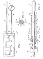
- FIG. 1 is a schematic elevational view of the component monitoring system embodying features of the invention.
- FIG. 2 is an enlarged elevational view of the monitoring system illustrating the sheath and biasing mechanism for urging the operative end of the monitor against the endometrium surface within the patient's uterine cavity.
- FIG. 3 is a partial transverse cross-sectional view of the system shown in FIG. 2 taken along the lines 3 - 3 .
- FIG. 4 is a schematic illustration of a female patient's reproductive organs with a component monitor shown in FIG. 1 deployed within the patient's uterine cavity.
- FIG. 5 is a schematic view illustrating a monitoring electrode extending into a patient's myometrium.
- FIG. 6 is a graph illustrating the pH changes to endometrial tissue and myometrial tissue due to uterine artery occlusion over a period of time.
- FIG. 1 illustrates a component monitoring system 10 embodying features of the invention which includes a permeable distal antimony electrode 11 , an elongated shaft 12 having a proximal end 13 secured to a pH monitoring unit 14 by a releasable connection 15 .
- a reference external electrode 16 adapted to be secured to an exterior portion of the patient, such as the patient's abdomen or thigh by suitable adhesive, has an elongated shaft 17 with a proximal end 18 secured to the pH monitoring unit 14 by a releasable connection 19 .
- the reference electrode is preferable a silver/silver chloride electrode.
- a suitable pH sensing electrode system is the Zinetics 24ME multi-use pH catheters from Medtronic, Inc.
- the pH monitoring unit 14 includes a personal digital assistant 20 , such as the Palm Pilot with a data acquisition system 21 , such as a DataStick DAQ adapter. Shafts 12 and 17 are secured to the manifold 22 by releasable connections 15 and 19 and transmission line 23 leads from the manifold 22 to a receiving port of the data acquisition system 21 to transmit the voltage differential between the active electrode 11 and the reference electrode 16 .
- a personal digital assistant 20 such as the Palm Pilot
- a data acquisition system 21 such as a DataStick DAQ adapter.
- Shafts 12 and 17 are secured to the manifold 22 by releasable connections 15 and 19 and transmission line 23 leads from the manifold 22 to a receiving port of the data acquisition system 21 to transmit the voltage differential between the active electrode 11 and the reference electrode 16 .
- the pH monitoring system 10 includes an introducing sheath 24 which is configured to extend through the patient's vaginal canal and into the uterine cavity.
- the distal end 25 of sheath 24 is preferably provided with a tapered non-traumatic tip.
- the proximal end 26 has a collar 27 , an attached spring 28 and Toughy-Borst adapter 29 secured to the proximal end of the spring 28 and configured to be releasably secured to the exterior of the shaft 12 of the active electrode 11 .
- FIGS. 2 and 3 illustrate in greater detail the introducer sheath 24 .
- the sheath 24 has an elongated shaft 32 with an inner lumen 33 configured to slidably receive the active electrode 11 and shaft 12 thereof, and a discharge port 34 at the distal end 25 of the sheath.
- the proximal end of the sheath has a pair of tabs or eyelets 35 for securing the proximal end to the patient, e.g. the patient's uterine cervix or vaginal labia, by a clip or suture or other suitable means.
- the active electrode 11 on the distal end of the shaft 12 is introduced into the patient's vaginal canal 34 , advanced into the uterine patient's uterine cavity 35 until the active electrode 11 is pressed against the endometrial fundus 36 of the patient's uterus 37 to monitor the pH of the endometrial tissue.
- the Toughy-borst adapter 29 on the sheath 24 is pulled proximally to elongate the spring 28 and then tightened about the exterior of the shaft 12 .
- the biasing action of the spring 28 urges the shaft 12 further into the patient's uterine cavity and presses the active electrode 11 against the fundus 30 to ensure proper contact for pH detection of the endometrial lining of the patient's uterus 31 .
- An exterior reference electrode 16 is secured to the patient's skin at a convenient place close to the patient's vaginal opening, e.g. the abdomen or thigh.
- the active electrode shaft 12 and the reference electrode shaft 17 are operatively connected to the manifold 22 of the monitor unit 14 at releasable connections 15 and 19 respectively.
- the electrical signals received from the active electrode 11 through conductor 37 and reference electrode 16 through conductor 38 are transmitted to the data acquisition system 21 through transmission line 23 .
- the endometrial tissue pH is displayed on the PDA.
- the active electrode 40 may be configured to penetrate the uterine wall through the endometrial layer 41 into the myometrial layer 42 .
- the pH signals tend to be a little more stable.
- a coller 43 should be provided to limit the penetration into the myometrial layer to prevent penetration through the uterine wall.
- an active electrode with a radio transmitter which can be implanted into the uterine wall without a catheter extending out of the patient's vaginal canal.
- a monitor is provided with a suitable receiver to receive the signal from the active electrode transmitter and display or record the received signal as a function of the pH (or other component parameter).
- the clips may be applied to occlude the patient's uterine artery with the pH monitor system installed to allow the patient sufficient mobility to leave the clinical setting without a catheter extending out o the patient's vaginal canal and return when the pH monitor indicates that the uterine artery has been occluded long enough so that the uterine clip should be removed.
- the active electrode and transmitter may also be removed.
- the temporary occlusion of uterine arteries for the treatment for uterine fibroids and uterine bleeding from a variety of causes and the monitoring of the concentration of components, e.g. pH, in the uterine tissue environment can be effective.
- the procedure is illustrated in the following example.
- the initial phase occurred over a period of about 5 to about 150 minutes, averaging 32 minutes.
- the pH of uterine lining tissue declined an average of about 0.5 pH units during the initial phase.
- the pH levels of the uterine tissue remained at essentially the reduced levels over a period of about 5 to about 150 minutes, averaging 53 minutes.
- the pH returned to the pre-occlusion levels from the low pH levels of the dwell phase over a period of about 30 to about 180 minutes, averaging about 114 minutes.
- FIG. 6 A typical display of the variation of pH of the endometrial and myometrial tissue with respect to time due to the ischemic effects of uterine artery occlusion is illustrated in FIG. 6 .
- porous antimony electrode was described as an active electrode a variety of electrodes may be used.
- reference electrode is described herein as being attached to the exterior of the patient. However other reference electrode locations may be employed.
- the shaft of the monitoring system may have the active electrode and a reference electrode.
Landscapes
- Health & Medical Sciences (AREA)
- Life Sciences & Earth Sciences (AREA)
- Physics & Mathematics (AREA)
- Surgery (AREA)
- General Health & Medical Sciences (AREA)
- Biophysics (AREA)
- Pathology (AREA)
- Engineering & Computer Science (AREA)
- Biomedical Technology (AREA)
- Heart & Thoracic Surgery (AREA)
- Medical Informatics (AREA)
- Molecular Biology (AREA)
- Veterinary Medicine (AREA)
- Animal Behavior & Ethology (AREA)
- Public Health (AREA)
- Optics & Photonics (AREA)
- Reproductive Health (AREA)
- Gynecology & Obstetrics (AREA)
- Chemical & Material Sciences (AREA)
- Chemical Kinetics & Catalysis (AREA)
- General Chemical & Material Sciences (AREA)
- Measuring And Recording Apparatus For Diagnosis (AREA)
- Surgical Instruments (AREA)
- Endoscopes (AREA)
Abstract
The invention provides a devices, methods and systems to measure and record uterine tissue environment components such as pH during the course of uterine artery occlusion. The uterus becomes ischemic due to the occlusion thereof, and its pH drops sharply within minutes of uterine artery occlusion and remains relatively low for a period of time. The return of normal pH is an indicator of return of blood to the ischemic tissue. In use, a catheter with a pH measuring tip is advanced through the patient's vaginal canal and into the patient's uterine cavity until the pH measuring active electrode on the distal end of the catheter contacts or penetrates the uterine fundus. The active electrode detects the pH and a signal representing pH is transmitted to a pH recording and monitoring device which preferably displays the pH. The signal may be transmitted through a conductor or by a radio transmitter. Components other than pH may be monitored such a pCO2, and pO2.
Description
- The invention relates generally to diagnostic measurements during the treatment of uterine disorders by the reduction of blood flow through a female patient's uterine artery.
- Hysterectomy (surgical removal of the uterus) is performed on approximately 600,000 women annually in the United. States. Hysterectomy is often the therapeutic choice for the treatment of a variety of uterine disorders such as cancer, adenomyosis, menorrhagia, uterine prolapse, dysfunctional uterine bleeding (abnormal menstrual bleeding that has no discrete anatomic explanation such as a tumor or growth), and muscular tumors of the uterus, known as leimyoma or uterine fibroids.
- However, hysterectomy is a drastic treatment, having many undesirable characteristics. Newer treatment methods have been developed for some diseases which avoid the need for a hysterectomy. For example, in 1995, it was demonstrated that uterine fibroids could be treated without hysterectomy using a non-surgical therapy, specifically comprising bilateral intraluminal occlusion of the uterine arteries (Ravina et al., “Arterial Embolization to Treat Uterine Myomata”, Lancet Sep. 9, 1995; Vol. 346; pp. 671-672, incorporated in its entirety herein). This technique is known as “uterine artery embolization”. In this technique, uterine arteries are accessed via a transvascular route from a common femoral artery into the left and right uterine arteries. The technique uses standard interventional radiology angiographic techniques and equipment, whereby the uterine arteries are accessed via a transvascular route from a common femoral artery into the left and right uterine arteries. Uterine artery embolization can be effectively used to control uterine bleeding from a variety of sources using coils placed in arterial and venous lumens (See U.S. Pat. Nos. 4,994,069, 5,226,911, and 5,549,824, all of which are incorporated in their entireties herein), or particles (GELFOAM pledgets, available from Upjohn, Kalamazoo, Mich. or IVALON particles, available from Boston Scientific).
- One of the key features for treating fibroids with uterine artery embolization is the fact that fibroids live a tenuous vascular life with very little ability to recruit a new blood supply from the host when the primary blood supply is compromised. The uterus on the other hand has a dual (or redundant) blood supply; the primary blood supply is from the bilateral uterine arteries, the secondary blood supply from the bilateral ovarian arteries. Consequently, when both uterine arteries are occluded, i.e., bilateral vessel occlusion, the uterus and the fibroids contained within the uterus are both deprived of their blood supply. However, as demonstrated by Ravina et al., the effect on the fibroids is greater than the effect on the uterus. In most instances, the fibroids wither and cease to cause clinical symptoms.
- The uterus has a dual (or redundant) blood supply, the primary blood supply being from the bilateral uterine arteries, and the secondary blood supply from the bilateral ovarian arteries. Consequently, when both uterine arteries are occluded, i.e. bilateral vessel occlusion; the uterus and the fibroids contained within the uterus are both deprived of their blood supply. However, as demonstrated by Ravina et al., the effect on the fibroid is greater than the effect on the uterus. In most instances, the fibroid withers and ceases to cause clinical symptoms. See also Burbank, et al., “Uterine Artery Occlusion by Embolization or Surgery for the Treatment of Fibroids: A Unifying Hypothesis—Transient Uterine Ischemia,” The Journal of the American Association of Gynecologic Laparoscopists, November 2000, Vol. 7, No. 4 Supplement, pp. S3-S49. U.S. Pat. No. 6,254,601, to Burbank et al. entitled “Methods for Occlusion of the Uterine Arteries,” describes numerous devices and methods useful for occluding a uterine artery.
- The current treatments offered to women for fibroid treatment or uterine bleeding focus on permanent or near permanent occlusion methods for the uterine artery. For example, embolizing with PVA particles causes uterine artery occlusion for 6 months to permanent in situ); embolizing with stainless steel coils causes permanent occlusion; embolizing with Gelfoam occludes for 3 to 4 weeks before degradation of the embolic particles; surgical ligation with metal vascular clips occlude permanently; and surgical ligation with RF ablation results in permanent occlusion.
- The prior art devices and methods are therefore aimed at permanent occlusion of the uterine artery, resulting in redirection of the blood flow to the uterus through collateral circulation. The patients who suffer most dramatically from uterine myomata are women of child bearing age who may desire to bear additional children. The current methods of embolizing or ligating uterine arteries are specifically contraindicated for women who desire to bear additional children. This is the realization of inadequate blood supply to the uterus because of the loss of the uterine arteries, the primary blood supply. While there have been reports of women who have undergone uterine artery embolization with PVA particles and who have subsequently become pregnant and deliver normal babies. Women who have undergone uterine artery embolization have also experienced premature menopause due to ovarian failure.
- Recent advances in non-permanent occlusion of uterine arteries for treating a variety of uterine disorders have relied upon time period to determine when to re-establish blood flow through the patient's uterine artery. However, the duration of the uterine artery occlusion does not always provide an accurate assessment of the treatment for a particular disorder, particularly uterine fibroids.
- The invention is directed to the detection of the extent of uterine artery occlusion in the treatment of uterine disorders in a female patient, and specifically, to monitoring of ionic components of the patient's uterine tissue such as the endometrium and myometrium.
- Occlusion of the uterine arteries alters the chemical environment of uterine tissue in a time dependent fashion, typically in three phases, an initial phase, a dwell phase and a return phase. Before the occlusion of the uterine arteries, the pH of the uterine tissue can range from about 6.0 to about 7.5, usually about 6.3 to about 7.0. Upon the occlusion of the patient's uterine arteries, the pH of the tissue begins to decline, the initial phase, as soon as it becomes ischemic which is a few minutes (typically about 2 to about 120 minutes, usually about 10 to about 60 minutes). The pH of the uterine tissue declines in the initial phase about 0.2 to about 1.0. pH units, usually about 0.3 to about 0.7 pH units. The pH stays at a lower level, the dwell phase, for about 10 to about 120 minutes, typically about 10 to about 80 minutes. After the dwell phase, the pH returns to normal or near normal pH values that existed prior to the uterine artery occlusion which is the return phase. The initial phase can last about 10 to about 90 minutes, but typically last about 10 to about 60 minutes. The increase of the uterine tissue pH after uterine artery occlusion is usually an indication that the ischemic tissue is receiving blood flow from collateral arteries such as the ovarian arteries, if the uterine arteries remain occluded. While the discussion herein refers primarily directed to monitoring the pH of uterine tissue, other species may be monitored such as pCO2, and pO2. Other components of the uterine tissue environment may be measured in the same or similar fashion. With pH monitoring the initial phase is a descent phase and the return phase is an ascent phase. When other components are monitored, the initial phase may be an ascent phase while the return phase is a descent phase.
- The time for effective uterine artery occlusion treatments may vary from patient to patient depending on a variety of factors, including the amount of collateral blood flow from the patient's ovarian and other arteries. However, the short term pH changes in the patient's uterine tissue, which result from the uterine artery occlusion, provide a more accurate indication of the progress of the procedure than existing methods.
- These and other advantages of the invention will be evident from the following detailed description of the invention hen taken in conjunction with the attached exemplary drawing.
-
FIG. 1 is a schematic elevational view of the component monitoring system embodying features of the invention. -
FIG. 2 is an enlarged elevational view of the monitoring system illustrating the sheath and biasing mechanism for urging the operative end of the monitor against the endometrium surface within the patient's uterine cavity. -
FIG. 3 is a partial transverse cross-sectional view of the system shown inFIG. 2 taken along the lines 3-3. -
FIG. 4 is a schematic illustration of a female patient's reproductive organs with a component monitor shown inFIG. 1 deployed within the patient's uterine cavity. -
FIG. 5 is a schematic view illustrating a monitoring electrode extending into a patient's myometrium. -
FIG. 6 is a graph illustrating the pH changes to endometrial tissue and myometrial tissue due to uterine artery occlusion over a period of time. -
FIG. 1 illustrates acomponent monitoring system 10 embodying features of the invention which includes a permeabledistal antimony electrode 11, anelongated shaft 12 having aproximal end 13 secured to apH monitoring unit 14 by areleasable connection 15. A referenceexternal electrode 16, adapted to be secured to an exterior portion of the patient, such as the patient's abdomen or thigh by suitable adhesive, has anelongated shaft 17 with aproximal end 18 secured to thepH monitoring unit 14 by areleasable connection 19. The reference electrode is preferable a silver/silver chloride electrode. A suitable pH sensing electrode system is the Zinetics 24ME multi-use pH catheters from Medtronic, Inc. - The
pH monitoring unit 14 includes a personaldigital assistant 20, such as the Palm Pilot with adata acquisition system 21, such as a DataStick DAQ adapter.Shafts releasable connections transmission line 23 leads from the manifold 22 to a receiving port of thedata acquisition system 21 to transmit the voltage differential between theactive electrode 11 and thereference electrode 16. - The
pH monitoring system 10 includes an introducingsheath 24 which is configured to extend through the patient's vaginal canal and into the uterine cavity. Thedistal end 25 ofsheath 24 is preferably provided with a tapered non-traumatic tip. Theproximal end 26 has acollar 27, an attachedspring 28 and Toughy-Borst adapter 29 secured to the proximal end of thespring 28 and configured to be releasably secured to the exterior of theshaft 12 of theactive electrode 11. -
FIGS. 2 and 3 illustrate in greater detail theintroducer sheath 24. Thesheath 24 has an elongatedshaft 32 with aninner lumen 33 configured to slidably receive theactive electrode 11 andshaft 12 thereof, and adischarge port 34 at thedistal end 25 of the sheath. The proximal end of the sheath has a pair of tabs or eyelets 35 for securing the proximal end to the patient, e.g. the patient's uterine cervix or vaginal labia, by a clip or suture or other suitable means. - As shown in
FIG. 4 , theactive electrode 11 on the distal end of theshaft 12 is introduced into the patient'svaginal canal 34, advanced into the uterine patient'suterine cavity 35 until theactive electrode 11 is pressed against the endometrial fundus 36 of the patient'suterus 37 to monitor the pH of the endometrial tissue. After the sheath is secured to the patient, the Toughy-borst adapter 29 on thesheath 24 is pulled proximally to elongate thespring 28 and then tightened about the exterior of theshaft 12. The biasing action of thespring 28 urges theshaft 12 further into the patient's uterine cavity and presses theactive electrode 11 against the fundus 30 to ensure proper contact for pH detection of the endometrial lining of the patient's uterus 31. - An
exterior reference electrode 16 is secured to the patient's skin at a convenient place close to the patient's vaginal opening, e.g. the abdomen or thigh. Theactive electrode shaft 12 and thereference electrode shaft 17 are operatively connected to themanifold 22 of themonitor unit 14 atreleasable connections active electrode 11 throughconductor 37 andreference electrode 16 throughconductor 38 are transmitted to thedata acquisition system 21 throughtransmission line 23. The endometrial tissue pH is displayed on the PDA. - As shown in
FIG. 5 , theactive electrode 40 may be configured to penetrate the uterine wall through theendometrial layer 41 into themyometrial layer 42. The pH signals tend to be a little more stable. However, acoller 43 should be provided to limit the penetration into the myometrial layer to prevent penetration through the uterine wall. - In some instances it may be desirable to provide an active electrode with a radio transmitter which can be implanted into the uterine wall without a catheter extending out of the patient's vaginal canal. A monitor is provided with a suitable receiver to receive the signal from the active electrode transmitter and display or record the received signal as a function of the pH (or other component parameter). This allow for the use of a uterine artery clip or clips such as described in co-pending applications Ser. No. 01/113,096, filed on Mar. 28, 2002 and Ser. No. 10/300,495, filed on Nov. 19, 2002, both of which have been assigned to the present assignee Vascular Control Systems, Inc. The clips may be applied to occlude the patient's uterine artery with the pH monitor system installed to allow the patient sufficient mobility to leave the clinical setting without a catheter extending out o the patient's vaginal canal and return when the pH monitor indicates that the uterine artery has been occluded long enough so that the uterine clip should be removed. The active electrode and transmitter may also be removed.
- The temporary occlusion of uterine arteries for the treatment for uterine fibroids and uterine bleeding from a variety of causes and the monitoring of the concentration of components, e.g. pH, in the uterine tissue environment can be effective. The procedure is illustrated in the following example.
- Ten women with menorrhagia, pelvic pain or pressure, and/or anemia had myomas and uteris larger than 13 weeks gestation were subjected to bilateral uterine artery occlusion. Prior to the uterine artery occlusion a pH monitoring probe (Zinetics 24ME multi-use pH catheter) was placed transcervically into the endometrium and myometrium. Before uterine artery occlusion, the uterine tissue pH value for the 10 patients averaged 6.7 pH units, ranging from 6.4 to 6.9. The average decline in pH during the initial phase was 0.5 pH units, ranging from 0.4 to 0.6. The uterine tissue of all of the patients exhibited a similar pattern of pH value changes over the monitoring period (less than 24 hours). The initial phase occurred over a period of about 5 to about 150 minutes, averaging 32 minutes. The pH of uterine lining tissue declined an average of about 0.5 pH units during the initial phase. In the dwell phase, the pH levels of the uterine tissue remained at essentially the reduced levels over a period of about 5 to about 150 minutes, averaging 53 minutes. In the return phase, the pH returned to the pre-occlusion levels from the low pH levels of the dwell phase over a period of about 30 to about 180 minutes, averaging about 114 minutes. A typical display of the variation of pH of the endometrial and myometrial tissue with respect to time due to the ischemic effects of uterine artery occlusion is illustrated in
FIG. 6 . - While particular forms of the invention have been illustrated and described herein, it will be apparent that various modifications and improvements can be made to the invention. For example, while a porous antimony electrode was described as an active electrode a variety of electrodes may be used. Moreover, the reference electrode is described herein as being attached to the exterior of the patient. However other reference electrode locations may be employed. For example, the shaft of the monitoring system may have the active electrode and a reference electrode.
- Individual features of embodiments of the invention may be shown in some drawings and not in others, but those skilled in the art will recognize that individual features of one embodiment of the invention can be combined with any or all the features of another embodiment. Accordingly, it is not intended that the invention be limited to the specific embodiments illustrated. It is therefore intended that this invention to be defined by the scope of the appended claims as broadly as the prior art will permit.
- Terms such a “element”, “member”, “device”, “sections”, “portion”, “section”, “steps” and words of similar import when used herein shall not be construed as invoking the provisions of 35 U.S.C. §112(6) unless the following claims expressly use the terms “means” or “step” followed by a particular function without specific structure or action. All patents and patent applications referred to above are hereby incorporated by reference in their entirety. Accordingly, it is not intended that the invention be limited, except as by the appended claims.
Claims (42)
1. A transvaginal device for measuring the pH of a female patient's uterine tissue, comprising:
a. an elongated introducer sheath which is configured to extend through a female patient's vaginal canal, uterine cervix and into the uterine cavity of the female patient's uterus and which has an elongated shaft, proximal and distal ends, a first port in the proximal end, a second port in the distal end and an inner lumen extending within the shaft in fluid communication with the first and second ports; and
b. a pH measuring catheter which is configured to be slidably disposed within the inner lumen of the introducer sheath and which has an elongated shaft with a proximal end and a distal end, which has an active electrode at the distal end of the shaft and which is configured to transmit a signal from the active electrode to a pH monitoring unit.
2. The transvaginal device of claim 1 including a biasing mechanism interconnecting the catheter and elongated sheath to urge the catheter distally with respect to the elongated sheath to ensure that the active electrode on the distal end of the catheter engages endometrial tissue within the uterine cavity.
3. The transvaginal device of claim 1 wherein the active electrode is a porous electrode.
4. The transvaginal device of claim 2 wherein the active electrode is an antimony based half cell electrode.
5. The transvaginal device of claim 1 including a reference electrode which is configured to be secured to an exterior location on the patient's body.
6. The transvaginal device of claim 1 including a reference electrode which is located on the shaft proximal to the active electrode.
7. The transvaginal device of claim 1 wherein the pH catheter is electrically connected to a pH monitoring unit by an electrical conductor for transmitting an electrical signal representing the voltage sensed by the active electrode.
8. The transvaginal device of claim 7 including a reference electrode which is configured to be secured to an exterior location on the patient's body and a connected signal transmitting member for transmitting a signal representing the voltage sensed by the reference electrode.
9. The transvaginal device of claim 1 wherein the pH catheter is provided with a transmitter to transmit a signal from the active electrode representing a uterine tissue parameter to the pH monitor which converts the signal to a pH value.
10. The transvaginal device of claim 8 wherein the pH monitor includes a manifold which develops a signal representing a difference between voltage sensed by the active electrode and voltage sensed by the reference electrode which is a measure of the pH of the uterine lining tissue.
11. The transvaginal device of claim 5 wherein the reference electrode is a silver/silver chloride half cell.
12. The transvaginal device of claim 1 wherein the sheath configured to provide access to the patient's uterine cavity is configured to be secured at least in part to the patient.
13. The transvaginal device of claim 12 wherein the sheath is in part secured within the patient's vaginal canal.
14. The transvaginal device of claim 2 wherein the biasing mechanism is configured to be releasably secured to the catheter shaft.
15. The transvaginal device of claim 14 wherein a Toughy-Borst adapter is provided on a proximal end of the biasing mechanism to releasably secure the proximal end of the biasing mechanism to the catheter shaft.
16. The transvaginal device of claim 2 wherein the biasing mechanism is a spring.
17. A method of monitoring the pH of tissue lining a female patient's uterine cavity during a medical treatment which includes occluding one or more uterine arteries, comprising:
a. providing a pH measuring catheter having an elongated shaft with proximal and distal end and an active electrode on the distal end thereof;
b. advancing the pH measuring catheter through the patient's vaginal canal, into the female patient's uterine cavity;
c. urging the active electrode on the distal end of the pH measuring catheter into contact with uterine tissue; and
d. generating a signal representing a pH parameter of the uterine tissue environment.
18. The method of claim 17 wherein a pH signal is generated representing the difference between the signal representing the pH parameter of the body cavity and a signal received from a reference electrode secured to an exterior portion of the patient.
19. The method of claim 17 wherein the difference signal is transmitted to a monitoring unit which displays pH units.
20. The method of claim 17 wherein an introducer sheath is introduced through the patient's vaginal canal and into the patient's uterine cavity and is secured to the patient.
21. The method of claim 20 wherein a biasing spring is secured between the introducer sheath and the pH measuring catheter to urge the active electrode into contact with uterine tissue.
22. The method of claim 21 wherein the uterine tissue is endometrial tissue.
23. The method of claim 21 wherein the uterine tissue is myometrial tissue.
24. The method of claim 17 wherein the pH of the patient's uterine lining tissue is monitored for at least 0.5 hour.
25. The method of claim 17 wherein the pH of the patient's uterine lining tissue is monitored for a period of up to about 48 hours.
26. A device for monitoring a component of a female patient's uterine tissue, comprising:
b. an elongated introducer sheath which is configured to extend through a female patient's vaginal canal, uterine cervix and into the uterine cavity of the female patient's uterus and which has an elongated shaft, proximal and distal ends, a first port in the proximal end, a second port in the distal end and an inner lumen extending within the shaft in fluid communication with the first and second ports; and
b. a component monitoring catheter which is configured to be slidably disposed within the inner lumen of the introducer sheath, which has an elongated shaft with a proximal end and a distal end, which has an active electrode at the distal end of the shaft, which is configured to transmit a signal representing the component concentration in the tissue environment the active electrode with a pH monitoring unit.
27. The device of claim 26 wherein a reference electrode is held in contact with the patient's body to generate a reference signal by the reference electrode is compared with the signal representing the component concentration in the tissue environment.
28. The device of claim 27 wherein a manifold is provided to generate a difference signal between the reference signal and the signal from the active electrode.
29. The device of claim 28 wherein a conductor is provided to transmit the difference signal to the pH monitor.
30. A method of treating a female patient's uterus, comprising:
a. occluding one or more of the patient's uterine arteries, comprising:
b. detecting the concentration of a component of uterine tissue environment,
c. monitoring the concentration of the detected component;
d. re-establishing blood flow in the patient's uterine artery after the concentration of the detected component returns to pre-occlusion levels or near pre-occlusion levels.
31. The method of claim 30 wherein the component is the hydrogen ion concentration.
32. The method of claim 31 wherein the hydrogen ion concentration is detected by a pH detecting electrode.
33. The method of claim 32 wherein a pH measuring catheter having an elongated shaft with proximal and distal end and an active electrode on the distal end thereof is advanced through the patient's vaginal canal, into the female patient's uterine cavity and the active electrode is urged into contact with uterine tissue.
34. The method of claim 33 wherein a signal is generated which represents a pH parameter of the uterine tissue environment.
35. The method of claim 34 wherein a pH signal is generated representing the difference between the signal representing the pH parameter of the body cavity and a signal received from a reference electrode secured to the patient
36. The method of claim 35 wherein the difference signal is transmitted to a monitoring unit which displays pH units relating to the signal received.
37. The method of claim 33 wherein an introducer sheath is introduced through the patient's vaginal canal and into the patient's uterine cavity and is secured to the patient.
38. The method of claim 37 wherein a biasing spring is secured between the introducer sheath and the pH measuring catheter to urge the active electrode into contact with uterine tissue.
39. The method of claim 33 the active electrode is urged against endometrial uterine tissue.
40. The method of claim 33 wherein the uterine tissue is myometrial tissue.
41. The method of claim 32 wherein the pH of the patient's uterine lining tissue is monitored for at least 0.5 hour.
42. The method of claim 32 wherein the pH of the patient's uterine lining tissue is monitored for a period of up to about 48 hours.
Priority Applications (1)
| Application Number | Priority Date | Filing Date | Title |
|---|---|---|---|
| US11/413,260 US7616979B2 (en) | 2003-03-28 | 2006-04-28 | Uterine tissue monitoring device and method |
Applications Claiming Priority (2)
| Application Number | Priority Date | Filing Date | Title |
|---|---|---|---|
| US10/402,892 US7333844B2 (en) | 2003-03-28 | 2003-03-28 | Uterine tissue monitoring device and method |
| US11/413,260 US7616979B2 (en) | 2003-03-28 | 2006-04-28 | Uterine tissue monitoring device and method |
Related Parent Applications (1)
| Application Number | Title | Priority Date | Filing Date |
|---|---|---|---|
| US10/402,892 Division US7333844B2 (en) | 2003-03-28 | 2003-03-28 | Uterine tissue monitoring device and method |
Publications (2)
| Publication Number | Publication Date |
|---|---|
| US20060241337A1 true US20060241337A1 (en) | 2006-10-26 |
| US7616979B2 US7616979B2 (en) | 2009-11-10 |
Family
ID=32989837
Family Applications (2)
| Application Number | Title | Priority Date | Filing Date |
|---|---|---|---|
| US10/402,892 Expired - Fee Related US7333844B2 (en) | 2003-03-28 | 2003-03-28 | Uterine tissue monitoring device and method |
| US11/413,260 Expired - Fee Related US7616979B2 (en) | 2003-03-28 | 2006-04-28 | Uterine tissue monitoring device and method |
Family Applications Before (1)
| Application Number | Title | Priority Date | Filing Date |
|---|---|---|---|
| US10/402,892 Expired - Fee Related US7333844B2 (en) | 2003-03-28 | 2003-03-28 | Uterine tissue monitoring device and method |
Country Status (1)
| Country | Link |
|---|---|
| US (2) | US7333844B2 (en) |
Cited By (6)
| Publication number | Priority date | Publication date | Assignee | Title |
|---|---|---|---|---|
| US20090054915A1 (en) * | 2007-08-23 | 2009-02-26 | Peter Meier | Obstruction of uterine arteries to treat uterine fibroids using mechanical instruments to twist the vessels |
| US20090054916A1 (en) * | 2007-08-23 | 2009-02-26 | Peter Meier | Clip-based method for treatment of uterine fibroids by obstruction of the uterine arteries |
| US20090062827A1 (en) * | 2007-08-31 | 2009-03-05 | Peter Meier | Vacuum-based method for obstruction of uterine arteries to treat uterine fibroids |
| US20090318950A1 (en) * | 2006-07-24 | 2009-12-24 | Yossi Gross | Fibroid treatment apparatus and method |
| US20110022073A1 (en) * | 2009-07-27 | 2011-01-27 | Fibro Control, Inc. | Balloon with rigid tube for occluding the uterine artery |
| RU2666604C1 (en) * | 2017-12-19 | 2018-09-11 | Владислав Геннадьевич Ившин | Method of estimation of acid-base balance of female genital organs (options) |
Families Citing this family (32)
| Publication number | Priority date | Publication date | Assignee | Title |
|---|---|---|---|---|
| US7223279B2 (en) * | 2000-04-21 | 2007-05-29 | Vascular Control Systems, Inc. | Methods for minimally-invasive, non-permanent occlusion of a uterine artery |
| US7875036B2 (en) * | 2004-10-27 | 2011-01-25 | Vascular Control Systems, Inc. | Short term treatment for uterine disorder |
| US7918795B2 (en) | 2005-02-02 | 2011-04-05 | Gynesonics, Inc. | Method and device for uterine fibroid treatment |
| FR2895667B3 (en) * | 2006-01-05 | 2008-04-04 | Rene Vinci | FERTILITY DETECTOR FOR MAMMALS |
| US11259825B2 (en) | 2006-01-12 | 2022-03-01 | Gynesonics, Inc. | Devices and methods for treatment of tissue |
| US7874986B2 (en) * | 2006-04-20 | 2011-01-25 | Gynesonics, Inc. | Methods and devices for visualization and ablation of tissue |
| US9357977B2 (en) | 2006-01-12 | 2016-06-07 | Gynesonics, Inc. | Interventional deployment and imaging system |
| US10058342B2 (en) | 2006-01-12 | 2018-08-28 | Gynesonics, Inc. | Devices and methods for treatment of tissue |
| US7815571B2 (en) | 2006-04-20 | 2010-10-19 | Gynesonics, Inc. | Rigid delivery systems having inclined ultrasound and needle |
| US8206300B2 (en) | 2008-08-26 | 2012-06-26 | Gynesonics, Inc. | Ablation device with articulated imaging transducer |
| US10595819B2 (en) | 2006-04-20 | 2020-03-24 | Gynesonics, Inc. | Ablation device with articulated imaging transducer |
| US8088072B2 (en) | 2007-10-12 | 2012-01-03 | Gynesonics, Inc. | Methods and systems for controlled deployment of needles in tissue |
| US20090163856A1 (en) * | 2007-12-19 | 2009-06-25 | Searete Llc, A Limited Liability Corporation Of The State Of Delaware | Treatment indications informed by a prior implant information |
| US9717896B2 (en) * | 2007-12-18 | 2017-08-01 | Gearbox, Llc | Treatment indications informed by a priori implant information |
| US8636670B2 (en) * | 2008-05-13 | 2014-01-28 | The Invention Science Fund I, Llc | Circulatory monitoring systems and methods |
| US20090292222A1 (en) * | 2008-05-14 | 2009-11-26 | Searete Llc | Circulatory monitoring systems and methods |
| US20100036263A1 (en) * | 2008-08-07 | 2010-02-11 | Searete Llc, A Limited Liability Corporation Of The State Of Delaware | Circulatory monitoring systems and methods |
| US20090292213A1 (en) * | 2008-05-21 | 2009-11-26 | Searete Llc, A Limited Liability Corporation Of The State Of Delaware | Circulatory monitoring systems and methods |
| US20090292214A1 (en) * | 2008-05-22 | 2009-11-26 | Searete Llc, A Limited Liability Corporation Of The State Of Delaware | Circulatory monitoring systems and methods |
| US20090287120A1 (en) | 2007-12-18 | 2009-11-19 | Searete Llc, A Limited Liability Corporation Of The State Of Delaware | Circulatory monitoring systems and methods |
| CN102170820A (en) * | 2008-08-05 | 2011-08-31 | Ph值诊断公司 | Apparatus, method and system for determining a physiological condition within a mammal |
| US8262574B2 (en) | 2009-02-27 | 2012-09-11 | Gynesonics, Inc. | Needle and tine deployment mechanism |
| US10751031B2 (en) | 2013-08-29 | 2020-08-25 | Mrinal K. Sanyal | Retrieval of biological materials from the human uterus, ovary and cervix by suction |
| US9808225B2 (en) | 2013-08-29 | 2017-11-07 | Mrinal K. Sanyal | Retrieval of biological materials from the human uterus, ovary and cervix by suction |
| US10105070B2 (en) | 2014-11-17 | 2018-10-23 | 3VO Medical, Inc. | Intrauterine access catheter for delivering and facilitating operation of a medical apparatus for assisting parturition |
| CN104970770A (en) * | 2015-06-12 | 2015-10-14 | 赵峰 | Gynecological uterus diagnosis rehabilitation device |
| EP4156204A1 (en) | 2016-11-11 | 2023-03-29 | Gynesonics, Inc. | Controlled treatment of tissue and dynamic interaction with, and comparison of, tissue and/or treatment data |
| GB2566463A (en) * | 2017-09-13 | 2019-03-20 | Univ Southampton | pH Sensor and Calibration method |
| RU184214U1 (en) * | 2018-03-23 | 2018-10-18 | Владислав Геннадьевич Ившин | Probe for ionometry |
| CN109528182A (en) * | 2019-01-09 | 2019-03-29 | 西安汇智医疗集团有限公司 | A kind of puerpera's patient monitor for implementing fetal monitoring based on uterine neck sacculus dilating catheter inner sensor |
| EP3946039A1 (en) * | 2019-04-05 | 2022-02-09 | Julius Georgiou | Method and apparatus for direct in-vivo, electrical and chemical monitoring and stimulation of the endometrial cavity |
| WO2021076940A1 (en) | 2019-10-16 | 2021-04-22 | Balman James Robert | Apparatus and method for determining physiological parameters of an infant in-utero |
Citations (99)
| Publication number | Priority date | Publication date | Assignee | Title |
|---|---|---|---|---|
| US2168867A (en) * | 1937-08-05 | 1939-08-08 | Takamine Ferment Company | Method and apparatus for testing the contents of the stomach and other body cavities |
| US2400251A (en) * | 1943-07-29 | 1946-05-14 | Charles E Nagel | Gynecological instrument |
| US3123067A (en) * | 1964-03-03 | Process and apparatus for determining the presence | ||
| US3209753A (en) * | 1962-05-04 | 1965-10-05 | Donald B Hawkins | Intestinal clamps and the like |
| US3973555A (en) * | 1973-10-16 | 1976-08-10 | Moeller Willi | Electrode cell assembly for the continuous determination of ion concentrations in living tissues |
| US4292960A (en) * | 1979-04-30 | 1981-10-06 | Rca Corporation | Apparatus and method for application of radioactive and microwave energy to the body |
| US4326535A (en) * | 1980-05-13 | 1982-04-27 | Akron City Hospital | Circuit and method for the radiotelemetry of esophageal pH in an ECG radiotelemetry system |
| US4428374A (en) * | 1978-12-20 | 1984-01-31 | Auburn Robert M | Umbilical cord clamping assembly |
| US4428379A (en) * | 1982-01-07 | 1984-01-31 | Technicare Corporation | Passive ultrasound needle probe locator |
| US4440620A (en) * | 1981-12-22 | 1984-04-03 | Olympus Optical Co., Ltd. | Measuring electrode device |
| US4509528A (en) * | 1981-12-16 | 1985-04-09 | Harvinder Sahota | Hemostat with blood flow sensor |
| US4546436A (en) * | 1983-07-06 | 1985-10-08 | The Johns Hopkins University | Portable pH data collector |
| US4650466A (en) * | 1985-11-01 | 1987-03-17 | Angiobrade Partners | Angioplasty device |
| US4757823A (en) * | 1987-01-27 | 1988-07-19 | Hofmeister John F | Method and apparatus for measuring uterine blood flow |
| US4840891A (en) * | 1986-09-03 | 1989-06-20 | Genetic Engineering, Inc. | Encapsulation of sperm for artificial insemination |
| US4945896A (en) * | 1989-01-24 | 1990-08-07 | Gade George F | Surgical retractor assembly having tissue viability sensor embedded therein |
| US4991588A (en) * | 1986-07-21 | 1991-02-12 | Pfizer Hospital Products Group, Inc. | Doppler guide wire |
| US4994069A (en) * | 1988-11-02 | 1991-02-19 | Target Therapeutics | Vaso-occlusion coil and method |
| US5037430A (en) * | 1986-01-06 | 1991-08-06 | Hasson Harrith M | Clamp for gynecological instruments |
| US5037433A (en) * | 1990-05-17 | 1991-08-06 | Wilk Peter J | Endoscopic suturing device and related method and suture |
| US5050297A (en) * | 1989-09-21 | 1991-09-24 | Becton, Dickinson And Company | Method for assembly of a directly exposed catheter sensor on a support tip |
| US5081997A (en) * | 1989-03-09 | 1992-01-21 | Vance Products Incorporated | Echogenic devices, material and method |
| US5105812A (en) * | 1990-07-25 | 1992-04-21 | Baylor College Of Medicine | Nasogastric tube with removable pH detector |
| US5108408A (en) * | 1990-04-20 | 1992-04-28 | Lally James J | Uterine-ring hysterectomy clamp |
| US5201314A (en) * | 1989-03-09 | 1993-04-13 | Vance Products Incorporated | Echogenic devices, material and method |
| US5226911A (en) * | 1991-10-02 | 1993-07-13 | Target Therapeutics | Vasoocclusion coil with attached fibrous element(s) |
| US5275166A (en) * | 1992-11-16 | 1994-01-04 | Ethicon, Inc. | Method and apparatus for performing ultrasonic assisted surgical procedures |
| US5289831A (en) * | 1989-03-09 | 1994-03-01 | Vance Products Incorporated | Surface-treated stent, catheter, cannula, and the like |
| US5336231A (en) * | 1992-05-01 | 1994-08-09 | Adair Edwin Lloyd | Parallel channel fixation, repair and ligation suture device |
| US5336229A (en) * | 1993-02-09 | 1994-08-09 | Laparomed Corporation | Dual ligating and dividing apparatus |
| US5383922A (en) * | 1993-03-15 | 1995-01-24 | Medtronic, Inc. | RF lead fixation and implantable lead |
| US5488958A (en) * | 1992-11-09 | 1996-02-06 | Vance Products Incorporated | Surgical cutting instrument for coring tissue affixed thereto |
| US5496331A (en) * | 1993-07-28 | 1996-03-05 | Terumo Kabushiki Kaisha | Knot-forming instrument and method of forming knots |
| US5507744A (en) * | 1992-04-23 | 1996-04-16 | Scimed Life Systems, Inc. | Apparatus and method for sealing vascular punctures |
| US5542944A (en) * | 1993-04-19 | 1996-08-06 | Bhatta; Krishan M. | Surgical device and method |
| US5549624A (en) * | 1994-06-24 | 1996-08-27 | Target Therapeutics, Inc. | Fibered vasooclusion coils |
| US5549824A (en) * | 1994-03-07 | 1996-08-27 | Ing. A. Maurer Sa | Filter apparatus including stacked intake and discharge plates |
| US5556396A (en) * | 1994-01-18 | 1996-09-17 | Endovascular, Inc. | Method for tubal electroligation |
| US5596988A (en) * | 1993-06-30 | 1997-01-28 | Biomedical Sensors, Ltd. | Multi-parameter sensor apparatus |
| US5598841A (en) * | 1993-09-24 | 1997-02-04 | Kowa Company Ltd. | Blood flow measurement system |
| US5614204A (en) * | 1995-01-23 | 1997-03-25 | The Regents Of The University Of California | Angiographic vascular occlusion agents and a method for hemostatic occlusion |
| US5658299A (en) * | 1995-07-20 | 1997-08-19 | Applied Medical Resources | Surgical ligating device and method for using same |
| US5662676A (en) * | 1992-06-24 | 1997-09-02 | K.U. Leuven Research & Development | Instrument set for laparoscopic hysterectomy |
| US5662680A (en) * | 1991-10-18 | 1997-09-02 | Desai; Ashvin H. | Endoscopic surgical instrument |
| US5665096A (en) * | 1995-03-07 | 1997-09-09 | Yoon; Inbae | Needle driving apparatus and methods of suturing tissue |
| US5672153A (en) * | 1992-08-12 | 1997-09-30 | Vidamed, Inc. | Medical probe device and method |
| US5672172A (en) * | 1994-06-23 | 1997-09-30 | Vros Corporation | Surgical instrument with ultrasound pulse generator |
| US5713896A (en) * | 1991-11-01 | 1998-02-03 | Medical Scientific, Inc. | Impedance feedback electrosurgical system |
| US5713942A (en) * | 1992-05-01 | 1998-02-03 | Vesta Medical, Inc. | Body cavity ablation apparatus and model |
| US5713371A (en) * | 1995-07-07 | 1998-02-03 | Sherman; Dani | Method of monitoring cervical dilatation during labor, and ultrasound transducer particularly useful in such method |
| US5716389A (en) * | 1995-11-13 | 1998-02-10 | Walinsky; Paul | Cardiac ablation catheter arrangement with movable guidewire |
| US5715832A (en) * | 1995-02-28 | 1998-02-10 | Boston Scientific Corporation | Deflectable biopsy catheter |
| US5720743A (en) * | 1996-06-07 | 1998-02-24 | Bischof; John C. | Thermally insulating surgical probe |
| US5749879A (en) * | 1989-08-16 | 1998-05-12 | Medtronic, Inc. | Device or apparatus for manipulating matter |
| US5759154A (en) * | 1996-12-23 | 1998-06-02 | C. R. Bard, Inc. | Print mask technique for echogenic enhancement of a medical device |
| US5766135A (en) * | 1995-03-08 | 1998-06-16 | Terwilliger; Richard A. | Echogenic needle tip |
| US5776129A (en) * | 1996-06-12 | 1998-07-07 | Ethicon Endo-Surgery, Inc. | Endometrial ablation apparatus and method |
| US5792059A (en) * | 1996-11-26 | 1998-08-11 | Esaote S.P.A. | Intraoperative probe, specifically intended for direct-contact observations |
| US5797397A (en) * | 1996-11-25 | 1998-08-25 | Hewlett-Packard Company | Ultrasound imaging system and method using intensity highlighting to facilitate tissue differentiation |
| US5800378A (en) * | 1992-08-12 | 1998-09-01 | Vidamed, Inc. | Medical probe device and method |
| US5895386A (en) * | 1996-12-20 | 1999-04-20 | Electroscope, Inc. | Bipolar coagulation apparatus and method for arthroscopy |
| US5895395A (en) * | 1997-07-17 | 1999-04-20 | Yeung; Teresa T. | Partial to full thickness suture device & method for endoscopic surgeries |
| US5899861A (en) * | 1995-03-31 | 1999-05-04 | Siemens Medical Systems, Inc. | 3-dimensional volume by aggregating ultrasound fields of view |
| US5904651A (en) * | 1996-10-28 | 1999-05-18 | Ep Technologies, Inc. | Systems and methods for visualizing tissue during diagnostic or therapeutic procedures |
| US5910484A (en) * | 1997-05-30 | 1999-06-08 | The General Hospital Corporation | Treatment of ischemic cardiac malfunction |
| US5911691A (en) * | 1996-05-21 | 1999-06-15 | Aloka Co., Ltd. | Ultrasound image processing apparatus and method of forming and displaying ultrasound images by the apparatus |
| US5916173A (en) * | 1997-02-26 | 1999-06-29 | Kirsner; Vaclav | Methods and apparatus for monitoring fertility status in the mammalian vagina |
| US5921933A (en) * | 1998-08-17 | 1999-07-13 | Medtronic, Inc. | Medical devices with echogenic coatings |
| US5941889A (en) * | 1997-10-14 | 1999-08-24 | Civco Medical Instruments Inc. | Multiple angle disposable needle guide system |
| US6013088A (en) * | 1998-11-17 | 2000-01-11 | Karavidas; Theocharis | Surgical clamp with removable tips |
| US6015541A (en) * | 1997-11-03 | 2000-01-18 | Micro Therapeutics, Inc. | Radioactive embolizing compositions |
| US6019724A (en) * | 1995-02-22 | 2000-02-01 | Gronningsaeter; Aage | Method for ultrasound guidance during clinical procedures |
| US6035238A (en) * | 1997-08-13 | 2000-03-07 | Surx, Inc. | Noninvasive devices, methods, and systems for shrinking of tissues |
| US6032673A (en) * | 1994-10-13 | 2000-03-07 | Femrx, Inc. | Methods and devices for tissue removal |
| US6033398A (en) * | 1996-03-05 | 2000-03-07 | Vnus Medical Technologies, Inc. | Method and apparatus for treating venous insufficiency using directionally applied energy |
| US6034477A (en) * | 1997-12-16 | 2000-03-07 | U.S. Philips Corporation | High-pressure discharge lamp |
| US6039693A (en) * | 1991-11-08 | 2000-03-21 | Mayo Foundation For Medical Education And Research | Volumetric image ultrasound transducer underfluid catheter system |
| US6045508A (en) * | 1997-02-27 | 2000-04-04 | Acuson Corporation | Ultrasonic probe, system and method for two-dimensional imaging or three-dimensional reconstruction |
| US6066139A (en) * | 1996-05-14 | 2000-05-23 | Sherwood Services Ag | Apparatus and method for sterilization and embolization |
| US6077257A (en) * | 1996-05-06 | 2000-06-20 | Vidacare, Inc. | Ablation of rectal and other internal body structures |
| US6080118A (en) * | 1999-02-25 | 2000-06-27 | Blythe; Cleveland | Vaginal probe and method of using same |
| US6096051A (en) * | 1998-03-20 | 2000-08-01 | Scimed Life Systems, Inc. | Endoscopic suture systems |
| US6104941A (en) * | 1998-03-26 | 2000-08-15 | Ge Marquette Medical Systems, Inc. | Physiological sensor |
| US6106473A (en) * | 1996-11-06 | 2000-08-22 | Sts Biopolymers, Inc. | Echogenic coatings |
| US6169914B1 (en) * | 1998-01-13 | 2001-01-02 | Urometrics, Inc. | Devices and methods for monitoring female arousal |
| US6175751B1 (en) * | 1999-03-16 | 2001-01-16 | Allen Maizes | Apparatus and method for sensing oxygen levels in a fetus |
| US6186947B1 (en) * | 1998-07-29 | 2001-02-13 | Asahi Kogaku Kogyo Kabushiki Kaisha | Sector scanning, intracavitary ultrasonic probe |
| US6210330B1 (en) * | 1999-08-04 | 2001-04-03 | Rontech Medical Ltd. | Apparatus, system and method for real-time endovaginal sonography guidance of intra-uterine, cervical and tubal procedures |
| US6231515B1 (en) * | 1999-01-13 | 2001-05-15 | Scimed Life Systems, Inc. | Safety mechanism and method to prevent rotating imaging guide device from exiting a catheter |
| US6254601B1 (en) * | 1998-12-08 | 2001-07-03 | Hysterx, Inc. | Methods for occlusion of the uterine arteries |
| US6261234B1 (en) * | 1998-05-07 | 2001-07-17 | Diasonics Ultrasound, Inc. | Method and apparatus for ultrasound imaging with biplane instrument guidance |
| US6280441B1 (en) * | 1997-12-15 | 2001-08-28 | Sherwood Services Ag | Apparatus and method for RF lesioning |
| US6368340B2 (en) * | 1995-04-03 | 2002-04-09 | William W. Malecki | Clamp assembly and method of use |
| US6425867B1 (en) * | 1998-09-18 | 2002-07-30 | University Of Washington | Noise-free real time ultrasonic imaging of a treatment site undergoing high intensity focused ultrasound therapy |
| US6567679B1 (en) * | 1999-05-28 | 2003-05-20 | E-Monitors, Inc. | Method of using a pH tissue monitor |
| US20030120286A1 (en) * | 2001-03-28 | 2003-06-26 | Vascular Control System | Luminal clip applicator with sensor |
| US20030120306A1 (en) * | 2000-04-21 | 2003-06-26 | Vascular Control System | Method and apparatus for the detection and occlusion of blood vessels |
| US6689056B1 (en) * | 1999-04-07 | 2004-02-10 | Medtronic Endonetics, Inc. | Implantable monitoring probe |
| US6905506B2 (en) * | 2001-03-28 | 2005-06-14 | Vascular Control Systems, Inc. | Multi-axial uterine artery identification, characterization, and occlusion pivoting devices and methods |
Family Cites Families (36)
| Publication number | Priority date | Publication date | Assignee | Title |
|---|---|---|---|---|
| US3411505A (en) | 1965-12-15 | 1968-11-19 | Paul D. Nobis | Device for interrupting arterial flow |
| US3777740A (en) | 1971-10-21 | 1973-12-11 | Administrator For Veterans Aff | Method and apparatus for non-invasively visualizing blood vessels |
| US4075572A (en) * | 1977-01-06 | 1978-02-21 | American Optical Corporation | Isolation amplifier having improved fidelity |
| DE2830033A1 (en) * | 1978-07-07 | 1980-01-17 | Hellige Gmbh | AMPLIFIER ARRANGEMENT WITH NOISE REDUCTION |
| US4200109A (en) * | 1978-09-07 | 1980-04-29 | Hewlett-Packard Company | Coupling circuit with driven guard |
| US4608987A (en) * | 1982-12-03 | 1986-09-02 | Physioventures, Inc. | Apparatus for transmitting ECG data |
| US4618929A (en) * | 1983-05-04 | 1986-10-21 | Akron City Hospital | Portable circuit and method for performing a time study and analysis of bodily ionic characteristics |
| US4610259A (en) * | 1983-08-31 | 1986-09-09 | Cns, Inc. | EEG signal analysis system |
| US4679002A (en) * | 1985-04-25 | 1987-07-07 | Westinghouse Electric Corp. | Electromagnetically shielded narrow band electroencephalographic amplifier |
| US5038782A (en) * | 1986-12-16 | 1991-08-13 | Sam Technology, Inc. | Electrode system for brain wave detection |
| US4803996A (en) * | 1987-09-28 | 1989-02-14 | Nippon Colin Co., Ltd. | Cardiovascular monitor |
| US4924875A (en) * | 1987-10-09 | 1990-05-15 | Biometrak Corporation | Cardiac biopotential analysis system and method |
| US4928704A (en) * | 1989-01-31 | 1990-05-29 | Mindcenter Corporation | EEG biofeedback method and system for training voluntary control of human EEG activity |
| JPH04357933A (en) * | 1990-11-20 | 1992-12-10 | Getsutsu:Kk | Biological signal processor |
| US5261409A (en) | 1991-05-27 | 1993-11-16 | Sulzer Brothers Limited | Puncturing device for blood vessels |
| US5275172A (en) * | 1992-04-20 | 1994-01-04 | Beth Israel Hospital Association | Electroencephalographic signal acquisition and processing system |
| WO1994006460A1 (en) | 1992-09-21 | 1994-03-31 | Vitaphore Corporation | Embolization plugs for blood vessels |
| US5368041A (en) * | 1992-10-15 | 1994-11-29 | Aspect Medical Systems, Inc. | Monitor and method for acquiring and processing electrical signals relating to bodily functions |
| US5392784A (en) * | 1993-08-20 | 1995-02-28 | Hewlett-Packard Company | Virtual right leg drive and augmented right leg drive circuits for common mode voltage reduction in ECG and EEG measurements |
| US5513649A (en) * | 1994-03-22 | 1996-05-07 | Sam Technology, Inc. | Adaptive interference canceler for EEG movement and eye artifacts |
| US5458596A (en) | 1994-05-06 | 1995-10-17 | Dorsal Orthopedic Corporation | Method and apparatus for controlled contraction of soft tissue |
| US5707349A (en) | 1994-05-09 | 1998-01-13 | Somnus Medical Technologies, Inc. | Method for treatment of air way obstructions |
| US5697942A (en) | 1994-07-31 | 1997-12-16 | Palti; Yoram | Internal vascular clamp |
| US5702407A (en) | 1994-11-29 | 1997-12-30 | Olympus Optical Co., Ltd. | Ligating apparatus |
| US5588960A (en) | 1994-12-01 | 1996-12-31 | Vidamed, Inc. | Transurethral needle delivery device with cystoscope and method for treatment of urinary incontinence |
| US5817022A (en) | 1995-03-28 | 1998-10-06 | Sonometrics Corporation | System for displaying a 2-D ultrasound image within a 3-D viewing environment |
| US5570692A (en) | 1995-05-19 | 1996-11-05 | Hayashi Denki Co. Ltd. | Ultrasonic doppler blood flow detector for hemorrhoid artery ligation |
| US5979453A (en) | 1995-11-09 | 1999-11-09 | Femrx, Inc. | Needle myolysis system for uterine fibriods |
| US5691314A (en) | 1996-03-18 | 1997-11-25 | The Medical College Of Hampton Roads | Adjunctive therapy |
| JPH09313487A (en) | 1996-05-29 | 1997-12-09 | Ge Yokogawa Medical Syst Ltd | Method and device for ultrasonic three-dimensional photographing |
| WO1999005962A1 (en) * | 1997-07-31 | 1999-02-11 | Case Western Reserve University | A system and method for non-invasive electrocardiographic imaging |
| US6549904B1 (en) * | 1999-06-25 | 2003-04-15 | Amazon.Com, Inc. | Auction notification system |
| US6681003B2 (en) * | 1999-10-05 | 2004-01-20 | Lifecor, Inc. | Data collection and system management for patient-worn medical devices |
| US6496705B1 (en) * | 2000-04-18 | 2002-12-17 | Motorola Inc. | Programmable wireless electrode system for medical monitoring |
| US6597942B1 (en) * | 2000-08-15 | 2003-07-22 | Cardiac Pacemakers, Inc. | Electrocardiograph leads-off indicator |
| US6823209B2 (en) * | 2001-10-19 | 2004-11-23 | Medtronic Physio-Control Corp. | Electrocardiogram filter |
-
2003
- 2003-03-28 US US10/402,892 patent/US7333844B2/en not_active Expired - Fee Related
-
2006
- 2006-04-28 US US11/413,260 patent/US7616979B2/en not_active Expired - Fee Related
Patent Citations (99)
| Publication number | Priority date | Publication date | Assignee | Title |
|---|---|---|---|---|
| US3123067A (en) * | 1964-03-03 | Process and apparatus for determining the presence | ||
| US2168867A (en) * | 1937-08-05 | 1939-08-08 | Takamine Ferment Company | Method and apparatus for testing the contents of the stomach and other body cavities |
| US2400251A (en) * | 1943-07-29 | 1946-05-14 | Charles E Nagel | Gynecological instrument |
| US3209753A (en) * | 1962-05-04 | 1965-10-05 | Donald B Hawkins | Intestinal clamps and the like |
| US3973555A (en) * | 1973-10-16 | 1976-08-10 | Moeller Willi | Electrode cell assembly for the continuous determination of ion concentrations in living tissues |
| US4428374A (en) * | 1978-12-20 | 1984-01-31 | Auburn Robert M | Umbilical cord clamping assembly |
| US4292960A (en) * | 1979-04-30 | 1981-10-06 | Rca Corporation | Apparatus and method for application of radioactive and microwave energy to the body |
| US4326535A (en) * | 1980-05-13 | 1982-04-27 | Akron City Hospital | Circuit and method for the radiotelemetry of esophageal pH in an ECG radiotelemetry system |
| US4509528A (en) * | 1981-12-16 | 1985-04-09 | Harvinder Sahota | Hemostat with blood flow sensor |
| US4440620A (en) * | 1981-12-22 | 1984-04-03 | Olympus Optical Co., Ltd. | Measuring electrode device |
| US4428379A (en) * | 1982-01-07 | 1984-01-31 | Technicare Corporation | Passive ultrasound needle probe locator |
| US4546436A (en) * | 1983-07-06 | 1985-10-08 | The Johns Hopkins University | Portable pH data collector |
| US4650466A (en) * | 1985-11-01 | 1987-03-17 | Angiobrade Partners | Angioplasty device |
| US5037430A (en) * | 1986-01-06 | 1991-08-06 | Hasson Harrith M | Clamp for gynecological instruments |
| US4991588A (en) * | 1986-07-21 | 1991-02-12 | Pfizer Hospital Products Group, Inc. | Doppler guide wire |
| US4840891A (en) * | 1986-09-03 | 1989-06-20 | Genetic Engineering, Inc. | Encapsulation of sperm for artificial insemination |
| US4757823A (en) * | 1987-01-27 | 1988-07-19 | Hofmeister John F | Method and apparatus for measuring uterine blood flow |
| US4994069A (en) * | 1988-11-02 | 1991-02-19 | Target Therapeutics | Vaso-occlusion coil and method |
| US4945896A (en) * | 1989-01-24 | 1990-08-07 | Gade George F | Surgical retractor assembly having tissue viability sensor embedded therein |
| US5201314A (en) * | 1989-03-09 | 1993-04-13 | Vance Products Incorporated | Echogenic devices, material and method |
| US5081997A (en) * | 1989-03-09 | 1992-01-21 | Vance Products Incorporated | Echogenic devices, material and method |
| US5289831A (en) * | 1989-03-09 | 1994-03-01 | Vance Products Incorporated | Surface-treated stent, catheter, cannula, and the like |
| US5749879A (en) * | 1989-08-16 | 1998-05-12 | Medtronic, Inc. | Device or apparatus for manipulating matter |
| US5050297A (en) * | 1989-09-21 | 1991-09-24 | Becton, Dickinson And Company | Method for assembly of a directly exposed catheter sensor on a support tip |
| US5108408A (en) * | 1990-04-20 | 1992-04-28 | Lally James J | Uterine-ring hysterectomy clamp |
| US5037433A (en) * | 1990-05-17 | 1991-08-06 | Wilk Peter J | Endoscopic suturing device and related method and suture |
| US5105812A (en) * | 1990-07-25 | 1992-04-21 | Baylor College Of Medicine | Nasogastric tube with removable pH detector |
| US5226911A (en) * | 1991-10-02 | 1993-07-13 | Target Therapeutics | Vasoocclusion coil with attached fibrous element(s) |
| US5662680A (en) * | 1991-10-18 | 1997-09-02 | Desai; Ashvin H. | Endoscopic surgical instrument |
| US5713896A (en) * | 1991-11-01 | 1998-02-03 | Medical Scientific, Inc. | Impedance feedback electrosurgical system |
| US6039693A (en) * | 1991-11-08 | 2000-03-21 | Mayo Foundation For Medical Education And Research | Volumetric image ultrasound transducer underfluid catheter system |
| US5507744A (en) * | 1992-04-23 | 1996-04-16 | Scimed Life Systems, Inc. | Apparatus and method for sealing vascular punctures |
| US5713942A (en) * | 1992-05-01 | 1998-02-03 | Vesta Medical, Inc. | Body cavity ablation apparatus and model |
| US5336231A (en) * | 1992-05-01 | 1994-08-09 | Adair Edwin Lloyd | Parallel channel fixation, repair and ligation suture device |
| US5662676A (en) * | 1992-06-24 | 1997-09-02 | K.U. Leuven Research & Development | Instrument set for laparoscopic hysterectomy |
| US5672153A (en) * | 1992-08-12 | 1997-09-30 | Vidamed, Inc. | Medical probe device and method |
| US5800378A (en) * | 1992-08-12 | 1998-09-01 | Vidamed, Inc. | Medical probe device and method |
| US5488958A (en) * | 1992-11-09 | 1996-02-06 | Vance Products Incorporated | Surgical cutting instrument for coring tissue affixed thereto |
| US5275166A (en) * | 1992-11-16 | 1994-01-04 | Ethicon, Inc. | Method and apparatus for performing ultrasonic assisted surgical procedures |
| US5336229A (en) * | 1993-02-09 | 1994-08-09 | Laparomed Corporation | Dual ligating and dividing apparatus |
| US5383922A (en) * | 1993-03-15 | 1995-01-24 | Medtronic, Inc. | RF lead fixation and implantable lead |
| US5542944A (en) * | 1993-04-19 | 1996-08-06 | Bhatta; Krishan M. | Surgical device and method |
| US5596988A (en) * | 1993-06-30 | 1997-01-28 | Biomedical Sensors, Ltd. | Multi-parameter sensor apparatus |
| US5496331A (en) * | 1993-07-28 | 1996-03-05 | Terumo Kabushiki Kaisha | Knot-forming instrument and method of forming knots |
| US5598841A (en) * | 1993-09-24 | 1997-02-04 | Kowa Company Ltd. | Blood flow measurement system |
| US5556396A (en) * | 1994-01-18 | 1996-09-17 | Endovascular, Inc. | Method for tubal electroligation |
| US5549824A (en) * | 1994-03-07 | 1996-08-27 | Ing. A. Maurer Sa | Filter apparatus including stacked intake and discharge plates |
| US5672172A (en) * | 1994-06-23 | 1997-09-30 | Vros Corporation | Surgical instrument with ultrasound pulse generator |
| US5549624A (en) * | 1994-06-24 | 1996-08-27 | Target Therapeutics, Inc. | Fibered vasooclusion coils |
| US6032673A (en) * | 1994-10-13 | 2000-03-07 | Femrx, Inc. | Methods and devices for tissue removal |
| US5614204A (en) * | 1995-01-23 | 1997-03-25 | The Regents Of The University Of California | Angiographic vascular occlusion agents and a method for hemostatic occlusion |
| US6019724A (en) * | 1995-02-22 | 2000-02-01 | Gronningsaeter; Aage | Method for ultrasound guidance during clinical procedures |
| US5715832A (en) * | 1995-02-28 | 1998-02-10 | Boston Scientific Corporation | Deflectable biopsy catheter |
| US5665096A (en) * | 1995-03-07 | 1997-09-09 | Yoon; Inbae | Needle driving apparatus and methods of suturing tissue |
| US5766135A (en) * | 1995-03-08 | 1998-06-16 | Terwilliger; Richard A. | Echogenic needle tip |
| US5899861A (en) * | 1995-03-31 | 1999-05-04 | Siemens Medical Systems, Inc. | 3-dimensional volume by aggregating ultrasound fields of view |
| US6368340B2 (en) * | 1995-04-03 | 2002-04-09 | William W. Malecki | Clamp assembly and method of use |
| US5713371A (en) * | 1995-07-07 | 1998-02-03 | Sherman; Dani | Method of monitoring cervical dilatation during labor, and ultrasound transducer particularly useful in such method |
| US5658299A (en) * | 1995-07-20 | 1997-08-19 | Applied Medical Resources | Surgical ligating device and method for using same |
| US5716389A (en) * | 1995-11-13 | 1998-02-10 | Walinsky; Paul | Cardiac ablation catheter arrangement with movable guidewire |
| US6033398A (en) * | 1996-03-05 | 2000-03-07 | Vnus Medical Technologies, Inc. | Method and apparatus for treating venous insufficiency using directionally applied energy |
| US6077257A (en) * | 1996-05-06 | 2000-06-20 | Vidacare, Inc. | Ablation of rectal and other internal body structures |
| US6066139A (en) * | 1996-05-14 | 2000-05-23 | Sherwood Services Ag | Apparatus and method for sterilization and embolization |
| US5911691A (en) * | 1996-05-21 | 1999-06-15 | Aloka Co., Ltd. | Ultrasound image processing apparatus and method of forming and displaying ultrasound images by the apparatus |
| US5720743A (en) * | 1996-06-07 | 1998-02-24 | Bischof; John C. | Thermally insulating surgical probe |
| US5776129A (en) * | 1996-06-12 | 1998-07-07 | Ethicon Endo-Surgery, Inc. | Endometrial ablation apparatus and method |
| US5904651A (en) * | 1996-10-28 | 1999-05-18 | Ep Technologies, Inc. | Systems and methods for visualizing tissue during diagnostic or therapeutic procedures |
| US6106473A (en) * | 1996-11-06 | 2000-08-22 | Sts Biopolymers, Inc. | Echogenic coatings |
| US5797397A (en) * | 1996-11-25 | 1998-08-25 | Hewlett-Packard Company | Ultrasound imaging system and method using intensity highlighting to facilitate tissue differentiation |
| US5792059A (en) * | 1996-11-26 | 1998-08-11 | Esaote S.P.A. | Intraoperative probe, specifically intended for direct-contact observations |
| US5895386A (en) * | 1996-12-20 | 1999-04-20 | Electroscope, Inc. | Bipolar coagulation apparatus and method for arthroscopy |
| US5759154A (en) * | 1996-12-23 | 1998-06-02 | C. R. Bard, Inc. | Print mask technique for echogenic enhancement of a medical device |
| US5916173A (en) * | 1997-02-26 | 1999-06-29 | Kirsner; Vaclav | Methods and apparatus for monitoring fertility status in the mammalian vagina |
| US6045508A (en) * | 1997-02-27 | 2000-04-04 | Acuson Corporation | Ultrasonic probe, system and method for two-dimensional imaging or three-dimensional reconstruction |
| US5910484A (en) * | 1997-05-30 | 1999-06-08 | The General Hospital Corporation | Treatment of ischemic cardiac malfunction |
| US5895395A (en) * | 1997-07-17 | 1999-04-20 | Yeung; Teresa T. | Partial to full thickness suture device & method for endoscopic surgeries |
| US6035238A (en) * | 1997-08-13 | 2000-03-07 | Surx, Inc. | Noninvasive devices, methods, and systems for shrinking of tissues |
| US5941889A (en) * | 1997-10-14 | 1999-08-24 | Civco Medical Instruments Inc. | Multiple angle disposable needle guide system |
| US6015541A (en) * | 1997-11-03 | 2000-01-18 | Micro Therapeutics, Inc. | Radioactive embolizing compositions |
| US6280441B1 (en) * | 1997-12-15 | 2001-08-28 | Sherwood Services Ag | Apparatus and method for RF lesioning |
| US6034477A (en) * | 1997-12-16 | 2000-03-07 | U.S. Philips Corporation | High-pressure discharge lamp |
| US6169914B1 (en) * | 1998-01-13 | 2001-01-02 | Urometrics, Inc. | Devices and methods for monitoring female arousal |
| US6096051A (en) * | 1998-03-20 | 2000-08-01 | Scimed Life Systems, Inc. | Endoscopic suture systems |
| US6104941A (en) * | 1998-03-26 | 2000-08-15 | Ge Marquette Medical Systems, Inc. | Physiological sensor |
| US6261234B1 (en) * | 1998-05-07 | 2001-07-17 | Diasonics Ultrasound, Inc. | Method and apparatus for ultrasound imaging with biplane instrument guidance |
| US6186947B1 (en) * | 1998-07-29 | 2001-02-13 | Asahi Kogaku Kogyo Kabushiki Kaisha | Sector scanning, intracavitary ultrasonic probe |
| US5921933A (en) * | 1998-08-17 | 1999-07-13 | Medtronic, Inc. | Medical devices with echogenic coatings |
| US6425867B1 (en) * | 1998-09-18 | 2002-07-30 | University Of Washington | Noise-free real time ultrasonic imaging of a treatment site undergoing high intensity focused ultrasound therapy |
| US6013088A (en) * | 1998-11-17 | 2000-01-11 | Karavidas; Theocharis | Surgical clamp with removable tips |
| US6254601B1 (en) * | 1998-12-08 | 2001-07-03 | Hysterx, Inc. | Methods for occlusion of the uterine arteries |
| US6231515B1 (en) * | 1999-01-13 | 2001-05-15 | Scimed Life Systems, Inc. | Safety mechanism and method to prevent rotating imaging guide device from exiting a catheter |
| US6080118A (en) * | 1999-02-25 | 2000-06-27 | Blythe; Cleveland | Vaginal probe and method of using same |
| US6175751B1 (en) * | 1999-03-16 | 2001-01-16 | Allen Maizes | Apparatus and method for sensing oxygen levels in a fetus |
| US6689056B1 (en) * | 1999-04-07 | 2004-02-10 | Medtronic Endonetics, Inc. | Implantable monitoring probe |
| US6567679B1 (en) * | 1999-05-28 | 2003-05-20 | E-Monitors, Inc. | Method of using a pH tissue monitor |
| US6210330B1 (en) * | 1999-08-04 | 2001-04-03 | Rontech Medical Ltd. | Apparatus, system and method for real-time endovaginal sonography guidance of intra-uterine, cervical and tubal procedures |
| US20030120306A1 (en) * | 2000-04-21 | 2003-06-26 | Vascular Control System | Method and apparatus for the detection and occlusion of blood vessels |
| US20030120286A1 (en) * | 2001-03-28 | 2003-06-26 | Vascular Control System | Luminal clip applicator with sensor |
| US6905506B2 (en) * | 2001-03-28 | 2005-06-14 | Vascular Control Systems, Inc. | Multi-axial uterine artery identification, characterization, and occlusion pivoting devices and methods |
Cited By (9)
| Publication number | Priority date | Publication date | Assignee | Title |
|---|---|---|---|---|
| US20090318950A1 (en) * | 2006-07-24 | 2009-12-24 | Yossi Gross | Fibroid treatment apparatus and method |
| US8357176B2 (en) | 2006-07-24 | 2013-01-22 | Fibro Control, Inc. | Fibroid treatment apparatus and method |
| US20090054915A1 (en) * | 2007-08-23 | 2009-02-26 | Peter Meier | Obstruction of uterine arteries to treat uterine fibroids using mechanical instruments to twist the vessels |
| US20090054916A1 (en) * | 2007-08-23 | 2009-02-26 | Peter Meier | Clip-based method for treatment of uterine fibroids by obstruction of the uterine arteries |
| US20090062827A1 (en) * | 2007-08-31 | 2009-03-05 | Peter Meier | Vacuum-based method for obstruction of uterine arteries to treat uterine fibroids |
| US20110022073A1 (en) * | 2009-07-27 | 2011-01-27 | Fibro Control, Inc. | Balloon with rigid tube for occluding the uterine artery |
| US8403953B2 (en) | 2009-07-27 | 2013-03-26 | Fibro Control, Inc. | Balloon with rigid tube for occluding the uterine artery |
| RU2666604C1 (en) * | 2017-12-19 | 2018-09-11 | Владислав Геннадьевич Ившин | Method of estimation of acid-base balance of female genital organs (options) |
| WO2019125210A1 (en) * | 2017-12-19 | 2019-06-27 | Владислав Геннадьевич ИВШИН | Method of evaluating the acid-base condition of female sex organs |
Also Published As
| Publication number | Publication date |
|---|---|
| US7333844B2 (en) | 2008-02-19 |
| US20040193028A1 (en) | 2004-09-30 |
| US7616979B2 (en) | 2009-11-10 |
Similar Documents
| Publication | Publication Date | Title |
|---|---|---|
| US7616979B2 (en) | Uterine tissue monitoring device and method | |
| US7229465B2 (en) | Method and apparatus for the detection and ligation of uterine arteries | |
| Pryor et al. | Varicocele | |
| Timor‐Tritsch et al. | Foley balloon catheter to prevent or manage bleeding during treatment for cervical and Cesarean scar pregnancy | |
| Kong et al. | Balloon tamponade for postpartum haemorrhage: case series and literature review | |
| Burn et al. | Embolization of uterine fibroids. | |
| CA2574269A1 (en) | Uterine artery occlusion staple | |
| US20040202694A1 (en) | Embolic occlusion of uterine arteries | |
| Cho et al. | Ultrasonographic visualization of balloon placement for uterine tamponade in massive primary postpartum hemorrhage | |
| US20150011856A1 (en) | Bleed detection technique | |
| Merce et al. | Intraovarian Doppler velocimetry in ovulatory, dysovulatory and anovulatory cycles | |
| Stober et al. | Direct evidence of hypertension and the possible role of post-menopause oestrogen deficiency in the pathogenesis of berry aneurysms | |
| Wechselberger et al. | Free‐flap monitoring with tissue‐oxygen measurement | |
| Penninckx et al. | Internal sphincter-saving in imperforate anus with or without fistula: A manometric study | |
| Takeda et al. | Temporary endovascular balloon occlusion of the bilateral internal iliac arteries for control of hemorrhage during laparoscopic-assisted myomectomy in a nulligravida with a large cervical myoma | |
| Ishikawa et al. | Therapeutic outcomes and postoperative courses in microwave endometrial ablation for menorrhagia | |
| East et al. | Fetal oxygen saturation monitoring in labour: an analysis of 118 cases | |
| Deigan et al. | Treatment of cervical intraepithelial neoplasia with electrocautery: A report of 776 cases | |
| Sroussi et al. | Cesarean scar pregnancy with deep serosal invasion at 16 weeks: uterus‐sparing surgery with posterior hysterotomy after transcatheter arterial embolization | |
| Makar et al. | Female sterilization failure after bipolar electrocoagulation: a 6 year retrospective study | |
| Manchanda et al. | Complications of hysteroscopy and management | |
| Tulandi et al. | Cervical pregnancy | |
| Othman | In the Treatment of Postpartum Haemorrhage, Bakri Balloon Should be the Second Line Measurement | |
| Naiknaware | Abnormal Uterine Bleeding: Revisited | |
| RU2161912C1 (en) | Method for determining functional consistency of female urethral sphincter |
Legal Events
| Date | Code | Title | Description |
|---|---|---|---|
| CC | Certificate of correction | ||
| REMI | Maintenance fee reminder mailed | ||
| LAPS | Lapse for failure to pay maintenance fees | ||
| STCH | Information on status: patent discontinuation |
Free format text: PATENT EXPIRED DUE TO NONPAYMENT OF MAINTENANCE FEES UNDER 37 CFR 1.362 |
|
| FP | Lapsed due to failure to pay maintenance fee |
Effective date: 20131110 |