JP5285278B2 - Flagellin related polypeptides and uses thereof - Google Patents
Flagellin related polypeptides and uses thereof Download PDFInfo
- Publication number
- JP5285278B2 JP5285278B2 JP2007548451A JP2007548451A JP5285278B2 JP 5285278 B2 JP5285278 B2 JP 5285278B2 JP 2007548451 A JP2007548451 A JP 2007548451A JP 2007548451 A JP2007548451 A JP 2007548451A JP 5285278 B2 JP5285278 B2 JP 5285278B2
- Authority
- JP
- Japan
- Prior art keywords
- flagellin
- hours
- mice
- irradiation
- seq
- Prior art date
- Legal status (The legal status is an assumption and is not a legal conclusion. Google has not performed a legal analysis and makes no representation as to the accuracy of the status listed.)
- Expired - Fee Related
Links
- 108010040721 Flagellin Proteins 0.000 title claims description 208
- 108090000765 processed proteins & peptides Proteins 0.000 title claims description 65
- 102000004196 processed proteins & peptides Human genes 0.000 title claims description 48
- 229920001184 polypeptide Polymers 0.000 title claims description 40
- 210000004027 cell Anatomy 0.000 claims description 94
- 230000000694 effects Effects 0.000 claims description 94
- 239000000203 mixture Substances 0.000 claims description 57
- 150000001413 amino acids Chemical class 0.000 claims description 48
- 230000006907 apoptotic process Effects 0.000 claims description 40
- 241000124008 Mammalia Species 0.000 claims description 39
- 102000004127 Cytokines Human genes 0.000 claims description 22
- 108090000695 Cytokines Proteins 0.000 claims description 22
- 230000001404 mediated effect Effects 0.000 claims description 19
- 230000035882 stress Effects 0.000 claims description 18
- 241000607142 Salmonella Species 0.000 claims description 12
- 210000000130 stem cell Anatomy 0.000 claims description 11
- 239000003963 antioxidant agent Substances 0.000 claims description 10
- 235000006708 antioxidants Nutrition 0.000 claims description 10
- 125000002924 primary amino group Chemical group [H]N([H])* 0.000 claims description 9
- 229940124553 radioprotectant Drugs 0.000 claims description 9
- 102000003972 Fibroblast growth factor 7 Human genes 0.000 claims description 8
- 108090000385 Fibroblast growth factor 7 Proteins 0.000 claims description 8
- -1 dioxoethylene-O Chemical class 0.000 claims description 8
- 239000003102 growth factor Substances 0.000 claims description 8
- 230000005847 immunogenicity Effects 0.000 claims description 8
- 125000003178 carboxy group Chemical group [H]OC(*)=O 0.000 claims description 7
- GVJHHUAWPYXKBD-UHFFFAOYSA-N (±)-α-Tocopherol Chemical compound OC1=C(C)C(C)=C2OC(CCCC(C)CCCC(C)CCCC(C)C)(C)CCC2=C1C GVJHHUAWPYXKBD-UHFFFAOYSA-N 0.000 claims description 6
- 230000003078 antioxidant effect Effects 0.000 claims description 6
- 229910052799 carbon Inorganic materials 0.000 claims description 6
- JKOQGQFVAUAYPM-UHFFFAOYSA-N amifostine Chemical compound NCCCNCCSP(O)(O)=O JKOQGQFVAUAYPM-UHFFFAOYSA-N 0.000 claims description 4
- 229960001097 amifostine Drugs 0.000 claims description 4
- QADHLRWLCPCEKT-LOVVWNRFSA-N androst-5-ene-3beta,17beta-diol Chemical compound C1[C@@H](O)CC[C@]2(C)[C@H]3CC[C@](C)([C@H](CC4)O)[C@@H]4[C@@H]3CC=C21 QADHLRWLCPCEKT-LOVVWNRFSA-N 0.000 claims description 4
- 239000000718 radiation-protective agent Substances 0.000 claims description 4
- 229930003427 Vitamin E Natural products 0.000 claims description 3
- WIGCFUFOHFEKBI-UHFFFAOYSA-N gamma-tocopherol Natural products CC(C)CCCC(C)CCCC(C)CCCC1CCC2C(C)C(O)C(C)C(C)C2O1 WIGCFUFOHFEKBI-UHFFFAOYSA-N 0.000 claims description 3
- 150000003431 steroids Chemical class 0.000 claims description 3
- 230000000451 tissue damage Effects 0.000 claims description 3
- 231100000827 tissue damage Toxicity 0.000 claims description 3
- 235000019165 vitamin E Nutrition 0.000 claims description 3
- 229940046009 vitamin E Drugs 0.000 claims description 3
- 239000011709 vitamin E Substances 0.000 claims description 3
- 230000004637 cellular stress Effects 0.000 claims description 2
- QGZKDVFQNNGYKY-UHFFFAOYSA-O Ammonium Chemical compound [NH4+] QGZKDVFQNNGYKY-UHFFFAOYSA-O 0.000 claims 1
- 102000005755 Intercellular Signaling Peptides and Proteins Human genes 0.000 claims 1
- 108010070716 Intercellular Signaling Peptides and Proteins Proteins 0.000 claims 1
- 241000699670 Mus sp. Species 0.000 description 120
- 108010057466 NF-kappa B Proteins 0.000 description 106
- 102000003945 NF-kappa B Human genes 0.000 description 106
- 238000011282 treatment Methods 0.000 description 87
- 239000012634 fragment Substances 0.000 description 77
- 206010028980 Neoplasm Diseases 0.000 description 59
- 235000001014 amino acid Nutrition 0.000 description 45
- 208000011580 syndromic disease Diseases 0.000 description 45
- 239000003795 chemical substances by application Substances 0.000 description 44
- 230000002496 gastric effect Effects 0.000 description 43
- 201000011510 cancer Diseases 0.000 description 41
- 229950009493 entolimod Drugs 0.000 description 41
- 108010079458 CBLB502 Proteins 0.000 description 40
- 230000034994 death Effects 0.000 description 38
- 230000005855 radiation Effects 0.000 description 37
- 102000008234 Toll-like receptor 5 Human genes 0.000 description 36
- 108010060812 Toll-like receptor 5 Proteins 0.000 description 36
- 230000004224 protection Effects 0.000 description 35
- WOVKYSAHUYNSMH-RRKCRQDMSA-N 5-bromodeoxyuridine Chemical compound C1[C@H](O)[C@@H](CO)O[C@H]1N1C(=O)NC(=O)C(Br)=C1 WOVKYSAHUYNSMH-RRKCRQDMSA-N 0.000 description 31
- 210000000813 small intestine Anatomy 0.000 description 31
- 230000004083 survival effect Effects 0.000 description 30
- 241000699666 Mus <mouse, genus> Species 0.000 description 29
- 239000000411 inducer Substances 0.000 description 29
- 102100025064 Cellular tumor antigen p53 Human genes 0.000 description 27
- 230000001950 radioprotection Effects 0.000 description 27
- 210000001519 tissue Anatomy 0.000 description 25
- 230000006378 damage Effects 0.000 description 23
- 238000000034 method Methods 0.000 description 23
- 230000003394 haemopoietic effect Effects 0.000 description 22
- 230000005865 ionizing radiation Effects 0.000 description 22
- 239000007924 injection Substances 0.000 description 21
- 238000002347 injection Methods 0.000 description 21
- 238000002474 experimental method Methods 0.000 description 20
- 108090000623 proteins and genes Proteins 0.000 description 20
- 230000003537 radioprotector Effects 0.000 description 20
- 102000002689 Toll-like receptor Human genes 0.000 description 19
- 108020000411 Toll-like receptor Proteins 0.000 description 19
- 125000003275 alpha amino acid group Chemical group 0.000 description 19
- 239000003814 drug Substances 0.000 description 19
- 235000018102 proteins Nutrition 0.000 description 18
- 102000004169 proteins and genes Human genes 0.000 description 18
- 229940079593 drug Drugs 0.000 description 17
- 230000034190 positive regulation of NF-kappaB transcription factor activity Effects 0.000 description 17
- 241001465754 Metazoa Species 0.000 description 16
- 230000027455 binding Effects 0.000 description 16
- 230000007246 mechanism Effects 0.000 description 16
- 238000010348 incorporation Methods 0.000 description 15
- 230000000968 intestinal effect Effects 0.000 description 15
- 230000030833 cell death Effects 0.000 description 14
- 210000004881 tumor cell Anatomy 0.000 description 14
- 230000012010 growth Effects 0.000 description 13
- 230000004913 activation Effects 0.000 description 12
- 238000002512 chemotherapy Methods 0.000 description 12
- 210000000056 organ Anatomy 0.000 description 12
- 230000004223 radioprotective effect Effects 0.000 description 12
- 238000001959 radiotherapy Methods 0.000 description 12
- 230000001580 bacterial effect Effects 0.000 description 11
- 210000004899 c-terminal region Anatomy 0.000 description 11
- 230000006698 induction Effects 0.000 description 11
- 239000003112 inhibitor Substances 0.000 description 11
- 239000002773 nucleotide Substances 0.000 description 11
- 125000003729 nucleotide group Chemical group 0.000 description 11
- 230000002829 reductive effect Effects 0.000 description 11
- 239000002158 endotoxin Substances 0.000 description 10
- 229920006008 lipopolysaccharide Polymers 0.000 description 10
- 238000012360 testing method Methods 0.000 description 10
- 108020004414 DNA Proteins 0.000 description 9
- 210000004347 intestinal mucosa Anatomy 0.000 description 9
- 230000037361 pathway Effects 0.000 description 9
- 239000000546 pharmaceutical excipient Substances 0.000 description 9
- 210000001035 gastrointestinal tract Anatomy 0.000 description 8
- 230000009036 growth inhibition Effects 0.000 description 8
- 210000000987 immune system Anatomy 0.000 description 8
- 231100000518 lethal Toxicity 0.000 description 8
- 230000001665 lethal effect Effects 0.000 description 8
- 102100020880 Kit ligand Human genes 0.000 description 7
- 230000004663 cell proliferation Effects 0.000 description 7
- 230000001413 cellular effect Effects 0.000 description 7
- 210000000981 epithelium Anatomy 0.000 description 7
- 230000001965 increasing effect Effects 0.000 description 7
- 238000006467 substitution reaction Methods 0.000 description 7
- 238000003556 assay Methods 0.000 description 6
- 210000001072 colon Anatomy 0.000 description 6
- 150000001875 compounds Chemical class 0.000 description 6
- 239000000824 cytostatic agent Substances 0.000 description 6
- 230000001472 cytotoxic effect Effects 0.000 description 6
- 230000001419 dependent effect Effects 0.000 description 6
- 210000000777 hematopoietic system Anatomy 0.000 description 6
- 238000007912 intraperitoneal administration Methods 0.000 description 6
- 239000003446 ligand Substances 0.000 description 6
- 244000052769 pathogen Species 0.000 description 6
- 230000035945 sensitivity Effects 0.000 description 6
- 239000003981 vehicle Substances 0.000 description 6
- IAKHMKGGTNLKSZ-INIZCTEOSA-N (S)-colchicine Chemical compound C1([C@@H](NC(C)=O)CC2)=CC(=O)C(OC)=CC=C1C1=C2C=C(OC)C(OC)=C1OC IAKHMKGGTNLKSZ-INIZCTEOSA-N 0.000 description 5
- 206010009944 Colon cancer Diseases 0.000 description 5
- 229940123457 Free radical scavenger Drugs 0.000 description 5
- 230000001640 apoptogenic effect Effects 0.000 description 5
- 208000029742 colonic neoplasm Diseases 0.000 description 5
- 231100000433 cytotoxic Toxicity 0.000 description 5
- 229940127089 cytotoxic agent Drugs 0.000 description 5
- 230000002950 deficient Effects 0.000 description 5
- 238000002337 electrophoretic mobility shift assay Methods 0.000 description 5
- 230000006870 function Effects 0.000 description 5
- 210000003780 hair follicle Anatomy 0.000 description 5
- 238000012744 immunostaining Methods 0.000 description 5
- 208000015181 infectious disease Diseases 0.000 description 5
- 230000005764 inhibitory process Effects 0.000 description 5
- 238000007918 intramuscular administration Methods 0.000 description 5
- 238000001990 intravenous administration Methods 0.000 description 5
- 231100000636 lethal dose Toxicity 0.000 description 5
- 210000004185 liver Anatomy 0.000 description 5
- 210000004072 lung Anatomy 0.000 description 5
- 239000008194 pharmaceutical composition Substances 0.000 description 5
- 238000006116 polymerization reaction Methods 0.000 description 5
- 230000002265 prevention Effects 0.000 description 5
- 230000001681 protective effect Effects 0.000 description 5
- 239000002516 radical scavenger Substances 0.000 description 5
- 150000003254 radicals Chemical class 0.000 description 5
- 239000003642 reactive oxygen metabolite Substances 0.000 description 5
- 230000001105 regulatory effect Effects 0.000 description 5
- 238000007920 subcutaneous administration Methods 0.000 description 5
- 239000000725 suspension Substances 0.000 description 5
- 230000005945 translocation Effects 0.000 description 5
- 201000004384 Alopecia Diseases 0.000 description 4
- 241000894006 Bacteria Species 0.000 description 4
- 108010037462 Cyclooxygenase 2 Proteins 0.000 description 4
- 102000003815 Interleukin-11 Human genes 0.000 description 4
- 108090000177 Interleukin-11 Proteins 0.000 description 4
- 108010002386 Interleukin-3 Proteins 0.000 description 4
- 102000000646 Interleukin-3 Human genes 0.000 description 4
- 206010058467 Lung neoplasm malignant Diseases 0.000 description 4
- 102100038280 Prostaglandin G/H synthase 2 Human genes 0.000 description 4
- 206010040047 Sepsis Diseases 0.000 description 4
- FAPWRFPIFSIZLT-UHFFFAOYSA-M Sodium chloride Chemical compound [Na+].[Cl-] FAPWRFPIFSIZLT-UHFFFAOYSA-M 0.000 description 4
- 102000036693 Thrombopoietin Human genes 0.000 description 4
- 108010041111 Thrombopoietin Proteins 0.000 description 4
- 208000036142 Viral infection Diseases 0.000 description 4
- 230000004071 biological effect Effects 0.000 description 4
- 210000001185 bone marrow Anatomy 0.000 description 4
- 210000003169 central nervous system Anatomy 0.000 description 4
- 230000001186 cumulative effect Effects 0.000 description 4
- 230000007812 deficiency Effects 0.000 description 4
- 230000001934 delay Effects 0.000 description 4
- 238000012217 deletion Methods 0.000 description 4
- 230000037430 deletion Effects 0.000 description 4
- 239000000839 emulsion Substances 0.000 description 4
- 210000003038 endothelium Anatomy 0.000 description 4
- 108700014844 flt3 ligand Proteins 0.000 description 4
- 238000009472 formulation Methods 0.000 description 4
- 210000003958 hematopoietic stem cell Anatomy 0.000 description 4
- 230000002209 hydrophobic effect Effects 0.000 description 4
- 230000001939 inductive effect Effects 0.000 description 4
- 229940074383 interleukin-11 Drugs 0.000 description 4
- 229940076264 interleukin-3 Drugs 0.000 description 4
- 210000003734 kidney Anatomy 0.000 description 4
- 230000007774 longterm Effects 0.000 description 4
- 201000005202 lung cancer Diseases 0.000 description 4
- 208000020816 lung neoplasm Diseases 0.000 description 4
- 210000004940 nucleus Anatomy 0.000 description 4
- 238000011275 oncology therapy Methods 0.000 description 4
- HAGVCKULCLQGRF-UHFFFAOYSA-N pifithrin Chemical compound [Br-].C1=CC(C)=CC=C1C(=O)CN1[C+](N)SC2=C1CCCC2 HAGVCKULCLQGRF-UHFFFAOYSA-N 0.000 description 4
- 231100000572 poisoning Toxicity 0.000 description 4
- 230000000607 poisoning effect Effects 0.000 description 4
- 230000035755 proliferation Effects 0.000 description 4
- 230000001737 promoting effect Effects 0.000 description 4
- 230000002633 protecting effect Effects 0.000 description 4
- 230000004044 response Effects 0.000 description 4
- 210000002966 serum Anatomy 0.000 description 4
- 230000035939 shock Effects 0.000 description 4
- 239000011780 sodium chloride Substances 0.000 description 4
- 230000004936 stimulating effect Effects 0.000 description 4
- 230000001629 suppression Effects 0.000 description 4
- 230000009885 systemic effect Effects 0.000 description 4
- RCINICONZNJXQF-MZXODVADSA-N taxol Chemical compound O([C@@H]1[C@@]2(C[C@@H](C(C)=C(C2(C)C)[C@H](C([C@]2(C)[C@@H](O)C[C@H]3OC[C@]3([C@H]21)OC(C)=O)=O)OC(=O)C)OC(=O)[C@H](O)[C@@H](NC(=O)C=1C=CC=CC=1)C=1C=CC=CC=1)O)C(=O)C1=CC=CC=C1 RCINICONZNJXQF-MZXODVADSA-N 0.000 description 4
- 210000005239 tubule Anatomy 0.000 description 4
- 208000035143 Bacterial infection Diseases 0.000 description 3
- 101001026137 Cavia porcellus Glutathione S-transferase A Proteins 0.000 description 3
- 101001071697 Cavia porcellus Glutathione S-transferase B Proteins 0.000 description 3
- CMSMOCZEIVJLDB-UHFFFAOYSA-N Cyclophosphamide Chemical compound ClCCN(CCCl)P1(=O)NCCCO1 CMSMOCZEIVJLDB-UHFFFAOYSA-N 0.000 description 3
- 230000033616 DNA repair Effects 0.000 description 3
- 102000004190 Enzymes Human genes 0.000 description 3
- 108090000790 Enzymes Proteins 0.000 description 3
- 206010073306 Exposure to radiation Diseases 0.000 description 3
- 108010070675 Glutathione transferase Proteins 0.000 description 3
- 102000005720 Glutathione transferase Human genes 0.000 description 3
- 108010017213 Granulocyte-Macrophage Colony-Stimulating Factor Proteins 0.000 description 3
- 102100039620 Granulocyte-macrophage colony-stimulating factor Human genes 0.000 description 3
- 206010061218 Inflammation Diseases 0.000 description 3
- 108060001084 Luciferase Proteins 0.000 description 3
- OKKJLVBELUTLKV-UHFFFAOYSA-N Methanol Chemical compound OC OKKJLVBELUTLKV-UHFFFAOYSA-N 0.000 description 3
- 229930012538 Paclitaxel Natural products 0.000 description 3
- 101000870531 Pleuronectes platessa Glutathione S-transferase A Proteins 0.000 description 3
- DNIAPMSPPWPWGF-UHFFFAOYSA-N Propylene glycol Chemical compound CC(O)CO DNIAPMSPPWPWGF-UHFFFAOYSA-N 0.000 description 3
- 101001026113 Rattus norvegicus Glutathione S-transferase alpha-1 Proteins 0.000 description 3
- 206010039491 Sarcoma Diseases 0.000 description 3
- 102000019197 Superoxide Dismutase Human genes 0.000 description 3
- 108010012715 Superoxide dismutase Proteins 0.000 description 3
- 208000027418 Wounds and injury Diseases 0.000 description 3
- 230000001154 acute effect Effects 0.000 description 3
- 238000004458 analytical method Methods 0.000 description 3
- 208000022362 bacterial infectious disease Diseases 0.000 description 3
- 230000015572 biosynthetic process Effects 0.000 description 3
- 230000000875 corresponding effect Effects 0.000 description 3
- 239000002254 cytotoxic agent Substances 0.000 description 3
- 231100000599 cytotoxic agent Toxicity 0.000 description 3
- 230000007423 decrease Effects 0.000 description 3
- 238000009826 distribution Methods 0.000 description 3
- OFDNQWIFNXBECV-VFSYNPLYSA-N dolastatin 10 Chemical compound CC(C)[C@H](N(C)C)C(=O)N[C@@H](C(C)C)C(=O)N(C)[C@@H]([C@@H](C)CC)[C@H](OC)CC(=O)N1CCC[C@H]1[C@H](OC)[C@@H](C)C(=O)N[C@H](C=1SC=CN=1)CC1=CC=CC=C1 OFDNQWIFNXBECV-VFSYNPLYSA-N 0.000 description 3
- 108010045524 dolastatin 10 Proteins 0.000 description 3
- 231100000673 dose–response relationship Toxicity 0.000 description 3
- 229940088598 enzyme Drugs 0.000 description 3
- 239000000284 extract Substances 0.000 description 3
- 239000000122 growth hormone Substances 0.000 description 3
- 229940088597 hormone Drugs 0.000 description 3
- 230000001976 improved effect Effects 0.000 description 3
- 230000004054 inflammatory process Effects 0.000 description 3
- 210000000936 intestine Anatomy 0.000 description 3
- 239000007927 intramuscular injection Substances 0.000 description 3
- 229940043355 kinase inhibitor Drugs 0.000 description 3
- 210000004324 lymphatic system Anatomy 0.000 description 3
- 238000005259 measurement Methods 0.000 description 3
- PSGAAPLEWMOORI-PEINSRQWSA-N medroxyprogesterone acetate Chemical compound C([C@@]12C)CC(=O)C=C1[C@@H](C)C[C@@H]1[C@@H]2CC[C@]2(C)[C@@](OC(C)=O)(C(C)=O)CC[C@H]21 PSGAAPLEWMOORI-PEINSRQWSA-N 0.000 description 3
- 201000001441 melanoma Diseases 0.000 description 3
- 229930014626 natural product Natural products 0.000 description 3
- 229960001592 paclitaxel Drugs 0.000 description 3
- 244000045947 parasite Species 0.000 description 3
- 210000001428 peripheral nervous system Anatomy 0.000 description 3
- 239000003757 phosphotransferase inhibitor Substances 0.000 description 3
- 230000003389 potentiating effect Effects 0.000 description 3
- 230000000770 proinflammatory effect Effects 0.000 description 3
- 230000002062 proliferating effect Effects 0.000 description 3
- 230000009467 reduction Effects 0.000 description 3
- 230000008439 repair process Effects 0.000 description 3
- 230000000717 retained effect Effects 0.000 description 3
- 238000012216 screening Methods 0.000 description 3
- 230000011664 signaling Effects 0.000 description 3
- 210000003491 skin Anatomy 0.000 description 3
- 150000003384 small molecules Chemical class 0.000 description 3
- 239000000243 solution Substances 0.000 description 3
- 239000000600 sorbitol Substances 0.000 description 3
- 235000010356 sorbitol Nutrition 0.000 description 3
- 239000000126 substance Substances 0.000 description 3
- 239000003826 tablet Substances 0.000 description 3
- RCINICONZNJXQF-XAZOAEDWSA-N taxol® Chemical compound O([C@@H]1[C@@]2(CC(C(C)=C(C2(C)C)[C@H](C([C@]2(C)[C@@H](O)C[C@H]3OC[C@]3(C21)OC(C)=O)=O)OC(=O)C)OC(=O)[C@H](O)[C@@H](NC(=O)C=1C=CC=CC=1)C=1C=CC=CC=1)O)C(=O)C1=CC=CC=C1 RCINICONZNJXQF-XAZOAEDWSA-N 0.000 description 3
- 230000001225 therapeutic effect Effects 0.000 description 3
- 238000002560 therapeutic procedure Methods 0.000 description 3
- 230000001052 transient effect Effects 0.000 description 3
- 230000004614 tumor growth Effects 0.000 description 3
- 230000009385 viral infection Effects 0.000 description 3
- DLMYFMLKORXJPO-FQEVSTJZSA-N (2R)-2-amino-3-[(triphenylmethyl)thio]propanoic acid Chemical compound C=1C=CC=CC=1C(C=1C=CC=CC=1)(SC[C@H](N)C(O)=O)C1=CC=CC=C1 DLMYFMLKORXJPO-FQEVSTJZSA-N 0.000 description 2
- 208000024893 Acute lymphoblastic leukemia Diseases 0.000 description 2
- 208000014697 Acute lymphocytic leukaemia Diseases 0.000 description 2
- 206010002198 Anaphylactic reaction Diseases 0.000 description 2
- CIWBSHSKHKDKBQ-JLAZNSOCSA-N Ascorbic acid Chemical compound OC[C@H](O)[C@H]1OC(=O)C(O)=C1O CIWBSHSKHKDKBQ-JLAZNSOCSA-N 0.000 description 2
- IJGRMHOSHXDMSA-UHFFFAOYSA-N Atomic nitrogen Chemical compound N#N IJGRMHOSHXDMSA-UHFFFAOYSA-N 0.000 description 2
- 208000031729 Bacteremia Diseases 0.000 description 2
- BPYKTIZUTYGOLE-IFADSCNNSA-N Bilirubin Chemical compound N1C(=O)C(C)=C(C=C)\C1=C\C1=C(C)C(CCC(O)=O)=C(CC2=C(C(C)=C(\C=C/3C(=C(C=C)C(=O)N\3)C)N2)CCC(O)=O)N1 BPYKTIZUTYGOLE-IFADSCNNSA-N 0.000 description 2
- 201000009030 Carcinoma Diseases 0.000 description 2
- 206010010144 Completed suicide Diseases 0.000 description 2
- UHDGCWIWMRVCDJ-CCXZUQQUSA-N Cytarabine Chemical compound O=C1N=C(N)C=CN1[C@H]1[C@@H](O)[C@H](O)[C@@H](CO)O1 UHDGCWIWMRVCDJ-CCXZUQQUSA-N 0.000 description 2
- FBPFZTCFMRRESA-FSIIMWSLSA-N D-Glucitol Natural products OC[C@H](O)[C@H](O)[C@@H](O)[C@H](O)CO FBPFZTCFMRRESA-FSIIMWSLSA-N 0.000 description 2
- 230000006820 DNA synthesis Effects 0.000 description 2
- 230000004568 DNA-binding Effects 0.000 description 2
- 102000004163 DNA-directed RNA polymerases Human genes 0.000 description 2
- 206010012335 Dependence Diseases 0.000 description 2
- OFDNQWIFNXBECV-UHFFFAOYSA-N Dolastatin 10 Natural products CC(C)C(N(C)C)C(=O)NC(C(C)C)C(=O)N(C)C(C(C)CC)C(OC)CC(=O)N1CCCC1C(OC)C(C)C(=O)NC(C=1SC=CN=1)CC1=CC=CC=C1 OFDNQWIFNXBECV-UHFFFAOYSA-N 0.000 description 2
- AOJJSUZBOXZQNB-TZSSRYMLSA-N Doxorubicin Chemical compound O([C@H]1C[C@@](O)(CC=2C(O)=C3C(=O)C=4C=CC=C(C=4C(=O)C3=C(O)C=21)OC)C(=O)CO)[C@H]1C[C@H](N)[C@H](O)[C@H](C)O1 AOJJSUZBOXZQNB-TZSSRYMLSA-N 0.000 description 2
- 241000588724 Escherichia coli Species 0.000 description 2
- LFQSCWFLJHTTHZ-UHFFFAOYSA-N Ethanol Chemical compound CCO LFQSCWFLJHTTHZ-UHFFFAOYSA-N 0.000 description 2
- 208000001382 Experimental Melanoma Diseases 0.000 description 2
- 108010010803 Gelatin Proteins 0.000 description 2
- 102000004269 Granulocyte Colony-Stimulating Factor Human genes 0.000 description 2
- 108010017080 Granulocyte Colony-Stimulating Factor Proteins 0.000 description 2
- 108010051696 Growth Hormone Proteins 0.000 description 2
- 208000032843 Hemorrhage Diseases 0.000 description 2
- 241000282412 Homo Species 0.000 description 2
- 206010020751 Hypersensitivity Diseases 0.000 description 2
- 102000004388 Interleukin-4 Human genes 0.000 description 2
- 108090000978 Interleukin-4 Proteins 0.000 description 2
- 235000010643 Leucaena leucocephala Nutrition 0.000 description 2
- 240000007472 Leucaena leucocephala Species 0.000 description 2
- 239000005089 Luciferase Substances 0.000 description 2
- 108091022875 Microtubule Proteins 0.000 description 2
- 102000029749 Microtubule Human genes 0.000 description 2
- 108091034117 Oligonucleotide Proteins 0.000 description 2
- 208000025174 PANDAS Diseases 0.000 description 2
- 208000021155 Paediatric autoimmune neuropsychiatric disorders associated with streptococcal infection Diseases 0.000 description 2
- 240000004718 Panda Species 0.000 description 2
- 235000016496 Panda oleosa Nutrition 0.000 description 2
- 208000006664 Precursor Cell Lymphoblastic Leukemia-Lymphoma Diseases 0.000 description 2
- VYPSYNLAJGMNEJ-UHFFFAOYSA-N Silicium dioxide Chemical compound O=[Si]=O VYPSYNLAJGMNEJ-UHFFFAOYSA-N 0.000 description 2
- 102100038803 Somatotropin Human genes 0.000 description 2
- 238000012288 TUNEL assay Methods 0.000 description 2
- NKANXQFJJICGDU-QPLCGJKRSA-N Tamoxifen Chemical compound C=1C=CC=CC=1C(/CC)=C(C=1C=CC(OCCN(C)C)=CC=1)/C1=CC=CC=C1 NKANXQFJJICGDU-QPLCGJKRSA-N 0.000 description 2
- MUMGGOZAMZWBJJ-DYKIIFRCSA-N Testostosterone Chemical compound O=C1CC[C@]2(C)[C@H]3CC[C@](C)([C@H](CC4)O)[C@@H]4[C@@H]3CCC2=C1 MUMGGOZAMZWBJJ-DYKIIFRCSA-N 0.000 description 2
- 102000056172 Transforming growth factor beta-3 Human genes 0.000 description 2
- 108090000097 Transforming growth factor beta-3 Proteins 0.000 description 2
- JXLYSJRDGCGARV-WWYNWVTFSA-N Vinblastine Natural products O=C(O[C@H]1[C@](O)(C(=O)OC)[C@@H]2N(C)c3c(cc(c(OC)c3)[C@]3(C(=O)OC)c4[nH]c5c(c4CCN4C[C@](O)(CC)C[C@H](C3)C4)cccc5)[C@@]32[C@H]2[C@@]1(CC)C=CCN2CC3)C JXLYSJRDGCGARV-WWYNWVTFSA-N 0.000 description 2
- 206010052428 Wound Diseases 0.000 description 2
- JLCPHMBAVCMARE-UHFFFAOYSA-N [3-[[3-[[3-[[3-[[3-[[3-[[3-[[3-[[3-[[3-[[3-[[5-(2-amino-6-oxo-1H-purin-9-yl)-3-[[3-[[3-[[3-[[3-[[3-[[5-(2-amino-6-oxo-1H-purin-9-yl)-3-[[5-(2-amino-6-oxo-1H-purin-9-yl)-3-hydroxyoxolan-2-yl]methoxy-hydroxyphosphoryl]oxyoxolan-2-yl]methoxy-hydroxyphosphoryl]oxy-5-(5-methyl-2,4-dioxopyrimidin-1-yl)oxolan-2-yl]methoxy-hydroxyphosphoryl]oxy-5-(6-aminopurin-9-yl)oxolan-2-yl]methoxy-hydroxyphosphoryl]oxy-5-(6-aminopurin-9-yl)oxolan-2-yl]methoxy-hydroxyphosphoryl]oxy-5-(6-aminopurin-9-yl)oxolan-2-yl]methoxy-hydroxyphosphoryl]oxy-5-(6-aminopurin-9-yl)oxolan-2-yl]methoxy-hydroxyphosphoryl]oxyoxolan-2-yl]methoxy-hydroxyphosphoryl]oxy-5-(5-methyl-2,4-dioxopyrimidin-1-yl)oxolan-2-yl]methoxy-hydroxyphosphoryl]oxy-5-(4-amino-2-oxopyrimidin-1-yl)oxolan-2-yl]methoxy-hydroxyphosphoryl]oxy-5-(5-methyl-2,4-dioxopyrimidin-1-yl)oxolan-2-yl]methoxy-hydroxyphosphoryl]oxy-5-(5-methyl-2,4-dioxopyrimidin-1-yl)oxolan-2-yl]methoxy-hydroxyphosphoryl]oxy-5-(6-aminopurin-9-yl)oxolan-2-yl]methoxy-hydroxyphosphoryl]oxy-5-(6-aminopurin-9-yl)oxolan-2-yl]methoxy-hydroxyphosphoryl]oxy-5-(4-amino-2-oxopyrimidin-1-yl)oxolan-2-yl]methoxy-hydroxyphosphoryl]oxy-5-(4-amino-2-oxopyrimidin-1-yl)oxolan-2-yl]methoxy-hydroxyphosphoryl]oxy-5-(4-amino-2-oxopyrimidin-1-yl)oxolan-2-yl]methoxy-hydroxyphosphoryl]oxy-5-(6-aminopurin-9-yl)oxolan-2-yl]methoxy-hydroxyphosphoryl]oxy-5-(4-amino-2-oxopyrimidin-1-yl)oxolan-2-yl]methyl [5-(6-aminopurin-9-yl)-2-(hydroxymethyl)oxolan-3-yl] hydrogen phosphate Polymers Cc1cn(C2CC(OP(O)(=O)OCC3OC(CC3OP(O)(=O)OCC3OC(CC3O)n3cnc4c3nc(N)[nH]c4=O)n3cnc4c3nc(N)[nH]c4=O)C(COP(O)(=O)OC3CC(OC3COP(O)(=O)OC3CC(OC3COP(O)(=O)OC3CC(OC3COP(O)(=O)OC3CC(OC3COP(O)(=O)OC3CC(OC3COP(O)(=O)OC3CC(OC3COP(O)(=O)OC3CC(OC3COP(O)(=O)OC3CC(OC3COP(O)(=O)OC3CC(OC3COP(O)(=O)OC3CC(OC3COP(O)(=O)OC3CC(OC3COP(O)(=O)OC3CC(OC3COP(O)(=O)OC3CC(OC3COP(O)(=O)OC3CC(OC3COP(O)(=O)OC3CC(OC3COP(O)(=O)OC3CC(OC3COP(O)(=O)OC3CC(OC3CO)n3cnc4c(N)ncnc34)n3ccc(N)nc3=O)n3cnc4c(N)ncnc34)n3ccc(N)nc3=O)n3ccc(N)nc3=O)n3ccc(N)nc3=O)n3cnc4c(N)ncnc34)n3cnc4c(N)ncnc34)n3cc(C)c(=O)[nH]c3=O)n3cc(C)c(=O)[nH]c3=O)n3ccc(N)nc3=O)n3cc(C)c(=O)[nH]c3=O)n3cnc4c3nc(N)[nH]c4=O)n3cnc4c(N)ncnc34)n3cnc4c(N)ncnc34)n3cnc4c(N)ncnc34)n3cnc4c(N)ncnc34)O2)c(=O)[nH]c1=O JLCPHMBAVCMARE-UHFFFAOYSA-N 0.000 description 2
- RJURFGZVJUQBHK-UHFFFAOYSA-N actinomycin D Natural products CC1OC(=O)C(C(C)C)N(C)C(=O)CN(C)C(=O)C2CCCN2C(=O)C(C(C)C)NC(=O)C1NC(=O)C1=C(N)C(=O)C(C)=C2OC(C(C)=CC=C3C(=O)NC4C(=O)NC(C(N5CCCC5C(=O)N(C)CC(=O)N(C)C(C(C)C)C(=O)OC4C)=O)C(C)C)=C3N=C21 RJURFGZVJUQBHK-UHFFFAOYSA-N 0.000 description 2
- 239000000556 agonist Substances 0.000 description 2
- 231100000360 alopecia Toxicity 0.000 description 2
- 208000003455 anaphylaxis Diseases 0.000 description 2
- 230000002424 anti-apoptotic effect Effects 0.000 description 2
- 230000000259 anti-tumor effect Effects 0.000 description 2
- 239000002246 antineoplastic agent Substances 0.000 description 2
- 238000003782 apoptosis assay Methods 0.000 description 2
- 125000004429 atom Chemical group 0.000 description 2
- 230000008901 benefit Effects 0.000 description 2
- 239000011230 binding agent Substances 0.000 description 2
- 230000000975 bioactive effect Effects 0.000 description 2
- 230000008827 biological function Effects 0.000 description 2
- 230000000740 bleeding effect Effects 0.000 description 2
- 210000004369 blood Anatomy 0.000 description 2
- 239000008280 blood Substances 0.000 description 2
- 238000009835 boiling Methods 0.000 description 2
- 239000011575 calcium Substances 0.000 description 2
- 210000000748 cardiovascular system Anatomy 0.000 description 2
- 230000032677 cell aging Effects 0.000 description 2
- 230000005779 cell damage Effects 0.000 description 2
- 230000032823 cell division Effects 0.000 description 2
- 230000010261 cell growth Effects 0.000 description 2
- 208000037887 cell injury Diseases 0.000 description 2
- 230000006727 cell loss Effects 0.000 description 2
- 210000000170 cell membrane Anatomy 0.000 description 2
- 230000008859 change Effects 0.000 description 2
- 230000008602 contraction Effects 0.000 description 2
- 210000001100 crypt cell Anatomy 0.000 description 2
- 229960004397 cyclophosphamide Drugs 0.000 description 2
- XUJNEKJLAYXESH-UHFFFAOYSA-N cysteine Natural products SCC(N)C(O)=O XUJNEKJLAYXESH-UHFFFAOYSA-N 0.000 description 2
- 235000018417 cysteine Nutrition 0.000 description 2
- 230000001085 cytostatic effect Effects 0.000 description 2
- 230000007123 defense Effects 0.000 description 2
- 230000007850 degeneration Effects 0.000 description 2
- 230000003111 delayed effect Effects 0.000 description 2
- 238000001514 detection method Methods 0.000 description 2
- 238000011161 development Methods 0.000 description 2
- 230000018109 developmental process Effects 0.000 description 2
- 230000008034 disappearance Effects 0.000 description 2
- 201000010099 disease Diseases 0.000 description 2
- 208000037265 diseases, disorders, signs and symptoms Diseases 0.000 description 2
- 239000007884 disintegrant Substances 0.000 description 2
- 238000009510 drug design Methods 0.000 description 2
- 239000003995 emulsifying agent Substances 0.000 description 2
- 210000002889 endothelial cell Anatomy 0.000 description 2
- 210000003989 endothelium vascular Anatomy 0.000 description 2
- HESCAJZNRMSMJG-HGYUPSKWSA-N epothilone A Natural products O=C1[C@H](C)[C@H](O)[C@H](C)CCC[C@H]2O[C@H]2C[C@@H](/C(=C\c2nc(C)sc2)/C)OC(=O)C[C@H](O)C1(C)C HESCAJZNRMSMJG-HGYUPSKWSA-N 0.000 description 2
- HESCAJZNRMSMJG-KKQRBIROSA-N epothilone A Chemical class C/C([C@@H]1C[C@@H]2O[C@@H]2CCC[C@@H]([C@@H]([C@@H](C)C(=O)C(C)(C)[C@@H](O)CC(=O)O1)O)C)=C\C1=CSC(C)=N1 HESCAJZNRMSMJG-KKQRBIROSA-N 0.000 description 2
- FRPJXPJMRWBBIH-RBRWEJTLSA-N estramustine Chemical compound ClCCN(CCCl)C(=O)OC1=CC=C2[C@H]3CC[C@](C)([C@H](CC4)O)[C@@H]4[C@@H]3CCC2=C1 FRPJXPJMRWBBIH-RBRWEJTLSA-N 0.000 description 2
- 229960001842 estramustine Drugs 0.000 description 2
- 239000000945 filler Substances 0.000 description 2
- 238000013467 fragmentation Methods 0.000 description 2
- 238000006062 fragmentation reaction Methods 0.000 description 2
- 230000004927 fusion Effects 0.000 description 2
- 239000008273 gelatin Substances 0.000 description 2
- 229920000159 gelatin Polymers 0.000 description 2
- 229940014259 gelatin Drugs 0.000 description 2
- 235000019322 gelatine Nutrition 0.000 description 2
- 235000011852 gelatine desserts Nutrition 0.000 description 2
- 231100000024 genotoxic Toxicity 0.000 description 2
- 230000001738 genotoxic effect Effects 0.000 description 2
- RWSXRVCMGQZWBV-WDSKDSINSA-N glutathione Chemical compound OC(=O)[C@@H](N)CCC(=O)N[C@@H](CS)C(=O)NCC(O)=O RWSXRVCMGQZWBV-WDSKDSINSA-N 0.000 description 2
- 230000013595 glycosylation Effects 0.000 description 2
- 238000006206 glycosylation reaction Methods 0.000 description 2
- 208000024963 hair loss Diseases 0.000 description 2
- 230000003676 hair loss Effects 0.000 description 2
- 238000007490 hematoxylin and eosin (H&E) staining Methods 0.000 description 2
- 230000011132 hemopoiesis Effects 0.000 description 2
- 239000005556 hormone Substances 0.000 description 2
- 238000002169 hydrotherapy Methods 0.000 description 2
- 229960001101 ifosfamide Drugs 0.000 description 2
- HOMGKSMUEGBAAB-UHFFFAOYSA-N ifosfamide Chemical compound ClCCNP1(=O)OCCCN1CCCl HOMGKSMUEGBAAB-UHFFFAOYSA-N 0.000 description 2
- 210000002865 immune cell Anatomy 0.000 description 2
- 230000002163 immunogen Effects 0.000 description 2
- 238000000338 in vitro Methods 0.000 description 2
- 230000002401 inhibitory effect Effects 0.000 description 2
- 210000005007 innate immune system Anatomy 0.000 description 2
- 229940028885 interleukin-4 Drugs 0.000 description 2
- 210000004966 intestinal stem cell Anatomy 0.000 description 2
- 238000010255 intramuscular injection Methods 0.000 description 2
- 239000007928 intraperitoneal injection Substances 0.000 description 2
- 210000000265 leukocyte Anatomy 0.000 description 2
- 150000002632 lipids Chemical group 0.000 description 2
- 239000000314 lubricant Substances 0.000 description 2
- 210000002540 macrophage Anatomy 0.000 description 2
- HQKMJHAJHXVSDF-UHFFFAOYSA-L magnesium stearate Chemical compound [Mg+2].CCCCCCCCCCCCCCCCCC([O-])=O.CCCCCCCCCCCCCCCCCC([O-])=O HQKMJHAJHXVSDF-UHFFFAOYSA-L 0.000 description 2
- 210000004962 mammalian cell Anatomy 0.000 description 2
- WKPWGQKGSOKKOO-RSFHAFMBSA-N maytansine Chemical compound CO[C@@H]([C@@]1(O)C[C@](OC(=O)N1)([C@H]([C@@H]1O[C@@]1(C)[C@@H](OC(=O)[C@H](C)N(C)C(C)=O)CC(=O)N1C)C)[H])\C=C\C=C(C)\CC2=CC(OC)=C(Cl)C1=C2 WKPWGQKGSOKKOO-RSFHAFMBSA-N 0.000 description 2
- 229960004616 medroxyprogesterone Drugs 0.000 description 2
- GLVAUDGFNGKCSF-UHFFFAOYSA-N mercaptopurine Chemical compound S=C1NC=NC2=C1NC=N2 GLVAUDGFNGKCSF-UHFFFAOYSA-N 0.000 description 2
- CXKWCBBOMKCUKX-UHFFFAOYSA-M methylene blue Chemical compound [Cl-].C1=CC(N(C)C)=CC2=[S+]C3=CC(N(C)C)=CC=C3N=C21 CXKWCBBOMKCUKX-UHFFFAOYSA-M 0.000 description 2
- 229960000907 methylthioninium chloride Drugs 0.000 description 2
- 210000004688 microtubule Anatomy 0.000 description 2
- 230000004048 modification Effects 0.000 description 2
- 238000012986 modification Methods 0.000 description 2
- 230000000877 morphologic effect Effects 0.000 description 2
- 230000035772 mutation Effects 0.000 description 2
- 229910052757 nitrogen Inorganic materials 0.000 description 2
- 235000015097 nutrients Nutrition 0.000 description 2
- 230000035764 nutrition Effects 0.000 description 2
- 235000016709 nutrition Nutrition 0.000 description 2
- 238000007911 parenteral administration Methods 0.000 description 2
- 239000002245 particle Substances 0.000 description 2
- 230000001717 pathogenic effect Effects 0.000 description 2
- 230000001575 pathological effect Effects 0.000 description 2
- 230000000144 pharmacologic effect Effects 0.000 description 2
- BASFCYQUMIYNBI-UHFFFAOYSA-N platinum Chemical compound [Pt] BASFCYQUMIYNBI-UHFFFAOYSA-N 0.000 description 2
- 239000000843 powder Substances 0.000 description 2
- 239000002243 precursor Substances 0.000 description 2
- 239000003755 preservative agent Substances 0.000 description 2
- 230000000861 pro-apoptotic effect Effects 0.000 description 2
- 230000008569 process Effects 0.000 description 2
- 230000005522 programmed cell death Effects 0.000 description 2
- 150000003180 prostaglandins Chemical class 0.000 description 2
- 238000011084 recovery Methods 0.000 description 2
- 230000008929 regeneration Effects 0.000 description 2
- 238000011069 regeneration method Methods 0.000 description 2
- 230000008844 regulatory mechanism Effects 0.000 description 2
- 230000003938 response to stress Effects 0.000 description 2
- 210000001732 sebaceous gland Anatomy 0.000 description 2
- 230000009758 senescence Effects 0.000 description 2
- 241000894007 species Species 0.000 description 2
- 235000020357 syrup Nutrition 0.000 description 2
- 239000006188 syrup Substances 0.000 description 2
- 210000001685 thyroid gland Anatomy 0.000 description 2
- WYWHKKSPHMUBEB-UHFFFAOYSA-N tioguanine Chemical compound N1C(N)=NC(=S)C2=C1N=CN2 WYWHKKSPHMUBEB-UHFFFAOYSA-N 0.000 description 2
- 230000001988 toxicity Effects 0.000 description 2
- 231100000419 toxicity Toxicity 0.000 description 2
- 238000013518 transcription Methods 0.000 description 2
- 230000035897 transcription Effects 0.000 description 2
- 238000013519 translation Methods 0.000 description 2
- KDQAABAKXDWYSZ-PNYVAJAMSA-N vinblastine sulfate Chemical compound OS(O)(=O)=O.C([C@H](C[C@]1(C(=O)OC)C=2C(=CC3=C([C@]45[C@H]([C@@]([C@H](OC(C)=O)[C@]6(CC)C=CCN([C@H]56)CC4)(O)C(=O)OC)N3C)C=2)OC)C[C@@](C2)(O)CC)N2CCC2=C1NC1=CC=CC=C21 KDQAABAKXDWYSZ-PNYVAJAMSA-N 0.000 description 2
- AQTQHPDCURKLKT-JKDPCDLQSA-N vincristine sulfate Chemical compound OS(O)(=O)=O.C([C@@H](C[C@]1(C(=O)OC)C=2C(=CC3=C([C@]45[C@H]([C@@]([C@H](OC(C)=O)[C@]6(CC)C=CCN([C@H]56)CC4)(O)C(=O)OC)N3C=O)C=2)OC)C[C@@](C2)(O)CC)N2CCC2=C1NC1=CC=CC=C21 AQTQHPDCURKLKT-JKDPCDLQSA-N 0.000 description 2
- XLYOFNOQVPJJNP-UHFFFAOYSA-N water Substances O XLYOFNOQVPJJNP-UHFFFAOYSA-N 0.000 description 2
- 239000000080 wetting agent Substances 0.000 description 2
- DIGQNXIGRZPYDK-WKSCXVIASA-N (2R)-6-amino-2-[[2-[[(2S)-2-[[2-[[(2R)-2-[[(2S)-2-[[(2R,3S)-2-[[2-[[(2S)-2-[[2-[[(2S)-2-[[(2S)-2-[[(2R)-2-[[(2S,3S)-2-[[(2R)-2-[[(2S)-2-[[(2S)-2-[[(2S)-2-[[2-[[(2S)-2-[[(2R)-2-[[2-[[2-[[2-[(2-amino-1-hydroxyethylidene)amino]-3-carboxy-1-hydroxypropylidene]amino]-1-hydroxy-3-sulfanylpropylidene]amino]-1-hydroxyethylidene]amino]-1-hydroxy-3-sulfanylpropylidene]amino]-1,3-dihydroxypropylidene]amino]-1-hydroxyethylidene]amino]-1-hydroxypropylidene]amino]-1,3-dihydroxypropylidene]amino]-1,3-dihydroxypropylidene]amino]-1-hydroxy-3-sulfanylpropylidene]amino]-1,3-dihydroxybutylidene]amino]-1-hydroxy-3-sulfanylpropylidene]amino]-1-hydroxypropylidene]amino]-1,3-dihydroxypropylidene]amino]-1-hydroxyethylidene]amino]-1,5-dihydroxy-5-iminopentylidene]amino]-1-hydroxy-3-sulfanylpropylidene]amino]-1,3-dihydroxybutylidene]amino]-1-hydroxy-3-sulfanylpropylidene]amino]-1,3-dihydroxypropylidene]amino]-1-hydroxyethylidene]amino]-1-hydroxy-3-sulfanylpropylidene]amino]-1-hydroxyethylidene]amino]hexanoic acid Chemical compound C[C@@H]([C@@H](C(=N[C@@H](CS)C(=N[C@@H](C)C(=N[C@@H](CO)C(=NCC(=N[C@@H](CCC(=N)O)C(=NC(CS)C(=N[C@H]([C@H](C)O)C(=N[C@H](CS)C(=N[C@H](CO)C(=NCC(=N[C@H](CS)C(=NCC(=N[C@H](CCCCN)C(=O)O)O)O)O)O)O)O)O)O)O)O)O)O)O)N=C([C@H](CS)N=C([C@H](CO)N=C([C@H](CO)N=C([C@H](C)N=C(CN=C([C@H](CO)N=C([C@H](CS)N=C(CN=C(C(CS)N=C(C(CC(=O)O)N=C(CN)O)O)O)O)O)O)O)O)O)O)O)O DIGQNXIGRZPYDK-WKSCXVIASA-N 0.000 description 1
- HONKEGXLWUDTCF-YFKPBYRVSA-N (2s)-2-amino-2-methyl-4-phosphonobutanoic acid Chemical compound OC(=O)[C@](N)(C)CCP(O)(O)=O HONKEGXLWUDTCF-YFKPBYRVSA-N 0.000 description 1
- FPVKHBSQESCIEP-UHFFFAOYSA-N (8S)-3-(2-deoxy-beta-D-erythro-pentofuranosyl)-3,6,7,8-tetrahydroimidazo[4,5-d][1,3]diazepin-8-ol Natural products C1C(O)C(CO)OC1N1C(NC=NCC2O)=C2N=C1 FPVKHBSQESCIEP-UHFFFAOYSA-N 0.000 description 1
- FDKXTQMXEQVLRF-ZHACJKMWSA-N (E)-dacarbazine Chemical compound CN(C)\N=N\c1[nH]cnc1C(N)=O FDKXTQMXEQVLRF-ZHACJKMWSA-N 0.000 description 1
- LKJPYSCBVHEWIU-KRWDZBQOSA-N (R)-bicalutamide Chemical compound C([C@@](O)(C)C(=O)NC=1C=C(C(C#N)=CC=1)C(F)(F)F)S(=O)(=O)C1=CC=C(F)C=C1 LKJPYSCBVHEWIU-KRWDZBQOSA-N 0.000 description 1
- ZORQXIQZAOLNGE-UHFFFAOYSA-N 1,1-difluorocyclohexane Chemical compound FC1(F)CCCCC1 ZORQXIQZAOLNGE-UHFFFAOYSA-N 0.000 description 1
- NWUYHJFMYQTDRP-UHFFFAOYSA-N 1,2-bis(ethenyl)benzene;1-ethenyl-2-ethylbenzene;styrene Chemical compound C=CC1=CC=CC=C1.CCC1=CC=CC=C1C=C.C=CC1=CC=CC=C1C=C NWUYHJFMYQTDRP-UHFFFAOYSA-N 0.000 description 1
- NLMDJJTUQPXZFG-UHFFFAOYSA-N 1,4,10,13-tetraoxa-7,16-diazacyclooctadecane Chemical compound C1COCCOCCNCCOCCOCCN1 NLMDJJTUQPXZFG-UHFFFAOYSA-N 0.000 description 1
- IQFYYKKMVGJFEH-OYDXRQHMSA-N 1-[(2r,4s,5s)-4-hydroxy-5-(hydroxymethyl)oxolan-2-yl]-5-methylpyrimidine-2,4-dione Chemical compound O=C1NC(=O)C(C)=CN1[C@@H]1O[C@H]([14CH2]O)[C@@H](O)C1 IQFYYKKMVGJFEH-OYDXRQHMSA-N 0.000 description 1
- IIZPXYDJLKNOIY-JXPKJXOSSA-N 1-palmitoyl-2-arachidonoyl-sn-glycero-3-phosphocholine Chemical compound CCCCCCCCCCCCCCCC(=O)OC[C@H](COP([O-])(=O)OCC[N+](C)(C)C)OC(=O)CCC\C=C/C\C=C/C\C=C/C\C=C/CCCCC IIZPXYDJLKNOIY-JXPKJXOSSA-N 0.000 description 1
- FPIPGXGPPPQFEQ-UHFFFAOYSA-N 13-cis retinol Natural products OCC=C(C)C=CC=C(C)C=CC1=C(C)CCCC1(C)C FPIPGXGPPPQFEQ-UHFFFAOYSA-N 0.000 description 1
- BFPYWIDHMRZLRN-UHFFFAOYSA-N 17alpha-ethynyl estradiol Natural products OC1=CC=C2C3CCC(C)(C(CC4)(O)C#C)C4C3CCC2=C1 BFPYWIDHMRZLRN-UHFFFAOYSA-N 0.000 description 1
- GCKMFJBGXUYNAG-UHFFFAOYSA-N 17alpha-methyltestosterone Natural products C1CC2=CC(=O)CCC2(C)C2C1C1CCC(C)(O)C1(C)CC2 GCKMFJBGXUYNAG-UHFFFAOYSA-N 0.000 description 1
- DBPWSSGDRRHUNT-CEGNMAFCSA-N 17α-hydroxyprogesterone Chemical compound C1CC2=CC(=O)CC[C@]2(C)[C@@H]2[C@@H]1[C@@H]1CC[C@@](C(=O)C)(O)[C@@]1(C)CC2 DBPWSSGDRRHUNT-CEGNMAFCSA-N 0.000 description 1
- NDMPLJNOPCLANR-UHFFFAOYSA-N 3,4-dihydroxy-15-(4-hydroxy-18-methoxycarbonyl-5,18-seco-ibogamin-18-yl)-16-methoxy-1-methyl-6,7-didehydro-aspidospermidine-3-carboxylic acid methyl ester Natural products C1C(CC)(O)CC(CC2(C(=O)OC)C=3C(=CC4=C(C56C(C(C(O)C7(CC)C=CCN(C67)CC5)(O)C(=O)OC)N4C)C=3)OC)CN1CCC1=C2NC2=CC=CC=C12 NDMPLJNOPCLANR-UHFFFAOYSA-N 0.000 description 1
- AOJJSUZBOXZQNB-VTZDEGQISA-N 4'-epidoxorubicin Chemical compound O([C@H]1C[C@@](O)(CC=2C(O)=C3C(=O)C=4C=CC=C(C=4C(=O)C3=C(O)C=21)OC)C(=O)CO)[C@H]1C[C@H](N)[C@@H](O)[C@H](C)O1 AOJJSUZBOXZQNB-VTZDEGQISA-N 0.000 description 1
- FWMNVWWHGCHHJJ-SKKKGAJSSA-N 4-amino-1-[(2r)-6-amino-2-[[(2r)-2-[[(2r)-2-[[(2r)-2-amino-3-phenylpropanoyl]amino]-3-phenylpropanoyl]amino]-4-methylpentanoyl]amino]hexanoyl]piperidine-4-carboxylic acid Chemical compound C([C@H](C(=O)N[C@H](CC(C)C)C(=O)N[C@H](CCCCN)C(=O)N1CCC(N)(CC1)C(O)=O)NC(=O)[C@H](N)CC=1C=CC=CC=1)C1=CC=CC=C1 FWMNVWWHGCHHJJ-SKKKGAJSSA-N 0.000 description 1
- IDPUKCWIGUEADI-UHFFFAOYSA-N 5-[bis(2-chloroethyl)amino]uracil Chemical compound ClCCN(CCCl)C1=CNC(=O)NC1=O IDPUKCWIGUEADI-UHFFFAOYSA-N 0.000 description 1
- VVIAGPKUTFNRDU-UHFFFAOYSA-N 6S-folinic acid Natural products C1NC=2NC(N)=NC(=O)C=2N(C=O)C1CNC1=CC=C(C(=O)NC(CCC(O)=O)C(O)=O)C=C1 VVIAGPKUTFNRDU-UHFFFAOYSA-N 0.000 description 1
- STQGQHZAVUOBTE-UHFFFAOYSA-N 7-Cyan-hept-2t-en-4,6-diinsaeure Natural products C1=2C(O)=C3C(=O)C=4C(OC)=CC=CC=4C(=O)C3=C(O)C=2CC(O)(C(C)=O)CC1OC1CC(N)C(O)C(C)O1 STQGQHZAVUOBTE-UHFFFAOYSA-N 0.000 description 1
- 208000031261 Acute myeloid leukaemia Diseases 0.000 description 1
- 208000036762 Acute promyelocytic leukaemia Diseases 0.000 description 1
- NMKUAEKKJQYLHK-UHFFFAOYSA-N Allocolchicine Natural products CC(=O)NC1CCC2=CC(OC)=C(OC)C(OC)=C2C2=CC=C(C(=O)OC)C=C21 NMKUAEKKJQYLHK-UHFFFAOYSA-N 0.000 description 1
- 235000019489 Almond oil Nutrition 0.000 description 1
- 102000010565 Apoptosis Regulatory Proteins Human genes 0.000 description 1
- 108010063104 Apoptosis Regulatory Proteins Proteins 0.000 description 1
- 241000416162 Astragalus gummifer Species 0.000 description 1
- 206010003571 Astrocytoma Diseases 0.000 description 1
- NOWKCMXCCJGMRR-UHFFFAOYSA-N Aziridine Chemical class C1CN1 NOWKCMXCCJGMRR-UHFFFAOYSA-N 0.000 description 1
- 208000003950 B-cell lymphoma Diseases 0.000 description 1
- 208000032791 BCR-ABL1 positive chronic myelogenous leukemia Diseases 0.000 description 1
- 241000193738 Bacillus anthracis Species 0.000 description 1
- 108020004513 Bacterial RNA Proteins 0.000 description 1
- 206010005003 Bladder cancer Diseases 0.000 description 1
- 108010006654 Bleomycin Proteins 0.000 description 1
- 206010006187 Breast cancer Diseases 0.000 description 1
- 208000026310 Breast neoplasm Diseases 0.000 description 1
- 101800001415 Bri23 peptide Proteins 0.000 description 1
- 208000011691 Burkitt lymphomas Diseases 0.000 description 1
- COVZYZSDYWQREU-UHFFFAOYSA-N Busulfan Chemical compound CS(=O)(=O)OCCCCOS(C)(=O)=O COVZYZSDYWQREU-UHFFFAOYSA-N 0.000 description 1
- 102400000107 C-terminal peptide Human genes 0.000 description 1
- 101800000655 C-terminal peptide Proteins 0.000 description 1
- 101100507655 Canis lupus familiaris HSPA1 gene Proteins 0.000 description 1
- GAGWJHPBXLXJQN-UHFFFAOYSA-N Capecitabine Natural products C1=C(F)C(NC(=O)OCCCCC)=NC(=O)N1C1C(O)C(O)C(C)O1 GAGWJHPBXLXJQN-UHFFFAOYSA-N 0.000 description 1
- GAGWJHPBXLXJQN-UORFTKCHSA-N Capecitabine Chemical compound C1=C(F)C(NC(=O)OCCCCC)=NC(=O)N1[C@H]1[C@H](O)[C@H](O)[C@@H](C)O1 GAGWJHPBXLXJQN-UORFTKCHSA-N 0.000 description 1
- 241000283707 Capra Species 0.000 description 1
- 229920002134 Carboxymethyl cellulose Polymers 0.000 description 1
- 208000005623 Carcinogenesis Diseases 0.000 description 1
- DLGOEMSEDOSKAD-UHFFFAOYSA-N Carmustine Chemical compound ClCCNC(=O)N(N=O)CCCl DLGOEMSEDOSKAD-UHFFFAOYSA-N 0.000 description 1
- 241000606161 Chlamydia Species 0.000 description 1
- 208000010833 Chronic myeloid leukaemia Diseases 0.000 description 1
- 229920002261 Corn starch Polymers 0.000 description 1
- ZZZCUOFIHGPKAK-UHFFFAOYSA-N D-erythro-ascorbic acid Natural products OCC1OC(=O)C(O)=C1O ZZZCUOFIHGPKAK-UHFFFAOYSA-N 0.000 description 1
- FBPFZTCFMRRESA-JGWLITMVSA-N D-glucitol Chemical compound OC[C@H](O)[C@@H](O)[C@H](O)[C@H](O)CO FBPFZTCFMRRESA-JGWLITMVSA-N 0.000 description 1
- 230000005778 DNA damage Effects 0.000 description 1
- 231100000277 DNA damage Toxicity 0.000 description 1
- 108090000626 DNA-directed RNA polymerases Proteins 0.000 description 1
- 108010092160 Dactinomycin Proteins 0.000 description 1
- 239000004338 Dichlorodifluoromethane Substances 0.000 description 1
- 238000002965 ELISA Methods 0.000 description 1
- 201000011001 Ebola Hemorrhagic Fever Diseases 0.000 description 1
- 206010014418 Electrolyte imbalance Diseases 0.000 description 1
- 206010048554 Endothelial dysfunction Diseases 0.000 description 1
- 208000037487 Endotoxemia Diseases 0.000 description 1
- PLAPMLGJVGLZOV-UHFFFAOYSA-N Epi-orientin Natural products OC1C(O)C(O)C(CO)OC1C1=C(O)C=C(O)C2=C1OC(C=1C=C(O)C(O)=CC=1)=CC2=O PLAPMLGJVGLZOV-UHFFFAOYSA-N 0.000 description 1
- 102400001368 Epidermal growth factor Human genes 0.000 description 1
- 101800003838 Epidermal growth factor Proteins 0.000 description 1
- HTIJFSOGRVMCQR-UHFFFAOYSA-N Epirubicin Natural products COc1cccc2C(=O)c3c(O)c4CC(O)(CC(OC5CC(N)C(=O)C(C)O5)c4c(O)c3C(=O)c12)C(=O)CO HTIJFSOGRVMCQR-UHFFFAOYSA-N 0.000 description 1
- QXRSDHAAWVKZLJ-OXZHEXMSSA-N Epothilone B Natural products O=C1[C@H](C)[C@H](O)[C@@H](C)CCC[C@@]2(C)O[C@H]2C[C@@H](/C(=C\c2nc(C)sc2)/C)OC(=O)C[C@H](O)C1(C)C QXRSDHAAWVKZLJ-OXZHEXMSSA-N 0.000 description 1
- 102000003951 Erythropoietin Human genes 0.000 description 1
- 108090000394 Erythropoietin Proteins 0.000 description 1
- 101100108327 Escherichia coli (strain K12) melA gene Proteins 0.000 description 1
- BFPYWIDHMRZLRN-SLHNCBLASA-N Ethinyl estradiol Chemical compound OC1=CC=C2[C@H]3CC[C@](C)([C@](CC4)(O)C#C)[C@@H]4[C@@H]3CCC2=C1 BFPYWIDHMRZLRN-SLHNCBLASA-N 0.000 description 1
- 201000008808 Fibrosarcoma Diseases 0.000 description 1
- 206010016654 Fibrosis Diseases 0.000 description 1
- GHASVSINZRGABV-UHFFFAOYSA-N Fluorouracil Chemical compound FC1=CNC(=O)NC1=O GHASVSINZRGABV-UHFFFAOYSA-N 0.000 description 1
- 206010017993 Gastrointestinal neoplasms Diseases 0.000 description 1
- 208000032612 Glial tumor Diseases 0.000 description 1
- 206010018338 Glioma Diseases 0.000 description 1
- WQZGKKKJIJFFOK-GASJEMHNSA-N Glucose Natural products OC[C@H]1OC(O)[C@H](O)[C@@H](O)[C@@H]1O WQZGKKKJIJFFOK-GASJEMHNSA-N 0.000 description 1
- 108010024636 Glutathione Proteins 0.000 description 1
- BLCLNMBMMGCOAS-URPVMXJPSA-N Goserelin Chemical compound C([C@@H](C(=O)N[C@H](COC(C)(C)C)C(=O)N[C@@H](CC(C)C)C(=O)N[C@@H](CCCN=C(N)N)C(=O)N1[C@@H](CCC1)C(=O)NNC(N)=O)NC(=O)[C@H](CO)NC(=O)[C@H](CC=1C2=CC=CC=C2NC=1)NC(=O)[C@H](CC=1NC=NC=1)NC(=O)[C@H]1NC(=O)CC1)C1=CC=C(O)C=C1 BLCLNMBMMGCOAS-URPVMXJPSA-N 0.000 description 1
- 108010069236 Goserelin Proteins 0.000 description 1
- 229940125497 HER2 kinase inhibitor Drugs 0.000 description 1
- ZBLLGPUWGCOJNG-UHFFFAOYSA-N Halichondrin B Natural products CC1CC2(CC(C)C3OC4(CC5OC6C(CC5O4)OC7CC8OC9CCC%10OC(CC(C(C9)C8=C)C%11%12CC%13OC%14C(OC%15CCC(CC(=O)OC7C6C)OC%15C%14O%11)C%13O%12)CC%10=C)CC3O2)OC%16OC(CC1%16)C(O)CC(O)CO ZBLLGPUWGCOJNG-UHFFFAOYSA-N 0.000 description 1
- 208000017604 Hodgkin disease Diseases 0.000 description 1
- 208000021519 Hodgkin lymphoma Diseases 0.000 description 1
- 208000010747 Hodgkins lymphoma Diseases 0.000 description 1
- 101000616438 Homo sapiens Microtubule-associated protein 4 Proteins 0.000 description 1
- 101000763579 Homo sapiens Toll-like receptor 1 Proteins 0.000 description 1
- 101000831567 Homo sapiens Toll-like receptor 2 Proteins 0.000 description 1
- 101000611183 Homo sapiens Tumor necrosis factor Proteins 0.000 description 1
- 239000004354 Hydroxyethyl cellulose Substances 0.000 description 1
- 229920000663 Hydroxyethyl cellulose Polymers 0.000 description 1
- 102000026633 IL6 Human genes 0.000 description 1
- XDXDZDZNSLXDNA-TZNDIEGXSA-N Idarubicin Chemical compound C1[C@H](N)[C@H](O)[C@H](C)O[C@H]1O[C@@H]1C2=C(O)C(C(=O)C3=CC=CC=C3C3=O)=C3C(O)=C2C[C@@](O)(C(C)=O)C1 XDXDZDZNSLXDNA-TZNDIEGXSA-N 0.000 description 1
- XDXDZDZNSLXDNA-UHFFFAOYSA-N Idarubicin Natural products C1C(N)C(O)C(C)OC1OC1C2=C(O)C(C(=O)C3=CC=CC=C3C3=O)=C3C(O)=C2CC(O)(C(C)=O)C1 XDXDZDZNSLXDNA-UHFFFAOYSA-N 0.000 description 1
- 206010062016 Immunosuppression Diseases 0.000 description 1
- 102000014150 Interferons Human genes 0.000 description 1
- 108010050904 Interferons Proteins 0.000 description 1
- 102000000589 Interleukin-1 Human genes 0.000 description 1
- 108010002352 Interleukin-1 Proteins 0.000 description 1
- 108010065805 Interleukin-12 Proteins 0.000 description 1
- 102000013462 Interleukin-12 Human genes 0.000 description 1
- 108090001005 Interleukin-6 Proteins 0.000 description 1
- 101710177504 Kit ligand Proteins 0.000 description 1
- XUJNEKJLAYXESH-REOHCLBHSA-N L-Cysteine Chemical compound SC[C@H](N)C(O)=O XUJNEKJLAYXESH-REOHCLBHSA-N 0.000 description 1
- FBOZXECLQNJBKD-ZDUSSCGKSA-N L-methotrexate Chemical compound C=1N=C2N=C(N)N=C(N)C2=NC=1CN(C)C1=CC=C(C(=O)N[C@@H](CCC(O)=O)C(O)=O)C=C1 FBOZXECLQNJBKD-ZDUSSCGKSA-N 0.000 description 1
- GUBGYTABKSRVRQ-QKKXKWKRSA-N Lactose Natural products OC[C@H]1O[C@@H](O[C@H]2[C@H](O)[C@@H](O)C(O)O[C@@H]2CO)[C@H](O)[C@@H](O)[C@H]1O GUBGYTABKSRVRQ-QKKXKWKRSA-N 0.000 description 1
- 208000031671 Large B-Cell Diffuse Lymphoma Diseases 0.000 description 1
- 240000002959 Leea rubra Species 0.000 description 1
- PTNJRKBWIYNFSY-UHFFFAOYSA-N Lirinin-O-methyl-ether Natural products COc1ccc-2c(CC3N(C)CCc4cc(OC)c(OC)c-2c34)c1 PTNJRKBWIYNFSY-UHFFFAOYSA-N 0.000 description 1
- 206010024769 Local reaction Diseases 0.000 description 1
- GQYIWUVLTXOXAJ-UHFFFAOYSA-N Lomustine Chemical compound ClCCN(N=O)C(=O)NC1CCCCC1 GQYIWUVLTXOXAJ-UHFFFAOYSA-N 0.000 description 1
- 102000008072 Lymphokines Human genes 0.000 description 1
- 108010074338 Lymphokines Proteins 0.000 description 1
- 206010025323 Lymphomas Diseases 0.000 description 1
- 102000043136 MAP kinase family Human genes 0.000 description 1
- 108091054455 MAP kinase family Proteins 0.000 description 1
- 229930126263 Maytansine Natural products 0.000 description 1
- 102000018697 Membrane Proteins Human genes 0.000 description 1
- 108010052285 Membrane Proteins Proteins 0.000 description 1
- 102000003792 Metallothionein Human genes 0.000 description 1
- 108090000157 Metallothionein Proteins 0.000 description 1
- FQISKWAFAHGMGT-SGJOWKDISA-M Methylprednisolone sodium succinate Chemical compound [Na+].C([C@@]12C)=CC(=O)C=C1[C@@H](C)C[C@@H]1[C@@H]2[C@@H](O)C[C@]2(C)[C@@](O)(C(=O)COC(=O)CCC([O-])=O)CC[C@H]21 FQISKWAFAHGMGT-SGJOWKDISA-M 0.000 description 1
- GCKMFJBGXUYNAG-HLXURNFRSA-N Methyltestosterone Chemical compound C1CC2=CC(=O)CC[C@]2(C)[C@@H]2[C@@H]1[C@@H]1CC[C@](C)(O)[C@@]1(C)CC2 GCKMFJBGXUYNAG-HLXURNFRSA-N 0.000 description 1
- 229920000168 Microcrystalline cellulose Polymers 0.000 description 1
- 102100021794 Microtubule-associated protein 4 Human genes 0.000 description 1
- 102000004232 Mitogen-Activated Protein Kinase Kinases Human genes 0.000 description 1
- 108090000744 Mitogen-Activated Protein Kinase Kinases Proteins 0.000 description 1
- 229930192392 Mitomycin Natural products 0.000 description 1
- 241000204031 Mycoplasma Species 0.000 description 1
- 201000003793 Myelodysplastic syndrome Diseases 0.000 description 1
- 208000033761 Myelogenous Chronic BCR-ABL Positive Leukemia Diseases 0.000 description 1
- 208000033776 Myeloid Acute Leukemia Diseases 0.000 description 1
- 102000010168 Myeloid Differentiation Factor 88 Human genes 0.000 description 1
- 108010077432 Myeloid Differentiation Factor 88 Proteins 0.000 description 1
- NWIBSHFKIJFRCO-WUDYKRTCSA-N Mytomycin Chemical compound C1N2C(C(C(C)=C(N)C3=O)=O)=C3[C@@H](COC(N)=O)[C@@]2(OC)[C@@H]2[C@H]1N2 NWIBSHFKIJFRCO-WUDYKRTCSA-N 0.000 description 1
- LKJPYSCBVHEWIU-UHFFFAOYSA-N N-[4-cyano-3-(trifluoromethyl)phenyl]-3-[(4-fluorophenyl)sulfonyl]-2-hydroxy-2-methylpropanamide Chemical compound C=1C=C(C#N)C(C(F)(F)F)=CC=1NC(=O)C(O)(C)CS(=O)(=O)C1=CC=C(F)C=C1 LKJPYSCBVHEWIU-UHFFFAOYSA-N 0.000 description 1
- 108010052419 NF-KappaB Inhibitor alpha Proteins 0.000 description 1
- 102100039337 NF-kappa-B inhibitor alpha Human genes 0.000 description 1
- 206010029260 Neuroblastoma Diseases 0.000 description 1
- KYRVNWMVYQXFEU-UHFFFAOYSA-N Nocodazole Chemical compound C1=C2NC(NC(=O)OC)=NC2=CC=C1C(=O)C1=CC=CS1 KYRVNWMVYQXFEU-UHFFFAOYSA-N 0.000 description 1
- 208000015914 Non-Hodgkin lymphomas Diseases 0.000 description 1
- 108091028043 Nucleic acid sequence Proteins 0.000 description 1
- 244000136948 Ocimum sanctum Species 0.000 description 1
- 235000004072 Ocimum sanctum Nutrition 0.000 description 1
- RBVAFYCFAFADAG-UHFFFAOYSA-N Orientin Natural products OCC1OC(C(O)c2c(O)cc(O)c3C(=O)C=C(Oc23)c4ccc(O)c(O)c4)C(O)C1O RBVAFYCFAFADAG-UHFFFAOYSA-N 0.000 description 1
- 241000283973 Oryctolagus cuniculus Species 0.000 description 1
- 206010061902 Pancreatic neoplasm Diseases 0.000 description 1
- 102000007079 Peptide Fragments Human genes 0.000 description 1
- 108010033276 Peptide Fragments Proteins 0.000 description 1
- 206010057249 Phagocytosis Diseases 0.000 description 1
- 108091000080 Phosphotransferase Proteins 0.000 description 1
- 102100024616 Platelet endothelial cell adhesion molecule Human genes 0.000 description 1
- 208000005374 Poisoning Diseases 0.000 description 1
- 239000002202 Polyethylene glycol Substances 0.000 description 1
- 241001085205 Prenanthella exigua Species 0.000 description 1
- 102400001018 Proadrenomedullin N-20 terminal peptide Human genes 0.000 description 1
- 101800000795 Proadrenomedullin N-20 terminal peptide Proteins 0.000 description 1
- XBDQKXXYIPTUBI-UHFFFAOYSA-M Propionate Chemical compound CCC([O-])=O XBDQKXXYIPTUBI-UHFFFAOYSA-M 0.000 description 1
- 206010060862 Prostate cancer Diseases 0.000 description 1
- 208000000236 Prostatic Neoplasms Diseases 0.000 description 1
- 206010037660 Pyrexia Diseases 0.000 description 1
- 206010068142 Radiation sickness syndrome Diseases 0.000 description 1
- 102000007056 Recombinant Fusion Proteins Human genes 0.000 description 1
- 108010008281 Recombinant Fusion Proteins Proteins 0.000 description 1
- 108700005075 Regulator Genes Proteins 0.000 description 1
- 230000018199 S phase Effects 0.000 description 1
- 101100120237 Salmonella dublin fliC gene Proteins 0.000 description 1
- 201000010208 Seminoma Diseases 0.000 description 1
- 206010041067 Small cell lung cancer Diseases 0.000 description 1
- DBMJMQXJHONAFJ-UHFFFAOYSA-M Sodium laurylsulphate Chemical compound [Na+].CCCCCCCCCCCCOS([O-])(=O)=O DBMJMQXJHONAFJ-UHFFFAOYSA-M 0.000 description 1
- 229920002472 Starch Polymers 0.000 description 1
- 235000021355 Stearic acid Nutrition 0.000 description 1
- 108010039445 Stem Cell Factor Proteins 0.000 description 1
- 206010042971 T-cell lymphoma Diseases 0.000 description 1
- 208000027585 T-cell non-Hodgkin lymphoma Diseases 0.000 description 1
- 210000001744 T-lymphocyte Anatomy 0.000 description 1
- 108700012920 TNF Proteins 0.000 description 1
- BPEGJWRSRHCHSN-UHFFFAOYSA-N Temozolomide Chemical compound O=C1N(C)N=NC2=C(C(N)=O)N=CN21 BPEGJWRSRHCHSN-UHFFFAOYSA-N 0.000 description 1
- 102100027010 Toll-like receptor 1 Human genes 0.000 description 1
- 102100024333 Toll-like receptor 2 Human genes 0.000 description 1
- 229920001615 Tragacanth Polymers 0.000 description 1
- 102000004887 Transforming Growth Factor beta Human genes 0.000 description 1
- 108090001012 Transforming Growth Factor beta Proteins 0.000 description 1
- 102100040247 Tumor necrosis factor Human genes 0.000 description 1
- FIAAVMJLAGNUKW-UHFFFAOYSA-N UNPD109131 Natural products OC1C(O)C(O)C(CO)OC1C1=C(O)C(C2C(C(O)C(O)C(CO)O2)O)=C(OC(=CC2=O)C=3C=CC(O)=CC=3)C2=C1O FIAAVMJLAGNUKW-UHFFFAOYSA-N 0.000 description 1
- LQSNPVIQIPKOGP-UHFFFAOYSA-N UNPD159785 Natural products OC1C(O)C(O)C(CO)OC1OC1=C(O)C=C(O)C2=C1OC(C=1C=C(O)C(O)=CC=1)=CC2=O LQSNPVIQIPKOGP-UHFFFAOYSA-N 0.000 description 1
- 208000007097 Urinary Bladder Neoplasms Diseases 0.000 description 1
- 208000024248 Vascular System injury Diseases 0.000 description 1
- 208000012339 Vascular injury Diseases 0.000 description 1
- 229940122803 Vinca alkaloid Drugs 0.000 description 1
- FPIPGXGPPPQFEQ-BOOMUCAASA-N Vitamin A Natural products OC/C=C(/C)\C=C\C=C(\C)/C=C/C1=C(C)CCCC1(C)C FPIPGXGPPPQFEQ-BOOMUCAASA-N 0.000 description 1
- 229930003268 Vitamin C Natural products 0.000 description 1
- 201000006083 Xeroderma Pigmentosum Diseases 0.000 description 1
- VSJCJBQENUBFJC-UHFFFAOYSA-N [NH4+].[Cl-].Cl[Te]1(Cl)OCCO1 Chemical compound [NH4+].[Cl-].Cl[Te]1(Cl)OCCO1 VSJCJBQENUBFJC-UHFFFAOYSA-N 0.000 description 1
- 230000003187 abdominal effect Effects 0.000 description 1
- 230000002159 abnormal effect Effects 0.000 description 1
- 238000009825 accumulation Methods 0.000 description 1
- RJURFGZVJUQBHK-IIXSONLDSA-N actinomycin D Chemical compound C[C@H]1OC(=O)[C@H](C(C)C)N(C)C(=O)CN(C)C(=O)[C@@H]2CCCN2C(=O)[C@@H](C(C)C)NC(=O)[C@H]1NC(=O)C1=C(N)C(=O)C(C)=C2OC(C(C)=CC=C3C(=O)N[C@@H]4C(=O)N[C@@H](C(N5CCC[C@H]5C(=O)N(C)CC(=O)N(C)[C@@H](C(C)C)C(=O)O[C@@H]4C)=O)C(C)C)=C3N=C21 RJURFGZVJUQBHK-IIXSONLDSA-N 0.000 description 1
- 230000003213 activating effect Effects 0.000 description 1
- 239000012190 activator Substances 0.000 description 1
- 231100000215 acute (single dose) toxicity testing Toxicity 0.000 description 1
- 231100000403 acute toxicity Toxicity 0.000 description 1
- 230000007059 acute toxicity Effects 0.000 description 1
- 239000000654 additive Substances 0.000 description 1
- 239000002487 adenosine deaminase inhibitor Substances 0.000 description 1
- 239000002671 adjuvant Substances 0.000 description 1
- 230000002411 adverse Effects 0.000 description 1
- 239000000443 aerosol Substances 0.000 description 1
- 230000032683 aging Effects 0.000 description 1
- 229940100198 alkylating agent Drugs 0.000 description 1
- 239000002168 alkylating agent Substances 0.000 description 1
- FPIPGXGPPPQFEQ-OVSJKPMPSA-N all-trans-retinol Chemical compound OC\C=C(/C)\C=C\C=C(/C)\C=C\C1=C(C)CCCC1(C)C FPIPGXGPPPQFEQ-OVSJKPMPSA-N 0.000 description 1
- 208000030961 allergic reaction Diseases 0.000 description 1
- 239000008168 almond oil Substances 0.000 description 1
- CEGOLXSVJUTHNZ-UHFFFAOYSA-K aluminium tristearate Chemical compound [Al+3].CCCCCCCCCCCCCCCCCC([O-])=O.CCCCCCCCCCCCCCCCCC([O-])=O.CCCCCCCCCCCCCCCCCC([O-])=O CEGOLXSVJUTHNZ-UHFFFAOYSA-K 0.000 description 1
- 229940063655 aluminum stearate Drugs 0.000 description 1
- ROBVIMPUHSLWNV-UHFFFAOYSA-N aminoglutethimide Chemical compound C=1C=C(N)C=CC=1C1(CC)CCC(=O)NC1=O ROBVIMPUHSLWNV-UHFFFAOYSA-N 0.000 description 1
- 229960003437 aminoglutethimide Drugs 0.000 description 1
- 230000036783 anaphylactic response Effects 0.000 description 1
- 229960002932 anastrozole Drugs 0.000 description 1
- YBBLVLTVTVSKRW-UHFFFAOYSA-N anastrozole Chemical compound N#CC(C)(C)C1=CC(C(C)(C#N)C)=CC(CN2N=CN=C2)=C1 YBBLVLTVTVSKRW-UHFFFAOYSA-N 0.000 description 1
- 239000003098 androgen Substances 0.000 description 1
- 208000007502 anemia Diseases 0.000 description 1
- 239000004037 angiogenesis inhibitor Substances 0.000 description 1
- 208000022338 anthrax infection Diseases 0.000 description 1
- 239000003242 anti bacterial agent Substances 0.000 description 1
- 229940046836 anti-estrogen Drugs 0.000 description 1
- 230000001833 anti-estrogenic effect Effects 0.000 description 1
- 230000000340 anti-metabolite Effects 0.000 description 1
- 230000000118 anti-neoplastic effect Effects 0.000 description 1
- 230000005809 anti-tumor immunity Effects 0.000 description 1
- 229940088710 antibiotic agent Drugs 0.000 description 1
- 230000000890 antigenic effect Effects 0.000 description 1
- 229940100197 antimetabolite Drugs 0.000 description 1
- 239000002256 antimetabolite Substances 0.000 description 1
- 229940045695 antineooplastic colchicine derivative Drugs 0.000 description 1
- 229940045719 antineoplastic alkylating agent nitrosoureas Drugs 0.000 description 1
- 210000002403 aortic endothelial cell Anatomy 0.000 description 1
- 230000005775 apoptotic pathway Effects 0.000 description 1
- 238000013459 approach Methods 0.000 description 1
- 229960003272 asparaginase Drugs 0.000 description 1
- VSRXQHXAPYXROS-UHFFFAOYSA-N azanide;cyclobutane-1,1-dicarboxylic acid;platinum(2+) Chemical compound [NH2-].[NH2-].[Pt+2].OC(=O)C1(C(O)=O)CCC1 VSRXQHXAPYXROS-UHFFFAOYSA-N 0.000 description 1
- 244000052616 bacterial pathogen Species 0.000 description 1
- 230000009286 beneficial effect Effects 0.000 description 1
- 229960000997 bicalutamide Drugs 0.000 description 1
- 230000001588 bifunctional effect Effects 0.000 description 1
- 230000008512 biological response Effects 0.000 description 1
- 230000000513 bioprotective effect Effects 0.000 description 1
- 229960001561 bleomycin Drugs 0.000 description 1
- OYVAGSVQBOHSSS-UAPAGMARSA-O bleomycin A2 Chemical compound N([C@H](C(=O)N[C@H](C)[C@@H](O)[C@H](C)C(=O)N[C@@H]([C@H](O)C)C(=O)NCCC=1SC=C(N=1)C=1SC=C(N=1)C(=O)NCCC[S+](C)C)[C@@H](O[C@H]1[C@H]([C@@H](O)[C@H](O)[C@H](CO)O1)O[C@@H]1[C@H]([C@@H](OC(N)=O)[C@H](O)[C@@H](CO)O1)O)C=1N=CNC=1)C(=O)C1=NC([C@H](CC(N)=O)NC[C@H](N)C(N)=O)=NC(N)=C1C OYVAGSVQBOHSSS-UAPAGMARSA-O 0.000 description 1
- 210000000988 bone and bone Anatomy 0.000 description 1
- 210000002798 bone marrow cell Anatomy 0.000 description 1
- 238000010322 bone marrow transplantation Methods 0.000 description 1
- 210000000481 breast Anatomy 0.000 description 1
- 229960002092 busulfan Drugs 0.000 description 1
- 210000004900 c-terminal fragment Anatomy 0.000 description 1
- 239000001506 calcium phosphate Substances 0.000 description 1
- 229910000389 calcium phosphate Inorganic materials 0.000 description 1
- 235000011010 calcium phosphates Nutrition 0.000 description 1
- 230000036952 cancer formation Effects 0.000 description 1
- 230000000711 cancerogenic effect Effects 0.000 description 1
- 229960004117 capecitabine Drugs 0.000 description 1
- 239000002775 capsule Substances 0.000 description 1
- 125000004432 carbon atom Chemical group C* 0.000 description 1
- JJWKPURADFRFRB-UHFFFAOYSA-N carbonyl sulfide Chemical compound O=C=S JJWKPURADFRFRB-UHFFFAOYSA-N 0.000 description 1
- 229960004562 carboplatin Drugs 0.000 description 1
- 239000001768 carboxy methyl cellulose Substances 0.000 description 1
- 235000010948 carboxy methyl cellulose Nutrition 0.000 description 1
- 239000008112 carboxymethyl-cellulose Substances 0.000 description 1
- 231100000504 carcinogenesis Toxicity 0.000 description 1
- 229960005243 carmustine Drugs 0.000 description 1
- 229940097647 casodex Drugs 0.000 description 1
- 230000015556 catabolic process Effects 0.000 description 1
- 230000020411 cell activation Effects 0.000 description 1
- 230000006369 cell cycle progression Effects 0.000 description 1
- 238000000006 cell growth inhibition assay Methods 0.000 description 1
- 239000013592 cell lysate Substances 0.000 description 1
- 230000012292 cell migration Effects 0.000 description 1
- 230000003822 cell turnover Effects 0.000 description 1
- 108091092356 cellular DNA Proteins 0.000 description 1
- 230000036755 cellular response Effects 0.000 description 1
- 230000010094 cellular senescence Effects 0.000 description 1
- 230000005754 cellular signaling Effects 0.000 description 1
- 210000003850 cellular structure Anatomy 0.000 description 1
- 238000005119 centrifugation Methods 0.000 description 1
- 229960004630 chlorambucil Drugs 0.000 description 1
- JCKYGMPEJWAADB-UHFFFAOYSA-N chlorambucil Chemical compound OC(=O)CCCC1=CC=C(N(CCCl)CCCl)C=C1 JCKYGMPEJWAADB-UHFFFAOYSA-N 0.000 description 1
- BFPSDSIWYFKGBC-UHFFFAOYSA-N chlorotrianisene Chemical compound C1=CC(OC)=CC=C1C(Cl)=C(C=1C=CC(OC)=CC=1)C1=CC=C(OC)C=C1 BFPSDSIWYFKGBC-UHFFFAOYSA-N 0.000 description 1
- 229960002559 chlorotrianisene Drugs 0.000 description 1
- 230000001886 ciliary effect Effects 0.000 description 1
- DQLATGHUWYMOKM-UHFFFAOYSA-L cisplatin Chemical compound N[Pt](N)(Cl)Cl DQLATGHUWYMOKM-UHFFFAOYSA-L 0.000 description 1
- 229960004316 cisplatin Drugs 0.000 description 1
- 238000004140 cleaning Methods 0.000 description 1
- 238000003776 cleavage reaction Methods 0.000 description 1
- 238000010367 cloning Methods 0.000 description 1
- 239000013599 cloning vector Substances 0.000 description 1
- 229940110456 cocoa butter Drugs 0.000 description 1
- 235000019868 cocoa butter Nutrition 0.000 description 1
- 239000003240 coconut oil Substances 0.000 description 1
- 235000019864 coconut oil Nutrition 0.000 description 1
- 229960001338 colchicine Drugs 0.000 description 1
- 230000000112 colonic effect Effects 0.000 description 1
- 238000009833 condensation Methods 0.000 description 1
- 230000005494 condensation Effects 0.000 description 1
- 230000003624 condensation of chromatin Effects 0.000 description 1
- 230000021615 conjugation Effects 0.000 description 1
- 239000008120 corn starch Substances 0.000 description 1
- 230000002596 correlated effect Effects 0.000 description 1
- 229940111134 coxibs Drugs 0.000 description 1
- 239000006071 cream Substances 0.000 description 1
- 239000003255 cyclooxygenase 2 inhibitor Substances 0.000 description 1
- UFULAYFCSOUIOV-UHFFFAOYSA-N cysteamine Chemical compound NCCS UFULAYFCSOUIOV-UHFFFAOYSA-N 0.000 description 1
- 229960002433 cysteine Drugs 0.000 description 1
- 229960000684 cytarabine Drugs 0.000 description 1
- 229960003901 dacarbazine Drugs 0.000 description 1
- 229960000640 dactinomycin Drugs 0.000 description 1
- 229960000975 daunorubicin Drugs 0.000 description 1
- STQGQHZAVUOBTE-VGBVRHCVSA-N daunorubicin Chemical compound O([C@H]1C[C@@](O)(CC=2C(O)=C3C(=O)C=4C=CC=C(C=4C(=O)C3=C(O)C=21)OC)C(C)=O)[C@H]1C[C@H](N)[C@H](O)[C@H](C)O1 STQGQHZAVUOBTE-VGBVRHCVSA-N 0.000 description 1
- 230000003247 decreasing effect Effects 0.000 description 1
- 238000006731 degradation reaction Methods 0.000 description 1
- 239000003405 delayed action preparation Substances 0.000 description 1
- 230000002939 deleterious effect Effects 0.000 description 1
- 238000013461 design Methods 0.000 description 1
- PXBRQCKWGAHEHS-UHFFFAOYSA-N dichlorodifluoromethane Chemical compound FC(F)(Cl)Cl PXBRQCKWGAHEHS-UHFFFAOYSA-N 0.000 description 1
- 235000019404 dichlorodifluoromethane Nutrition 0.000 description 1
- RGLYKWWBQGJZGM-ISLYRVAYSA-N diethylstilbestrol Chemical compound C=1C=C(O)C=CC=1C(/CC)=C(\CC)C1=CC=C(O)C=C1 RGLYKWWBQGJZGM-ISLYRVAYSA-N 0.000 description 1
- 229960000452 diethylstilbestrol Drugs 0.000 description 1
- 230000004069 differentiation Effects 0.000 description 1
- 230000029087 digestion Effects 0.000 description 1
- 210000002249 digestive system Anatomy 0.000 description 1
- 239000003085 diluting agent Substances 0.000 description 1
- 239000002270 dispersing agent Substances 0.000 description 1
- 239000003534 dna topoisomerase inhibitor Substances 0.000 description 1
- 229960004679 doxorubicin Drugs 0.000 description 1
- 238000001647 drug administration Methods 0.000 description 1
- 230000004064 dysfunction Effects 0.000 description 1
- 239000008157 edible vegetable oil Substances 0.000 description 1
- 229940121647 egfr inhibitor Drugs 0.000 description 1
- 238000001493 electron microscopy Methods 0.000 description 1
- 230000008030 elimination Effects 0.000 description 1
- 238000003379 elimination reaction Methods 0.000 description 1
- 230000008694 endothelial dysfunction Effects 0.000 description 1
- 230000003511 endothelial effect Effects 0.000 description 1
- 229940116977 epidermal growth factor Drugs 0.000 description 1
- 102000052116 epidermal growth factor receptor activity proteins Human genes 0.000 description 1
- 108700015053 epidermal growth factor receptor activity proteins Proteins 0.000 description 1
- 229960001904 epirubicin Drugs 0.000 description 1
- 229930013356 epothilone Natural products 0.000 description 1
- QXRSDHAAWVKZLJ-PVYNADRNSA-N epothilone B Chemical compound C/C([C@@H]1C[C@@H]2O[C@]2(C)CCC[C@@H]([C@@H]([C@@H](C)C(=O)C(C)(C)[C@@H](O)CC(=O)O1)O)C)=C\C1=CSC(C)=N1 QXRSDHAAWVKZLJ-PVYNADRNSA-N 0.000 description 1
- 229940105423 erythropoietin Drugs 0.000 description 1
- 210000003238 esophagus Anatomy 0.000 description 1
- 150000002148 esters Chemical class 0.000 description 1
- 229940011871 estrogen Drugs 0.000 description 1
- 239000000262 estrogen Substances 0.000 description 1
- 239000000328 estrogen antagonist Substances 0.000 description 1
- 235000019441 ethanol Nutrition 0.000 description 1
- BEFDCLMNVWHSGT-UHFFFAOYSA-N ethenylcyclopentane Chemical compound C=CC1CCCC1 BEFDCLMNVWHSGT-UHFFFAOYSA-N 0.000 description 1
- VJJPUSNTGOMMGY-MRVIYFEKSA-N etoposide Chemical compound COC1=C(O)C(OC)=CC([C@@H]2C3=CC=4OCOC=4C=C3[C@@H](O[C@H]3[C@@H]([C@@H](O)[C@@H]4O[C@H](C)OC[C@H]4O3)O)[C@@H]3[C@@H]2C(OC3)=O)=C1 VJJPUSNTGOMMGY-MRVIYFEKSA-N 0.000 description 1
- 229960005420 etoposide Drugs 0.000 description 1
- 239000013604 expression vector Substances 0.000 description 1
- 235000019197 fats Nutrition 0.000 description 1
- 230000004761 fibrosis Effects 0.000 description 1
- 238000001914 filtration Methods 0.000 description 1
- 238000013100 final test Methods 0.000 description 1
- 210000003495 flagella Anatomy 0.000 description 1
- 229930003935 flavonoid Natural products 0.000 description 1
- 150000002215 flavonoids Chemical class 0.000 description 1
- 235000017173 flavonoids Nutrition 0.000 description 1
- 229960000961 floxuridine Drugs 0.000 description 1
- ODKNJVUHOIMIIZ-RRKCRQDMSA-N floxuridine Chemical compound C1[C@H](O)[C@@H](CO)O[C@H]1N1C(=O)NC(=O)C(F)=C1 ODKNJVUHOIMIIZ-RRKCRQDMSA-N 0.000 description 1
- GIUYCYHIANZCFB-FJFJXFQQSA-N fludarabine phosphate Chemical compound C1=NC=2C(N)=NC(F)=NC=2N1[C@@H]1O[C@H](COP(O)(O)=O)[C@@H](O)[C@@H]1O GIUYCYHIANZCFB-FJFJXFQQSA-N 0.000 description 1
- 229960005304 fludarabine phosphate Drugs 0.000 description 1
- 229960002949 fluorouracil Drugs 0.000 description 1
- 239000004052 folic acid antagonist Substances 0.000 description 1
- 235000008191 folinic acid Nutrition 0.000 description 1
- 239000011672 folinic acid Substances 0.000 description 1
- VVIAGPKUTFNRDU-ABLWVSNPSA-N folinic acid Chemical compound C1NC=2NC(N)=NC(=O)C=2N(C=O)C1CNC1=CC=C(C(=O)N[C@@H](CCC(O)=O)C(O)=O)C=C1 VVIAGPKUTFNRDU-ABLWVSNPSA-N 0.000 description 1
- 125000000524 functional group Chemical group 0.000 description 1
- 210000000232 gallbladder Anatomy 0.000 description 1
- 210000003736 gastrointestinal content Anatomy 0.000 description 1
- 231100000414 gastrointestinal toxicity Toxicity 0.000 description 1
- 239000000499 gel Substances 0.000 description 1
- 229960005277 gemcitabine Drugs 0.000 description 1
- SDUQYLNIPVEERB-QPPQHZFASA-N gemcitabine Chemical compound O=C1N=C(N)C=CN1[C@H]1C(F)(F)[C@H](O)[C@@H](CO)O1 SDUQYLNIPVEERB-QPPQHZFASA-N 0.000 description 1
- 230000002068 genetic effect Effects 0.000 description 1
- 239000008103 glucose Substances 0.000 description 1
- 235000001727 glucose Nutrition 0.000 description 1
- 229960003180 glutathione Drugs 0.000 description 1
- 125000005456 glyceride group Chemical group 0.000 description 1
- 239000003966 growth inhibitor Substances 0.000 description 1
- FXNFULJVOQMBCW-VZBLNRDYSA-N halichondrin b Chemical compound O([C@@H]1[C@@H](C)[C@@H]2O[C@@H]3C[C@@]4(O[C@H]5[C@@H](C)C[C@@]6(C[C@@H]([C@@H]7O[C@@H](C[C@@H]7O6)[C@@H](O)C[C@@H](O)CO)C)O[C@H]5C4)O[C@@H]3C[C@@H]2O[C@H]1C[C@@H]1C(=C)[C@H](C)C[C@@H](O1)CC[C@H]1C(=C)C[C@@H](O1)CC1)C(=O)C[C@H](O2)CC[C@H]3[C@H]2[C@H](O2)[C@@H]4O[C@@H]5C[C@@]21O[C@@H]5[C@@H]4O3 FXNFULJVOQMBCW-VZBLNRDYSA-N 0.000 description 1
- 230000036541 health Effects 0.000 description 1
- 201000005787 hematologic cancer Diseases 0.000 description 1
- 208000019691 hematopoietic and lymphoid cell neoplasm Diseases 0.000 description 1
- 208000024200 hematopoietic and lymphoid system neoplasm Diseases 0.000 description 1
- 230000002008 hemorrhagic effect Effects 0.000 description 1
- 239000000833 heterodimer Substances 0.000 description 1
- 208000021760 high fever Diseases 0.000 description 1
- 231100000086 high toxicity Toxicity 0.000 description 1
- 239000000710 homodimer Substances 0.000 description 1
- 235000019447 hydroxyethyl cellulose Nutrition 0.000 description 1
- 229960002899 hydroxyprogesterone Drugs 0.000 description 1
- 229960000908 idarubicin Drugs 0.000 description 1
- 230000005931 immune cell recruitment Effects 0.000 description 1
- 230000028993 immune response Effects 0.000 description 1
- 230000001506 immunosuppresive effect Effects 0.000 description 1
- 238000002513 implantation Methods 0.000 description 1
- 238000001727 in vivo Methods 0.000 description 1
- 230000010661 induction of programmed cell death Effects 0.000 description 1
- 230000028709 inflammatory response Effects 0.000 description 1
- 238000001802 infusion Methods 0.000 description 1
- 239000007972 injectable composition Substances 0.000 description 1
- 208000014674 injury Diseases 0.000 description 1
- 230000015788 innate immune response Effects 0.000 description 1
- 238000003780 insertion Methods 0.000 description 1
- 230000037431 insertion Effects 0.000 description 1
- 230000010354 integration Effects 0.000 description 1
- 230000003993 interaction Effects 0.000 description 1
- 230000002452 interceptive effect Effects 0.000 description 1
- 229940047124 interferons Drugs 0.000 description 1
- 230000021995 interleukin-8 production Effects 0.000 description 1
- 210000002490 intestinal epithelial cell Anatomy 0.000 description 1
- 230000004207 intestinal integrity Effects 0.000 description 1
- 238000001361 intraarterial administration Methods 0.000 description 1
- 230000003834 intracellular effect Effects 0.000 description 1
- 238000007913 intrathecal administration Methods 0.000 description 1
- 238000010253 intravenous injection Methods 0.000 description 1
- 239000003456 ion exchange resin Substances 0.000 description 1
- 229920003303 ion-exchange polymer Polymers 0.000 description 1
- UWKQSNNFCGGAFS-XIFFEERXSA-N irinotecan Chemical compound C1=C2C(CC)=C3CN(C(C4=C([C@@](C(=O)OC4)(O)CC)C=4)=O)C=4C3=NC2=CC=C1OC(=O)N(CC1)CCC1N1CCCCC1 UWKQSNNFCGGAFS-XIFFEERXSA-N 0.000 description 1
- 239000013038 irreversible inhibitor Substances 0.000 description 1
- FIAAVMJLAGNUKW-VQVVXJKKSA-N isovitexin 8-C-beta-glucoside Chemical compound O[C@@H]1[C@@H](O)[C@H](O)[C@@H](CO)O[C@H]1C1=C(O)C([C@H]2[C@@H]([C@@H](O)[C@H](O)[C@@H](CO)O2)O)=C(OC(=CC2=O)C=3C=CC(O)=CC=3)C2=C1O FIAAVMJLAGNUKW-VQVVXJKKSA-N 0.000 description 1
- 210000001985 kidney epithelial cell Anatomy 0.000 description 1
- 150000002596 lactones Chemical class 0.000 description 1
- 239000008101 lactose Substances 0.000 description 1
- 210000000867 larynx Anatomy 0.000 description 1
- 230000002045 lasting effect Effects 0.000 description 1
- 235000010445 lecithin Nutrition 0.000 description 1
- 239000000787 lecithin Substances 0.000 description 1
- 229940067606 lecithin Drugs 0.000 description 1
- 229960001691 leucovorin Drugs 0.000 description 1
- 208000032839 leukemia Diseases 0.000 description 1
- 230000000670 limiting effect Effects 0.000 description 1
- 239000007788 liquid Substances 0.000 description 1
- 239000012669 liquid formulation Substances 0.000 description 1
- 229960002247 lomustine Drugs 0.000 description 1
- 239000006210 lotion Substances 0.000 description 1
- 239000007937 lozenge Substances 0.000 description 1
- 210000004698 lymphocyte Anatomy 0.000 description 1
- 210000005210 lymphoid organ Anatomy 0.000 description 1
- 235000019359 magnesium stearate Nutrition 0.000 description 1
- 208000015486 malignant pancreatic neoplasm Diseases 0.000 description 1
- 238000004519 manufacturing process Methods 0.000 description 1
- 239000003550 marker Substances 0.000 description 1
- 239000000463 material Substances 0.000 description 1
- 239000011159 matrix material Substances 0.000 description 1
- 239000003771 matrix metalloproteinase inhibitor Substances 0.000 description 1
- 229940121386 matrix metalloproteinase inhibitor Drugs 0.000 description 1
- 229960004961 mechlorethamine Drugs 0.000 description 1
- HAWPXGHAZFHHAD-UHFFFAOYSA-N mechlorethamine Chemical compound ClCCN(C)CCCl HAWPXGHAZFHHAD-UHFFFAOYSA-N 0.000 description 1
- 229960002985 medroxyprogesterone acetate Drugs 0.000 description 1
- RQZAXGRLVPAYTJ-GQFGMJRRSA-N megestrol acetate Chemical compound C1=C(C)C2=CC(=O)CC[C@]2(C)[C@@H]2[C@@H]1[C@@H]1CC[C@@](C(C)=O)(OC(=O)C)[C@@]1(C)CC2 RQZAXGRLVPAYTJ-GQFGMJRRSA-N 0.000 description 1
- 229960004296 megestrol acetate Drugs 0.000 description 1
- 229960001924 melphalan Drugs 0.000 description 1
- SGDBTWWWUNNDEQ-LBPRGKRZSA-N melphalan Chemical compound OC(=O)[C@@H](N)CC1=CC=C(N(CCCl)CCCl)C=C1 SGDBTWWWUNNDEQ-LBPRGKRZSA-N 0.000 description 1
- 210000004379 membrane Anatomy 0.000 description 1
- 239000012528 membrane Substances 0.000 description 1
- 229960003151 mercaptamine Drugs 0.000 description 1
- 229960001428 mercaptopurine Drugs 0.000 description 1
- 230000001394 metastastic effect Effects 0.000 description 1
- 206010061289 metastatic neoplasm Diseases 0.000 description 1
- 229960000485 methotrexate Drugs 0.000 description 1
- NMKUAEKKJQYLHK-KRWDZBQOSA-N methyl (7s)-7-acetamido-1,2,3-trimethoxy-6,7-dihydro-5h-dibenzo[5,3-b:1',2'-e][7]annulene-9-carboxylate Chemical compound CC(=O)N[C@H]1CCC2=CC(OC)=C(OC)C(OC)=C2C2=CC=C(C(=O)OC)C=C21 NMKUAEKKJQYLHK-KRWDZBQOSA-N 0.000 description 1
- 229920000609 methyl cellulose Polymers 0.000 description 1
- 125000002496 methyl group Chemical group [H]C([H])([H])* 0.000 description 1
- 235000010270 methyl p-hydroxybenzoate Nutrition 0.000 description 1
- 239000004292 methyl p-hydroxybenzoate Substances 0.000 description 1
- 239000001923 methylcellulose Substances 0.000 description 1
- 235000010981 methylcellulose Nutrition 0.000 description 1
- 229960004584 methylprednisolone Drugs 0.000 description 1
- 229960001566 methyltestosterone Drugs 0.000 description 1
- 239000008108 microcrystalline cellulose Substances 0.000 description 1
- 229940016286 microcrystalline cellulose Drugs 0.000 description 1
- 235000019813 microcrystalline cellulose Nutrition 0.000 description 1
- 230000005012 migration Effects 0.000 description 1
- 238000013508 migration Methods 0.000 description 1
- CFCUWKMKBJTWLW-BKHRDMLASA-N mithramycin Chemical compound O([C@@H]1C[C@@H](O[C@H](C)[C@H]1O)OC=1C=C2C=C3C[C@H]([C@@H](C(=O)C3=C(O)C2=C(O)C=1C)O[C@@H]1O[C@H](C)[C@@H](O)[C@H](O[C@@H]2O[C@H](C)[C@H](O)[C@H](O[C@@H]3O[C@H](C)[C@@H](O)[C@@](C)(O)C3)C2)C1)[C@H](OC)C(=O)[C@@H](O)[C@@H](C)O)[C@H]1C[C@@H](O)[C@H](O)[C@@H](C)O1 CFCUWKMKBJTWLW-BKHRDMLASA-N 0.000 description 1
- 229960004857 mitomycin Drugs 0.000 description 1
- 230000011278 mitosis Effects 0.000 description 1
- 229960001156 mitoxantrone Drugs 0.000 description 1
- KKZJGLLVHKMTCM-UHFFFAOYSA-N mitoxantrone Chemical compound O=C1C2=C(O)C=CC(O)=C2C(=O)C2=C1C(NCCNCCO)=CC=C2NCCNCCO KKZJGLLVHKMTCM-UHFFFAOYSA-N 0.000 description 1
- 210000003097 mucus Anatomy 0.000 description 1
- 231100000350 mutagenesis Toxicity 0.000 description 1
- 238000002703 mutagenesis Methods 0.000 description 1
- 208000025113 myeloid leukemia Diseases 0.000 description 1
- DAZSWUUAFHBCGE-KRWDZBQOSA-N n-[(2s)-3-methyl-1-oxo-1-pyrrolidin-1-ylbutan-2-yl]-3-phenylpropanamide Chemical compound N([C@@H](C(C)C)C(=O)N1CCCC1)C(=O)CCC1=CC=CC=C1 DAZSWUUAFHBCGE-KRWDZBQOSA-N 0.000 description 1
- XTSSXTWGEJTWBM-FQEVSTJZSA-N n-[(7s)-1,2,3,10-tetramethoxy-9-oxo-6,7-dihydro-5h-benzo[a]heptalen-7-yl]benzamide Chemical compound N([C@H]1CCC=2C=C(C(=C(OC)C=2C2=CC=C(OC)C(=O)C=C21)OC)OC)C(=O)C1=CC=CC=C1 XTSSXTWGEJTWBM-FQEVSTJZSA-N 0.000 description 1
- CMEGANPVAXDBPL-INIZCTEOSA-N n-[(7s)-1,2,3-trimethoxy-10-methylsulfanyl-9-oxo-6,7-dihydro-5h-benzo[a]heptalen-7-yl]acetamide Chemical compound C1([C@@H](NC(C)=O)CC2)=CC(=O)C(SC)=CC=C1C1=C2C=C(OC)C(OC)=C1OC CMEGANPVAXDBPL-INIZCTEOSA-N 0.000 description 1
- YOHYSYJDKVYCJI-UHFFFAOYSA-N n-[3-[[6-[3-(trifluoromethyl)anilino]pyrimidin-4-yl]amino]phenyl]cyclopropanecarboxamide Chemical compound FC(F)(F)C1=CC=CC(NC=2N=CN=C(NC=3C=C(NC(=O)C4CC4)C=CC=3)C=2)=C1 YOHYSYJDKVYCJI-UHFFFAOYSA-N 0.000 description 1
- 210000004898 n-terminal fragment Anatomy 0.000 description 1
- 239000013642 negative control Substances 0.000 description 1
- 230000006654 negative regulation of apoptotic process Effects 0.000 description 1
- PEFNSGRTCBGNAN-UHFFFAOYSA-N nephrocizin Natural products OC1C(O)C(O)C(CO)OC1OC1=CC(O)=C2C(=O)C=C(C=3C=C(O)C(O)=CC=3)OC2=C1 PEFNSGRTCBGNAN-UHFFFAOYSA-N 0.000 description 1
- 208000007538 neurilemmoma Diseases 0.000 description 1
- 230000004770 neurodegeneration Effects 0.000 description 1
- 229950006344 nocodazole Drugs 0.000 description 1
- 208000002154 non-small cell lung carcinoma Diseases 0.000 description 1
- 230000005937 nuclear translocation Effects 0.000 description 1
- QIQXTHQIDYTFRH-UHFFFAOYSA-N octadecanoic acid Chemical compound CCCCCCCCCCCCCCCCCC(O)=O QIQXTHQIDYTFRH-UHFFFAOYSA-N 0.000 description 1
- OQCDKBAXFALNLD-UHFFFAOYSA-N octadecanoic acid Natural products CCCCCCCC(C)CCCCCCCCC(O)=O OQCDKBAXFALNLD-UHFFFAOYSA-N 0.000 description 1
- 239000003921 oil Substances 0.000 description 1
- 235000019198 oils Nutrition 0.000 description 1
- 239000002674 ointment Substances 0.000 description 1
- 238000005457 optimization Methods 0.000 description 1
- 210000003463 organelle Anatomy 0.000 description 1
- 230000006365 organism survival Effects 0.000 description 1
- PLAPMLGJVGLZOV-VPRICQMDSA-N orientin Chemical compound O[C@@H]1[C@@H](O)[C@H](O)[C@@H](CO)O[C@H]1C1=C(O)C=C(O)C2=C1OC(C=1C=C(O)C(O)=CC=1)=CC2=O PLAPMLGJVGLZOV-VPRICQMDSA-N 0.000 description 1
- 201000008968 osteosarcoma Diseases 0.000 description 1
- 210000001672 ovary Anatomy 0.000 description 1
- 125000004430 oxygen atom Chemical group O* 0.000 description 1
- 229940094443 oxytocics prostaglandins Drugs 0.000 description 1
- 229940118537 p53 inhibitor Drugs 0.000 description 1
- PIRWNASAJNPKHT-SHZATDIYSA-N pamp Chemical compound C([C@@H](C(=O)N[C@@H](CCCNC(N)=N)C(=O)N[C@@H](CCCCN)C(=O)N[C@@H](CCCCN)C(=O)N[C@@H](CC=1C2=CC=CC=C2NC=1)C(=O)N[C@@H](CC(N)=O)C(=O)N[C@@H](CCCCN)C(=O)N[C@@H](CC=1C2=CC=CC=C2NC=1)C(=O)N[C@@H](C)C(=O)N[C@@H](CC(C)C)C(=O)N[C@@H](CO)C(=O)N[C@@H](CCCNC(N)=N)C(N)=O)NC(=O)[C@H](CCC(O)=O)NC(=O)[C@H](CO)NC(=O)[C@H](C)NC(=O)[C@@H](NC(=O)[C@H](CC(O)=O)NC(=O)[C@H](CC(C)C)NC(=O)[C@H](CCCNC(N)=N)NC(=O)[C@H](C)N)C(C)C)C1=CC=CC=C1 PIRWNASAJNPKHT-SHZATDIYSA-N 0.000 description 1
- 210000000496 pancreas Anatomy 0.000 description 1
- 201000002528 pancreatic cancer Diseases 0.000 description 1
- 208000008443 pancreatic carcinoma Diseases 0.000 description 1
- 239000012188 paraffin wax Substances 0.000 description 1
- 230000036961 partial effect Effects 0.000 description 1
- 239000006072 paste Substances 0.000 description 1
- 231100000915 pathological change Toxicity 0.000 description 1
- 230000036285 pathological change Effects 0.000 description 1
- WVUNYSQLFKLYNI-AATRIKPKSA-N pelitinib Chemical group C=12C=C(NC(=O)\C=C\CN(C)C)C(OCC)=CC2=NC=C(C#N)C=1NC1=CC=C(F)C(Cl)=C1 WVUNYSQLFKLYNI-AATRIKPKSA-N 0.000 description 1
- 230000035515 penetration Effects 0.000 description 1
- 229960002340 pentostatin Drugs 0.000 description 1
- FPVKHBSQESCIEP-JQCXWYLXSA-N pentostatin Chemical compound C1[C@H](O)[C@@H](CO)O[C@H]1N1C(N=CNC[C@H]2O)=C2N=C1 FPVKHBSQESCIEP-JQCXWYLXSA-N 0.000 description 1
- 210000005259 peripheral blood Anatomy 0.000 description 1
- 239000011886 peripheral blood Substances 0.000 description 1
- 230000008782 phagocytosis Effects 0.000 description 1
- 230000009038 pharmacological inhibition Effects 0.000 description 1
- 239000012660 pharmacological inhibitor Substances 0.000 description 1
- 102000020233 phosphotransferase Human genes 0.000 description 1
- 230000008288 physiological mechanism Effects 0.000 description 1
- 239000004033 plastic Substances 0.000 description 1
- 229920003023 plastic Polymers 0.000 description 1
- 229910052697 platinum Inorganic materials 0.000 description 1
- 229960003171 plicamycin Drugs 0.000 description 1
- 229920001223 polyethylene glycol Polymers 0.000 description 1
- 229920000642 polymer Polymers 0.000 description 1
- 239000001267 polyvinylpyrrolidone Substances 0.000 description 1
- 229920000036 polyvinylpyrrolidone Polymers 0.000 description 1
- 235000013855 polyvinylpyrrolidone Nutrition 0.000 description 1
- 239000013641 positive control Substances 0.000 description 1
- 230000029279 positive regulation of transcription, DNA-dependent Effects 0.000 description 1
- 230000004481 post-translational protein modification Effects 0.000 description 1
- OXCMYAYHXIHQOA-UHFFFAOYSA-N potassium;[2-butyl-5-chloro-3-[[4-[2-(1,2,4-triaza-3-azanidacyclopenta-1,4-dien-5-yl)phenyl]phenyl]methyl]imidazol-4-yl]methanol Chemical compound [K+].CCCCC1=NC(Cl)=C(CO)N1CC1=CC=C(C=2C(=CC=CC=2)C2=N[N-]N=N2)C=C1 OXCMYAYHXIHQOA-UHFFFAOYSA-N 0.000 description 1
- 229920001592 potato starch Polymers 0.000 description 1
- 238000001556 precipitation Methods 0.000 description 1
- 229960005205 prednisolone Drugs 0.000 description 1
- OIGNJSKKLXVSLS-VWUMJDOOSA-N prednisolone Chemical compound O=C1C=C[C@]2(C)[C@H]3[C@@H](O)C[C@](C)([C@@](CC4)(O)C(=O)CO)[C@@H]4[C@@H]3CCC2=C1 OIGNJSKKLXVSLS-VWUMJDOOSA-N 0.000 description 1
- XOFYZVNMUHMLCC-ZPOLXVRWSA-N prednisone Chemical compound O=C1C=C[C@]2(C)[C@H]3C(=O)C[C@](C)([C@@](CC4)(O)C(=O)CO)[C@@H]4[C@@H]3CCC2=C1 XOFYZVNMUHMLCC-ZPOLXVRWSA-N 0.000 description 1
- 229960004618 prednisone Drugs 0.000 description 1
- 238000002360 preparation method Methods 0.000 description 1
- 230000001686 pro-survival effect Effects 0.000 description 1
- CPTBDICYNRMXFX-UHFFFAOYSA-N procarbazine Chemical compound CNNCC1=CC=C(C(=O)NC(C)C)C=C1 CPTBDICYNRMXFX-UHFFFAOYSA-N 0.000 description 1
- 229960000624 procarbazine Drugs 0.000 description 1
- 230000000750 progressive effect Effects 0.000 description 1
- 230000002035 prolonged effect Effects 0.000 description 1
- 239000003380 propellant Substances 0.000 description 1
- 235000010232 propyl p-hydroxybenzoate Nutrition 0.000 description 1
- 239000004405 propyl p-hydroxybenzoate Substances 0.000 description 1
- QELSKZZBTMNZEB-UHFFFAOYSA-N propylparaben Chemical compound CCCOC(=O)C1=CC=C(O)C=C1 QELSKZZBTMNZEB-UHFFFAOYSA-N 0.000 description 1
- 210000002307 prostate Anatomy 0.000 description 1
- 230000004853 protein function Effects 0.000 description 1
- 150000003212 purines Chemical class 0.000 description 1
- 239000002510 pyrogen Substances 0.000 description 1
- 150000003235 pyrrolidines Chemical class 0.000 description 1
- 239000012217 radiopharmaceutical Substances 0.000 description 1
- 229940121896 radiopharmaceutical Drugs 0.000 description 1
- 230000002799 radiopharmaceutical effect Effects 0.000 description 1
- 102000005962 receptors Human genes 0.000 description 1
- 108020003175 receptors Proteins 0.000 description 1
- 210000000664 rectum Anatomy 0.000 description 1
- 230000001172 regenerating effect Effects 0.000 description 1
- 230000025915 regulation of apoptotic process Effects 0.000 description 1
- 230000026267 regulation of growth Effects 0.000 description 1
- 231100000812 repeated exposure Toxicity 0.000 description 1
- 230000001850 reproductive effect Effects 0.000 description 1
- 210000004994 reproductive system Anatomy 0.000 description 1
- 108091008020 response regulators Proteins 0.000 description 1
- 230000008458 response to injury Effects 0.000 description 1
- 201000006845 reticulosarcoma Diseases 0.000 description 1
- 208000029922 reticulum cell sarcoma Diseases 0.000 description 1
- 201000009410 rhabdomyosarcoma Diseases 0.000 description 1
- OWPCHSCAPHNHAV-QIPOKPRISA-N rhizoxin Chemical compound C/C([C@@H]([C@@H](C)[C@H]1OC(=O)[C@@H]2O[C@H]2C[C@@H]2C[C@@H](OC(=O)C2)[C@H](C)/C=C/[C@H]2O[C@]2(C)[C@@H](O)C1)OC)=C\C=C\C(\C)=C\C1=COC(C)=N1 OWPCHSCAPHNHAV-QIPOKPRISA-N 0.000 description 1
- 150000003839 salts Chemical class 0.000 description 1
- 238000003118 sandwich ELISA Methods 0.000 description 1
- 206010039667 schwannoma Diseases 0.000 description 1
- 230000007017 scission Effects 0.000 description 1
- 208000011571 secondary malignant neoplasm Diseases 0.000 description 1
- 230000028327 secretion Effects 0.000 description 1
- 230000009528 severe injury Effects 0.000 description 1
- 230000019491 signal transduction Effects 0.000 description 1
- 239000000377 silicon dioxide Substances 0.000 description 1
- 235000020374 simple syrup Nutrition 0.000 description 1
- 208000000587 small cell lung carcinoma Diseases 0.000 description 1
- 238000002415 sodium dodecyl sulfate polyacrylamide gel electrophoresis Methods 0.000 description 1
- 235000019333 sodium laurylsulphate Nutrition 0.000 description 1
- 239000008109 sodium starch glycolate Substances 0.000 description 1
- 229940079832 sodium starch glycolate Drugs 0.000 description 1
- 229920003109 sodium starch glycolate Polymers 0.000 description 1
- 239000002904 solvent Substances 0.000 description 1
- 235000010199 sorbic acid Nutrition 0.000 description 1
- 239000004334 sorbic acid Substances 0.000 description 1
- 229940075582 sorbic acid Drugs 0.000 description 1
- 239000001593 sorbitan monooleate Substances 0.000 description 1
- 235000011069 sorbitan monooleate Nutrition 0.000 description 1
- 229940035049 sorbitan monooleate Drugs 0.000 description 1
- 229960002920 sorbitol Drugs 0.000 description 1
- 210000000952 spleen Anatomy 0.000 description 1
- 206010041823 squamous cell carcinoma Diseases 0.000 description 1
- 102000009076 src-Family Kinases Human genes 0.000 description 1
- 108010087686 src-Family Kinases Proteins 0.000 description 1
- 230000006641 stabilisation Effects 0.000 description 1
- 238000011105 stabilization Methods 0.000 description 1
- 239000003381 stabilizer Substances 0.000 description 1
- 238000010186 staining Methods 0.000 description 1
- 239000008107 starch Substances 0.000 description 1
- 235000019698 starch Nutrition 0.000 description 1
- 229940032147 starch Drugs 0.000 description 1
- 239000008117 stearic acid Substances 0.000 description 1
- 230000000638 stimulation Effects 0.000 description 1
- 210000002784 stomach Anatomy 0.000 description 1
- 229960001052 streptozocin Drugs 0.000 description 1
- ZSJLQEPLLKMAKR-GKHCUFPYSA-N streptozocin Chemical compound O=NN(C)C(=O)N[C@H]1[C@@H](O)O[C@H](CO)[C@@H](O)[C@@H]1O ZSJLQEPLLKMAKR-GKHCUFPYSA-N 0.000 description 1
- 238000010254 subcutaneous injection Methods 0.000 description 1
- 239000007929 subcutaneous injection Substances 0.000 description 1
- 235000000346 sugar Nutrition 0.000 description 1
- 230000009469 supplementation Effects 0.000 description 1
- 239000000829 suppository Substances 0.000 description 1
- 239000002511 suppository base Substances 0.000 description 1
- 239000000375 suspending agent Substances 0.000 description 1
- 230000002459 sustained effect Effects 0.000 description 1
- 230000002195 synergetic effect Effects 0.000 description 1
- 238000003786 synthesis reaction Methods 0.000 description 1
- 239000000454 talc Substances 0.000 description 1
- 229910052623 talc Inorganic materials 0.000 description 1
- 235000012222 talc Nutrition 0.000 description 1
- 229960001603 tamoxifen Drugs 0.000 description 1
- 229960001674 tegafur Drugs 0.000 description 1
- WFWLQNSHRPWKFK-ZCFIWIBFSA-N tegafur Chemical compound O=C1NC(=O)C(F)=CN1[C@@H]1OCCC1 WFWLQNSHRPWKFK-ZCFIWIBFSA-N 0.000 description 1
- 229960004964 temozolomide Drugs 0.000 description 1
- NRUKOCRGYNPUPR-QBPJDGROSA-N teniposide Chemical compound COC1=C(O)C(OC)=CC([C@@H]2C3=CC=4OCOC=4C=C3[C@@H](O[C@H]3[C@@H]([C@@H](O)[C@@H]4O[C@@H](OC[C@H]4O3)C=3SC=CC=3)O)[C@@H]3[C@@H]2C(OC3)=O)=C1 NRUKOCRGYNPUPR-QBPJDGROSA-N 0.000 description 1
- 229960001278 teniposide Drugs 0.000 description 1
- 208000001608 teratocarcinoma Diseases 0.000 description 1
- 210000001550 testis Anatomy 0.000 description 1
- 229960003604 testosterone Drugs 0.000 description 1
- 229940124597 therapeutic agent Drugs 0.000 description 1
- 150000003573 thiols Chemical class 0.000 description 1
- 208000030901 thyroid gland follicular carcinoma Diseases 0.000 description 1
- 229960003087 tioguanine Drugs 0.000 description 1
- 230000000699 topical effect Effects 0.000 description 1
- 229940044693 topoisomerase inhibitor Drugs 0.000 description 1
- XFCLJVABOIYOMF-QPLCGJKRSA-N toremifene Chemical compound C1=CC(OCCN(C)C)=CC=C1C(\C=1C=CC=CC=1)=C(\CCCl)C1=CC=CC=C1 XFCLJVABOIYOMF-QPLCGJKRSA-N 0.000 description 1
- 229960005026 toremifene Drugs 0.000 description 1
- 239000000196 tragacanth Substances 0.000 description 1
- 235000010487 tragacanth Nutrition 0.000 description 1
- 229940116362 tragacanth Drugs 0.000 description 1
- 230000009495 transient activation Effects 0.000 description 1
- 238000011269 treatment regimen Methods 0.000 description 1
- IUCJMVBFZDHPDX-UHFFFAOYSA-N tretamine Chemical compound C1CN1C1=NC(N2CC2)=NC(N2CC2)=N1 IUCJMVBFZDHPDX-UHFFFAOYSA-N 0.000 description 1
- 229950001353 tretamine Drugs 0.000 description 1
- 229960005294 triamcinolone Drugs 0.000 description 1
- GFNANZIMVAIWHM-OBYCQNJPSA-N triamcinolone Chemical compound O=C1C=C[C@]2(C)[C@@]3(F)[C@@H](O)C[C@](C)([C@@]([C@H](O)C4)(O)C(=O)CO)[C@@H]4[C@@H]3CCC2=C1 GFNANZIMVAIWHM-OBYCQNJPSA-N 0.000 description 1
- 150000004654 triazenes Chemical class 0.000 description 1
- QORWJWZARLRLPR-UHFFFAOYSA-H tricalcium bis(phosphate) Chemical compound [Ca+2].[Ca+2].[Ca+2].[O-]P([O-])([O-])=O.[O-]P([O-])([O-])=O QORWJWZARLRLPR-UHFFFAOYSA-H 0.000 description 1
- CYRMSUTZVYGINF-UHFFFAOYSA-N trichlorofluoromethane Chemical compound FC(Cl)(Cl)Cl CYRMSUTZVYGINF-UHFFFAOYSA-N 0.000 description 1
- 229940029284 trichlorofluoromethane Drugs 0.000 description 1
- 230000004565 tumor cell growth Effects 0.000 description 1
- 208000029729 tumor suppressor gene on chromosome 11 Diseases 0.000 description 1
- 238000013042 tunel staining Methods 0.000 description 1
- 229960001055 uracil mustard Drugs 0.000 description 1
- 201000005112 urinary bladder cancer Diseases 0.000 description 1
- VBEQCZHXXJYVRD-GACYYNSASA-N uroanthelone Chemical compound C([C@@H](C(=O)N[C@H](C(=O)N[C@@H](CS)C(=O)N[C@@H](CC(N)=O)C(=O)N[C@@H](CS)C(=O)N[C@H](C(=O)N[C@@H]([C@@H](C)CC)C(=O)NCC(=O)N[C@@H](CC=1C=CC(O)=CC=1)C(=O)N[C@@H](CO)C(=O)NCC(=O)N[C@@H](CC(O)=O)C(=O)N[C@@H](CCCNC(N)=N)C(=O)N[C@@H](CS)C(=O)N[C@@H](CCC(N)=O)C(=O)N[C@@H]([C@@H](C)O)C(=O)N[C@@H](CCCNC(N)=N)C(=O)N[C@@H](CC(O)=O)C(=O)N[C@@H](CC(C)C)C(=O)N[C@@H](CCCNC(N)=N)C(=O)N[C@@H](CC=1C2=CC=CC=C2NC=1)C(=O)N[C@@H](CC=1C2=CC=CC=C2NC=1)C(=O)N[C@@H](CCC(O)=O)C(=O)N[C@@H](CC(C)C)C(=O)N[C@@H](CCCNC(N)=N)C(O)=O)C(C)C)[C@@H](C)O)NC(=O)[C@H](CO)NC(=O)[C@H](CC(O)=O)NC(=O)[C@H](CC(C)C)NC(=O)[C@H](CO)NC(=O)[C@H](CCC(O)=O)NC(=O)[C@@H](NC(=O)[C@H](CC=1NC=NC=1)NC(=O)[C@H](CCSC)NC(=O)[C@H](CS)NC(=O)[C@@H](NC(=O)CNC(=O)CNC(=O)[C@H](CC(N)=O)NC(=O)[C@H](CC(C)C)NC(=O)[C@H](CS)NC(=O)[C@H](CC=1C=CC(O)=CC=1)NC(=O)CNC(=O)[C@H](CC(O)=O)NC(=O)[C@H](CC=1C=CC(O)=CC=1)NC(=O)[C@H](CO)NC(=O)[C@H](CO)NC(=O)[C@H]1N(CCC1)C(=O)[C@H](CS)NC(=O)CNC(=O)[C@H]1N(CCC1)C(=O)[C@H](CC=1C=CC(O)=CC=1)NC(=O)[C@H](CO)NC(=O)[C@@H](N)CC(N)=O)C(C)C)[C@@H](C)CC)C1=CC=C(O)C=C1 VBEQCZHXXJYVRD-GACYYNSASA-N 0.000 description 1
- 210000003556 vascular endothelial cell Anatomy 0.000 description 1
- 239000002525 vasculotropin inhibitor Substances 0.000 description 1
- 230000007332 vesicle formation Effects 0.000 description 1
- 229960003048 vinblastine Drugs 0.000 description 1
- 229960004982 vinblastine sulfate Drugs 0.000 description 1
- JXLYSJRDGCGARV-XQKSVPLYSA-N vincaleukoblastine Chemical compound C([C@@H](C[C@]1(C(=O)OC)C=2C(=CC3=C([C@]45[C@H]([C@@]([C@H](OC(C)=O)[C@]6(CC)C=CCN([C@H]56)CC4)(O)C(=O)OC)N3C)C=2)OC)C[C@@](C2)(O)CC)N2CCC2=C1NC1=CC=CC=C21 JXLYSJRDGCGARV-XQKSVPLYSA-N 0.000 description 1
- 229960004528 vincristine Drugs 0.000 description 1
- OGWKCGZFUXNPDA-UHFFFAOYSA-N vincristine Natural products C1C(CC)(O)CC(CC2(C(=O)OC)C=3C(=CC4=C(C56C(C(C(OC(C)=O)C7(CC)C=CCN(C67)CC5)(O)C(=O)OC)N4C=O)C=3)OC)CN1CCC1=C2NC2=CC=CC=C12 OGWKCGZFUXNPDA-UHFFFAOYSA-N 0.000 description 1
- OGWKCGZFUXNPDA-XQKSVPLYSA-N vincristine Chemical compound C([N@]1C[C@@H](C[C@]2(C(=O)OC)C=3C(=CC4=C([C@]56[C@H]([C@@]([C@H](OC(C)=O)[C@]7(CC)C=CCN([C@H]67)CC5)(O)C(=O)OC)N4C=O)C=3)OC)C[C@@](C1)(O)CC)CC1=C2NC2=CC=CC=C12 OGWKCGZFUXNPDA-XQKSVPLYSA-N 0.000 description 1
- 229960002110 vincristine sulfate Drugs 0.000 description 1
- 229960004355 vindesine Drugs 0.000 description 1
- UGGWPQSBPIFKDZ-KOTLKJBCSA-N vindesine Chemical compound C([C@@H](C[C@]1(C(=O)OC)C=2C(=CC3=C([C@]45[C@H]([C@@]([C@H](O)[C@]6(CC)C=CCN([C@H]56)CC4)(O)C(N)=O)N3C)C=2)OC)C[C@@](C2)(O)CC)N2CCC2=C1N=C1[C]2C=CC=C1 UGGWPQSBPIFKDZ-KOTLKJBCSA-N 0.000 description 1
- 230000003612 virological effect Effects 0.000 description 1
- 235000019155 vitamin A Nutrition 0.000 description 1
- 239000011719 vitamin A Substances 0.000 description 1
- 235000019154 vitamin C Nutrition 0.000 description 1
- 239000011718 vitamin C Substances 0.000 description 1
- 229940045997 vitamin a Drugs 0.000 description 1
- 231100000054 whole-body exposure Toxicity 0.000 description 1
- 229940033942 zoladex Drugs 0.000 description 1
Images


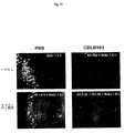


Classifications
-
- A—HUMAN NECESSITIES
- A61—MEDICAL OR VETERINARY SCIENCE; HYGIENE
- A61K—PREPARATIONS FOR MEDICAL, DENTAL OR TOILETRY PURPOSES
- A61K38/00—Medicinal preparations containing peptides
- A61K38/16—Peptides having more than 20 amino acids; Gastrins; Somatostatins; Melanotropins; Derivatives thereof
- A61K38/164—Peptides having more than 20 amino acids; Gastrins; Somatostatins; Melanotropins; Derivatives thereof from bacteria
-
- A—HUMAN NECESSITIES
- A61—MEDICAL OR VETERINARY SCIENCE; HYGIENE
- A61K—PREPARATIONS FOR MEDICAL, DENTAL OR TOILETRY PURPOSES
- A61K39/00—Medicinal preparations containing antigens or antibodies
- A61K39/02—Bacterial antigens
- A61K39/025—Enterobacteriales, e.g. Enterobacter
- A61K39/0275—Salmonella
-
- A—HUMAN NECESSITIES
- A61—MEDICAL OR VETERINARY SCIENCE; HYGIENE
- A61K—PREPARATIONS FOR MEDICAL, DENTAL OR TOILETRY PURPOSES
- A61K45/00—Medicinal preparations containing active ingredients not provided for in groups A61K31/00 - A61K41/00
- A61K45/06—Mixtures of active ingredients without chemical characterisation, e.g. antiphlogistics and cardiaca
-
- A—HUMAN NECESSITIES
- A61—MEDICAL OR VETERINARY SCIENCE; HYGIENE
- A61P—SPECIFIC THERAPEUTIC ACTIVITY OF CHEMICAL COMPOUNDS OR MEDICINAL PREPARATIONS
- A61P17/00—Drugs for dermatological disorders
- A61P17/02—Drugs for dermatological disorders for treating wounds, ulcers, burns, scars, keloids, or the like
-
- A—HUMAN NECESSITIES
- A61—MEDICAL OR VETERINARY SCIENCE; HYGIENE
- A61P—SPECIFIC THERAPEUTIC ACTIVITY OF CHEMICAL COMPOUNDS OR MEDICINAL PREPARATIONS
- A61P31/00—Antiinfectives, i.e. antibiotics, antiseptics, chemotherapeutics
- A61P31/04—Antibacterial agents
-
- A—HUMAN NECESSITIES
- A61—MEDICAL OR VETERINARY SCIENCE; HYGIENE
- A61P—SPECIFIC THERAPEUTIC ACTIVITY OF CHEMICAL COMPOUNDS OR MEDICINAL PREPARATIONS
- A61P31/00—Antiinfectives, i.e. antibiotics, antiseptics, chemotherapeutics
- A61P31/12—Antivirals
-
- A—HUMAN NECESSITIES
- A61—MEDICAL OR VETERINARY SCIENCE; HYGIENE
- A61P—SPECIFIC THERAPEUTIC ACTIVITY OF CHEMICAL COMPOUNDS OR MEDICINAL PREPARATIONS
- A61P35/00—Antineoplastic agents
-
- A—HUMAN NECESSITIES
- A61—MEDICAL OR VETERINARY SCIENCE; HYGIENE
- A61P—SPECIFIC THERAPEUTIC ACTIVITY OF CHEMICAL COMPOUNDS OR MEDICINAL PREPARATIONS
- A61P39/00—General protective or antinoxious agents
-
- A—HUMAN NECESSITIES
- A61—MEDICAL OR VETERINARY SCIENCE; HYGIENE
- A61P—SPECIFIC THERAPEUTIC ACTIVITY OF CHEMICAL COMPOUNDS OR MEDICINAL PREPARATIONS
- A61P39/00—General protective or antinoxious agents
- A61P39/02—Antidotes
-
- A—HUMAN NECESSITIES
- A61—MEDICAL OR VETERINARY SCIENCE; HYGIENE
- A61P—SPECIFIC THERAPEUTIC ACTIVITY OF CHEMICAL COMPOUNDS OR MEDICINAL PREPARATIONS
- A61P43/00—Drugs for specific purposes, not provided for in groups A61P1/00-A61P41/00
-
- C—CHEMISTRY; METALLURGY
- C07—ORGANIC CHEMISTRY
- C07K—PEPTIDES
- C07K14/00—Peptides having more than 20 amino acids; Gastrins; Somatostatins; Melanotropins; Derivatives thereof
- C07K14/195—Peptides having more than 20 amino acids; Gastrins; Somatostatins; Melanotropins; Derivatives thereof from bacteria
- C07K14/24—Peptides having more than 20 amino acids; Gastrins; Somatostatins; Melanotropins; Derivatives thereof from bacteria from Enterobacteriaceae (F), e.g. Citrobacter, Serratia, Proteus, Providencia, Morganella, Yersinia
- C07K14/255—Salmonella (G)
-
- Y—GENERAL TAGGING OF NEW TECHNOLOGICAL DEVELOPMENTS; GENERAL TAGGING OF CROSS-SECTIONAL TECHNOLOGIES SPANNING OVER SEVERAL SECTIONS OF THE IPC; TECHNICAL SUBJECTS COVERED BY FORMER USPC CROSS-REFERENCE ART COLLECTIONS [XRACs] AND DIGESTS
- Y02—TECHNOLOGIES OR APPLICATIONS FOR MITIGATION OR ADAPTATION AGAINST CLIMATE CHANGE
- Y02A—TECHNOLOGIES FOR ADAPTATION TO CLIMATE CHANGE
- Y02A50/00—TECHNOLOGIES FOR ADAPTATION TO CLIMATE CHANGE in human health protection, e.g. against extreme weather
- Y02A50/30—Against vector-borne diseases, e.g. mosquito-borne, fly-borne, tick-borne or waterborne diseases whose impact is exacerbated by climate change
-
- Y—GENERAL TAGGING OF NEW TECHNOLOGICAL DEVELOPMENTS; GENERAL TAGGING OF CROSS-SECTIONAL TECHNOLOGIES SPANNING OVER SEVERAL SECTIONS OF THE IPC; TECHNICAL SUBJECTS COVERED BY FORMER USPC CROSS-REFERENCE ART COLLECTIONS [XRACs] AND DIGESTS
- Y10—TECHNICAL SUBJECTS COVERED BY FORMER USPC
- Y10S—TECHNICAL SUBJECTS COVERED BY FORMER USPC CROSS-REFERENCE ART COLLECTIONS [XRACs] AND DIGESTS
- Y10S530/00—Chemistry: natural resins or derivatives; peptides or proteins; lignins or reaction products thereof
- Y10S530/82—Proteins from microorganisms
- Y10S530/825—Bacteria
Landscapes
- Health & Medical Sciences (AREA)
- Life Sciences & Earth Sciences (AREA)
- Chemical & Material Sciences (AREA)
- Medicinal Chemistry (AREA)
- General Health & Medical Sciences (AREA)
- Animal Behavior & Ethology (AREA)
- Veterinary Medicine (AREA)
- Public Health (AREA)
- Pharmacology & Pharmacy (AREA)
- Organic Chemistry (AREA)
- Nuclear Medicine, Radiotherapy & Molecular Imaging (AREA)
- General Chemical & Material Sciences (AREA)
- Chemical Kinetics & Catalysis (AREA)
- Epidemiology (AREA)
- Bioinformatics & Cheminformatics (AREA)
- Engineering & Computer Science (AREA)
- Proteomics, Peptides & Aminoacids (AREA)
- Gastroenterology & Hepatology (AREA)
- Immunology (AREA)
- Oncology (AREA)
- Toxicology (AREA)
- Communicable Diseases (AREA)
- Biophysics (AREA)
- Biochemistry (AREA)
- Genetics & Genomics (AREA)
- Molecular Biology (AREA)
- Microbiology (AREA)
- Mycology (AREA)
- Dermatology (AREA)
- Virology (AREA)
- Medicines That Contain Protein Lipid Enzymes And Other Medicines (AREA)
- Peptides Or Proteins (AREA)
Description
関連出願への相互参照
本願は、2004年12月22日に出願された米国仮特許出願第60/639,826号の利益を主張する。米国仮特許出願第60/639,826号は、本明細書中に参考として援用される。
This application claims the benefit of US Provisional Patent Application No. 60 / 639,826, filed Dec. 22, 2004. US Provisional Patent Application No. 60 / 639,826 is hereby incorporated by reference.
発明の分野
本発明は、アポトーシスの影響から哺乳動物を防護するためのフラゲリン関連ポリペプチドの使用に関する。より詳細には、本発明は、放射線および癌治療などのストレスへの曝露から哺乳動物を防護するためのフラゲリン関連ポリペプチドの使用に関する。
The present invention relates to the use of flagellin-related polypeptides to protect mammals from the effects of apoptosis. More particularly, the present invention relates to the use of flagellin-related polypeptides to protect mammals from exposure to stress, such as radiation and cancer treatment.
発明の背景
正常細胞から腫瘍細胞への進行は、負の成長調節機構の喪失(成長阻害刺激に対する耐性が含まれる)ならびに成長因子およびホルモンへの依存の欠如を伴う。放射線または細胞障害薬に基づく従来の癌治療は、正常細胞および悪生細胞の成長調節の相違に依存する。従来の癌治療は、細胞を重篤な遺伝動性ストレスに供する。これらの状態下で、大部分の正常細胞は成長が停止するようになり、それにより、生き残る一方で、腫瘍細胞は分裂し続け、死滅する。
BACKGROUND OF THE INVENTION Progression from normal cells to tumor cells is accompanied by a loss of negative growth regulatory mechanisms (including resistance to growth inhibitory stimuli) and a lack of dependence on growth factors and hormones. Traditional cancer therapies based on radiation or cytotoxic drugs rely on differences in growth regulation between normal and evil cells. Conventional cancer treatments subject the cells to severe genetic dynamic stress. Under these conditions, most normal cells begin to stop growing, thereby surviving while tumor cells continue to divide and die.
しかし、従来の癌治療ストラテジーは、正常な急速に分割する組織またはアポトーシス性組織を危険にさらす性質がある。これらの正常な急速に分割する細胞への損傷により、癌治療の周知の副作用(感受性組織:造血、小腸、毛包)が生じる。このような組織の天然の感受性は、癌細胞が頻繁に自殺(アポトーシス)機構を欠損し、治療手順が正常な感受性組織を死滅させるが癌細胞に損傷を与えなくなることがあるという事実によって複雑になる。癌療法の副作用を最小にするという従来の試みは、(a)腫瘍細胞の治療に対する感受性をより高めること、(b)腫瘍細胞に対する癌療法の特異性をより高めること、または(c)治療後(例えば、エリスロポイエチン、GM−CSF、およびKGF)の正常細胞の再生を促進することに基づく。 However, conventional cancer treatment strategies have the property of jeopardizing normal, rapidly dividing or apoptotic tissue. Damage to these normal, rapidly dividing cells causes well-known side effects of cancer treatment (sensitive tissues: hematopoiesis, small intestine, hair follicles). The natural susceptibility of such tissues is complicated by the fact that cancer cells frequently lack suicide (apoptosis) mechanisms and the treatment procedure can kill normal sensitive tissues but not damage the cancer cells. Become. Prior attempts to minimize the side effects of cancer therapy include (a) increasing the sensitivity of tumor cells to treatment, (b) increasing the specificity of cancer therapy for tumor cells, or (c) after treatment. Based on promoting the regeneration of normal cells (eg, erythropoietin, GM-CSF, and KGF).
癌治療における化学療法および放射線療法に関連する副作用を緩和するための治療薬が依然として必要とされている。本発明は、これらのニーズを満たし、他の関連する利点を提供する。 There remains a need for therapeutic agents to alleviate the side effects associated with chemotherapy and radiation therapy in cancer treatment. The present invention fulfills these needs and provides other related advantages.
発明の概要
薬学的有効量のフラゲリンを含む組成物を患者に投与する工程を含む、アポトーシスを誘発する1つまたは複数の治療または状態から哺乳動物を防護する方法。フラゲリンは、配列番号1、または配列番号1のフラグメント、改変体、アナログ、ホモログ、誘導体、またはその組み合わせを含み得る。フラゲリンは、TLR−5媒介活性を誘導し得る。
Summary of the Invention A method of protecting a mammal from one or more treatments or conditions that induce apoptosis, comprising administering to a patient a composition comprising a pharmaceutically effective amount of flagellin. Flagellin can comprise SEQ ID NO: 1, or a fragment, variant, analog, homolog, derivative, or combination thereof of SEQ ID NO: 1. Flagellin can induce TLR-5 mediated activity.
フラゲリンは、配列番号1のアミノ酸1〜174および418〜505と少なくとも30%同一であり得る。フラゲリンは、89位、90位、91位、95位、98位、101位、115位、422位、423位、426位、431位、436位、および452位からなる群から選択される少なくとも10個の保存アミノ酸を含み得る。フラゲリンは、配列番号1、8、10、12、30、32、34、36、38、40、42、44、46、48、50、および52からなる群から選択される配列を含み得る。 Flagellin can be at least 30% identical to amino acids 1-174 and 418-505 of SEQ ID NO: 1. Flagellin is at least selected from the group consisting of 89, 90, 91, 95, 98, 101, 115, 422, 423, 426, 431, 436, and 452. It may contain 10 conserved amino acids. The flagellin may comprise a sequence selected from the group consisting of SEQ ID NOs: 1, 8, 10, 12, 30, 32, 34, 36, 38, 40, 42, 44, 46, 48, 50, and 52.
フラゲリンを使用して、癌治療(化学療法または放射線療法であり得る)を受けている哺乳動物を治療することができる。フラゲリンを使用して、放射線を被爆した哺乳動物を治療することができる。フラゲリンを、放射線防護剤と組み合わせて投与することができる。フラゲリンを使用して、哺乳動物の創傷、中毒、細菌感染、ウイルス感染、および温度衝撃を治療することができる。フラゲリンを使用して、組織(胃腸管、肺、腎臓、肝臓、心血管系、血管内皮、中枢神経系、末梢神経系、造血前駆細胞、免疫系、および毛包が含まれる)のアポトーシスを防護することができる。フラゲリンを使用して、哺乳動物の敗血症を予防することもできる。 Flagellin can be used to treat a mammal undergoing cancer treatment, which can be chemotherapy or radiation therapy. Flagellin can be used to treat mammals exposed to radiation. Flagellin can be administered in combination with a radioprotector. Flagellin can be used to treat mammalian wounds, poisoning, bacterial infections, viral infections, and thermal shock. Use flagellin to protect the apoptosis of tissues (including gastrointestinal tract, lung, kidney, liver, cardiovascular system, vascular endothelium, central nervous system, peripheral nervous system, hematopoietic progenitor cells, immune system, and hair follicle) can do. Flagellin can also be used to prevent sepsis in mammals.
本発明は、薬学的に許容可能な量のNF−κBを誘導する薬剤を含む組成物を哺乳動物に投与する工程を含む、構成性に活性なNF−κB癌を罹患した哺乳動物の治療方法に関する。薬剤は、フラゲリンであり得る。薬剤を、癌治療前、同時、または癌治療後に投与することができる。治療は、化学療法または放射線療法であり得る。 The present invention relates to a method for treating a mammal suffering from constitutively active NF-κB cancer, comprising the step of administering to the mammal a composition comprising a pharmaceutically acceptable amount of an agent that induces NF-κB. About. The agent can be flagellin. The agent can be administered before, simultaneously with, or after cancer treatment. The treatment can be chemotherapy or radiation therapy.
本発明はまた、哺乳動物に薬学的に許容可能な量のNF−κBを誘導する薬剤を含む組成物を投与する工程を含む、癌治療に起因する正常組織に損傷を受けた哺乳動物の治療方法に関する。薬剤は、フラゲリンであり得る。薬剤を、癌治療前、同時、または癌治療後に投与することができる。治療は、化学療法または放射線療法であり得る。 The invention also treats a mammal damaged to normal tissue resulting from cancer treatment, comprising administering to the mammal a composition comprising a pharmaceutically acceptable amount of an agent that induces NF-κB. Regarding the method. The agent can be flagellin. The agent can be administered before, simultaneously with, or after cancer treatment. The treatment can be chemotherapy or radiation therapy.
本発明はまた、哺乳動物に薬学的に許容可能な量のNF−κBを誘導する薬剤を含む組成物を投与する工程を含む、ストレスに起因する正常組織に損傷を受けた哺乳動物の治療方法に関する。薬剤は、フラゲリンであり得る。薬剤を、哺乳動物が罹患した疾患の治療前、同時、または治療後に投与することができる。 The present invention also provides a method of treating a mammal damaged in normal tissue caused by stress, comprising the step of administering to the mammal a composition comprising a pharmaceutically acceptable amount of an agent that induces NF-κB. About. The agent can be flagellin. The agent can be administered before, simultaneously with, or after treatment of a disease that affects a mammal.
本発明はまた、哺乳動物に薬学的に許容可能な量のNF−κBを誘導する薬剤を含む組成物を投与する工程を含む、哺乳動物の細胞老化の調整方法に関する。薬剤は、フラゲリンであり得る。薬剤を、哺乳動物が罹患した疾患の治療前、同時、または治療後に投与することができる。 The present invention also relates to a method of regulating mammalian cell senescence comprising the step of administering to a mammal a composition comprising a pharmaceutically acceptable amount of an agent that induces NF-κB. The agent can be flagellin. The agent can be administered before, simultaneously with, or after treatment of a disease that affects a mammal.
本発明はまた、NF−κB活性を誘導する薬剤、化学療法薬、および任意選択的に薬学的に許容可能なアジュバント、希釈剤、またはキャリアを含む薬学的組成物に関する。薬剤は、フラゲリンであり得る。 The invention also relates to pharmaceutical compositions comprising an agent that induces NF-κB activity, a chemotherapeutic agent, and optionally a pharmaceutically acceptable adjuvant, diluent, or carrier. The agent can be flagellin.
本発明はまた、疑われる誘導因子をNF−κB活性化発現系に添加する工程と、コントロールをNF−κB活性化発現系に別で添加する工程と、それによりNF−κB活性化発現系レベルを増加させる能力によってNF−κBの誘導因子を同定することとを含む、NF−κBの誘導因子のスクリーニング方法に関する。 The present invention also includes a step of adding a suspected inducer to the NF-κB activation expression system, a step of separately adding a control to the NF-κB activation expression system, and thereby a level of the NF-κB activation expression system. Identifying an inducer of NF-κB by the ability to increase NF-κB.
本発明はまた、哺乳動物に薬学的有効量のNF−κBを誘導する薬剤を含む組成物を投与する工程を含む、哺乳動物を放射線の影響から防護する方法に関する。薬剤は、フラゲリンであってよく、これは、Salmonella属に由来し得る。組成物を、放射線防護剤と組み合わせて投与することができる。放射線防護剤は、抗酸化剤であってよく、これは、アミホスチン(amifostine)またはビタミンEであり得る。放射線防護剤はまた、サイトカインであってよく、これは、幹細胞刺激因子であり得る。 The invention also relates to a method of protecting a mammal from the effects of radiation, comprising administering to the mammal a composition comprising a pharmaceutically effective amount of an agent that induces NF-κB. The drug may be flagellin, which can be derived from the genus Salmonella. The composition can be administered in combination with a radioprotectant. The radioprotector may be an antioxidant, which may be amifostine or vitamin E. The radioprotector can also be a cytokine, which can be a stem cell stimulator.
本発明は、患者に薬学的有効量のNF−κBを誘導する薬剤を含む組成物を投与する工程を含む、アポトーシスを誘発する1つまたは複数の治療または状態から患者を防護する方法に関する。薬剤は、フラゲリンであってよく、これは、Salmonella属に由来し得る。治療は癌治療であってよく、これは、化学療法または放射線療法であり得る。状態はストレスであってよく、これは、放射線、創傷、中毒、感染、および温度衝撃であり得る。 The present invention relates to a method of protecting a patient from one or more treatments or conditions that induce apoptosis, comprising administering to the patient a composition comprising a pharmaceutically effective amount of an agent that induces NF-κB. The drug may be flagellin, which can be derived from the genus Salmonella. The treatment may be a cancer treatment, which may be chemotherapy or radiation therapy. The condition can be stress, which can be radiation, wounding, addiction, infection, and temperature shock.
本発明はまた、疑われるモジュレーターを細胞ベースのアポトーシス系に添加する工程と、コントロールを細胞ベースのアポトーシス系に別で添加する工程と、それによりアポトーシス率を変化させる能力によってアポトーシスのモジュレーターを同定することと、疑われるモジュレーターが哺乳動物の寄生虫または共生生物に由来することとを含む、アポトーシスのモジュレーターのスクリーニング方法に関する。 The invention also identifies modulators of apoptosis by adding a suspected modulator to a cell-based apoptotic system, adding a control separately to the cell-based apoptotic system, and thereby the ability to change the rate of apoptosis. And a method for screening for modulators of apoptosis, wherein the suspected modulator is derived from a mammalian parasite or commensal.
本発明はまた、疑われるモジュレーターをNF−κB活性化発現系に添加する工程と、コントロールをNF−κB活性化発現系に別で添加する工程と、それによりNF−κB活性化発現率を変化させる能力によってNF−κBのモジュレーターを同定することと、前記疑われるモジュレーターが、哺乳動物の寄生虫に由来することとを含む、NF−κBのモジュレーターのスクリーニング方法に関する。寄生虫は、Salmonella属、マイコプラズマ属、およびクラミジア属に由来し得るが、これらに限定されない。 The present invention also includes a step of adding a suspected modulator to the NF-κB activation expression system, a step of separately adding a control to the NF-κB activation expression system, and thereby changing the NF-κB activation expression rate. The present invention relates to a method of screening for a modulator of NF-κB, comprising identifying a modulator of NF-κB by the ability to cause and that the suspected modulator is derived from a mammalian parasite. Parasites can be derived from, but not limited to, Salmonella, Mycoplasma, and Chlamydia.
本発明はまた、本明細書中に記載の任意のスクリーニング方法によって同定されるモジュレーターに関する。本発明はまた、本明細書中に記載のモジュレーターを含む組成物に関する。組成物は、薬学的に許容可能な量の本明細書中に記載のモジュレーターを含む薬学的組成物であり得る。 The present invention also relates to modulators identified by any screening method described herein. The invention also relates to a composition comprising a modulator as described herein. The composition can be a pharmaceutical composition comprising a pharmaceutically acceptable amount of a modulator as described herein.
本発明はまた、アポトーシスを増強するモジュレーターを含む薬学的組成物をこのような治療を必要とする被験体に投与する工程を含む、癌の治療方法に関する。 The present invention also relates to a method of treating cancer comprising administering a pharmaceutical composition comprising a modulator that enhances apoptosis to a subject in need of such treatment.
本発明はまた、患者にアポトーシスを阻害するモジュレーターを含む薬学的組成物を投与する工程を含む、アポトーシスを誘発する1つまたは複数の治療から患者を防護する方法を提供する。1つまたは複数の治療は、癌治療であり得る。癌治療は、化学療法または
放射線療法であり得る。
The invention also provides a method of protecting a patient from one or more treatments that induce apoptosis, comprising administering to the patient a pharmaceutical composition comprising a modulator that inhibits apoptosis. The one or more treatments can be a cancer treatment. The cancer treatment can be chemotherapy or radiation therapy.
(詳細な説明)
本発明は、ストレス(化学療法、放射線療法、および照射が含まれるが、これらに限定されない)によって生じたアポトーシスからの正常な細胞および組織の防護に関する。細胞におけるアポトーシスの調節には以下の2つの主な機構が存在する:p53経路(アポトーシス促進性)およびNF−κB経路(抗アポトーシス性)。両経路は、腫瘍で頻繁に無秩序化する:通常p53が喪失する一方で、NF−κBが構成性に活性になる。したがって、正常細胞におけるp53の阻害およびNF−κBの活性化により、癌治療などのストレスによって生じる死滅から正常細胞を防護することができるが、腫瘍細胞はこれらの調節機構が無秩序化されているので、腫瘍細胞により高い治療耐性を持たせられないであろう。これは、p53およびNF−κBがそれぞれ活性化および抑制の標的と考えられているp53およびNF−κBにおける従来の見解に矛盾する。
(Detailed explanation)
The present invention relates to the protection of normal cells and tissues from apoptosis caused by stress, including but not limited to chemotherapy, radiation therapy, and irradiation. There are two main mechanisms for the regulation of apoptosis in cells: the p53 pathway (pro-apoptotic) and the NF-κB pathway (anti-apoptotic). Both pathways are frequently disordered in tumors: NF-κB becomes constitutively active while p53 is usually lost. Therefore, inhibition of p53 and activation of NF-κB in normal cells can protect normal cells from death caused by stress such as cancer treatment, but tumor cells have their regulatory mechanisms disordered The tumor cells will not be made more resistant to treatment. This contradicts the traditional view of p53 and NF-κB, where p53 and NF-κB are considered targets for activation and repression, respectively.
本発明は、アポトーシスから正常細胞を防護するためのNF−κB活性の誘導に関する。哺乳動物におけるNF−κB活性の誘導により、正常細胞を、癌治療および高熱、有害線量の放射線被爆(例えば、原子力発電所、防衛産業または放射性医薬品生産の労働者、および軍人)、および細胞の老化で生じる細胞ストレスに帰するアポトーシスから防護することができる。NF−κBが多数の腫瘍細胞で構成性に活性であるので、NF−κB活性の誘導により、腫瘍細胞に有利な効果を与えることなく正常な細胞をアポトーシスから防護することができる。一旦正常な細胞が修復されると、NF−κB活性を正常レベルに修復することができる。造血系(免疫系が含まれる)、腸上皮、および毛包などの照射および化学療法感受性組織を防護するためにNF−κB活性を誘導することができる。 The present invention relates to the induction of NF-κB activity to protect normal cells from apoptosis. By induction of NF-κB activity in mammals, normal cells are treated for cancer and high fever, harmful doses of radiation exposure (eg, workers in nuclear power plants, defense industry or radiopharmaceutical production, and military personnel), and cellular aging. Can protect against apoptosis resulting from cellular stress. Since NF-κB is constitutively active in many tumor cells, induction of NF-κB activity can protect normal cells from apoptosis without having a beneficial effect on the tumor cells. Once normal cells are repaired, NF-κB activity can be restored to normal levels. NF-κB activity can be induced to protect irradiated and chemosensitive tissues such as the hematopoietic system (including the immune system), intestinal epithelium, and hair follicles.
NF−κB活性の誘導因子を、いくつかの他の適用に使用することもできる。種々の重篤な状態(照射、創傷、中毒、感染、老化、および温度ショックが含まれるが、これらに限定されない)への哺乳動物の被爆によって生じる病的結果および死は、ストレス応答の正常な生理学的機構の活性(プログラム細胞死(アポトーシス)の誘導または生体活性タンパク質(サイトカイン)の放出など)に起因し得る。 Inducers of NF-κB activity can also be used for several other applications. The morbidity and death resulting from mammalian exposure to various serious conditions, including but not limited to irradiation, wounding, poisoning, infection, aging, and temperature shock, is normal for stress responses. It can be attributed to the activity of physiological mechanisms, such as induction of programmed cell death (apoptosis) or release of bioactive proteins (cytokines).
アポトーシスは、通常、創傷細胞および遺伝子損傷細胞由来の組織を「浄化する」ように機能する一方で、サイトカインは、病原体に対する生物の防護系を動員するように機能を果たす。しかし、重篤な損傷の条件下では、両ストレス応答機構自体が自滅の原因として作用し得る。例えば、照射による死亡は、造血系、免疫系、および消化系で起こる大規模なp53媒介性アポトーシスに起因し得る。NF−κBの妥当な薬理学的調節により、重篤なストレス条件下での生存率が増加し得る。これらの要因の調節により、損傷器官由来の細胞の炎症応答および生死の決断の両方を調節することが可能である。NF−κB誘導因子の投与によってアポトーシスから防護することができる組織には、胃腸管、肺、腎臓、肝臓、心血管系、血管内皮、中枢神経系、末梢神経系、造血前駆細胞、免疫系、および毛包が含まれるが、これらに限定されない。 Apoptosis normally functions to “cleanse” tissue from wound cells and genetically damaged cells, while cytokines function to mobilize organisms' defense systems against pathogens. However, under severe damage conditions, both stress response mechanisms themselves can act as a cause of self-destruction. For example, death from irradiation can be attributed to massive p53-mediated apoptosis that occurs in the hematopoietic, immune, and digestive systems. With reasonable pharmacological modulation of NF-κB, survival under severe stress conditions can be increased. By modulating these factors, it is possible to regulate both the inflammatory response and the life / death decision of cells from the damaged organ. Tissues that can be protected from apoptosis by administration of NF-κB inducer include gastrointestinal tract, lung, kidney, liver, cardiovascular system, vascular endothelium, central nervous system, peripheral nervous system, hematopoietic progenitor cell, immune system, And hair follicles, including but not limited to.
NF−κBの防護的役割は、以下をコードする複数の遺伝子の転写活性化によって媒介される:a)両方の主なアポトーシス経路を遮断する抗アポトーシスタンパク質、b)HPおよび他の幹細胞の増殖および生存を誘導するサイトカインおよび成長因子、およびc)強力なROS除去抗酸化タンパク質(MnSOD(SOD−2)など)。したがって、放射線防護のためのNF−κBの一過性活性化により、Toll様受容体の活性化を介してNF−κBの活性化が到達する場合に達成することができる同時免疫刺激効果によって癌患者のアポトーシスの抑制だけでなく、二次癌の発生率を減少させる能力を達成することが可能である。 The protective role of NF-κB is mediated by transcriptional activation of multiple genes encoding: a) anti-apoptotic proteins that block both major apoptotic pathways, b) proliferation of HP and other stem cells and Cytokines and growth factors that induce survival, and c) potent ROS-removal antioxidant proteins such as MnSOD (SOD-2). Thus, transient activation of NF-κB for radioprotection results in cancer by a co-immunostimulatory effect that can be achieved when NF-κB activation reaches via Toll-like receptor activation. It is possible to achieve not only the suppression of patient apoptosis, but also the ability to reduce the incidence of secondary cancers.
標的としてのNF−κB経路の別の魅力的な特性は、候補放射線防護剤と見なすことができる多数の天然因子によるその活性化である。これらのうちでは、複数の病原体関連分子パターン(PAMP)である。PAMPは、宿主生物で見出されない分子であり、巨大な病原体群を特徴とし、容易に変異することができない。これらは、Toll様受容体(TLR)(自然免疫の重要なセンサーエレメント)によって認識される。TLRは、直接またはサイトカイン放出を介した免疫細胞の移動および活性化の誘導による免疫系の第1の警告機構として作用する。TLRは、ホモ二量体およびヘテロ二量体として作用することが公知のI型膜タンパク質である。リガンド結合の際、TLRは、MyD88タンパク質(ほとんどのTLRの不可欠なシグナル伝達アダプター)を補充する。次に起こるシグナル伝達カスケードにより、(i)NF−κB経路の活性化、および(ii)MAPK(JunN末端キナーゼ(JNK)が含まれる)の活性化が含まれる効果が導かれる。Toll様受容体リガンドによるNF−κB経路の活性化により、リガンドが潜在的な放射線防護剤として魅力的になる。サイトカインと異なり、多数のPAMPはTLRの活性化以外はほとんど効果がないので、副作用を生じる可能性が低い。さらに、多数のPAMPは、ヒトに存在する。 Another attractive property of the NF-κB pathway as a target is its activation by a number of natural factors that can be considered candidate radioprotectors. Among these are multiple pathogen associated molecular patterns (PAMPs). PAMP is a molecule that is not found in the host organism, is characterized by a large group of pathogens and cannot be easily mutated. These are recognized by the Toll-like receptor (TLR), an important sensor element of innate immunity. TLRs act as a primary warning mechanism of the immune system by induction of immune cell migration and activation, either directly or through cytokine release. TLR is a type I membrane protein known to act as a homodimer and a heterodimer. Upon ligand binding, the TLR recruits MyD88 protein, an essential signaling adapter for most TLRs. The subsequent signaling cascade leads to effects including (i) activation of the NF-κB pathway, and (ii) activation of MAPK, which includes JunN-terminal kinase (JNK). Activation of the NF-κB pathway by Toll-like receptor ligands makes the ligands attractive as potential radioprotectors. Unlike cytokines, many PAMPs have little effect other than TLR activation and are therefore less likely to cause side effects. In addition, many PAMPs exist in humans.
その免疫細胞活性化機能と一致して、全てのTLRは、脾臓および末梢血白血球で発現し、他のリンパ器官およびリンパ球の一部でより多数のTLR特異的発現パターンが存在する。しかし、TLRは、体内の他の組織および器官でも発現する(例えば、TLR1は遍在的に発現し、TLR5は胃腸管上皮および内皮でも見出される一方で、TLR2、6、7、および8は、肺で発現することが公知である)。 Consistent with its immune cell activation function, all TLRs are expressed in spleen and peripheral blood leukocytes, and there are more TLR-specific expression patterns in other lymphoid organs and parts of lymphocytes. However, TLRs are also expressed in other tissues and organs in the body (eg, TLR1 is ubiquitously expressed and TLR5 is also found in gastrointestinal epithelium and endothelium, while TLR2, 6, 7, and 8 are It is known to be expressed in the lung).
1.定義
本明細書中で使用される専門用語は特定の実施形態を説明することのみを目的とし、本発明を制限しないと理解すべきである。明細書および添付の特許請求の範囲で使用する場合、文脈上で明確に別の記載を行わない限り、単数形「a」、「an」、および「the」には複数形が含まれることに留意しなければならない。
1. Definitions It should be understood that the terminology used herein is for the purpose of describing particular embodiments only and is not intended to limit the invention. As used in the specification and appended claims, the singular forms “a”, “an”, and “the” include plural referents unless the context clearly dictates otherwise. You have to be careful.
本明細書中で使用される、用語「投与」は、NF−κB活性を誘導する薬剤の投薬量を説明するために使用される場合、薬物の単回用量または複数回用量を意味する。 As used herein, the term “administration” when used to describe the dosage of an agent that induces NF-κB activity means a single dose or multiple doses of a drug.
本明細書中で使用される、用語「アナログ」は、ペプチドまたはポリペプチドの文脈で使用される場合、従来のアミノ酸組由来の1つまたは複数の非標準アミノ酸または他の構造変形形態を意味する。 As used herein, the term “analog” when used in the context of a peptide or polypeptide means one or more non-standard amino acids or other structural variations from a conventional set of amino acids. .
本明細書中で使用される、用語「抗体」は、IgG、IgM、IgA、IgD、もしくはIgEの抗体クラス、またはそのフラグメントもしくは誘導体(Fab、F(ab’)2、Fd、および単鎖抗体が含まれる)、ダイアボディ(diabody)、二重特異性抗体、二機能性抗体、およびその誘導体を意味する。抗体は、所望のエピトープまたはこれに由来する配列に対して十分な結合特異性を示すモノクローナル抗体、ポリクローナル抗体、親和性精製抗体、またはその混合物であり得る。抗体はまた、キメラ抗体であり得る。抗体を、当該分野で公知の1つまたは複数の化学的部分、ペプチド部分、またはポリペプチド部分の結合によって誘導体化することができる。抗体を、化学部分と抱合することができる。 As used herein, the term “antibody” refers to an antibody class of IgG, IgM, IgA, IgD, or IgE, or fragments or derivatives thereof (Fab, F (ab ′) 2 , Fd, and single chain antibodies ), Diabodies, bispecific antibodies, bifunctional antibodies, and derivatives thereof. The antibody can be a monoclonal antibody, a polyclonal antibody, an affinity purified antibody, or a mixture thereof that exhibits sufficient binding specificity for the desired epitope or sequence derived therefrom. The antibody can also be a chimeric antibody. An antibody can be derivatized by conjugation of one or more chemical moieties, peptide moieties, or polypeptide moieties known in the art. The antibody can be conjugated with a chemical moiety.
本明細書中で使用される、用語「アポトーシス」は、細胞小器官の完全性が保護される細胞容積の進行性の収縮、光学顕微鏡または電子顕微鏡によって認められるクロマチンの凝縮(すなわち、核凝縮)、および/または遠心分離沈殿アッセイによって決定されるヌクレオソームサイズのフラグメントへのDNAの切断を含む細胞死の形態をいう。食細胞によるインタクトな細胞フラグメント(「アポトーシス小体」)の貪食によって細胞膜の完全性が失われた(例えば、膜小胞形成)場合に細胞死が起こる。 As used herein, the term “apoptosis” refers to the progressive contraction of cell volume where the integrity of the organelle is protected, the condensation of chromatin as seen by light or electron microscopy (ie, nuclear condensation). , And / or a form of cell death involving the cleavage of DNA into nucleosome-sized fragments as determined by centrifugation precipitation assays. Cell death occurs when phagocytosis of intact cell fragments (“apoptotic bodies”) results in loss of cell membrane integrity (eg, membrane vesicle formation).
本明細書中で使用される、用語「癌」は、異常な無制御の細胞分裂に起因する任意の悪性の成長または腫瘍を意味し、リンパ系または血流を介して身体の他の部分に拡大し得る。 As used herein, the term “cancer” refers to any malignant growth or tumor resulting from abnormal uncontrolled cell division, and to other parts of the body via the lymphatic system or bloodstream. Can expand.
本明細書中で使用される、用語「癌治療」は、当該分野で公知の任意の癌治療(化学療法および放射線療法が含まれるが、これらに限定されない)を意味する。 As used herein, the term “cancer treatment” means any cancer treatment known in the art, including but not limited to chemotherapy and radiation therapy.
本明細書中で使用される、用語「〜との組み合わせ」は、NF−κB活性を誘導する薬剤の投与およびさらなる治療を説明するために使用する場合、薬剤を、さらなる治療前、同時、治療後、または規則正しく(metronomically)投与することができることを意味する。本明細書中で使用される、用語「〜共に」、「同時の」、または「同時に」は、本発明のさらなる治療および薬剤を、48時間以内、好ましくは24時間以内、より好ましくは12時間以内、さらにより好ましくは6時間以内、最も好ましくは3時間以内またはそれ未満で施すことを意味する。本明細書中で使用される、用語「規則正しく」は、さらなる治療と異なる時点且つ反復投与および/またはさらなる治療と比較して一定の頻度での薬剤の投与を意味する。 As used herein, the term “in combination with” when used to describe administration of an agent that induces NF-κB activity and further treatment, treats the agent prior to further treatment, concomitantly. It means that it can be administered later or regularly. As used herein, the terms “both”, “simultaneous”, or “simultaneously” refer to additional treatments and agents of the invention within 48 hours, preferably within 24 hours, more preferably 12 hours. Within, even more preferably within 6 hours, most preferably within 3 hours or less. As used herein, the term “regularly” refers to administration of a drug at a different time than the further treatment and at a certain frequency compared to repeated administration and / or further treatment.
薬剤を、さらなる治療前の任意の時点(さらなる治療の約48時間前、46時間前、44時間前、42時間前、40時間前、38時間前、36時間前、34時間前、32時間前、30時間前、28時間前、26時間前、24時間前、22時間前、20時間前、18時間前、16時間前、14時間前、12時間前、10時間前、8時間前、6時間前、4時間前、3時間前、2時間前、または1時間前が含まれるが、これらに限定されない)で投与することができる。薬剤を、さらなる治療後の一定の時点(被爆から約1時間後、2時間後、3時間後、4時間後、6時間後、8時間後、10時間後、12時間後、14時間後、16時間後、18時間後、20時間後、22時間後、24時間後、26時間後、28時間後、30時間後、32時間後、34時間後、36時間後、38時間後、40時間後、42時間後、44時間後、46時間後、または48時間後が含まれるが、これらに限定されない)で投与することができる。 The drug can be administered at any time before further treatment (approximately 48 hours, 46 hours, 44 hours, 42 hours, 40 hours, 38 hours, 36 hours, 36 hours, 34 hours, 32 hours before further treatment). 30 hours, 28 hours, 26 hours, 24 hours, 22 hours, 20 hours, 18 hours, 16 hours, 14 hours, 12 hours, 10 hours, 8 hours, 6 hours Including, but not limited to, 4 hours prior, 3 hours prior, 2 hours prior, or 1 hour prior). The drug was administered at a fixed time after further treatment (approximately 1 hour, 2 hours, 3 hours, 4 hours, 6 hours, 8 hours, 10 hours, 12 hours, 14 hours after exposure) 16 hours, 18 hours, 20 hours, 22 hours, 24 hours, 26 hours, 28 hours, 30 hours, 32 hours, 34 hours, 36 hours, 36 hours, 38 hours, 40 hours , 42 hours, 44 hours, 46 hours, or 48 hours later, including but not limited to).
本明細書中で使用される、用語「誘導体」は、ペプチドまたはポリペプチドの文脈で使用される場合、一次構造以外が異なるペプチドまたはポリペプチド(アミノ酸およびアミノ酸アナログ)を意味する。例として、誘導体は、グリコシル化(翻訳後修飾の一形態)が異なり得る。例えば、ペプチドまたはポリペプチドは、異種系における発現に起因するグリコシル化パターンを示し得る。少なくとも1つの生物活性が保持される場合、これらのペプチドまたはポリペプチドは、本発明の誘導体である。他の誘導体には、共有結合的に修飾されたN末端またはC末端を有するペプチドまたは融合ポリペプチド、PEG化ペプチドまたはポリペプチド、脂質部分に会合したペプチドまたはポリペプチド、アルキル化ペプチドまたはポリペプチド、アミノ酸側鎖の官能基を介して他のペプチドに結合したペプチドまたはポリペプチド、ポリペプチドまたはキメラ、および当該分野で理解されているさらなる修飾物が含まれるが、これらに限定されない。 As used herein, the term “derivative”, when used in the context of a peptide or polypeptide, means a peptide or polypeptide (amino acids and amino acid analogs) that differ in terms other than primary structure. As an example, derivatives may differ in glycosylation (a form of post-translational modification). For example, a peptide or polypeptide can exhibit a glycosylation pattern resulting from expression in a heterologous system. If at least one biological activity is retained, these peptides or polypeptides are derivatives of the invention. Other derivatives include covalently modified N- or C-terminal peptides or fusion polypeptides, PEGylated peptides or polypeptides, peptides or polypeptides associated with lipid moieties, alkylated peptides or polypeptides, Peptides or polypeptides linked to other peptides through amino acid side chain functional groups, polypeptides or chimeras, and additional modifications understood in the art, but are not limited to these.
本明細書中で使用される、用語「フラグメント」は、ペプチドまたはポリペプチドの文脈で使用される場合、基準ペプチドまたはポリペプチドの一部を意味する。 As used herein, the term “fragment” when used in the context of a peptide or polypeptide means a portion of a reference peptide or polypeptide.
本明細書中で使用される、用語「ホモログ」は、ペプチドまたはポリペプチドの文脈で使用される場合、共通の進化上の祖先を有するペプチドまたはポリペプチドを意味する。 As used herein, the term “homolog” when used in the context of a peptide or polypeptide means a peptide or polypeptide that has a common evolutionary ancestry.
本明細書中で使用される、用語「処置する」または「処置」は、状態からの哺乳動物の防護をいう場合、状態の予防、抑制、防止、または消失を意味する。状態の予防は、状態の発症前に本発明の組成物を哺乳動物に投与する工程を含む。状態の抑制は、状態の誘導後であるがその臨床的外観前に哺乳動物に本発明の組成物を投与する工程を含む。状態の防止は、状態が軽減または維持されるように状態の臨床的外観後に哺乳動物に本発明の組成物を投与する工程を含む。溶媒の消失は、哺乳動物がもはや状態を罹患しないように状態の臨床的外観後に哺乳動物に本発明の組成物を投与する工程を含む。 As used herein, the term “treating” or “treatment” when referring to the protection of a mammal from a condition means prevention, suppression, prevention, or elimination of the condition. Prevention of the condition involves administering the composition of the invention to the mammal prior to the onset of the condition. Suppression of the condition involves administering the composition of the invention to the mammal after induction of the condition but before its clinical appearance. Prevention of the condition involves administering the composition of the invention to the mammal after the clinical appearance of the condition so that the condition is reduced or maintained. The disappearance of the solvent involves administering the composition of the invention to the mammal after the clinical appearance of the condition so that the mammal no longer suffers from the condition.
本明細書中で使用される、用語「腫瘍細胞」は、癌に関連する任意の細胞を意味する。 As used herein, the term “tumor cell” means any cell associated with cancer.
本明細書中で使用される、用語「改変体」は、ペプチドまたはポリペプチドの文脈で使用される場合、アミノ酸の挿入、欠失、または保存的置換によってアミノ酸配列が異なるが、少なくとも1つの生物活性を保持するペプチドまたはポリペプチドを意味する。「生物活性」の代表例には、TLR5に結合する能力および特異的抗体によって結合される能力が含まれるが、これらに限定されない。アミノ酸の保存的置換(すなわち、アミノ酸の類似の性質(例えば、親水性、荷電領域の程度および分布)を有する異なるアミノ酸への置換)は、当該分野で典型的には小さな変化と認識されている。これらの小さな変化を、当該分野で理解されているように、アミノ酸のヒドロパシー指標を考慮することによって一部を同定することができる。Kyte et al,J.MoI.Biol.157:105−132(1982)。アミノ酸のヒドロパシー指標は、その疎水性および電荷の考慮に基づく。類似のヒドロパシー指標のアミノ酸を置換することができ、且つタンパク質機能を保持することができることが当該分野で公知である。1つの態様では、ヒドロパシー指標が±2のアミノ酸を置換する。アミノ酸の親水性を使用して置換することができ、これにより、生物機能が保持されたタンパク質が得られるであろう。ペプチドにおけるアミノ酸の親水性の考慮により、このペプチドの最も高い局所平均疎水性(抗原性および免疫原性と十分に相関すると報告されている有用な基準)を計算することが可能である(米国特許第4,554,101号(本明細書中で参考として援用される))。当該分野で理解されているように、類似の親水性値を有するアミノ酸の置換により、生物活性(例えば、免疫原性)が保持されたペプチドを得ることができる。1つの態様では、互いに親水性値が±2以内のアミノ酸と置換することができる。アミノ酸の疎水性指標および親水性値は共に、そのアミノ酸の特定の側鎖に影響を受ける。その所見と一致して、生物機能と適合するアミノ酸置換は、疎水性、親水性、電荷、サイズ、および他の性質によって明らかとなるアミノ酸の相対的類似性、特に、そのアミノ酸の側鎖に依存すると理解されている。 As used herein, the term “variant”, when used in the context of a peptide or polypeptide, differs in amino acid sequence by amino acid insertion, deletion, or conservative substitution, but at least one organism. A peptide or polypeptide that retains activity is meant. Representative examples of “biological activity” include, but are not limited to, the ability to bind to TLR5 and the ability to be bound by a specific antibody. Conservative substitutions of amino acids (ie, substitutions for different amino acids with similar properties of amino acids (eg, hydrophilicity, charged region extent and distribution)) are typically recognized in the art as minor changes . These small changes can be identified in part by considering the amino acid hydropathy index, as is understood in the art. Kyte et al, J.A. MoI. Biol. 157: 105-132 (1982). The hydropathic index of an amino acid is based on consideration of its hydrophobicity and charge. It is known in the art that amino acids with similar hydropathic indices can be substituted and protein function can be retained. In one embodiment, amino acids with a hydropathy index of ± 2 are substituted. Substitutions can be made using the hydrophilicity of amino acids, which will result in proteins that retain biological function. By considering the hydrophilicity of the amino acids in the peptide, it is possible to calculate the highest local average hydrophobicity of this peptide (a useful criterion that has been reported to correlate well with antigenicity and immunogenicity) (US Patent) 4,554,101 (incorporated herein by reference)). As understood in the art, substitution of amino acids with similar hydrophilicity values can yield peptides that retain biological activity (eg, immunogenicity). In one embodiment, amino acids having hydrophilicity values within ± 2 of each other can be substituted. Both the hydrophobicity index and the hydrophilicity value of an amino acid are affected by the specific side chain of that amino acid. Consistent with that finding, amino acid substitutions that are compatible with biological function depend on the relative similarity of amino acids, especially the side chains of the amino acids, as revealed by hydrophobicity, hydrophilicity, charge, size, and other properties It is understood.
2.治療方法
a.構成性に活性なNF−κB腫瘍
本発明は、治療有効量のNF−κB活性を誘導する薬剤を含む組成物を哺乳動物に投与する工程を含む、構成性に活性なNF−κB癌を罹患した哺乳動物を治療する方法に関する。NF−κB活性を誘導する薬剤を、癌治療(化学療法および放射線療法など)と組み合わせて投与することができる。
2. Treatment method a. The present invention relates to constitutively active NF-κB cancer comprising the step of administering to a mammal a composition comprising a therapeutically effective amount of an agent that induces NF-κB activity. Relates to a method of treating a mammal. Agents that induce NF-κB activity can be administered in combination with cancer treatments (such as chemotherapy and radiation therapy).
癌治療は、細胞傷害薬、細胞増殖抑制薬、またはその組み合わせの投与を含み得る。細胞傷害薬は、(1)細胞のDNA複製能力の妨害、および(2)癌細胞の細胞死および/またはアポトーシスの誘導によって癌細胞の増殖を予防する。細胞傷害薬は、細胞シグナル伝達過程の調整、妨害、または阻害を介して作用し、細胞増殖をしばしば低継続レベルで調節する。 Cancer treatment can include the administration of cytotoxic drugs, cytostatic drugs, or combinations thereof. Cytotoxic drugs prevent cancer cell growth by (1) interfering with the cell's ability to replicate DNA and (2) inducing cell death and / or apoptosis of the cancer cell. Cytotoxic agents act through the modulation, interference, or inhibition of cell signaling processes and often regulate cell proliferation at low sustained levels.
細胞傷害薬として使用することができる化合物クラスには、以下が含まれるが、これらに限定されない:アルキル化薬(ナイトロジェンマスタード、エチレンイミン誘導体、アルキルスルホン酸、ニトロソ尿素、およびトリアゼンが含まれるが、これらに限定されない):ウラシルマスタード、クロルメチン、シクロホスファミド(Cytoxan(登録商標))、イホスファミド、メルファラン、クロラムブシル、ピポブロマン、トリエチレン−メラミン、トリエチレンチオホスホラミン(triethylenethiophosphoramine)、ブスルファン、カルムスチン、ロムスチン、ストレプトゾシン、ダカルバジン、およびテモゾロミド;代謝拮抗薬(葉酸アンタゴニスト、ピロリジンアナログ、プリンアナログ、およびアデノシンデアミナーゼインヒビターが含まれるが、これらに限定されない):メトトレキサート、5−フルオロウラシル、フロクスウリジン、シタラビン、6メルカプトプリン、6−チオグアニン、リン酸フルダラビン、ペントスタチン、およびゲムシタビン;天然物およびその誘導体(例えば、ビンカアルカロイド、抗腫瘍性抗生物質、酵素、リンホカイン、およびエピポドフィロトキシン(epipodophyllotoxins)):ビンブラスチン、ビンクリスチン、ビンデシン、ブレオマイシン、ダクチノマイシン、ダウノルビシン、ドキソルビシン、エピルビシン、イダルビシン、ara−c、パクリタキセル(パクリタキセルは、タキソール(登録商標)として市販されている)、ミトラマイシン、デオキシコホルマイシン、マイトマイシン−c、1−アスパラギナーゼ、インターフェロン(好ましくは、インターフェロン−α)、エトポシド、およびテニポシド。他の増殖性細胞傷害薬は、ナベルベン(navelbene)、CPT−11、アナストラゾール(anastrazole)、レトラゾール(letrazole)、カペシタビン、レオキサフィン(reloxafine)、シクロホスファミド、イフォスファミド、およびドロロキサフィン(droloxafine)である。 Compound classes that can be used as cytotoxic agents include, but are not limited to: alkylating agents (including nitrogen mustards, ethyleneimine derivatives, alkyl sulfonic acids, nitrosoureas, and triazenes). Uracil mustard, chlormethine, cyclophosphamide (Cytoxan®), ifosfamide, melphalan, chlorambucil, piperobroman, triethylene-melamine, triethylenethiophosphoramine, busulfan, carmustine , Lomustine, streptozocin, dacarbazine, and temozolomide; antimetabolites (folic acid antagonists, pyrrolidine analogs, purine analogs, and Including but not limited to adenosine deaminase inhibitors): methotrexate, 5-fluorouracil, floxuridine, cytarabine, 6 mercaptopurine, 6-thioguanine, fludarabine phosphate, pentostatin, and gemcitabine; natural products and derivatives thereof ( For example, vinca alkaloids, antitumor antibiotics, enzymes, lymphokines, and epipodophyllotoxins): vinblastine, vincristine, vindesine, bleomycin, dactinomycin, daunorubicin, doxorubicin, epirubicin, idarubicin, ara-c, Paclitaxel (Paclitaxel is commercially available as Taxol®), mitramycin, deoxycoformy Emissions, mitomycin -c, 1-asparaginase, interferons (preferably, interferon-.alpha.), etoposide, and teniposide. Other proliferative cytotoxic drugs include navelben, CPT-11, anastrazole, letrazol, capecitabine, reloxafine, cyclophosphamide, ifosfamide, and droloxafine ).
微小管作用薬は細胞の有糸分裂を妨害し、その細胞傷害活性が当該分野で周知である。本発明で有用な微小管作用薬には、アロコルヒチン(allocolchicine)(NSC 406042)、ハリコンドリンB(NSC 609395)、コルヒチン(NSC 757)、コルヒチン誘導体(例えば、NSC 33410)、ドラスタチン10(dolastatin 10)(NSC 376128)、マイタンシン(maytansine)(NSC 153858)、リゾキシン(NSC 332598)、パクリタキセル(Taxol(登録商標)、NSC 125973)、タキソール(登録商標)誘導体(例えば、誘導体(例えば、NSC 608832)、チオコルヒチン(thiocolchicine)(NSC 361792)、トリチルシステイン(NSC 83265)、硫酸ビンブラスチン(NSC 49842)、硫酸ビンクリスチン(NSC 67574)、天然および合成エポシロン(エポシロンA、エポシロンB、およびディスコデルモリド(Service,(1996)Science,274:2009を参照のこと)が含まれるが、これらに限定されない)、エストラムスチン、ノコダゾール、およびMAP4などが含まれるが、これらに限定されない。このような薬剤の例は、Bulinski(1997)J.Cell Sci.110:3055 3064;Panda(1997)Proc.Natl.Acad.Sci.USA 94:10560−10564;Muhlradt(1997)Cancer Res.57:3344−3346;Nicolaou(1997)Nature 387:268−272;Vasquez(1997)MoI.Biol.Cell.8:973−985;およびPanda(1996)J.Biol.Chem 271:29807−29812にも記載されている。
Microtubule agonists interfere with cell mitosis and their cytotoxic activity is well known in the art. Microtubule agonists useful in the present invention include allocolchicine (NSC 406042), halichondrin B (NSC 609395), colchicine (NSC 757), colchicine derivatives (eg, NSC 33410), dolastatin 10 (
エピドフィロトキシン(epidophyllotoxin)などの細胞傷害薬;抗新生物酵素;トポイソメラーゼインヒビター;プロカルバジン;ミトキサントロン;シスプラチンおよびカルボプラチンなどの白金配位複合体;生物学的応答調節物質;成長抑制物質;抗ホルモン治療薬;ロイコボリン(leucovorin);テガフール;および造血成長因子などの細胞傷害薬も適切である。 Cytotoxic drugs such as epiphyllotoxin; antineoplastic enzymes; topoisomerase inhibitors; procarbazine; mitoxantrone; platinum coordination complexes such as cisplatin and carboplatin; biological response regulators; growth inhibitors; Cytotoxic agents such as hormone therapeutics; leucovorin; tegafur; and hematopoietic growth factors are also suitable.
使用することができる細胞増殖抑制薬には、ホルモンおよびステロイド(合成アナログが含まれる):17α−エチニルエストラジオール、ジエチルスチルベストロール、テストステロン、プレドニゾン、フルオキシメステロン、プロピオン酸ドロモスタノロン、テストラクトン、メゲストロラセテート(megestrolacetate)、メチルプレドニゾロン、メチルテストステロン、プレドニゾロン、トリアムシノロン、クロロトリアニセン(hlorotrianisene)、ヒドロキシプロゲステロン、アミノグルテチミド、エストラムスチン、酢酸メドロキシプロゲストロン(medroxyprogesterone acetate)、リュープロリド、フルタミド、トレミフェン、ゾラデックス(zoladex)が含まれるが、これらに限定されない。 Cytostatics that can be used include hormones and steroids (including synthetic analogs): 17α-ethynylestradiol, diethylstilbestrol, testosterone, prednisone, fluxoxymesterone, drmostanolone propionate, test lactone, mege Strolacetate, methylprednisolone, methyltestosterone, prednisolone, triamcinolone, chlorotrianisene, hydroxyprogesterone, aminoglutethimide, estramustine, medroxyprogesterone acetate, medroxyprogesterone, medroxyprogesterone , Toremifene, zoladex Including, but not limited to.
他の細胞増殖抑制薬は、マトリックスメタロプロテイナーゼインヒビターなどの抗血管新生薬、抗VEGF抗体などの他のVEGFインヒビター、ZD6474およびSU6668などの小分子である。Genetechの抗Her2抗体も使用することができる。適切なEGFRインヒビターは、EKB−569(不可逆性インヒビター)である。EGFRに免疫特異的なImclone抗体C225およびsrcインヒビターも含まれる。 Other cytostatic drugs are anti-angiogenic drugs such as matrix metalloproteinase inhibitors, other VEGF inhibitors such as anti-VEGF antibodies, small molecules such as ZD6474 and SU6668. Genetech's anti-Her2 antibody can also be used. A suitable EGFR inhibitor is EKB-569 (an irreversible inhibitor). Also included are Imclone antibody C225 and src inhibitors immunospecific for EGFR.
アンドロゲン依存性癌腫を非増殖性にするカソデックス(登録商標)(bicalutamide,Astra Zeneca)も細胞増殖抑制薬としての使用に適切である。細胞増殖抑制薬のさらに別の例は、エストロゲン依存性乳癌の増殖または成長を阻害する抗エストロゲンであるタモキシフェン(登録商標)である。細胞増殖シグナル伝達のインヒビターは、細胞増殖抑制薬である。代表例には、上皮細胞成長因子インヒビター、Her−2インヒビター、MEK−I キナーゼインヒビター、MAPKキナーゼインヒビター、PI3インヒビター、Srcキナーゼインヒビター、およびPDGFインヒビターが含まれる。 Casodex® (bicalutamide, Astra Zeneca), which renders androgen-dependent carcinoma nonproliferative, is also suitable for use as a cytostatic drug. Yet another example of a cytostatic drug is Tamoxifen®, an anti-estrogen that inhibits the growth or growth of estrogen-dependent breast cancer. Inhibitors of cell proliferation signaling are cytostatic drugs. Representative examples include epidermal growth factor inhibitors, Her-2 inhibitors, MEK-I kinase inhibitors, MAPK kinase inhibitors, PI3 inhibitors, Src kinase inhibitors, and PDGF inhibitors.
種々の癌を本発明にしたがって治療することができ、癌には、以下が含まれるが、これらに限定されない:膀胱(進行性および転移性膀胱癌が含まれる)、乳房、結腸(直腸結腸癌が含まれる)、腎臓、肝臓、肺(小細胞肺癌および非小細胞肺癌)、卵巣、前立腺、精巣、尿生殖路、リンパ系、直腸、喉頭、膵臓(外分泌膵癌が含まれる)、食道、胃、胆嚢、頚部、甲状腺、皮膚(扁平上皮癌が含まれる)の癌腫;リンパ系の造血器腫瘍(白血病、急性リンパ性白血病、急性リンパ性白血病、B細胞リンパ腫、T細胞リンパ腫、ホジキンリンパ腫、非−ホジキンリンパ腫、毛様細胞リンパ腫、組織球性リンパ腫、バーキットリンパ腫が含まれる);骨髄細胞系列の造血器腫瘍(急性および慢性骨髄性白血病、骨髄異形成症候群、骨髄性白血病、および前骨髄球性白血病が含まれる);中枢神経系および末梢神経系の腫瘍(星細胞腫、神経芽細胞腫、神経膠腫、シュワン腫が含まれる);間葉系起源の腫瘍(線維肉腫、横紋筋肉縮、および骨肉腫が含まれる);ならびに他の腫瘍(メラノーマ、色素性乾皮症(xenoderma pigmentosum)、ケラトアクタンソーマ(keratoactanthoma)、精上皮腫、甲状腺濾胞癌、奇形癌腫、および胃腸管または腹骨盤腔の癌。 A variety of cancers can be treated according to the present invention, including but not limited to: bladder (including advanced and metastatic bladder cancer), breast, colon (rectal colon cancer) ), Kidney, liver, lung (small cell lung cancer and non-small cell lung cancer), ovary, prostate, testis, urogenital tract, lymphatic system, rectum, larynx, pancreas (including exocrine pancreatic cancer), esophagus, stomach Gallbladder, cervical, thyroid, skin (including squamous cell carcinoma) carcinoma; lymphoid hematopoietic tumor (leukemia, acute lymphoblastic leukemia, acute lymphoblastic leukemia, B cell lymphoma, T cell lymphoma, Hodgkin lymphoma, non -Hodgkin lymphoma, ciliary cell lymphoma, histiocytic lymphoma, Burkitt lymphoma); myeloid lineage hematopoietic tumors (acute and chronic myelogenous leukemia, myelodysplastic syndrome, myelogenous leukemia) And promyelocytic leukemia); tumors of the central and peripheral nervous system (including astrocytoma, neuroblastoma, glioma, Schwannoma); tumors of mesenchymal origin (fibrosarcoma) , Rhabdomyosarcoma, and osteosarcoma); and other tumors (melanoma, xeroderma pigmentosum, keratoactantoma, seminoma, thyroid follicular carcinoma, teratocarcinoma, and Cancer of the gastrointestinal tract or abdominal pelvic cavity.
b.癌治療由来の副作用の治療
本発明はまた、哺乳動物に治療有効量のNF−κB活性を誘導する薬剤を含む組成物を投与する工程を含む、構成性に活性なNF−κB癌の治療に起因する正常組織に対する損傷を被った哺乳動物の治療方法に関する。NF−κB活性を誘導する薬剤を、上記の癌治療と組み合わせて投与することができる。
b. Treatment of side effects derived from cancer treatment The present invention is also directed to the treatment of constitutively active NF-κB cancer comprising the step of administering to a mammal a composition comprising a therapeutically effective amount of an agent that induces NF-κB activity. The present invention relates to a method for treating a mammal that has suffered damage to normal tissue. Agents that induce NF-κB activity can be administered in combination with the cancer treatment described above.
c.細胞老化の調節
本発明はまた、哺乳動物に治療有効量のNF−κB活性を誘導する薬剤を投与する工程を含む、哺乳動物の細胞老化を調整する方法に関する。NF−κB活性を誘導する薬剤を、他の治療と組み合わせて投与することができる。
c. Modulation of Cell Senescence The present invention also relates to a method of modulating cellular senescence in a mammal comprising administering to the mammal a therapeutically effective amount of an agent that induces NF-κB activity. Agents that induce NF-κB activity can be administered in combination with other therapies.
d.ストレスの治療
本発明はまた、哺乳動物に薬学的に治療有効量のNF−κB活性を誘導する薬剤を含む組成物を投与する工程を含む、ストレスに起因する正常組織に損傷を受けた哺乳動物の治療方法に関する。NF−κB活性を誘導する薬剤を、他の治療と組み合わせて投与することができる。ストレスは、任意の起源(照射、創傷、中毒、感染、および温度ショックが含まれるが、これらに限定されない)に起因し得る。
d. Treatment of Stress The present invention also includes a mammal damaged by normal tissue resulting from stress comprising the step of administering to the mammal a composition comprising a pharmaceutically therapeutically effective amount of an agent that induces NF-κB activity. Relates to a method of treatment. Agents that induce NF-κB activity can be administered in combination with other therapies. Stress can be due to any source, including but not limited to irradiation, wounding, addiction, infection, and temperature shock.
e.照射
本発明はまた、放射線の被爆効果からの細胞の防護に関する。電離放射線由来の正常細胞の損傷および死滅は、被爆細胞の直接的な照射誘導性損傷と自殺による死滅またはアポトーシスを引き起こす照射誘導性ストレスに対する活性な遺伝的プログラム細胞反応との組み合わせである。アポトーシスは、いくつかの放射線感受性器官(すなわち、造血系および免疫系、消化管の上皮など)で生じる大規模な細胞喪失で重要な役割を果たし、その不全が器官の一般的な放射線感受性を決定づける。
e. Irradiation The present invention also relates to protection of cells from radiation exposure effects. Normal cell damage and death from ionizing radiation is a combination of direct radiation-induced damage to the exposed cells and an active genetically programmed cell response to radiation-induced stress that causes death or apoptosis by suicide. Apoptosis plays an important role in the massive cell loss that occurs in some radiosensitive organs (ie, the hematopoietic and immune systems, the epithelium of the gastrointestinal tract, etc.), and its failure determines the general radiosensitivity of the organ .
電離放射線(IR)への被爆は短期間でも長期間でもよく、単回または複数回の線量として、全身または局所に適用することができる。したがって、原発事故または軍事攻撃は、単回の高線量の全身被爆(時折、その後の放射性同位体での長期中毒)を含み得る。造血器官を宿主血液前駆体から「浄化する」ことによってドナー骨髄のために造血器官を調製することが必要である場合の骨髄移植患者の前処置(適用線量を厳格に調節した)についても同様である。癌治療は、全身照射として適用する場合致死線量を非常に超える局所照射の複数回線量を含み得る。放射性同位体での中毒または治療は、標的化器官(例えば、125I吸入の場合の甲状腺)の照射によって長期間局所被爆させる。最後に、生物学的影響の重症度が有意に異なる電離放射線の物理的形態が多数存在する。 Exposure to ionizing radiation (IR) may be short or long term and may be applied systemically or locally as single or multiple doses. Thus, a nuclear accident or military attack can include a single high dose of whole body exposure (sometimes with long-term poisoning with subsequent radioisotopes). The same applies to bone marrow transplant patient pretreatment (applied doses are tightly adjusted) when it is necessary to prepare hematopoietic organs for donor bone marrow by “cleaning” hematopoietic organs from host blood precursors. is there. Cancer treatment may involve multiple line doses of local irradiation that greatly exceed the lethal dose when applied as whole body irradiation. Radioisotope poisoning or treatment is locally exposed for long periods by irradiation of targeted organs (eg, thyroid in the case of 125I inhalation). Finally, there are many physical forms of ionizing radiation that differ significantly in the severity of biological effects.
分子レベルおよび細胞レベルで、放射粒子(radiation particle)は、DNA、タンパク質、細胞膜、および他の高分子構造中の破損および架橋を精製することができる。電離放射線はまた、フリーラジカルおよび活性酸素種(ROS)の生成によって細胞成分に対する二次的損傷を誘導する。複数の修復系(DNAの完全性および忠実性を修復するいくつかのDNA修復経路ならびにフリーラジカルおよびROSを除去して酸化タンパク質および脂質を還元する抗酸化化学物質および酵素など)によってこの損傷を無効にする。細胞チェックポイント系(checkpoint system)は、DNAの欠損を検出し、損傷が修復されるか、細胞に成長停止またはプログラム細胞死(アポトーシス)を実行させる決定に到達するまで細胞周期の進行を遅延させる。 At the molecular and cellular level, radiation particles can purify breaks and crosslinks in DNA, proteins, cell membranes, and other macromolecular structures. Ionizing radiation also induces secondary damage to cellular components through the generation of free radicals and reactive oxygen species (ROS). Multiple repair systems (such as several DNA repair pathways that repair DNA integrity and fidelity, and antioxidant chemicals and enzymes that remove free radicals and ROS to reduce oxidized proteins and lipids) counteract this damage To. The cell checkpoint system detects DNA deficiency and delays cell cycle progression until the damage is repaired or a decision is reached to cause the cell to perform growth arrest or programmed cell death (apoptosis) .
照射により、哺乳動物の器官が低線量での軽度の変異誘発および発癌効果から高線量によるほとんど即時の死滅までの範囲の損傷を受け得る。成分の全放射線感受性を、いくつかの感受性組織(造血系、生殖系、および細胞代謝回転率が高い異なる上皮が含まれる)で生じた病理学的変化によって決定する。 Irradiation can damage mammalian organs ranging from mild mutagenesis and carcinogenic effects at low doses to almost immediate death at high doses. The total radiosensitivity of the components is determined by pathological changes that occur in several sensitive tissues, including the hematopoietic system, the reproductive system, and different epithelia with high cell turnover rates.
死亡を誘導するγ線照射の急性の病理学的結果は、線量によって異なり、特定の各線量に対する生物の感受性の閾値を規定する一定の器官の不全によって決定される。したがって、低線量での死亡は骨髄形成不全から生じる一方で、中程度の線量では胃腸管(GI)症候群の誘導によってより早く死亡する。非常に高い線量の照射により、神経変性の誘発によってほぼ即死し得る。 The acute pathological consequences of gamma irradiation that induce death are dose dependent and are determined by the failure of certain organs that define a threshold of organism sensitivity to each particular dose. Thus, death at low doses results from myelodysplastic dysfunction, while moderate doses die earlier due to induction of gastrointestinal tract (GI) syndrome. With very high doses of irradiation, death can be almost immediate by induction of neurodegeneration.
照射による一定の急性毒性期間を生存する生物は、長期遠隔性の結果(照射から数ヵ月後および数年後の被爆器官(例えば、腎臓、肝臓、または肺)における照射誘導性発癌および線維症が含まれる)を罹患し得る。 Organisms that survive certain periods of acute toxicity from irradiation have long-term distant consequences (radiation-induced carcinogenesis and fibrosis in exposed organs (eg, kidney, liver, or lung) months and years after irradiation). Included).
細胞DNAは、直接および間接的(フリーラジカルベースの)機構による種々のDNA損傷型(遺伝毒性ストレス)を生じるIRの主な標的である。全生物は、照射損傷DNAの有効な回復が可能なDNA修復系を維持するが、DNA修復過程のエラーによって変異し得る。 Cellular DNA is the primary target of IR that produces a variety of DNA damage types (genotoxic stress) by direct and indirect (free radical-based) mechanisms. All organisms maintain a DNA repair system capable of effective recovery of radiation damaged DNA, but can be mutated by errors in the DNA repair process.
腫瘍は、一般に、γ線照射により感受性が高く、正常組織に対する損傷が比較的低い低線量で複数回処置することができる。それにもかかわらず、いくつかの例では、正常組織の損傷は、癌治療のためのγ線照射の適用を制限する因子である。従来の三次元原体照射またはさらにより集束したBeamCath送達による癌療法におけるγ線照射の使用はまた、照射の蓄積作用および急速に新生される正常組織(骨髄および胃腸(GI)管など)の幹細胞の損傷の誘導に起因する毒性のために線量が制限される。 Tumors are generally sensitive to gamma irradiation and can be treated multiple times with low doses with relatively low damage to normal tissue. Nevertheless, in some instances, normal tissue damage is a factor that limits the application of gamma irradiation for cancer treatment. The use of gamma irradiation in cancer therapy with conventional three-dimensional conformal irradiation or even more focused BeamCath delivery is also the cumulative effect of irradiation and stem cells of rapidly emerging normal tissues such as bone marrow and gastrointestinal (GI) tract The dose is limited due to toxicity due to the induction of damage.
高線量では、照射誘導性死亡は、いわゆる造血器および胃腸管の放射線症候群に関連する。造血器症候群は、血液およびリンパ系の再生を不可能にする造血細胞およびその前駆細胞の喪失によって特徴づけられる。通常、感染(免疫抑制の結果)、出血、および/または貧血の結果として死亡する。GI症候群は、主に小腸の腸上皮の大規模な細胞死およびその後の腸壁の崩壊ならびに菌血症および敗血症由来の死滅に起因する。造血器症候群は、通常、低線量の照射で発症し、GI症候群よりも遅れて死滅する。 At high doses, radiation-induced death is associated with so-called hematopoietic and gastrointestinal radiation syndrome. Hematopoietic syndrome is characterized by the loss of hematopoietic cells and their progenitor cells, which makes it impossible to regenerate the blood and lymphatic system. Death usually results from infection (results of immunosuppression), bleeding, and / or anemia. GI syndrome is primarily due to massive cell death of the intestinal epithelium of the small intestine and subsequent collapse of the intestinal wall and death from bacteremia and sepsis. Hematopoietic syndrome usually develops with low dose irradiation and dies later than GI syndrome.
以前は、放射線防護剤は、典型的には、抗酸化剤(合成および天然の両方)であった。最近になって、サイトカインおよび成長因子が放射線防護剤のリストに加わっている。その放射線防護剤の機構は、感受性組織の再生効果の促進の結果であるとみなされている。両放射線防護剤群の間に明確な機能の差は存在しないが、いくつかのサイトカインは、細胞抗酸化タンパク質(マンガンスーパーオキシドジスムターゼ(MnSOD)およびメタロチオネインなど)の発現を誘導する。 Previously, radioprotectors were typically antioxidants (both synthetic and natural). More recently, cytokines and growth factors have been added to the list of radioprotectors. The mechanism of the radioprotectant is considered to be the result of promoting the regenerative effect of sensitive tissues. Although there is no clear functional difference between the two radioprotectant groups, some cytokines induce the expression of cellular antioxidant proteins such as manganese superoxide dismutase (MnSOD) and metallothionein.
特定の薬剤の防護基準を、線量修飾係数(DMFまたはDRF)によって示す。DMFは、放射線防護剤処置被験体および未処置コントロール被験体への一定範囲の放射線量での照射およびその後の生存またはいくつかの他の評価項目の比較によって決定される。DMFは、一般に、30日生存(LD50/30薬物処置からLD50/30賦形剤処置を割る)を計算し、造血系の防護を定量する。胃腸系防護を評価するために、6日生存または7日生存についてLD50およびDMFを計算する。他で示さない限り、本明細書中に提供したDMF値は30日である。 The protection criteria for a particular drug is indicated by a dose modification factor (DMF or DRF). DMF is determined by irradiation of a range of radiation doses to radioprotectant-treated and untreated control subjects and subsequent survival or comparison of several other endpoints. DMF generally calculates 30 day survival (LD50 / 30 drug treatment divided by LD50 / 30 vehicle treatment) and quantifies protection of the hematopoietic system. To assess gastrointestinal protection, LD50 and DMF are calculated for 6 or 7 day survival. Unless otherwise indicated, the DMF value provided herein is 30 days.
以下に示すように、NF−κBの誘導因子は、細胞レベルおよび生物全体として強い生存促進(pro−survival)活性を有する。超致死線量の照射に応答して、NF−κBの誘導因子は、胃腸系および造血系症候群の両方を阻害するが、これが急性放射線被爆による死亡の主な原因である。これらの特性の結果として、NF−κBの誘導因子を使用して、天然の照射事象および原発事故の影響を治療することができる。さらに、NF−κBの誘導因子が現在公知の全ての放射線防護剤と異なる機構を介して作用するので、これらを、他の放射線防護剤と組み合わせて使用し、それにより、電離放射線からの防護規模を劇的に増大させることができる。 As shown below, the inducer of NF-κB has strong pro-survival activity at the cellular level and the whole organism. In response to ultra-lethal doses of radiation, NF-κB inducers inhibit both gastrointestinal and hematopoietic syndromes, which are a major cause of death from acute radiation exposure. As a result of these properties, inducers of NF-κB can be used to treat the effects of natural irradiation events and nuclear accidents. Furthermore, since the inducers of NF-κB act through a different mechanism than all currently known radioprotectors, they are used in combination with other radioprotectors, thereby providing a protection scale from ionizing radiation. Can be dramatically increased.
従来の放射線防護剤(例えば、フリーラジカルの捕捉剤)と対照的に、NF−κB活性の誘導因子は、一次照射媒介性損傷を軽減することができないが、二次事象(一次損傷に対する活性細胞反応を含む)に対して作用することができるので、既存の一連の防護を補足することができる。ピフィスリンα(p53の薬理学的インヒビター)(哺乳動物細胞における照射反応の重要なメディエーター)は、この新規の放射線防護剤クラスの例である。しかし、p53インヒビターの活性は、造血系の防護に限定され、消化管(胃腸管症候群)に防護効果を示さないので、これらの化合物の治療価値が減少する。広範な活性を示す抗アポトーシス性医薬品が非常に必要とされている。 In contrast to conventional radioprotectors (eg free radical scavengers), inducers of NF-κB activity are not able to reduce primary radiation-mediated damage, but secondary events (active cells against primary damage) Can be supplemented with an existing set of protections. Pyphithrin alpha (a pharmacological inhibitor of p53), an important mediator of the irradiation response in mammalian cells, is an example of this novel class of radioprotectors. However, the activity of p53 inhibitors is limited to the protection of the hematopoietic system and has no protective effect on the gastrointestinal tract (gastrointestinal syndrome), thus reducing the therapeutic value of these compounds. There is a great need for anti-apoptotic drugs that exhibit a wide range of activities.
NF−κBの誘導因子を放射線防護剤として使用して、現在利用可能な手段(遮蔽および既存の生物防護剤(bioprotective agent)の適用)によって達成可能なレベルを超える放射線耐性に増加させることによって許容照射範囲を拡大し、例えば、発電所内の原発事故または宇宙飛行中の大規模態様粒子現象における生存機会を増大させることができる。1.5を超えるほぼDMF(30日生存)では、NF−κB誘導因子であるフラゲリンは、任意の現在報告されている天然化合物よりも有効である。 Acceptable by using an inducer of NF-κB as a radioprotectant and increasing the radiation resistance beyond that achievable by currently available means (shielding and application of existing bioprotective agents) The irradiation range can be expanded, for example, increasing the chances of survival in a nuclear accident in a power plant or large-scale particle phenomenon during space flight. At near DMF above 1.5 (30 day survival), the NF-κB inducer flagellin is more effective than any currently reported natural compound.
NF−κBの誘導因子はまた、例えば、中枢神経系および生殖器官における低線量照射に起因する代替不可能な細胞の喪失の治療に有用であり得る。NF−κBの誘導因子を、癌化学療法時に使用して、化学療法に関連する副作用(脱毛症が含まれる)を治療することもできる。 Inducers of NF-κB may also be useful in the treatment of irreplaceable cell loss due to, for example, low dose irradiation in the central nervous system and reproductive organs. NF-κB inducers can also be used during cancer chemotherapy to treat side effects associated with chemotherapy, including alopecia.
1つの実施形態では、治療有効量のNF−κBの誘導因子を含む組成物を哺乳動物に投与して被爆した哺乳動物を治療する。NF−κBの誘導因子を含む組成物を、1つまたは複数の放射線防護剤と組み合わせて投与することができる。1つまたは複数の放射線防護剤は、被爆の影響を処置する任意の薬剤(抗酸化剤、フリーラジカル捕捉剤、およびサイトカイン)であり得る。 In one embodiment, the exposed mammal is treated by administering to the mammal a composition comprising a therapeutically effective amount of an inducer of NF-κB. A composition comprising an inducer of NF-κB can be administered in combination with one or more radioprotectors. The one or more radioprotectors can be any agent that treats the effects of exposure (antioxidants, free radical scavengers, and cytokines).
NF−κBの誘導因子は、DNAおよび他の細胞構造の損傷に応答した照射誘導性プログラム細胞死を阻害することができるが、NF−κBの誘導因子は、細胞レベルでの損傷に対応することができず、変異を防止することができない。フリーラジカルおよび活性酸素種(ROS)は、変異および他の細胞内損傷の主な原因である。抗酸化剤およびフリーラジカル捕捉剤は、フリーラジカルによる損傷の防止で有効である。NF−κBの誘導因子と抗酸化剤またはフリーラジカル捕捉剤との組み合わせにより、被爆による損傷の拡大を減少させ、生存率を高め、健康を改善することができる。本発明の実施で使用することができる抗酸化剤およびフリーラジカル捕捉剤には、チオール(システイン、システアミン、グルタチオン、およびビリルビンなど);アミフォスチン(WR−2721);ビタミンA;ビタミンC;ビタミンE;およびフラボノイド(インディアンホリーバジル(オシマム・サンクタム(Ocimum sanctum))由来のオリエンチン(orientin)およびビセニン(vicenin)など)が含まれるが、これらに限定されない。 NF-κB inducers can inhibit radiation-induced programmed cell death in response to damage to DNA and other cellular structures, whereas NF-κB inducers correspond to damage at the cellular level Cannot be prevented. Free radicals and reactive oxygen species (ROS) are the main causes of mutations and other intracellular damage. Antioxidants and free radical scavengers are effective in preventing free radical damage. A combination of an inducer of NF-κB and an antioxidant or a free radical scavenger can reduce the spread of damage due to exposure, increase the survival rate, and improve health. Antioxidants and free radical scavengers that can be used in the practice of the present invention include thiols (such as cysteine, cysteamine, glutathione, and bilirubin); amifostine (WR-2721); vitamin A; vitamin C; vitamin E; And flavonoids, such as, but not limited to, orientin and vicenin from Indian Holly Basil (Ocimum sanctum).
NF−κBの誘導因子を、放射線感受性幹細胞集団の補充および/または防護によって放射線防護性を付与する多数のサイトカインおよび成長因子と組み合わせて投与することもできる。幹細胞因子(SCF、c−kitリガンド)、Flt−3リガンド、およびインターロイキン1フラグメントであるIL−1b−rdの使用によって副作用を最小にした放射線防護を達成することができる。幹細胞(記載の全サイトカイン)の増殖の誘導およびそのアポトーシス(SCF)の防止によって防護を達成することができる。処置により、照射前に白血球およびその前駆体を蓄積することが可能であり、それにより、照射後に免疫系をより迅速に再構成することが可能である。SCFは、1.3〜1.35の範囲のDMFの致死線量照射マウスを有効に救出し、且つ胃腸管症候群にも有効である。Flt−3リガンドもまた、マウス(LD100/30で70〜80%の30日生存、DMF>1.2に相当)およびウサギで強い防護を示す。
The inducer of NF-κB can also be administered in combination with a number of cytokines and growth factors that confer radioprotection by supplementation and / or protection of the radiosensitive stem cell population. The use of stem cell factor (SCF, c-kit ligand), Flt-3 ligand, and IL-1b-rd, an
さらに、以下などのサイトカインの組み合わせによって放射線防護を増強することができる:インターロイキン4(IL−4)および/またはインターロイキン11(IL−11)と組み合わせたTPO;IL−3と組み合わせたGM−CSF;Flt−3リガンドと組み合わせたG−CSF;4F組み合わせ:SCF、Flt−3リガンド、TPO、およびIL−3;ならびに5F組み合わせ:SDF−1を加えた4F。 In addition, radioprotection can be enhanced by combinations of cytokines such as: TPO in combination with interleukin 4 (IL-4) and / or interleukin 11 (IL-11); GM- in combination with IL-3 CSF; G-CSF in combination with Flt-3 ligand; 4F combination: SCF, Flt-3 ligand, TPO, and IL-3; and 5F combination: 4F with SDF-1.
さらに、胃腸管放射線防護剤(形質転換成長因子β3(TGFb3)、インターロイキン11(IL−11)、および記載のケラチノサイト成長因子(KGF)が含まれる)を使用することができる。これらの放射線防護剤も腸を防護するが、これらは、フラゲリンまたはフラゲリン関連ポリペプチドと相乗効果を示す可能性が高い。なぜなら、下記の結果がフラゲリンおよびフラゲリン関連ポリペプチドが内皮を防護する一方で、これらの胃腸管放射線防護剤がGI管の上皮を防護することを示すからである。 In addition, gastrointestinal radioprotectors (including transforming growth factor β3 (TGFb3), interleukin 11 (IL-11), and the described keratinocyte growth factor (KGF)) can be used. Although these radioprotectors also protect the intestines, they are likely to be synergistic with flagellin or flagellin-related polypeptides. This is because the following results indicate that flagellin and flagellin-related polypeptides protect the endothelium while these gastrointestinal radioprotectors protect the GI tract epithelium.
いくつかの因子(本来はサイトカインではないが)は、免疫細胞の増殖を刺激し、NF−κBの誘導因子と組み合わせて使用することができる。例えば、5−AED(5−アンドロステンジオール)は、サイトカインの発現を刺激して細菌感染およびウイルス感染への耐性を増加させるステロイドである。照射24時間前のマウスへの5−AEDの皮下注射により、生存がDMF=1.26に改善された。合成化合物(アンモニウムトリクロロ(ジオキソエチレン−O,O’−)テルレート(AS−101)など)を使用して、多数のサイトカインの分泌を誘導し、NF−κBの誘導因子と組み合わせることもできる。さらなる放射線防護剤には、成長ホルモン(GH)、トロンボポイエチン(TPO)、インターロイキン3(IL−3)、顆粒球−マクロファージコロニー刺激因子(GM−CSF)、顆粒球コロニー刺激因子(G−CSF)、および間質由来因子1(stromal derived factor−1)(SDF−1)が含まれる。
Several factors (though not intrinsically cytokines) stimulate immune cell proliferation and can be used in combination with NF-κB inducers. For example, 5-AED (5-androstenediol) is a steroid that stimulates cytokine expression to increase resistance to bacterial and viral infections. Subcutaneous injection of 5-AED in
成長因子およびサイトカインを使用して、胃腸管症候群から防護することもできる。ケラチノサイト成長因子(KGF)は、腸粘膜の増殖および分化を促進し、腸陰窩中の照射後細胞生存を増加させる。造血サイトカインおよび放射線防護性SCFは、腸幹細胞の生存および関連する短期生物生存も増加させることができる。 Growth factors and cytokines can also be used to protect against gastrointestinal syndrome. Keratinocyte growth factor (KGF) promotes intestinal mucosal proliferation and differentiation and increases post-irradiation cell survival in the intestinal crypts. Hematopoietic cytokines and radioprotective SCF can also increase intestinal stem cell survival and associated short-term organism survival.
NF−κBの誘導因子は、胃腸(GI)症候群および造血器症候群からの防護を付与することができる。15Gyの全身致死線量照射を受けたマウスのほとんどがGI症候群によって死亡するので、NF−κBの誘導因子および1つまたは複数のGI症候群のインヒビターを含む組成物はより有効であり得る。本発明の実施で使用することができるGI症候群のインヒビターには、SCFおよびKGFなどのサイトカインが含まれるが、これらに限定されない。 Inducers of NF-κB can confer protection from gastrointestinal (GI) syndrome and hematopoietic syndrome. Compositions comprising an inducer of NF-κB and one or more inhibitors of GI syndrome may be more effective since most mice that receive 15 Gy of systemic lethal dose die from GI syndrome. Inhibitors of GI syndrome that can be used in the practice of the present invention include, but are not limited to, cytokines such as SCF and KGF.
NF−κBの誘導因子を含む組成物を、被爆前の任意の時点(被爆の約48時間前、46時間前、44時間前、42時間前、40時間前、38時間前、36時間前、34時間前、32時間前、30時間前、28時間前、26時間前、24時間前、22時間前、20時間前、18時間前、16時間前、14時間前、12時間前、10時間前、8時間前、6時間前、4時間前、3時間前、2時間前、または1時間前が含まれるが、これらに限定されない)で投与することができる。NF−κBの誘導因子を含む組成物を、被爆後の任意の時点(被爆から約1時間後、2時間後、3時間後、4時間後、6時間後、8時間後、10時間後、12時間後、14時間後、16時間後、18時間後、20時間後、22時間後、24時間後、26時間後、28時間後、30時間後、32時間後、34時間後、36時間後、38時間後、40時間後、42時間後、44時間後、46時間後、または48時間後が含まれるが、これらに限定されない)で投与することができる。 A composition comprising an inducer of NF-κB can be applied at any time prior to exposure (approximately 48 hours, 46 hours, 44 hours, 42 hours, 40 hours, 38 hours, 36 hours, 34 hours, 32 hours, 30 hours, 28 hours, 26 hours, 24 hours, 22 hours, 20 hours, 18 hours, 16 hours, 14 hours, 12 hours, 10 hours Including, but not limited to, before, 8 hours, 6 hours, 4 hours, 3 hours, 2 hours, or 1 hour). A composition containing an inducer of NF-κB can be applied at any time after exposure (approximately 1 hour, 2 hours, 3 hours, 4 hours, 6 hours, 8 hours, 10 hours after exposure, 12 hours, 14 hours, 16 hours, 18 hours, 20 hours, 22 hours, 24 hours, 26 hours, 28 hours, 30 hours, 32 hours, 34 hours, 36 hours , 38 hours, 40 hours, 42 hours, 44 hours, 46 hours, or 48 hours later, including but not limited to.
f.敗血症
本発明はまた、治療有効量のNF−κB活性を誘導する薬剤を含む組成物を哺乳動物に投与する工程を含む、哺乳動物の敗血症の予防方法に関する。NF−κB活性を誘導する薬剤を、他の治療と組み合わせて投与することができる。
f. Sepsis The present invention also relates to a method for preventing sepsis in mammals, comprising the step of administering to the mammal a composition comprising a therapeutically effective amount of an agent that induces NF-κB activity. Agents that induce NF-κB activity can be administered in combination with other therapies.
ウイルス感染または細菌感染は、Toll様受容体(TLR)リガンドを介した自然免疫系を刺激し得る。マクロファージを、その表面上のTLR5の存在によるフラゲリンおよびフラゲリン関連ポリペプチドによって防護および/または刺激することができる。例えば、炭疽菌感染症の発症における重要段階は、炭疽菌内でのマクロファージの死滅である。フラゲリンおよびフラゲリン関連ポリペプチドを使用した種々のストレスからの腸内皮の防護により、GI細胞死を予防し、病原菌のGI壁の透過を予防することもでき、それにより、エボラなどの感染によって生じるGI出血を予防することができる。他の出血性ウイルス感染症を、内皮および胃腸管上皮の救出によって予防することもできる。 Viral or bacterial infections can stimulate the innate immune system via Toll-like receptor (TLR) ligands. Macrophages can be protected and / or stimulated by flagellin and flagellin related polypeptides due to the presence of TLR5 on their surface. For example, an important step in the development of anthrax infection is the death of macrophages within the anthrax. Protection of intestinal endothelium from various stresses using flagellin and flagellin-related polypeptides can also prevent GI cell death and prevent GI wall penetration of pathogens, thereby causing GI caused by infections such as Ebola Can prevent bleeding. Other hemorrhagic viral infections can also be prevented by rescue of endothelium and gastrointestinal epithelium.
3.薬剤
本発明はまた、NF−κB活性を誘導する薬剤に関する。薬剤は、人工的に合成された化合物または天然に存在する化合物であり得る。薬剤は、低分子量の化合物、ポリペプチド、ペプチド、そのフラグメント、アナログ、ホモログ、改変体、または誘導体であり得る。
3. Agents The present invention also relates to agents that induce NF-κB activity. The agent can be an artificially synthesized compound or a naturally occurring compound. The agent can be a low molecular weight compound, polypeptide, peptide, fragment thereof, analog, homolog, variant, or derivative.
薬剤はまた、NF−κB誘導サイトカイン(IL12、IL6、TNF、およびTGFβが含まれるが、これらに限定されない)であり得る。薬剤はまた、プロスタグランジンであり得る。薬剤はまた、成長因子(KGFおよびPDGFが含まれるが、これらに限定されない)であり得る。薬剤はまた、NF−κB活性を誘導する抗体であり得る。 The agent can also be an NF-κB-inducing cytokine, including but not limited to IL12, IL6, TNF, and TGFβ. The agent can also be a prostaglandin. The agent can also be a growth factor, including but not limited to KGF and PDGF. The agent can also be an antibody that induces NF-κB activity.
a.フラゲリン
1つの実施形態では、NF−κB活性を誘導する薬剤はフラゲリンである。以下の実施例に示すように、フラゲリンおよびフラゲリン関連ポリペプチドは、細胞レベルおよび生物全体として強い生存促進活性を有する。興味深いことに、フラゲリンはまた、抗腫瘍免疫の主成分であるナチュラルキラー(NK)細胞およびTリンパ球を刺激する(Tsujimoto H,et.al.,J Leukoc Biol.2005 Oct;78(4):888−97;Caron G.,et.al.,J Immunol.2005 Aug l;175(3):1551−7;Honko AN & Mizel SB,Immunol Res.2005;33(l):83−101)。結果として、フラゲリンを、癌治療で放射線防護剤として使用することができる。
a. Flagellin In one embodiment, the agent that induces NF-κB activity is flagellin. As shown in the following examples, flagellin and flagellin-related polypeptides have strong survival promoting activity at the cellular level and the whole organism. Interestingly, flagellin also stimulates natural killer (NK) cells and T lymphocytes, which are the main components of anti-tumor immunity (Tsujimoto H, et. Al., J Leukoc Biol. 2005 Oct; 78 (4): 888-97; Caron G., et.al., J Immunol.2005 August; 175 (3): 1551-7; Honko AN & Mizel SB, Immunol Res.2005; 33 (l): 83-101). As a result, flagellin can be used as a radioprotectant in cancer treatment.
本発明はまた、フラゲリン関連ポリペプチド(本明細書中に記載のポリペプチドなど)に関する。本明細書中で使用される、用語「フラゲリン」は、任意の供給源(グラム陽性細菌種およびグラム陰性細菌種が含まれる)由来のフラゲリンまたはフラゲリン関連ポリペプチドを意味することを意図する。23種の細菌由来のフラゲリンのアミノ酸配列は、米国特許出願番号2003/0044429号(その内容が本明細書中で参考として援用される)の図7に示されている。米国特許出願番号2003/0044429号の図7に列挙したフラゲリンポリペプチドをコードするヌクレオチド配列は、供給源(NCBI Genbankデータベースが含まれる)から公的に利用可能である。 The present invention also relates to flagellin related polypeptides, such as the polypeptides described herein. As used herein, the term “flagellin” is intended to mean a flagellin or a flagellin-related polypeptide from any source, including gram positive and gram negative bacterial species. The amino acid sequences of flagellins from 23 bacteria are shown in FIG. 7 of US Patent Application No. 2003/0044429, the contents of which are incorporated herein by reference. Nucleotide sequences encoding flagellin polypeptides listed in FIG. 7 of US Patent Application No. 2003/0044429 are publicly available from sources (including the NCBI Genbank database).
フラゲリンは、細菌鞭毛の主成分である。フラゲリンは、3つのドメインから構成される(図9)。ドメイン1(D1)およびドメイン(D2)は、不連続であり、アミノ末端およびカルボキシ末端中の残基がヘアピン構造の形成によって並列した場合に形成される。D1ドメインおよびD2ドメインを含むアミノ末端およびカルボキシ末端が最も保存されるのに対して、中間の超可変ドメイン(D3)は、可変性が非常に高い。Escherichia coliヒンジによって分離されたアミノD1およびD2ならびにカルボキシD1およびD2を含む組換えタンパク質(ND1−2/ECH/CD2)を使用した研究は、ECHエレメントと結合した場合にD1およびD2が生物活性を示すことを示す。このキメラ(ヒンジのみではない)は、2つの腸上皮細胞株においてIκBαの分解、NF−κB活性化、ならびにNOおよびIL−8の産生を誘導した。非保存D3ドメインは、鞭毛フィラメントの表面上に存在し、主な抗原エピトープを含む。フラゲリンの強力な炎症促進活性は、高度に保存されたNおよびCのD1およびD2領域に存在し得る。 Flagellin is the main component of bacterial flagella. Flagellin is composed of three domains (Figure 9). Domain 1 (D1) and domain (D2) are discontinuous and are formed when residues in the amino and carboxy termini are juxtaposed by the formation of a hairpin structure. The amino and carboxy terminus containing the D1 and D2 domains are most conserved, whereas the intermediate hypervariable domain (D3) is very variable. Studies using amino acids D1 and D2 separated by an Escherichia coli hinge and a recombinant protein containing carboxy D1 and D2 (ND1-2 / ECH / CD2) show that D1 and D2 are biologically active when bound to the ECH element. Indicates to show. This chimera (not just the hinge) induced IκBα degradation, NF-κB activation, and NO and IL-8 production in two intestinal epithelial cell lines. A non-conserved D3 domain is present on the surface of flagellar filaments and contains the main antigenic epitope. Flagellin's potent pro-inflammatory activity may be present in the highly conserved N and C D1 and D2 regions.
フラゲリンは、Toll様受容体5(TLR5)への結合によってNF−κB活性を誘導する。TLRファミリーは、少なくとも10メンバーから構成され、病原体に対する自然免疫防護で不可欠である。自然免疫系は、細菌病原体上で保存された病原体関連分子パターン(PAMP)を認識する。TLRは、細菌フラゲリンに特別な保存構造を認識し得る。保存構造は、アミノ酸含有量の変動がいくらか許容される巨大な残基群から構成され得る。Smith et al.,Nat Immunol.4:1247−53(2003)は、TLR5によって認識される保存構造の一部であるフラゲリン中の13個の保存アミノ酸を同定した。TLR5活性に重要なフラゲリンの13個の保存アミノ酸を、図24に示す。 Flagellin induces NF-κB activity by binding to Toll-like receptor 5 (TLR5). The TLR family consists of at least 10 members and is essential for innate immune protection against pathogens. The innate immune system recognizes pathogen-associated molecular patterns (PAMPs) conserved on bacterial pathogens. TLRs can recognize special conserved structures in bacterial flagellin. Conserved structures can be composed of a large group of residues that allow some variation in amino acid content. Smith et al. Nat Immunol. 4: 1247-53 (2003) identified 13 conserved amino acids in flagellin that are part of the conserved structure recognized by TLR5. The 13 conserved amino acids of flagellin that are important for TLR5 activity are shown in FIG.
好ましい実施形態では、フラゲリンは、Salmonella属に由来し、その代表例は、S.dublin(GenBankアクセッション番号M84972によってコードされる)(配列番号1)である。別の好ましい実施形態では、フラゲリン関連ポリペプチドは、TLR5に結合してTLR5媒介活性(NF−κB活性の活性化など)を誘導する配列番号1のフラグメント、改変体、アナログ、ホモログ、誘導体、またはその組み合わせである。フラゲリンのフラグメント、改変体、アナログ、ホモログ、または誘導体を、フラゲリンのドメイン構造およびTLR5によって認識される保存構造に基づいた理論ベースのデザインによって得ることができる。 In a preferred embodiment, flagellin is from the genus Salmonella, representative examples of which are S. cerevisiae. doublelin (encoded by GenBank accession number M84972) (SEQ ID NO: 1). In another preferred embodiment, the flagellin-related polypeptide binds to TLR5 and induces TLR5-mediated activity (such as activation of NF-κB activity), a fragment, variant, analog, homolog, derivative, or That combination. Flagellin fragments, variants, analogs, homologs, or derivatives can be obtained by a theory-based design based on the flagellin domain structure and the conserved structure recognized by TLR5.
より好ましい実施形態では、配列番号1のフラグメント、改変体、アナログ、ホモログ、誘導体、またはその組み合わせは、図24に示す少なくとも10、11、12、または13個の保存アミノ酸(89位、90位、91位、95位、98位、101位、115位、422位、423位、426位、431位、436位、および452位)を含む。別のより好ましい実施形態では、配列番号1のフラグメント、改変体、アナログ、ホモログ、誘導体、またはその組み合わせのアミノ末端およびカルボキシ末端は、配列番号1のアミノ酸1〜174および418〜505と少なくとも30%、35%、40%、45%、50%、55%、60%、65%、70%、75%、80%、85%、90%、95%、97%、98%、または99%同一である。図26は、配列番号1と比較した既知のTLR−5刺激活性を有するフラゲリンのアミノ末端およびカルボキシ末端の同一率を列挙する。
In a more preferred embodiment, the fragment, variant, analog, homolog, derivative, or combination thereof of SEQ ID NO: 1 is at least 10, 11, 12, or 13 conserved amino acids (
フラゲリンホモログは、任意のグラム陽性細菌種およびグラム陰性細菌種由来のフラゲリンポリペプチド(米国特許出願番号2003/000044429号(その内容が本明細書中で参考として援用される)に開示のフラゲリンポリペプチドおよび図25に示すBLASTの結果に列挙したアクセッション番号に対応するフラゲリンペプチドが含まれるが、これらに限定されない)であり得る。フラゲリンホモログのフラグメント、改変体、アナログ、ホモログ、および誘導体も意図される。 Flagellin homologues are flagellain polypeptides from any Gram-positive and Gram-negative bacterial species (disclosed in US Patent Application No. 2003/000044429, the contents of which are incorporated herein by reference). Phosphopolypeptides and flagellin peptides corresponding to the accession numbers listed in the BLAST results shown in FIG. 25 may be included). Fragments, variants, analogs, homologs, and derivatives of flagellin homologs are also contemplated.
フラゲリンフラグメントは、TLR5活性を刺激するフラゲリンポリペプチドの一部であり得る。少なくともいくらかのTLR5刺激活性を保持する多数のフラゲリンの欠失変異体が作製されている。本明細書中の実施例に開示の欠失変異体に加えて、代表的な欠失変異体には、アミノ酸185〜306または444〜492を欠くGenBankアクセッション番号D13689の翻訳物およびアミノ酸179〜415を欠くGenBankアクセッション番号M84973の翻訳物が含まれる。フラゲリンフラグメントのホモログ、改変体、アナログ、および誘導体も意図される。 The flagellin fragment can be part of a flagellin polypeptide that stimulates TLR5 activity. A number of flagellin deletion mutants have been made that retain at least some TLR5 stimulating activity. In addition to the deletion mutants disclosed in the Examples herein, representative deletion mutants include a translation of GenBank accession number D13689, which lacks amino acids 185-306 or 444-492, and amino acids 179- A translation of GenBank accession number M84973 lacking 415 is included. Homologs, variants, analogs, and derivatives of flagellin fragments are also contemplated.
フラゲリン改変体には、トランスポゾンが挿入され、可変D3ドメインに変化したフラゲリンポリペプチドが含まれる。改変体がTLR5活性を刺激するようにD3ドメインの一部または全部をD1およびD2ドメインを適切に折り畳むことが可能なヒンジまたはリンカーポリペプチドに置換することができる。変異ヒンジエレメントの代表例は、Escherichia coliMukBタンパク質ならびに配列番号3および4に見出すことができる。フラゲリン改変体のフラグメント、ホモログ、アナログ、および誘導体も意図される。 Flagellin variants include flagellin polypeptides in which a transposon has been inserted and changed to a variable D3 domain. Some or all of the D3 domain can be replaced with a hinge or linker polypeptide that can properly fold the D1 and D2 domains so that the variant stimulates TLR5 activity. Representative examples of mutant hinge elements can be found in the Escherichia coli MukB protein and SEQ ID NOs: 3 and 4. Fragment, homologues, analogs, and derivatives of flagellin variants are also contemplated.
4.組成物
本発明はまた、治療有効量のNF−κBの誘導因子を含む組成物に関する。組成物は、当該分野で周知の方法を使用して生成することができる薬学的組成物であり得る。上記のように、NF−κBの誘導因子を含む組成物を、アポトーシスに関連する状態(被爆、癌治療の副作用、ストレス、および細胞老化が含まれるが、これらに限定されない)の治療のために哺乳動物に投与することができる。組成物はまた、さらなる薬剤(放射線防護剤または化学療法薬が含まれるが、これらに限定されない)を含み得る。
4). Compositions The present invention also relates to compositions comprising a therapeutically effective amount of an inducer of NF-κB. The composition can be a pharmaceutical composition that can be produced using methods well known in the art. As described above, a composition comprising an inducer of NF-κB may be used for the treatment of conditions associated with apoptosis, including but not limited to exposure, side effects of cancer treatment, stress, and cellular aging. It can be administered to a mammal. The composition can also include additional agents, including but not limited to radioprotective or chemotherapeutic agents.
a.投与
本発明の組成物を、任意の様式で(経口、非経口、舌下、経皮、直腸、経粘膜、局所、吸入、口腔投与、胸腔内、またはその組み合わせが含まれるが、これらに限定されない)投与することができる。非経口投与には、静脈内、動脈内、腹腔内、皮下、筋肉内、鞘内、および関節内が含まれるが、これらに限定されない。経粘膜投与には、鼻腔内投与が含まれるが、これらに限定されない。動物のために、組成物を、通常の獣医学診療に関する適切に許容可能な処方物として投与することができる。獣医は、特定の動物に最も適切な投与計画および投与経路を容易に決定することができる。
a. Administration The compositions of the present invention may be applied in any manner (including, but not limited to, oral, parenteral, sublingual, transdermal, rectal, transmucosal, topical, inhalation, buccal administration, intrathoracic, or combinations thereof) Not) can be administered. Parenteral administration includes, but is not limited to, intravenous, intraarterial, intraperitoneal, subcutaneous, intramuscular, intrathecal, and intraarticular. Transmucosal administration includes, but is not limited to, intranasal administration. For animals, the composition can be administered as a suitably acceptable formulation for normal veterinary practice. The veterinarian can readily determine the dosing regimen and route of administration that is most appropriate for a particular animal.
組成物を、アポトーシスを誘発するストレスまたはその組み合わせの前、後、または同時に投与することができる。組成物を、アポトーシスを誘発するストレスへの曝露の約1
〜約48時間前または後に投与することができる。
The composition can be administered before, after, or simultaneously with stress or a combination thereof that induces apoptosis. The composition is about 1 of exposure to stress that induces apoptosis.
Can be administered before or after about 48 hours.
b.処方物
本発明の組成物は、従来の様式で処方された錠剤またはロゼンジの形態であり得る。例えば、経口投与のための錠剤およびカプセルは、従来の賦形剤(結合剤、充填剤、潤滑剤、崩壊剤、および湿潤剤が含まれるが、これらに限定されない)を含み得る。結合剤には、シロップ、アカシア、ゼラチン、ソルビトール、トラガカント、デンプンの粘液、およびポリビニルピロリドンが含まれるが、これらに限定されない。充填剤には、ラクトース、糖、微結晶性セルロース、トウモロコシデンプン、リン酸カルシウム、およびソルビトールが含まれるが、これらに限定されない。潤滑剤には、ステアリン酸マグネシウム、ステアリン酸、タルク、ポリエチレングリコール、およびシリカが含まれるが、これらに限定されない。崩壊剤には、ジャガイモデンプンおよびデンプングリコール酸ナトリウムが含まれるが、これらに限定されない。湿潤剤には、ラウリル硫酸ナトリウムが含まれるが、これらに限定されない。錠剤を、当該分野で周知の方法によってコーティングすることができる。
b. Formulations The compositions of the present invention may be in the form of tablets or lozenges formulated in a conventional manner. For example, tablets and capsules for oral administration can contain conventional excipients, including, but not limited to, binders, fillers, lubricants, disintegrants, and wetting agents. Binders include, but are not limited to, syrup, acacia, gelatin, sorbitol, tragacanth, starch mucus, and polyvinylpyrrolidone. Fillers include but are not limited to lactose, sugar, microcrystalline cellulose, corn starch, calcium phosphate, and sorbitol. Lubricants include, but are not limited to, magnesium stearate, stearic acid, talc, polyethylene glycol, and silica. Disintegrants include, but are not limited to, potato starch and sodium starch glycolate. Wetting agents include, but are not limited to, sodium lauryl sulfate. Tablets can be coated by methods well known in the art.
本発明の組成物はまた、液体処方物(水性または油性懸濁液、溶液、乳濁液、シロップ、およびエリキシルが含まれるが、これらに限定されない)であり得る。組成物を、使用前に水または他の適切な賦形剤を使用して構成するための乾燥生成物として処方することもできる。このような液体調製物は、添加剤(懸濁剤、乳化剤、非水性賦形剤、および防腐剤が含まれるが、これらに限定されない)を含み得る。懸濁剤には、ソルビトールシロップ、メチルセルロース、グルコース/糖シロップ、ゼラチン、ヒドロキシエチルセルロース、カルボキシメチルセルロース、ステアリン酸アルミニウムゲル、および食用硬化脂肪が含まれるが、これらに限定されない。乳化剤には、レシチン、モノオレイン酸ソルビタン、およびアカシアが含まれるが、これらに限定されない。非水性賦形剤には、食用油、アーモンド油、分留ヤシ油、油性エステル、プロピレングリコール、およびエチルアルコールが含まれるが、これらに限定されない。防腐剤には、p−ヒドロキシ安息香酸メチルまたはプロピル、およびソルビン酸が含まれるが、これらに限定されない。 The compositions of the present invention can also be liquid formulations, including but not limited to aqueous or oily suspensions, solutions, emulsions, syrups, and elixirs. The composition can also be formulated as a dry product for constitution using water or other suitable excipients prior to use. Such liquid preparations can contain additives, including but not limited to suspending agents, emulsifying agents, non-aqueous excipients, and preservatives. Suspensions include, but are not limited to, sorbitol syrup, methyl cellulose, glucose / sugar syrup, gelatin, hydroxyethyl cellulose, carboxymethyl cellulose, aluminum stearate gel, and edible hardened fat. Emulsifiers include but are not limited to lecithin, sorbitan monooleate, and acacia. Non-aqueous excipients include, but are not limited to, edible oils, almond oil, fractionated coconut oil, oily esters, propylene glycol, and ethyl alcohol. Preservatives include, but are not limited to, methyl or propyl p-hydroxybenzoate, and sorbic acid.
本発明の組成物を座剤として処方することもでき、この組成物は、座剤基剤(カカオバターまたはグリセリドが含まれるが、これらに限定されない)を含み得る。本発明の組成物を吸入のために処方することもでき、この組成物は、乾燥粉末として投与することができるか、ジクロロジフルオロメタンまたはトリクロロフルオロメタンなどの噴射剤を使用したエアゾールの形態であり得る溶液、懸濁液、または乳濁液の形態(これらに限定されない)であり得る。本発明の組成物を、水性または非水性賦形剤(クリーム、軟膏、ローション、ペースト、薬用プラスター、パッチ、または膜が含まれるが、これらに限定されない)を含む経皮処方物として処方することができる。 The compositions of the present invention can also be formulated as suppositories, and the composition can include a suppository base, including but not limited to cocoa butter or glycerides. The composition of the invention can also be formulated for inhalation, which can be administered as a dry powder or is in the form of an aerosol using a propellant such as dichlorodifluoromethane or trichlorofluoromethane. It can be in the form of, but not limited to, the resulting solution, suspension, or emulsion. Formulating the composition of the present invention as a transdermal formulation comprising an aqueous or non-aqueous excipient (including but not limited to creams, ointments, lotions, pastes, medicinal plasters, patches, or films). Can do.
本発明の組成物を、非経口投与(注射または持続注入が含まれるが、これらに限定されない)のために処方することもできる。注射用処方物は、懸濁液、溶液、または油性もしくは水性賦形剤の乳濁液の形態であってよく、処方剤(formulation agent)(懸濁剤、安定剤、および分散剤が含まれるが、これらに限定されない)を含み得る。組成物を、適切な賦形剤(滅菌無発熱物質水が含まれるが、これらに限定されない)で再構成するための粉末形態で提供することもできる。 The compositions of the present invention can also be formulated for parenteral administration, including but not limited to injection or continuous infusion. Injectable formulations may be in the form of suspensions, solutions, or emulsions of oily or aqueous excipients, including formulation agents (suspensions, stabilizers, and dispersants) (But not limited to). The composition can also be provided in a powder form for reconstitution with a suitable excipient, including but not limited to sterile pyrogen water.
本発明の組成物を、移植または筋肉内注射によって投与することができるデポー調製物として処方することもできる。組成物を、適切な高分子または疎水性材料(例えば、許容可能なオイルの乳濁液として)、イオン交換樹脂、またはやや溶けにくい誘導体(例えば、やや溶けにくい塩として)を使用して処方することができる。 The compositions of the invention can also be formulated as a depot preparation that can be administered by implantation or intramuscular injection. The composition is formulated using a suitable polymer or hydrophobic material (eg, as an acceptable oil emulsion), an ion exchange resin, or a slightly less soluble derivative (eg, as a slightly less soluble salt). be able to.
c.投薬量
治療に必要な薬剤の治療有効量は、治療条件、NF−κB活性の誘導に望ましい期間、ならびに患者の年齢および状態によって変化し、最終的に、担当医によって決定される。しかし、一般に、成人のヒトに使用される用量は、典型的には、0.001mg/kg/日〜約200mg/kg/日の範囲である。用量は、約1μg/kg/日〜約100μg/kg/日であり得る。望ましい用量を、単回用量で都合よく投与するか、適切な間隔で(例えば、1日に2回、3回、4回、またはそれ以上の分割用量)複数回用量として投与することができる。一旦薬剤を投与しなくなると正常細胞におけるNF−κB活性が減少し得るので、しばしば、複数の用量が望ましいか必要である。
c. Dosage The therapeutically effective amount of the drug required for treatment will vary depending on the treatment conditions, the period desired for induction of NF-κB activity, and the age and condition of the patient and will ultimately be determined by the attending physician. In general, however, doses used for adult humans typically range from 0.001 mg / kg / day to about 200 mg / kg / day. The dose can be from about 1 μg / kg / day to about 100 μg / kg / day. The desired dose can be conveniently administered in a single dose or can be administered as multiple doses at appropriate intervals (eg, twice, three times, four times or more divided doses per day). Multiple doses are often desirable or necessary because NF-κB activity in normal cells can be reduced once the drug is no longer administered.
NF−κBの誘導因子の投薬量は、任意の投薬量(1μg/kg、25μg/kg、50μg/kg、75μg/kg、100μg/kg、125μg/kg、150μg/kg、175μg/kg、200μg/kg、225μg/kg、250μg/kg、275μg/kg、300μg/kg、325μg/kg、350μg/kg、375μg/kg、400μg/kg、425μg/kg、450μg/kg、475μg/kg、500μg/kg、525μg/kg、550μg/kg、575μg/kg、600μg/kg、625μg/kg、650μg/kg、675μg/kg、700μg/kg、725μg/kg、750μg/kg、775μg/kg、800μg/kg、825μg/kg、850μg/kg、875μg/kg、900μg/kg、925μg/kg、950μg/kg、975μg/kg、または1mg/kgが含まれるが、これらに限定されない)であり得る。 The dosage of the inducer of NF-κB is arbitrary dosage (1 μg / kg, 25 μg / kg, 50 μg / kg, 75 μg / kg, 100 μg / kg, 125 μg / kg, 150 μg / kg, 175 μg / kg, 200 μg / kg). kg, 225 μg / kg, 250 μg / kg, 275 μg / kg, 300 μg / kg, 325 μg / kg, 350 μg / kg, 375 μg / kg, 400 μg / kg, 425 μg / kg, 450 μg / kg, 475 μg / kg, 500 μg / kg, 525 μg / kg, 550 μg / kg, 575 μg / kg, 600 μg / kg, 625 μg / kg, 650 μg / kg, 675 μg / kg, 700 μg / kg, 725 μg / kg, 750 μg / kg, 775 μg / kg, 800 μg / kg, 825 μg / kg kg, 850 μg / kg, 875 μg / kg, 900 μ g / kg, 925 μg / kg, 950 μg / kg, 975 μg / kg, or 1 mg / kg).
本発明は、以下の非限定的な実施例によって示される複数の態様を有する。 The present invention has multiple aspects as illustrated by the following non-limiting examples.
(実施例1 マウスにおけるGI症候群のp53欠損促進性の発症)
哺乳動物の電離放射線(IR)に起因する主な死亡原因は、放射線量に依存する。9〜10Gyまでの線量で、マウスは、主に致死性骨髄減少造血器(HP)症候群(lethal bone marrow depletion−hematopoietic(HP) syndrome)のために12〜20日後に死亡する。この線量では、照射マウスを、骨髄移植によって死亡から救出することができる。15Gy超を被爆した動物は、処置から7〜12日後に(造血器症候群によって死亡し得る前に)、小腸−胃腸(GI)症候群に対する損傷による合併症によって死亡する。HPおよびGI症候群の両方では、組織の致死的損傷は、大規模なp53依存性アポトーシスから開始される。この所見により、p53が照射誘導死の決定要因であり得るとより早く示唆することが可能である。常に、p53欠損マウスは、HP症候群によって死亡する放射線量に耐性を示し、6〜11Gyのγ線照射を受けた野生型動物の死亡を、小分子p53インヒビターであるピフィスリン(pifithrin)−α(PFT)によるp53の一過性の薬理学的阻害によって減少させることができる(Komarov et al 1999)。遺伝毒性ストレスに対して組織を感作する因子としてのp53の同定を、実験的化学療法または照射の結果として生じる抜け毛(脱毛症)のp53依存性の証明によってさらに強化した。したがって、前の所見に基づいて、p53がより高い線量のIR後の致死的GI症候群の発症に重要な役割を果たし続けると予想されるであろう。驚いたことに、p53欠損は、より高い線量のIRに対してマウスを感作し、致死的胃腸症候群を発症する(図1)。IR後のp53欠損上皮の陰窩における連続的細胞増殖は、損傷陰窩細胞死の加速および絨毛の急速な破壊と相関する。p53は、小腸陰窩における成長停止を誘導し、それによって腸の完全性を保存することによって延命する(図2)。したがって、p53のアポトーシス促進機能が造血器症候群を促進する一方で、その成長停止機能が胃腸症候群の発症を遅延させる。
(Example 1 p53 deficiency-promoting onset of GI syndrome in mice)
The main cause of death due to ionizing radiation (IR) in mammals depends on the radiation dose. At doses up to 9-10 Gy, mice die 12-20 days mainly due to lethal bone marine depletion-hematopoietic (HP) syndrome. At this dose, irradiated mice can be rescued from death by bone marrow transplantation. Animals exposed to> 15 Gy die 7 to 12 days after treatment (before they can die from hematopoietic syndrome) due to complications due to damage to the small intestine-gastrointestinal (GI) syndrome. In both HP and GI syndromes, lethal tissue damage begins with massive p53-dependent apoptosis. This observation can suggest earlier that p53 may be a determinant of radiation-induced death. At all times, p53-deficient mice are resistant to radiation doses that die from HP syndrome, and the death of wild-type animals that have received 6-11 Gy of γ-irradiation has been reduced to the small molecule p53 inhibitor pifithrin-α (PFT). ) By transient pharmacological inhibition of p53 (Komarov et al 1999). The identification of p53 as a factor that sensitizes tissues to genotoxic stress was further enhanced by demonstration of p53 dependence of hair loss (alopecia) resulting from experimental chemotherapy or irradiation. Therefore, based on previous findings, it would be expected that p53 will continue to play an important role in the development of lethal GI syndrome after higher doses of IR. Surprisingly, p53 deficiency sensitizes mice to higher doses of IR and develops fatal gastrointestinal syndrome (FIG. 1). Continuous cell proliferation in the crypts of p53-deficient epithelium after IR correlates with accelerated accelerated crypt cell death and rapid destruction of the villi. p53 proliferates by inducing growth arrest in the small intestinal crypt, thereby preserving intestinal integrity (FIG. 2). Thus, the pro-apoptotic function of p53 promotes hematopoietic syndrome, while its growth arresting function delays the onset of gastrointestinal syndrome.
小腸における細胞集団の動力学を、非常に詳細に分析した。腸上皮中の細胞増殖は、幹細胞および初期増殖前駆細胞が存在する陰窩に限定される。2回の細胞分裂後、既に分化した陰窩幹細胞の子孫は、絨毛に移動し、絨毛先端から吹き出す。マウスの小腸では、細胞の全「航海(trip)」(増殖性区画から絨毛先端への)は、通常、3〜5日を要する。γ線照射に対する小腸の反応が病理形態学的レベルで十分に試験されているにもかかわらず、GI死亡の正確な原因(一次事象が含まれる)が何であるかは不明確なままである。上皮陰窩細胞損傷およびその後の絨毛の露出を直接的結果とし、流動物と電解質との不均衡、菌血症、および内毒素血症を発症して死亡し得る。炎症および間質応答に加えて、内皮機能不全は死亡に寄与する重要な要因のようである。まとめると、IR誘導性HP症候群からの防護方法として非常に有効であることが示されたp53の薬理学的抑制は、GI症候群に(有害ではないが)役立たない。したがって、別の機構(例えば、NF−κBの活性化およびその後の細胞死の阻害など)に依存する小腸上皮の放射線防護に対する別のアプローチを開発する必要がある。 The dynamics of the cell population in the small intestine was analyzed in great detail. Cell proliferation in the intestinal epithelium is limited to crypts where stem cells and early progenitor cells are present. After two cell divisions, the progeny of already differentiated crypt stem cells migrate to the villi and blow out of the villi tip. In the mouse small intestine, the entire “trip” of cells (from the proliferative compartment to the villi tip) usually takes 3-5 days. Despite the well-tested response of the small intestine to gamma irradiation at the pathomorphological level, it remains unclear what the exact cause of GI death (including primary events) is. Epithelial crypt cell damage and subsequent villous exposure can be a direct consequence and can result in fluid and electrolyte imbalance, bacteremia, and endotoxemia and death. In addition to inflammation and stromal response, endothelial dysfunction appears to be an important factor contributing to death. In summary, the pharmacological suppression of p53, which has been shown to be very effective as a protection against IR-induced HP syndrome, does not help (but not deleterious) GI syndrome. Therefore, there is a need to develop another approach to radioprotection of the small intestinal epithelium that relies on other mechanisms, such as activation of NF-κB and subsequent inhibition of cell death.
(実施例2 フラゲリンはIR誘導性GI症候群に起因するマウスの死亡を遅延する)
GI管損傷を誘導した従来の照射モデルの場合、15Gyのγ線照射を使用したマウスの全身照射により、GI症候群のために8日以内に死亡した。フラゲリンがIRからGI上皮を防護することができるかどうかを試験するために、本発明者らは、15Gyの照射後のマウスの致死性の動力学に対するi.v.注射したフラゲリンの効果を試験した。本発明者らは、一定範囲のフラゲリン用量を使用し、その全ては文献から公知の最も高い許容用量(300μg/マウス)よりも有意に低かった。処置から4時間後に照射した。代表例の結果を、図4に示す。予想通り、コントロール照射マウス(PBSをi.v.投与した)は、処置から5日後と8日後との間に死亡した。フラゲリンを投与した動物は、有意により長く生存し、動物生存の延長はフラゲリンの用量と相関した。照射7日後の小腸の病理形態学的分析により、フラゲリン処置群とコントロール群との間の劇的な相違が明らかとなった(図5)。0.2mg/kgのフラゲリンの静脈内、腹腔内、および皮下送達ならびにその後の13Gyの照射によって同程度に防護され、85〜90%のマウスが30日間生存した(データ示さず)。理論に拘束されないが、フラゲリンは、NF−κBの活性化による放射線防護剤であり得、恐らくアポトーシス死のインヒビターとして作用する。
Example 2 Flagellin delays mouse death due to IR-induced GI syndrome)
In the case of a conventional irradiation model that induced GI tract injury, whole body irradiation of mice using 15 Gy of gamma irradiation died within 8 days due to GI syndrome. In order to test whether flagellin can protect the GI epithelium from IR, we have investigated the lethal kinetics of mice after 15 Gy irradiation. v. The effect of injected flagellin was tested. We used a range of flagellin doses, all of which were significantly lower than the highest tolerated dose known from the literature (300 μg / mouse).
(実施例3 致死性IR誘導性造血器症候群からのマウスのフラゲリン救出)
本発明者らは、次に、フラゲリンが、致死性GI毒性を生じることができないより低い放射線量(通常、11Gyまで)によって実験的に誘導されたHP症候群由来のマウスのIR誘導死に効果があるかどうかを試験した。上記実験と同様に実験を行ったが(図14および15)、15Gyの代わりに、マウスに10Gy(13日目までにコントロール群が100%死亡する線量)を照射した(図6)。フラゲリン処置群(5μg/マウス)は、この線量のIRから完全に防護され、驚いたことに、フラゲリン媒介性放射線防護はGIだけでなくHP IR誘導性症候群に対しても作用することを示す。
Example 3 Rescue of flagellin in mice from lethal IR-induced hematopoietic syndrome
We next find that flagellin is effective in IR-induced death of HP syndrome-derived mice experimentally induced by lower radiation doses (usually up to 11 Gy) that are not capable of producing lethal GI toxicity. Whether it was tested. Experiments were performed in the same manner as the above experiment (FIGS. 14 and 15), but instead of 15 Gy, mice were irradiated with 10 Gy (the dose at which the control group died 100% by day 13) (FIG. 6). The flagellin-treated group (5 μg / mouse) is fully protected from this dose of IR and surprisingly shows that flagellin-mediated radioprotection acts not only on GI but also on HP IR-induced syndromes.
( 実施例4 フラゲリンの防護効果への時間の依存)
マウスに、次に、13Gyのγ線照射前の異なる時間にフラゲリンを投与した。このような実験の1つの結果を、図7に示す。得られた結果は、処置の1〜4時間前に注射した場合、フラゲリンが13Gyからの放射線防護剤として有効であることを示す。
(Example 4 Dependence of time on the protective effect of flagellin)
Mice were then administered flagellin at different times prior to 13 Gy gamma irradiation. One result of such an experiment is shown in FIG. The results obtained indicate that flagellin is effective as a radioprotectant from 13 Gy when injected 1 to 4 hours prior to treatment.
処置時間に対するフラゲリンの放射線防護活性の依存をさらに評価するために、γ線照射時に対していくつかの時点でマウスに注射した。5μg/マウス(0.2mg/kg)の全長フラゲリン、またはコントロールマウスには5μg/マウス(0.2mg/kg)の細菌RNAポリメラーゼの腹腔内注射を使用して、本質的に上記説明のように実験を行った。NIH−Swissマウス系統を使用して実験を行った。結果は、処置の1〜2時間前にフラゲリンを注射した場合、13Gyの照射後にフラゲリンによって約90%が生存することを示す(図7)。明確にするために−1時間のグラフのみを示すが、両時点(−1時間および−2時間)は、類似の程度および動力学の生存を示す。4時間で、いくらかより低い防護を示す。照射の24時間前に注射したフラゲリンは、13Gyで誘導される死に防護効果を示さなかった。 To further assess the dependence of flagellin's radioprotective activity on treatment time, mice were injected at several time points relative to the time of gamma irradiation. Using intraperitoneal injection of 5 μg / mouse (0.2 mg / kg) full-length flagellin, or 5 μg / mouse (0.2 mg / kg) bacterial RNA polymerase for control mice, essentially as described above The experiment was conducted. Experiments were performed using the NIH-Swiss mouse strain. The results show that about 90% are survived by flagellin after 13 Gy irradiation when injected with flagellin 1-2 hours prior to treatment (FIG. 7). Only the -1 hour graph is shown for clarity, but both time points (-1 hour and -2 hours) show a similar degree and kinetic survival. 4 hours shows somewhat lower protection. Flagellin injected 24 hours prior to irradiation showed no protective effect on death induced by 13 Gy.
興味深いことに、10Gyのγ線照射の24時間前のフラゲリンの投与は100%の防護を示した。マウスへの13Gyの照射によって主にGI症候群に起因する死亡を誘導する一方で、10Gyで誘導される死は、ほとんど造血器症候群によって媒介される。したがって、このような10Gy照射からの長期防護を、フラゲリンおよび/または長寿命二次サイトカインによって誘導される造血幹細胞の増殖または生存の増強によって媒介し得る。
Interestingly, administration of
(実施例5 フラゲリンのLD50/30、LD50/7、およびDMFの決定)
本発明者らは、次に、フラゲリンの放射線量依存性防護を評価した。上に示すように(図7)、フラゲリンでの処置は、10Gyのγ線照射に対する100%防護に十分であり(この線量によって造血器症候群で死亡する)、13Gyで90%が30日間生存した(造血器症候群およびGI症候群の両方)。照射の1時間前に腹腔内注射したフラゲリン5μg/マウス(0.2mg/kg)を使用して、上記のように実験を行った。
Example 5 Determination of LD 50/30 , LD 50/7 and DMF of flagellin
We next evaluated the dose-dependent protection of flagellin. As shown above (FIG. 7), treatment with flagellin is sufficient for 100% protection against 10 Gy gamma irradiation (this dose dies in hematopoietic syndrome) and 90% survived for 30 days at 13 Gy. (Both hematopoietic and GI syndromes). Experiments were performed as described above using 5 μg / mouse (0.2 mg / kg) of flagellin injected intraperitoneally 1 hour before irradiation.
しかし、15Gyでは、100%が7日間生存し、13日後に死亡が遅延する一方で(30日生存は0%)、コントロール群は7日目までにGI症候群によって完全に死亡した(図8)。15Gy照射後のフラゲリン処置群死亡率の動態は、10Gyのコントロール群に類似しており、造血器症候群に起因する死亡が示唆される。この結果により、フラゲリンのLD50/30が13.5〜14Gyであり、DMF30が約1.75〜1.8であると評価される。この程度の放射線防護は、天然化合物についての任意の報告よりも有意に高かった。 However, at 15 Gy, 100% survived for 7 days and death was delayed after 13 days (30 days survival was 0%), while the control group died completely by GI syndrome by day 7 (FIG. 8). . The kinetics of the flagellin-treated group mortality after 15 Gy irradiation is similar to the 10 Gy control group, suggesting death due to hematopoietic syndrome. This result, LD 50/30 of flagellin is 13.5~14Gy, is evaluated as DMF 30 is about 1.75 to 1.8. This degree of radioprotection was significantly higher than any report on natural compounds.
(実施例6 フラゲリンフラグメントの合理的デザインおよびクローニング)
FliC遺伝子によってコードされるSalmonellaフラゲリン(配列番号2)は、生存促進NF−κB経路の強いアクチベーターである。これは、その放射線防護作用の最も可能性が高い機構である。以前の研究では、細胞表面上のToll様受容体5(TLR5)へのフラゲリンの結合がNF−κBの活性化に必要な工程であることが示されている。Salmonellaフラゲリンのドメイン構造は、文献に十分に詳細に記載されている(図9)。さらに、フラゲリン−TLR5複合体の以前の構造研究(図10)により、結合(すなわち、NF−κB活性化)に不可欠であるか重要でないドメインを区別することができる。タンパク質最小化により、フラゲリン関連ポリペプチドの反復投与後の免疫応答を減少させることができる。低分子量のタンパク質および少数の利用可能な免疫原性エピトープによる免疫原性の低下に一部起因して、これを達成することができる。
Example 6 Rational Design and Cloning of Flagellin Fragment
Salmonella flagellin (SEQ ID NO: 2) encoded by the FliC gene is a strong activator of the survival promoting NF-κB pathway. This is the most likely mechanism for its radioprotective action. Previous studies have shown that flagellin binding to Toll-like receptor 5 (TLR5) on the cell surface is a necessary step for NF-κB activation. The domain structure of Salmonella flagellin has been described in sufficient detail in the literature (FIG. 9). Furthermore, previous structural studies of the flagellin-TLR5 complex (FIG. 10) can distinguish domains that are essential or unimportant for binding (ie, NF-κB activation). Protein minimization can reduce the immune response after repeated administration of flagellin-related polypeptides. This can be achieved in part due to reduced immunogenicity due to low molecular weight proteins and a small number of available immunogenic epitopes.
TLR5結合に必要なドメインを、進化的に保存された細菌フラゲリンのN末端ドメインおよびC末端ドメイン中に排他的に配置することができる。超可変ドメイン(アミノ酸178〜402)は、TLR5と密接に接触しなくなる。前に証明したように、このドメインの可動性リンカーペプチドへの置換は、TLR5への結合を破壊しなかった。さらに、N末端およびC末端のコイルドコイル重合ドメイン(アミノ酸1〜55、456〜505)は、TLR5に結合せず、重要でない可能性が高い(それぞれ本明細書中に示す、修飾N末端およびC末端BおよびB’参照)。また、実質的にTLR5に結合する主なN末端のαヘリックス以外の全ドメインを欠く別のフラグメント末端(アミノ酸56〜100)は、結合に十分であり得る。 The domains required for TLR5 binding can be placed exclusively in the evolutionary conserved bacterial flagellin N-terminal and C-terminal domains. The hypervariable domain (amino acids 178-402) loses intimate contact with TLR5. As demonstrated previously, substitution of this domain with a flexible linker peptide did not break binding to TLR5. In addition, the N-terminal and C-terminal coiled-coil polymerization domains (amino acids 1-55, 456-505) do not bind to TLR5 and are likely not critical (the modified N-terminal and C-terminals shown herein, respectively). B and B ′). Alternatively, another fragment end (amino acids 56-100) lacking the entire domain other than the main N-terminal alpha helix that binds substantially to TLR5 may be sufficient for binding.
したがって、pGEX−KGクローニングベクター(配列番号5および6)から採取した可動性リンカー(配列番号3および4)と連結した3つのN末端型(A、B、C)および2つのC末端型(A’、B’)を発現構築物に組み合わせて、いくつかの可能なフラゲリンフラグメントを産生した(表1)。さらに、個別のN末端(A、B、C)およびC末端のグルタチオン−S−トランスフェラーゼ(GST)融合物(GST−A’、GST−B’)を示す構築物を調製した。全構築物を、pRSETb細菌発現ベクター中にクローン化し、6xHisタグ化タンパク質を産生し、さらなる実験のために精製した(図11)。 Thus, three N-terminal types (A, B, C) and two C-terminal types (A) linked to a mobile linker (SEQ ID NOs: 3 and 4) taken from the pGEX-KG cloning vector (SEQ ID NOs: 5 and 6). ', B') was combined with the expression construct to produce several possible flagellin fragments (Table 1). In addition, constructs were prepared that showed individual N-terminal (A, B, C) and C-terminal glutathione-S-transferase (GST) fusions (GST-A ', GST-B'). The entire construct was cloned into the pRSETb bacterial expression vector to produce a 6xHis tagged protein and purified for further experiments (Figure 11).
表1 Table 1

フラゲリンの放射線防護活性がNF−κB依存性であるようであるので、本発明者らは、フラゲリンフラグメントが核にNF−κBを転位置し、DNA中のその標的部位への結合を誘導する能力を試験した。フラゲリンおよびフラグメント処置A549肺癌細胞由来の核抽出物および標識した合成NF−κB結合kBオリゴヌクレオチドを使用した電気泳動移動度シフトアッセイ(EMSA)によってこれを試験した。
Since the radioprotective activity of flagellin appears to be NF-κB dependent, we have translocated NF-κB to the nucleus and induced binding to its target site in DNA. The ability was tested. This was tested by electrophoretic mobility shift assay (EMSA) using nuclear extracts from flagellin and fragment treated A549 lung cancer cells and labeled synthetic NF-κB binding kB oligonucleotides.
フラゲリン自体ならびにフラグメントAA’、AB’、およびBA’のみでNF−κB転位置を誘導することができた(図12)。転位置レベルは、フラゲリンならびにフラグメントAA’、AB’、およびBA’で類似している。超可変ドメインは、NF−κB転位置に必要でないようである一方で、少なくとも1つの重合ドメイン、N末端、またはC末端が存在する必要がある。N末端およびC末端フラグメントの混合物(A+A’、A+B’)は不活性であった。 Flagellin itself and fragments AA ', AB', and BA 'alone were able to induce NF-κB translocation (Figure 12). The translocation level is similar for flagellin and fragments AA ', AB', and BA '. While the hypervariable domain does not appear to be necessary for NF-κB translocation, at least one polymerization domain, N-terminus, or C-terminus must be present. A mixture of N-terminal and C-terminal fragments (A + A ', A + B') was inactive.
NF−κBの核への転位置がアポトーシスのNF−κB調節インヒビターの誘導における重要な工程である一方で、それ自体では十分でない。選択されたフラグメントがNF−κB調節遺伝子の発現を誘導する能力を直接試験するために、本発明者らは、レポーターアッセイ実験を行った。フラゲリンならびにAA’、BB’、A’、およびB’フラグメントを、NF−κB応答性プロモーター下でのルシフェラーゼ遺伝子を保有するH116ヒト結腸癌細胞の処置のために使用した。レポーター構築物は、細胞のNF−κB状態の検出のために日常的に使用されているHsp70最小プロモーターを組み合わせたE選択プロモーター由来の3つのNF−κB結合部位を含んでいた。フラゲリンまたはその短縮フラグメントの培地への添加から6時間後の細胞溶解物中のルシフェラーゼ活性を測定した。正のコントロールとしてTNFを使用した。各実験結果を図13に示し、フラゲリンおよびフラグメントAA’がNF−κBを活性化することができるのに対して、フラグメントBB’、GST−A’、およびGST−B’はできないことを示す。 While translocation of NF-κB to the nucleus is an important step in the induction of NF-κB-regulated inhibitors of apoptosis, it is not sufficient in itself. In order to directly test the ability of selected fragments to induce expression of NF-κB regulatory genes, we performed reporter assay experiments. Flagellin and AA ', BB', A ', and B' fragments were used for the treatment of H116 human colon cancer cells carrying the luciferase gene under an NF-κB responsive promoter. The reporter construct contained three NF-κB binding sites derived from the E selection promoter combined with the Hsp70 minimal promoter routinely used for detection of cellular NF-κB status. Luciferase activity in the cell lysate was measured 6 hours after addition of flagellin or its truncated fragment to the medium. TNF was used as a positive control. The results of each experiment are shown in FIG. 13 and show that flagellin and fragment AA 'can activate NF-κB, whereas fragments BB', GST-A ', and GST-B' cannot.
(実施例8 フラゲリンフラグメントのさらなる最適化)
本発明者らは、そのN末端の半分からペプチドフラグメントを段階的に除去することによるさらなるフラグメントの産生によってAA’フラゲリンフラグメントをさらに最小化した(表2)。フラゲリンおよびフラグメント処置HT29ヒト結腸癌細胞由来の核抽出物および標識した合成NF−κB結合κBオリゴヌクレオチドを使用して、上記のように電気泳動移動度シフトアッセイを行った。HT29細胞中のNF−κB結合活性を、TNFa(10ng/ml)またはフラゲリンフラグメント(1mg/ml)で15分間刺激した。図14に示すように、フラグメントAA’nl−170、AA’n54−170、AA’nl−163、およびAA’n54−163はそれぞれNF−κB転位置を誘導し、そのレベルは、AA’nl−170、AA’n54−170、およびAA’nl−163のフラゲリンに匹敵する。
Example 8 Further Optimization of Flagellin Fragment
We further minimized the AA ′ flagellin fragment by producing additional fragments by stepwise removal of the peptide fragment from its N-terminal half (Table 2). Electrophoretic mobility shift assays were performed as described above using nuclear extracts from flagellin and fragment-treated HT29 human colon cancer cells and labeled synthetic NF-κB binding κB oligonucleotides. NF-κB binding activity in HT29 cells was stimulated with TNFa (10 ng / ml) or flagellin fragment (1 mg / ml) for 15 minutes. As shown in FIG. 14, fragments AA'nl-170, AA'n54-170, AA'nl-163, and AA'n54-163 each induce NF-κB translocation, the level of which is AA'nl -170, AA'n54-170, and AA'nl-163 flagellin comparable.
表2 Table 2
上記実験(EMSAおよびレポーター活性化アッセイ)を、30分間のボイルおよび細胞への適用前の再生に供したフラゲリンおよびAA’フラグメントを使用して繰り返した。結果は、ボイルしないで得た結果に匹敵した(データ示さず)。これは、認められたフラゲリンフラグメント活性の相違がタンパク質安定性の変化によって生じ得ないことを示す。 The above experiment (EMSA and reporter activation assay) was repeated using flagellin and AA 'fragments that were subjected to 30 minutes of boiling and regeneration prior to application to cells. The results were comparable to those obtained without boiling (data not shown). This indicates that the observed difference in flagellin fragment activity cannot be caused by changes in protein stability.
(実施例9 フラゲリンおよびフラゲリンフラグメントの放射線防護性のin vivoでの比較)
上記のように、全長フラゲリンは造血器症候群および胃腸症候群の両方を防護する。11Gy(マウスの造血器症候群関連死亡を誘導する線量)または14Gy(GI症候群由来の死亡を生じる線量)でのγ線照射後に、フラゲリンフラグメントの放射線防護性を同様に試験した。マウス(10匹/群)に、5.0μg/マウス(0.2mg/kg)のフラゲリンおよびそのフラグメント(AA’またはBB’)を皮下注射し、1時間後にγ線を照射した。
Example 9 In vivo comparison of radioprotection of flagellin and flagellin fragments
As mentioned above, full-length flagellin protects both hematopoietic and gastrointestinal syndromes. The radioprotective properties of flagellin fragments were similarly tested after gamma irradiation at 11 Gy (dose that induces hematopoietic syndrome-related death in mice) or 14 Gy (dose resulting in death from GI syndrome). Mice (10 / group) were injected subcutaneously with 5.0 μg / mouse (0.2 mg / kg) flagellin and its fragments (AA ′ or BB ′) and irradiated with γ-
AA’フラグメントによって示された放射線防護度は、少なくとも全長フラゲリンに匹敵する(図16)。AA’フラグメントおよび全長フラゲリンの両方は、11Gyおよび14Gyで照射したマウスについて100%の30日生存を示した。一方で、BB’フラグメントを注射したマウスの30日生存は0%であった。BB’フラグメントがin vitroでNF−κBを誘導することができないので、このことは予想される。これらの結果は、放射線防護度を減少させることなくフラゲリンのサイズを有意に減少させることができる(約40%の除去)ことを示す。さらに、in vitroでのNF−κB活性化の結果から放射線防護の可能性を予想することができることが確認される。 The degree of radioprotection exhibited by the AA 'fragment is at least comparable to full-length flagellin (Figure 16). Both AA 'fragment and full-length flagellin showed 100% 30-day survival for mice irradiated with 11 Gy and 14 Gy. On the other hand, the 30-day survival of mice injected with the BB 'fragment was 0%. This is expected because the BB 'fragment is unable to induce NF-κB in vitro. These results show that the size of flagellin can be significantly reduced without reducing the degree of radioprotection (about 40% removal). Furthermore, it is confirmed that the possibility of radiation protection can be predicted from the results of NF-κB activation in vitro.
(実施例10 フラゲリン媒介性放射線防護の細胞標的の同定)
14Gyでの照射から5日後に、フラゲリンで前処置したマウスおよびコントロールマウスから腸粘膜の組織サンプルを採取した。コントロール動物を、5.0μg/マウス(0.2mg/kg)の細菌RNA−ポリメラーゼで処置した。小腸の病理形態学的分析により、コントロールマウスの陰窩および絨毛のサイズの減少ならびに多数の凝縮アポトーシス核を有する細胞ならびに処置マウスにおけるほぼ正常な形態が明らかとなった(データ示さず)。AA’フラグメントで処置したマウスからも組織サンプル(小腸および背中由来の皮膚)を得た。図17に示す結果は、少なくとも3匹のマウス組で認められた典型的な形態の領域である。フラゲリンおよびAA’フラグメントでの処置後、マウスは、絨毛/陰窩構造を保持したほぼ正常な腸形態を示した(図17A)。
Example 10 Identification of Cell Targets for Flagellin-Mediated Radioprotection
Tissue samples of intestinal mucosa were obtained from mice pretreated with flagellin and
細胞の死および生存における純粋に組織学的な観察に加えて、本発明者らは、アポトーシス関連DNA断片化を検出するTUNELアッセイを使用した腸組織におけるアポトーシス細胞死のより特殊化した試験を行った。これらの実験により、より高い確率で照射によって枯渇し、フラゲリンフラグメント処置によって救出された細胞集団を定義することができた。IR処置後の小腸で検出可能な最も迅速な照射誘導性変化は、絨毛の血管内皮細胞で生じるアポトーシスであり、これは、処置から5時間の速さで認められる(図17B)。小腸の放射線感受性に重要であると考えられるこのアポトーシスは、AA’フラグメントで前処置したマウスではほとんど完全に遮断された(図17Bの下のパネル)。処置から数日以内に生じ、フラグメントAA’処置動物で非常に抑制される絨毛および陰窩の変性は、血管損傷の結果として起こる。フラゲリンフラグメントによる小腸内皮細胞の有効な防護は、これらの細胞中でのTLR5の発現に起因し得る。 In addition to purely histological observations on cell death and survival, we have conducted more specialized tests of apoptotic cell death in intestinal tissue using the TUNEL assay to detect apoptosis-related DNA fragmentation. It was. These experiments were able to define cell populations that were more depleted by irradiation and rescued by flagellin fragment treatment. The most rapid radiation-induced change detectable in the small intestine after IR treatment is apoptosis occurring in villi vascular endothelial cells, which is observed as fast as 5 hours after treatment (FIG. 17B). This apoptosis, thought to be important for small intestinal radiosensitivity, was almost completely blocked in mice pretreated with the AA 'fragment (lower panel in FIG. 17B). Villous and crypt degeneration that occurs within a few days of treatment and is highly suppressed in fragment AA 'treated animals occurs as a result of vascular injury. Effective protection of small intestinal endothelial cells by flagellin fragments may be due to the expression of TLR5 in these cells.
著しく、AA’フラグメントおよびフラゲリンも、体毛の毛包の基底に存在する脂腺の照射誘導性消失を予防した(図27C)。これらの結果により、照射誘導性脱毛の放射線防護および予防に対するAA’フラグメントの適切性がさらに確認される。 Notably, AA 'fragment and flagellin also prevented radiation-induced loss of sebaceous glands present in the base of hair follicles (Fig. 27C). These results further confirm the suitability of the AA 'fragment for radiation protection and prevention of radiation induced hair loss.
(実施例11 超致死照射からの防護)
AA’フラグメントによって得られた放射線防護限度を調査するために、本発明者らは、マウスに、17Gyおよび20Gyの単回線量の全身γ線照射を行った。負のコントロールとして不活性フラゲリンフラグメント(CB)を使用して、上記のように実験を行った。
(Example 11 Protection from super lethal irradiation)
In order to investigate the radioprotection limits obtained with the AA ′ fragment, we performed single-line dose whole body gamma irradiation of 17 Gy and 20 Gy on mice. Experiments were performed as described above using an inactive flagellin fragment (CB) as a negative control.
予想通り、本発明者らは、17Gyおよび20Gyの両群で100%の死亡を認めた(図18)。しかし、AA’フラグメントの投与によってその両方で死亡が有意に遅延された。最も顕著には、コントロールマウスにおける17Gyでの死亡の動態はGI症候群と一致する一方で(6〜7日での死亡)、AA’フラグメントで処置したマウスの死亡は造血器症候群に媒介されるようである(10〜15日で死亡)。これは、フラゲリンおよびフラゲリンフラグメントが17Gyもの線量でのGI症候群から防護することができることを示す。さらに、これは、造血器放射線防護剤と組み合わせたフラゲリンおよびフラゲリンフラグメントによってなおさらなる放射線防護を得ることができることを示す。 As expected, we found 100% death in both the 17 Gy and 20 Gy groups (FIG. 18). However, administration of the AA 'fragment significantly delayed mortality in both. Most notably, the kinetics of death at 17 Gy in control mice is consistent with GI syndrome (6-7 day death), while death in mice treated with AA 'fragment appears to be mediated by hematopoietic syndrome (Died in 10-15 days). This indicates that flagellin and flagellin fragments can protect against GI syndrome at doses as high as 17 Gy. Furthermore, this indicates that still further radioprotection can be obtained with flagellin and flagellin fragments in combination with hematopoietic radioprotectors.
(実施例12 免疫原性および反復投与の研究)
タンパク質の全免疫原性は、反復使用に対するその適切性によって決定することができる。免疫系によって生成された抗体はタンパク質の治療活性を減少させることができ、タンパク質に対してIgE抗体が産生された場合には第2の曝露時にアナフィラキシー反応も誘導し得る。したがって、全長フラゲリンと比較した抗体の量および多様性の任意の減少によって改善する。したがって、フラゲリンおよびそのフラグメントの反復導入後、本発明者らは、a)第2の暴露後に得られる放射線防護の有効性、b)局所および全身アレルギー反応、およびc)抗体力価をモニタリングした。
Example 12 Immunogenicity and repeated dose studies
The total immunogenicity of a protein can be determined by its suitability for repeated use. Antibodies produced by the immune system can reduce the therapeutic activity of the protein and can also induce an anaphylactic reaction upon the second exposure if IgE antibodies are produced against the protein. Thus, it is improved by any reduction in the amount and diversity of antibodies compared to full length flagellin. Thus, after repeated introduction of flagellin and its fragments, we monitored a) the effectiveness of radioprotection obtained after the second exposure, b) local and systemic allergic reactions, and c) antibody titers.
本発明者らは、AA’がAA’に曝露されたマウスを防護する能力を試験した。20匹のNIH−Swissマウス群に、5μg/マウス(0.2mg/kg)のAA’を皮下注射した。等用量のAA’の第2の注射を、21日後(10匹)および28日後(別の10匹)に行い、抗体形成に十分な時間を置いた。第2のAA’注射後に、13Gyの全身γ照射を行った(注射から1時間後)。以前にAA’に曝露していなかったマウスで認められたように、両群で100%の30日生存が認められた(データ示さず)。これらの結果は、長期にわたる反復投与でもAA’の活性が減少せず、多目的適用の可能性が再確認されることを示す。また、フラゲリンまたはAA’のいずれにおいても局所アレルギー反応またはアナフィラキシーは認められなかった。 We tested the ability of AA 'to protect mice exposed to AA'. A group of 20 NIH-Swiss mice was injected subcutaneously with 5 μg / mouse (0.2 mg / kg) AA '. A second injection of an equal dose of AA 'was given 21 days (10 animals) and 28 days (another 10 animals), allowing sufficient time for antibody formation. After the second AA 'injection, 13 Gy whole body gamma irradiation was performed (1 hour after injection). As seen in mice that had not previously been exposed to AA ', 100% 30-day survival was observed in both groups (data not shown). These results indicate that repeated administration over a long period does not reduce the activity of AA ', reaffirming the possibility of multipurpose application. In addition, no local allergic reaction or anaphylaxis was observed in either flagellin or AA '.
本発明者らは、AA’が生物の免疫状態に与える影響を定量するための抗体力価のELISAによる決定も行った。96ウェルプレートを、20mg/mlのフラゲリンまたはAA’(50ml/ウェル)でコーティングし、+4℃で一晩インキュベートした。マウスから回収した血清サンプルを、数倍に希釈してウェルに添加し、一晩インキュベートし、その後、二次ヤギ抗マウスIgG HPO−抱合体抗体と6時間反応させた。414nmのフィルターを使用した分光光度計を使用して測定を行った。各マウスについて決定した抗体力価および平均力価を、図19および図20に示す。 The inventors also determined by ELISA the antibody titer to quantify the effect of AA 'on the immune status of the organism. 96-well plates were coated with 20 mg / ml flagellin or AA '(50 ml / well) and incubated overnight at + 4 ° C. Serum samples collected from mice were diluted several fold and added to wells, incubated overnight, and then reacted with secondary goat anti-mouse IgG HPO-conjugate antibody for 6 hours. Measurements were made using a spectrophotometer with a 414 nm filter. The antibody titers and average titers determined for each mouse are shown in FIGS. 19 and 20.
AA’は、マウスで非常に低いレベルの抗体を誘導し(図19)、21日目に約0.8mg/ml血清および28日目に約10%超である。他方では(図20)、フラゲリンは高力価の抗体を誘導し、21日目および28日目の両方で約20mg/mlである。概して、これは、超可変ドメインの除去が元のタンパク質と比較してAA’の免疫原性を激しく減少させる(約25倍)ことを示す。図19は、大部分のAA’特異的抗体がフラゲリンを認識することができることも示す。これにより、AA’の合理的デザインによって新規の免疫原性エピトープはあまり産生されない一方で、元のタンパク質の95%を超える免疫原性が除去されることが確認される。
AA 'induces very low levels of antibody in mice (Figure 19), about 0.8 mg / ml serum on
(実施例13 フラゲリンおよびAA’の急性毒性研究)
Salmonellaフラゲリンの致死用量は、1mg/kg(全身性炎症)と10mg/kg(100%死亡)との間である。本発明者らは、0.5、1、2、4、および8mg/kgの漸増用量のAA’をマウス(4匹/用量群)に皮下投与した。より低い(約60%)分子量により、8mg/kgのAA’は、モル換算で13.3mg/kgのフラゲリンの用量に相当する。全用量の投与から数日後に、目に見える有害な影響(死亡率、罹患率、または活性の減少および熱などの全身性炎症の兆候など)は認められなかった。これは、全長フラゲリンと比較してAA’の炎症促進効果を無視することができることを示し、特に、AA’が0.2mg/kgで有効な放射線防護を示すと見なされる。毒性の減少は、AA’フラグメント中の主要な炎症促進ドメインの不在に起因し得る。
Example 13 Acute toxicity study of flagellin and AA ′
The lethal dose of Salmonella flagellin is between 1 mg / kg (systemic inflammation) and 10 mg / kg (100% death). We administered 0.5, 1, 2, 4, and 8 mg / kg escalating doses of AA 'subcutaneously to mice (4 animals / dose group). With a lower (about 60%) molecular weight, 8 mg / kg AA ′ corresponds to a dose of 13.3 mg / kg flagellin in terms of moles. No visible adverse effects (such as mortality, morbidity, or decreased activity and signs of systemic inflammation such as fever) were observed several days after administration of the full dose. This indicates that the pro-inflammatory effect of AA ′ can be ignored compared to full-length flagellin, and in particular, AA ′ is considered to show effective radioprotection at 0.2 mg / kg. The reduction in toxicity can be attributed to the absence of the major pro-inflammatory domain in the AA ′ fragment.
(実施例14 AA’による分割照射からの防護)
短期間内の反復照射は、例えば、宇宙照射事象および臨床的放射治療レジメン(regimen)と共通し得る。本発明者らは、AA’フラゲリンフラグメントがマウスを分割γ線照射の亜致死レジメン(3Gyで4回の処置)および100%致死レジメン(4Gyで4回の処置)から防護する能力を試験した。フラグメントAA’または生理食塩水を、各照射前に(1日1回を4日間)NIH−Swiss雌マウスに投与した。単回線量照射について上記のように、AA’を投与した(5μg/マウス、照射1時間前に皮下投与)。
(Example 14 Protection from split irradiation by AA ')
Repeated irradiation within a short period of time can be common with, for example, cosmic irradiation events and clinical radiation therapy regimens. We tested the ability of AA 'flagellin fragment to protect mice from a sub-lethal regimen of 3 gamma irradiation (4 treatments with 3 Gy) and a 100% lethal regimen (4 treatments with 4 Gy). . Fragment AA ′ or saline was administered to NIH-Swiss female mice before each irradiation (once daily for 4 days). AA ′ was administered as described above for single-line dose irradiation (5 μg / mouse, administered subcutaneously 1 hour before irradiation).
図21の結果は、AA’が短い時間枠内の反復被爆量を有意に防護することを示す。AA’処置後の100%30日生存に依然として適合する分割照射の累積線量は、単回線量照射シナリオで得られた累積線量に匹敵する。 The results in FIG. 21 show that AA 'significantly protects against repeated exposures within a short time frame. The cumulative dose of fractionated irradiation still compatible with 100% 30 day survival after AA 'treatment is comparable to the cumulative dose obtained in the single line dose scenario.
(実施例15 AA’は、照射の抗腫瘍治療効果を損なうことなく正常組織を防護する)
癌治療における潜在的な放射線防護剤の最終試験は、腫瘍選択性(腫瘍を全くまたはほとんど防護することなく正常組織を防護する能力)である。本発明者らは、10匹のNIH−Swissマウスの両側腹(全部で20腫瘍)に、2×10⁶細胞の同系肉腫細胞株モデル(NIH3T3に由来し、ドミナントネガティブインヒビターGSE56によって不活化されたp53で自発的に形質転換した肉腫)を皮下注射した。腫瘍の直径が5〜7mmのサイズに到達した場合(5日目)、マウスに、0.2mg/kgのAA’または生理食塩水の賦形剤を皮下注射し、1時間後に4Gyの全身γ線照射(3×4.3Gy=12.9Gyの総線量)を行った。5、6、および7日目に注射および照射を行った。
Example 15 AA ′ protects normal tissue without compromising the anti-tumor therapeutic effect of irradiation
The final test for potential radioprotectors in cancer treatment is tumor selectivity (the ability to protect normal tissues with little or no protection of the tumor). We have infused 10 NIH-Swiss mice on both flank (total 20 tumors) with a 2 × 10 6 cell syngeneic sarcoma cell line model (NIH3T3 and inactivated by dominant negative inhibitor GSE56). Sarcomas spontaneously transformed with p53) were injected subcutaneously. When the tumor diameter reached a size of 5-7 mm (day 5), mice were injected subcutaneously with 0.2 mg / kg AA ′ or saline vehicle and 4 Gy systemic γ one hour later. Irradiation (3 × 4.3 Gy = 12.9 Gy total dose) was performed. Injections and irradiation were performed on
結果を図11に示すように、AA’によって腫瘍の照射誘導性収縮が増強された。18日目までに、全ての照射された腫瘍保有マウスが急性照射毒性によって死亡したのに対して、放射線療法およびAA’の両方を受けたマウスの100%が治癒し、治療を生き延びた。別の同系腫瘍モデル(B16黒色腫細胞)を使用しても類似の結果が得られた(図11、右のパネル)。驚いたことに、非照射マウスでさえ、AA’投与によって腫瘍の成長速度が減少した。これは、Toll様受容体の他のリガンドによって生じることが公知のAA’誘導性免疫刺激に起因し得る。これらの結果は、AA’が2つの腫瘍型の放射線感受性に影響を与えずにマウスの照射耐性を増加させ、それにより、放射線療法とAA’との組み合わせが治療結果を改善する可能性を切り開くことを示す。
As shown in FIG. 11, the radiation-induced contraction of the tumor was enhanced by AA '. By
(実施例16 AA’およびLPSの放射線防護機構は異なる)
グラム陰性細菌のリポポリサッカリド(LPS)は、別のToll様受容体(TLR4)のリガンドである。LPSは、NF−κBの強力な誘導因子であり、サイトカインのその後のカスケードである。LPSは、放射線防護化合物として公知であるが、その高い毒性により、その使用が不可能である(放射線防護用量が致死用量と非常に近い)。LPSによる放射線防護の基礎をなす主な機構の1つは、GI防護プロスタグランジンの合成を駆動するシクロオキシゲナーゼ2(COX−2)の活性化である。TLR5による放射線防護もCOX−2活性に依存する可能性を、NIH−Swissマウスの照射の1時間前に、1mg/kgのNS398(合成COX−2インヒビター)または対応する賦形剤のi.p.注射と組み合わせたLPS(2mg/kg)、AA’(0.2mg/kg)、または賦形剤のs.c.投与によって試験した。次いで、マウスを、13Gyの全身γ線照射で処置した。NS398は、LPS媒介性放射線防護を完全に除去したが、AA’の放射線防護は除去しなかった(図12)。この結果は、AA’の活性がCOX−2に有意に依存せず、LPS媒介性防護機構と異なる機構によって放射線防護を誘導することを示す。
(Example 16 AA ′ and LPS have different radiation protection mechanisms)
The gram-negative bacterium lipopolysaccharide (LPS) is another Toll-like receptor (TLR4) ligand. LPS is a potent inducer of NF-κB and the subsequent cascade of cytokines. LPS is known as a radioprotective compound, but its high toxicity makes it impossible to use (the radioprotective dose is very close to the lethal dose). One of the main mechanisms underlying radiation protection by LPS is the activation of cyclooxygenase 2 (COX-2), which drives the synthesis of GI protected prostaglandins. The possibility that radioprotection by TLR5 may also depend on COX-2 activity, i.e., 1 mg / kg NS398 (synthetic COX-2 inhibitor) or the corresponding excipient i. H. One hour prior to irradiation of NIH-Swiss mice. p. LPS (2 mg / kg), AA ′ (0.2 mg / kg), or excipient s. c. Tested by administration. The mice were then treated with 13 Gy whole body gamma irradiation. NS398 completely removed LPS-mediated radioprotection, but not AA ′ radioprotection (FIG. 12). This result indicates that the activity of AA ′ is not significantly dependent on COX-2 and induces radioprotection by a mechanism different from the LPS-mediated protection mechanism.
(実施例17 AA’は複数のマウス系統を防護する)
本発明者らは、AA’がNIH−SwissマウスおよびICRマウスを照射から防護することを確認した。AA’の放射線防護活性が少数のマウス系統に制限されないことを確認するために、異なる起源のいくつかのさらなるマウス系統を、AA’:129/Sv、DBA/2(比較的放射線抵抗性を示す)、Balb/c(比較的放射線感受性を示す)、およびBalb/c×DBA/2 F1ハイブリッドCD2F1によって防護について試験した。実験群に照射の30分前に0.2mg/kgのAA’を注射する一方で、コントロール群に賦形剤(PBS)を注射した。
(Example 17 AA 'protects multiple mouse strains)
We have confirmed that AA ′ protects NIH-Swiss and ICR mice from irradiation. In order to confirm that the radioprotective activity of AA ′ is not restricted to a few mouse strains, several additional mouse strains of different origin were selected for AA ′: 129 / Sv, DBA / 2 (relatively radioresistant ), Balb / c (which is relatively radiosensitive), and Balb / c × DBA / 2 F1 hybrid CD2F1. The experimental group was injected with 0.2 mg / kg AA ′ 30 minutes before irradiation, while the control group was injected with vehicle (PBS).
全マウス群(各8〜10匹、8〜12週齢の雌)に、10Gyの単回線量を全身γ線照射した。10日目および30日目のマウスの生存を示す。結果を、円錐グラフとして図27に示す。10日目に、Balb/cマウスのみが死亡し、この死亡はAA’投与によって劇的に減少する(0%生存対100%生存)。30日目に、全試験株は、AA’投与後に生存率が改善された(0〜25%対50〜100%)。
All mouse groups (each 8-10 mice, 8-12 weeks old female) were irradiated with a single line dose of 10 Gy throughout the body. The survival of mice on
(実施例18 AA’の薬物動態)
薬物動態パラメーター(生物中に存在する薬物の有効濃度および持続時間)は、薬物投与の経路、用量、および時間に重要であり得る。したがって、CBLB502(AA’)の薬物動態を、以下の4つの一般的な注射経路について試験した:静脈内(i.v.)、皮下(s.c.)、腹腔内(i.p.)、または筋肉内(i.m.)。CBLB502(AA’)の放射線防護用量(0.2mg/kg)を、12〜15週齢のICRマウスに注射し、注射後の特定の時間に血漿サンプルを回収した(少なくとも3匹/測定点)。血漿中のCBLB502レベルを、較正のためにコントロールICR血漿中で急増させた既知濃度のCBLB502を使用したサンドイッチELISAによって測定した。結果を、図28および図29に示す。
Example 18 Pharmacokinetics of AA ′
Pharmacokinetic parameters (effective concentration and duration of drug present in the organism) can be important for the route, dose, and time of drug administration. Therefore, the pharmacokinetics of CBLB502 (AA ′) were tested for the following four general injection routes: intravenous (iv), subcutaneous (sc), intraperitoneal (ip). Or intramuscularly (im). A radioprotective dose (0.2 mg / kg) of CBLB502 (AA ′) was injected into 12-15 week old ICR mice and plasma samples were collected at specific times after injection (at least 3 animals / measuring point). . Plasma CBLB502 levels were measured by sandwich ELISA using known concentrations of CBLB502 spiked in control ICR plasma for calibration. The results are shown in FIGS. 28 and 29.
結果は、筋肉内または腹腔内注射によって血漿中のCBLB502が最も高いレベルおよび最も長い持続時間で存在することを示す。筋肉内注射後、3時間を超えてマウス血漿中に有意な(5ng/ml超)レベルのCBLB502が認められる。静脈内注射により、血流からCBLB502がより急速に消失する。
The results indicate that CBLB502 in plasma is present at the highest level and longest duration by intramuscular or intraperitoneal injection. Significant (> 5 ng / ml) levels of CBLB502 are observed in mouse plasma over 3 hours after intramuscular injection. Intravenous injection causes
(実施例19 γ線照射に対するAA’の影響は、A549細胞の細胞死および成長阻害を誘導する)
A549ヒト肺癌細胞株は、NF−κB DNA結合活性の活性化によってフラゲリンに応答すると報告されている(Tallant T.,et.al.,BMC Microbiol.2004 Aug 23;4:33)。本発明者らは、細胞成長阻害アッセイにおいてこの活性化がγ−IRからの細胞の防護に変換されるかどうかをチェックすることを決定した。
Example 19 Effect of AA ′ on γ-irradiation induces cell death and growth inhibition of A549 cells)
The A549 human lung cancer cell line has been reported to respond to flagellin by activating NF-κB DNA binding activity (Tallant T., et. Al., BMC Microbiol. 2004
腫瘍細胞を、3つの異なる密度で(単一の細胞、予備、または半接触層を産生する0.5×10⁴、1×10⁴、および2×10⁴細胞/ウェル)3つの96ウェルプレートのウェルに播種した。細胞をプラスチックに付着させた後、CBLB502(2μg/ml)を非照射細胞または7Gyもしくは10Gyのγ線照射の15分前にウェルに添加した。コントロールウェルに、同体積の賦形剤(PBS)を投与した。全ての時点を四連で行った。照射から72時間後、培地をメチレンブルーを含む50%メタノールに置換し、ウェル中の生細胞の相対数を、分光光度計を使用して650nmで測定した。結果を図30に示す。この実験を、固定用量の1×104A549細胞/ウェルを使用しても繰り返し、5、10、または15Gyのγ線照射の1時間前にCBLB502を添加した(データ示さず)。全ての試験した実験条件で類似のフラゲリン効果が認められた。
Tumor cells at three different densities (0.5 × 10 4 , 1 × 10 4 , and 2 × 10 4 cells / well producing a single cell, spare, or semi-contact layer) in three 96-well plates Of the wells. After the cells were attached to the plastic, CBLB502 (2 μg / ml) was added to the
γ線照射により、3つ全ての密度でプレートしたA549細胞数の線量依存性の減少が誘導された(非照射コントロール群と比較して60%まで)。CBLB502は、γ線照射使用の有無にかかわらず、細胞数に対する効果が無いかわずかであった。これは、腫瘍細胞がCBLB502(AA’)によって照射から有意に防護されないことを示す。この効果は、構成性に活性なNF−κB経路またはいくつかの他の機構を有する腫瘍細胞に起因し得る。 Gamma irradiation induced a dose-dependent decrease in the number of A549 cells plated at all three densities (up to 60% compared to the non-irradiated control group). CBLB502 had little or no effect on the number of cells regardless of the use of gamma irradiation. This indicates that tumor cells are not significantly protected from irradiation by CBLB502 (AA '). This effect may be attributed to tumor cells that have a constitutively active NF-κB pathway or some other mechanism.
(実施例20 γ線照射に対するAA’の影響は、複数の細胞株の細胞死および成長阻害を誘導する)
A549細胞の結果に基づいて、いくつかのさらなる腫瘍細胞株(ヒト黒色腫Mel−7およびMel−29、結腸癌HCT116、肺癌HT1080)、不死化腎臓上皮細胞(NEK)および正常マウス大動脈内皮細胞(MAEC)を、インタクトなコントロールと比較したCBLB502で前処置を行うか行わない10Gyおよび15Gyのγ線照射後の成長阻害アッセイで試験した。処置の前日に、細胞を96ウェルプレートに播種した。照射の4時間前、1時間前、または10分前にCBLB502(2μg/ml)をウェルに添加した(全ての時点を四連で行った)。48時間後、メチレンブルー染色を行い、ウェル中の生細胞の相対量を決定した。3つ全ての時点で同一の結果を示した(照射の1時間前に添加したCBLB502についての結果を図31に示す)。成長阻害率を、コントロール非照射ウェルにおけるOD650を0%阻害として計算した。
(Example 20 The effect of AA 'on gamma irradiation induces cell death and growth inhibition of multiple cell lines)
Based on the results of A549 cells, several additional tumor cell lines (human melanoma Mel-7 and Mel-29, colon cancer HCT116, lung cancer HT1080), immortalized kidney epithelial cells (NEK) and normal mouse aortic endothelial cells ( MAEC) was tested in a growth inhibition assay after 10 Gy and 15 Gy gamma irradiation with or without pretreatment with CBLB502 compared to intact controls. The day before treatment, cells were seeded in 96 well plates. CBLB502 (2 μg / ml) was added to the
ヒト黒色腫細胞株およびMAEC細胞の両方は、むしろγ線照射対耐性を示し、インタクトな細胞(0Gy)と比較して、10Gyおよび15Gy後にわずかな(20%未満)成長阻害のみを示した。NKE細胞、HT1080細胞、およびHCT116細胞は、γ線照射後に40%までの成長阻害を示した。顕著には、CBLB502は、照射の有無にかかわらず腫瘍細胞成長に対する阻害効果は無いかわずかにあるのみであった。実験を2回繰り返した。さらに、肺癌H1299細胞および前立腺癌CWR22において類似の結果を得た(データ示さず)。これは、CBLB502によって腫瘍細胞株が照射誘導性細胞死から有意に防護されないことを示す。 Both human melanoma cell lines and MAEC cells were rather resistant to gamma irradiation and showed only slight (less than 20%) growth inhibition after 10 Gy and 15 Gy compared to intact cells (0 Gy). NKE cells, HT1080 cells, and HCT116 cells showed growth inhibition up to 40% after gamma irradiation. Notably, CBLB502 had little or no inhibitory effect on tumor cell growth with or without irradiation. The experiment was repeated twice. Furthermore, similar results were obtained in lung cancer H1299 cells and prostate cancer CWR22 (data not shown). This indicates that CBLB502 does not significantly protect tumor cell lines from radiation-induced cell death.
(実施例21 小腸陰窩へのBrdU組み込みに対する照射およびAA’の影響)
アポトーシスの直接阻害に加え、一過性の増殖の半減(half)およびその後の修復は、放射線防護の別の機構であり得、TGF−β3などの他の放射線防護剤について記載されている(Booth D.,et.al.,Int J Cancer.2000 Apr l;86(l):53−9)。したがって、本発明者らは、最初の投与から数時間の小腸(照射するかしていない)中の細胞の増殖活性にCBLB502(AA’)が影響を与える可能性を試験することを決定した(図32)。CBLB502またはPBSをマウスにi.p.注射し、30分後に15Gyの照射を行った(使用する場合)。注射から2時間後(照射を適用した場合、照射から1.5時間後)、BrdUを腹腔内注射した。さらに1.5時間後に小腸サンプルを得た。
Example 21 Effect of irradiation and AA ′ on BrdU incorporation into small intestine crypts
In addition to direct inhibition of apoptosis, transient growth half and subsequent repair may be another mechanism of radioprotection, which has been described for other radioprotective agents such as TGF-β3 (Boot) D., et.al., Int J Cancer. 2000 Apr.; 86 (l): 53-9). Therefore, we decided to test the possibility that CBLB502 (AA ') affects the proliferative activity of cells in the small intestine (irradiated or not) several hours after the first administration ( FIG. 32). CBLB502 or PBS to mice i. p. Injection and 15
照射することなく、BrdUが高レベルで非処置NIH−Swissマウスの腸陰窩中の細胞の核に組み込まれたのに対して(図32の左上)、DNA合成(BrdU組み込みによって測定)はCBLB502処置マウスの陰窩でほぼ検出できなかった(図32の右上)。賦形剤処置照射マウスでは、コントロールマウスよりもBrdU組み込みが低かった。重要には、BrdU組み込みレベルはCBLB502によって非常に減少し、おそらく、より後期の(G2期)照射誘導性成長停止と対照的に、早い(S期)成長停止を示す。したがって、CBLB502またはフラゲリンの細胞増殖抑制活性は、小腸のさらなる放射線防護機構であり得る。 Without irradiation, BrdU was incorporated at high levels into the nucleus of cells in the intestinal crypts of untreated NIH-Swiss mice (upper left of FIG. 32), whereas DNA synthesis (measured by BrdU incorporation) was observed in CBLB502. Almost no detection was possible in the crypts of the treated mice (upper right of FIG. 32). In vehicle-treated irradiated mice, BrdU incorporation was lower than in control mice. Importantly, BrdU incorporation levels are greatly reduced by CBLB502, presumably indicating an early (S phase) growth arrest, as opposed to a later (G2) radiation-induced growth arrest. Thus, the cytostatic activity of CBLB502 or flagellin may be an additional radioprotective mechanism of the small intestine.
(実施例22 AA’媒介性成長停止の持続時間およびBrdU組み込みの減少)
本発明者らは、次に、小腸におけるCBLB502誘導性細胞停止の持続時間を決定した。CBLB502またはPBSをマウスにi.p.注射し、BrdUを1時間後または4時間後に注射し、さらに1.5時間後にいくつかのマウスから小腸サンプルを得た(3匹のマウス由来のサンプルを示す)(図33)。
Example 22 Reduced duration of AA'-mediated growth arrest and BrdU incorporation
We next determined the duration of CBLB502-induced cell arrest in the small intestine. CBLB502 or PBS to mice i. p. After injection, BrdU was injected 1 or 4 hours later, and an additional 1.5 hours later, small intestine samples were obtained from several mice (samples from 3 mice are shown) (FIG. 33).
腸内のBrdUの組み込みは、1時間後にBrdUを注射した場合、(2時間後にBrdUを注射した前の実験で示すように)コントロールと比較して減少した。NIH−Swissマウス、ICRマウス、およびBalb/cマウスは、類似の程度のBrdU組み込みのCBLB502媒介性遮断を示した(Balb/cサンプルを、図33に示す)。CBLB502注射から4時間後にBrdUを注射した場合、組み込み/DNA合成レベルは、コントロールで依然として高かった。これは、CBLB502による腸幹細胞増殖の阻害が一過性のもので迅速に(4時間までに)解決することができ、その後増殖し得る(おそらく、細胞の部分的同期化に起因する)ことを示す。
Intestinal BrdU incorporation was reduced when BrdU was injected 1 hour later (as shown in the previous experiment after
(実施例23 結腸陰窩へのBrdU組み込みに対するAA’の影響)
結腸は、小腸よりも放射線感受性がはるかに低い。小腸における増殖の減少と放射線防護との間の関係をさらに試験するために、本発明者らは、結腸へのBrdU組み込みに対するCBLB502の効果を決定した。CBLB502またはPBSをマウスにi.p.注射し、1時間後にBrdUを注射し、さらに1.5時間後に小腸サンプルを得た(図34)。
Example 23 Effect of AA 'on BrdU incorporation into colonic crypts
The colon is much less radiosensitive than the small intestine. To further test the relationship between reduced proliferation and radioprotection in the small intestine, we determined the effect of CBLB502 on BrdU incorporation into the colon. CBLB502 or PBS to mice i. p. Injection, one hour later BrdU was injected, and an additional 1.5 hours later small intestine samples were obtained (Figure 34).
小腸と異なり、CBLB502は結腸へのBrdU組み込み効果がなかった。両器官中にTLR5が豊富であるので、これは驚くべきである。効果の相違は、CBLB502によって誘導されるさらなるTLR5シグナル伝達効果を隠し得る結腸中のより多い共生細菌量に起因し得る。 Unlike the small intestine, CBLB502 had no effect of BrdU incorporation into the colon. This is surprising because both organs are rich in TLR5. The difference in effect may be due to the greater amount of commensal bacteria in the colon that may mask the additional TLR5 signaling effect induced by CBLB502.
(実施例24 投与経路による放射線防護可能性の比較)
本発明者らは、次に、以下のいくつかの経路を介して投与されたFliCフラゲリンによる放射線防護を試験した:静脈内(i.v.)、腹腔内(i.p.)、筋肉内(i.m.)、皮下(s.c.)、および細管栄養(gavage)。非経口(非細管栄養)経路のために、マウスにPBSまたは賦形剤に溶解した0.2mg/kgのFliCフラゲリンを注射し、1時間後に13Gyの照射を行った。細管栄養送達実験では、13Gyのγ線照射の1時間前に、5匹のマウスに増加した用量(50μg)のFliCを含む50μlのPBSを嚥下させた。8〜10週齢のメスNIH−Swissマウス(5〜10匹/群)で両実験を行った。
(Example 24: Comparison of radioprotection possibilities by administration route)
We next tested radioprotection by FliC flagellin administered via several routes: intravenous (iv), intraperitoneal (ip), intramuscular. (Im), subcutaneous (sc), and tubule nutrition. For the parenteral (nontubule feeding) route, mice were injected with 0.2 mg / kg FliC flagellin dissolved in PBS or vehicle and irradiated with 13 Gy one hour later. In the tubule nutrient delivery experiment, 5 mice were swallowed with 50 μl PBS containing an increased dose (50 μg) of FliC one hour prior to 13 Gy gamma irradiation. Both experiments were performed with 8-10 week old female NIH-Swiss mice (5-10 mice / group).
細管栄養を加えた全ての試験経路は、類似の程度の防護を示し、85〜90%のマウスが30日生存した(データ示さず)。細管栄養送達によって照射に対する防護は認められず、これは、胃腸環境によるタンパク質の消化に起因し得る。さらに、フラゲリン受容体(TLR5)は、腸内容物に曝露された腸上皮の管腔部位に存在しない(Gewirtz AT.,et.al.,J Immunol.2001 Aug 15;167(4):1882−5)。
All test routes with tubule nutrition showed a similar degree of protection, with 85-90% of mice surviving for 30 days (data not shown). There is no protection against irradiation by tubule nutrient delivery, which may be due to digestion of the protein by the gastrointestinal environment. Furthermore, the flagellin receptor (TLR5) is not present in the luminal site of the intestinal epithelium exposed to intestinal contents (Gewirtz AT., Et. Al., J Immunol. 2001
(実施例25 小腸の形態に対するAA’の効果)
フラゲリン(およびCBLB502)は、TLR5への結合を介してNF−κB活性を誘導し得る。したがって、CBLB502媒介性放射線防護は、TLR5の存在および活性に依存し得る。MOLF/Eiマウスは、TLR5欠損の公知の天然モデルである(Sebastiani G.,et.al.,Genomics.2000 Mar 15;64(3):230−40)。CBLB502媒介性放射線防護が実際にTLR5依存性であることを検証するために、本発明者らは、MOLF/EiマウスおよびNIH−SwissマウスにおけるCBLB502による照射からの小腸の防護を試験した(図35)。15Gyのγ線照射の0.5時間前に、両マウス系統に0.2mg/kgのCBLB502(AA’)またはPBSを投与した。照射から4日後に小腸サンプルを得て、ヘマトキシリン−エオシンで染色し、病理形態学的分析に供した。
(Example 25 Effect of AA 'on small intestine morphology)
Flagellin (and CBLB502) can induce NF-κB activity through binding to TLR5. Thus, CBLB502-mediated radioprotection may depend on the presence and activity of TLR5. MOLF / Ei mice are a known natural model of TLR5 deficiency (Sebastiani G., et.al., Genomics. 2000
NIH−Swiss(TLR5野生型)マウスでは、PBS処置マウスにおける短い絨毛および正常な陰窩構造の消失と比較して、CBLB502の前処置によって腸の形態が保存された(長い絨毛、正常な陰窩)。その一方で、TLR5欠損MOLF/Eiマウスでは、CBLB502の投与によって15Gyのγ線照射後の腸の形態に対する影響は改善されず、CBLB502使用の有無にかかわらず短い絨毛および正常な陰窩構造の破壊が認められた。これは、TLR5の存在が小腸におけるCBLB502媒介性放射線防護に必要であり得ることを示す。 In NIH-Swiss (TLR5 wild type) mice, pretreatment with CBLB502 preserved intestinal morphology (long villi, normal crypts) compared to the loss of short villi and normal crypt structures in PBS-treated mice. ). On the other hand, in TLR5-deficient MOLF / Ei mice, administration of CBLB502 did not improve the effect on intestinal morphology after 15 Gy γ-irradiation, with short villi and normal crypt structure destruction with or without CBLB502 Was recognized. This indicates that the presence of TLR5 may be necessary for CBLB502-mediated radioprotection in the small intestine.
(実施例26 フラゲリン誘導体)
図36に示すドメイン構造に基づいて、さらなるフラゲリン改変体を産生した。次いで、フラゲリン改変体を、上記で考察したいくつかの改変体と共に、NF−κB刺激活性について試験した(表3)。A549細胞を、刺激しないままにするか、表示のTNF(10ng/ml)または1μg/mlの精製フラゲリンもしくは種々の表示のフラゲリン誘導体で45分間刺激し、全細胞抽出物を実施例7に記載のように調製した。EMSAアッセイを行い、NF−κB DNA−タンパク質複合体を実施例7に記載のように検出した。
Example 26 Flagellin Derivative
Based on the domain structure shown in FIG. 36, further flagellin variants were produced. The flagellin variants were then tested for NF-κB stimulating activity along with some of the variants discussed above (Table 3). A549 cells were left unstimulated or stimulated with the indicated TNF (10 ng / ml) or 1 μg / ml of purified flagellin or various indicated flagellin derivatives for 45 minutes, and the whole cell extract described in Example 7 It was prepared as follows. An EMSA assay was performed and NF-κB DNA-protein complexes were detected as described in Example 7.
表3 Table 3
カルボキシル重合ドメイン(aa450〜505)が存在しない場合、活性にアミノ酸163と176との間の領域が必要であり得る。カルボキシル重合ドメインが存在する場合にこの領域は活性に重要でないので、この領域は誘導体の安定性に関与し得る。アミノ酸70と129との間の領域は活性化に重要であり、誘導体の安定化に関与し得る。アミノ酸402と450との間の領域も活性に必要であり得る。上記で同定したドメインは、3つの巨大なα−ヘリックス内に存在し(アミノ酸54〜129および402〜450内に存在する)、活性誘導体を産生するために、環様構造(重合ドメインを含むか含まない)を形成することが必要であり得る。
In the absence of the carboxyl polymerization domain (aa 450-505), a region between
Claims (6)
アミノ末端ドメイン−リンカー−カルボキシ末端ドメイン
のポリペプチドを含む、哺乳動物におけるアポトーシス媒介性の組織損傷を処置するための組成物であって、ここで、該アポトーシスは細胞ストレスに起因し、
ここで、
(a)該アミノ末端ドメインが、配列番号1のアミノ酸54〜129を含む、Salmonellaフラゲリンのアミノ末端ドメインに近接する領域であり;
(b)該カルボキシ末端ドメインが、配列番号1のアミノ酸402〜450を含む、Salmonellaフラゲリンのカルボキシ末端ドメインに近接する領域であり;そして
(c)該アミノ末端ドメインおよび該カルボキシ末端ドメインの少なくとも一方が、重合ドメインを含み、ここで、該アミノ末端重合ドメインは、配列番号1のアミノ酸1〜50を含み、そして、該カルボキシ末端重合ドメインは、配列番号1のアミノ酸450〜505を含み;
該ポリペプチドは、NF−κB活性を誘導し、そして、該ポリペプチドは、Salmonellaフラゲリンの5%未満の免疫原性を有し、
ここで、該リンカーは、直鎖炭素リンカー、分枝鎖炭素リンカー、複素環式炭素リンカー、ペプチドリンカー、配列番号3、および、配列番号4からなる群から選択される、組成物。 The following formula:
Amino terminal domain-linker-carboxy terminal domain
Including the polypeptide, a composition for treating tissue damage apoptosis mediated in a mammal, wherein the apoptosis is caused by cellular stress,
here,
(A) the amino-terminal domain comprises the amino acid 54 to 129 of SEQ ID NO: 1, be a region close to the amino end stand main Salmonella flagellin;
(B) the carboxy terminal domain comprises the amino acid 402-450 of SEQ ID NO: 1, be a region close to the carboxy end stand main Salmonella flagellin; and (c) at least the amino-terminal domain and the carboxy-terminal domain One comprises a polymerized domain, wherein the amino terminal polymerized domain comprises amino acids 1-50 of SEQ ID NO: 1 and the carboxy terminal polymerized domain comprises amino acids 450-505 of SEQ ID NO: 1 ;
The polypeptide induces NF-[kappa] B activity, and, 該Po Ripepuchido has less than 5% of the immunogenicity of Salmonella flagellin,
Wherein the linker is a straight chain carbon linker, a branched carbon linkers, heterocyclic carbon linkers, peptide linker, SEQ ID NO: 3, and Ru is selected from the group consisting of SEQ ID NO: 4, the composition.
Applications Claiming Priority (3)
| Application Number | Priority Date | Filing Date | Title |
|---|---|---|---|
| US63982604P | 2004-12-22 | 2004-12-22 | |
| US60/639,826 | 2004-12-22 | ||
| PCT/US2005/046485 WO2006069198A1 (en) | 2004-12-22 | 2005-12-22 | Flagellin related polypeptides and uses thereof |
Publications (2)
| Publication Number | Publication Date |
|---|---|
| JP2008525472A JP2008525472A (en) | 2008-07-17 |
| JP5285278B2 true JP5285278B2 (en) | 2013-09-11 |
Family
ID=36602096
Family Applications (1)
| Application Number | Title | Priority Date | Filing Date |
|---|---|---|---|
| JP2007548451A Expired - Fee Related JP5285278B2 (en) | 2004-12-22 | 2005-12-22 | Flagellin related polypeptides and uses thereof |
Country Status (5)
| Country | Link |
|---|---|
| US (12) | US8007812B2 (en) |
| EP (2) | EP2460534A1 (en) |
| JP (1) | JP5285278B2 (en) |
| IL (2) | IL184140A (en) |
| WO (1) | WO2006069198A1 (en) |
Families Citing this family (31)
| Publication number | Priority date | Publication date | Assignee | Title |
|---|---|---|---|---|
| US20030220258A1 (en) | 2001-12-21 | 2003-11-27 | Robbert Benner | Treatment of ischemic events |
| US8680059B2 (en) | 1998-05-20 | 2014-03-25 | Biotempt B.V. | Oligopeptide acetate and formulations thereof |
| US6844315B2 (en) | 1998-05-20 | 2005-01-18 | Erasmus Universiteit Rotterdam | Immunoregulator |
| USRE43279E1 (en) | 2000-03-29 | 2012-03-27 | Biotemp B.V. | Compositions capable of reducing elevated blood urea concentration |
| US7786084B2 (en) | 2001-12-21 | 2010-08-31 | Biotempt B.V. | Treatment of burns |
| US7638485B2 (en) * | 2003-12-02 | 2009-12-29 | Cleveland Biolabs, Inc. | Modulating apoptosis |
| JP4750716B2 (en) | 2003-12-02 | 2011-08-17 | クリーブランド クリニック ファウンデイション | How to protect against radiation using flagellin |
| US8007812B2 (en) | 2004-12-22 | 2011-08-30 | Cleveland Clinic Foundation | Flagellin related polypeptides and uses thereof |
| SG163567A1 (en) | 2005-07-05 | 2010-08-30 | Biotempt Bv | Treatment of tumors |
| EP1864692A1 (en) | 2006-06-07 | 2007-12-12 | Biotempt B.V. | Use of peptides for the control of radiation injury |
| RU2008152171A (en) * | 2006-07-05 | 2010-08-10 | Интермьюн, Инк. (Us) | NEW HEPATITIS C VIRAL REPLICATION INHIBITORS |
| EP2320948B1 (en) * | 2008-08-01 | 2013-03-06 | Cleveland Biolabs, Inc. | Methods for treating reperfusion injuries |
| CA2768794C (en) | 2009-07-22 | 2018-09-18 | E. I. Du Pont De Nemours And Company | Sequences and their use for detection and characterization of e. coli o157:h7 |
| WO2011038537A1 (en) * | 2009-09-29 | 2011-04-07 | 上海南方基因科技有限公司 | Methods and kits for the prediction, prevention and treatment of septicemia and septic shock |
| AU2010303464B2 (en) * | 2009-10-06 | 2014-05-01 | Panacela Labs Llc | Use of toll-like receptor and agonist for treating cancer |
| AU2012205681B2 (en) | 2011-01-10 | 2016-07-28 | Cleveland Biolabs, Inc. | Use of toll-like receptor agonist for treating cancer |
| EP2490021A1 (en) | 2011-02-18 | 2012-08-22 | Biotempt B.V. | Modulators of PRR and GPCR signalling |
| AU2012278744B2 (en) | 2011-07-05 | 2016-11-24 | Institute Of Radiation Medicine, Academy Of Military Medical Sciences, People's Liberation Army Of China | Use of Salmonella flagellin derivative in preparation of drug for preventing and treating inflammatory bowel diseases |
| EP3275464A1 (en) | 2011-08-01 | 2018-01-31 | Emory University | Vlps containing ligands and methods related thereto |
| US9296792B2 (en) * | 2012-05-23 | 2016-03-29 | The Board Of Trustees Of The Leland Stanford Junior University | Ordered flagellin array as an immunostimulant |
| US9861693B2 (en) | 2012-09-07 | 2018-01-09 | Emory University | HIV immune stimulating compositions comprising recombinantly expressed pili on bacteria and methods related thereto |
| WO2015080631A1 (en) * | 2013-11-27 | 2015-06-04 | Obschestvo S Ogranichennoy Otvetstvennost`Yu "Panacela Labs" | Improved expression vector for toll-like receptor and agonist and use for treating cancer |
| KR20170031251A (en) | 2014-07-30 | 2017-03-20 | 클리브랜드 바이오랩스, 아이엔씨. | Flagellin compositions and uses |
| WO2016054696A1 (en) | 2014-10-09 | 2016-04-14 | Lipotek Pty Ltd | Chimeric proteins |
| US10183056B2 (en) * | 2014-10-16 | 2019-01-22 | Cleveland Biolabs, Inc. | Methods and compositions for the treatment of radiation-related disorders |
| KR101634380B1 (en) * | 2015-06-23 | 2016-06-28 | 한국생산기술연구원 | Method for production of TLR5 agonist |
| US10717767B2 (en) | 2017-07-20 | 2020-07-21 | Spogen Biotech, Inc. | Bioactive polypeptides for improvements in plant protection, growth and productivity |
| US20210236594A1 (en) * | 2018-04-24 | 2021-08-05 | Genome Protection, Inc. | Methods for improving frailty and aging |
| CN110420317B (en) * | 2019-07-25 | 2022-11-22 | 中国人民解放军军事科学院军事医学研究院 | Application of CBLB502 protein in preventing and treating posttraumatic stress disorder |
| CN114502573A (en) * | 2019-08-30 | 2022-05-13 | 基因组保护股份有限公司 | Method for improving efficacy of vaccines |
| CA3123340A1 (en) * | 2021-06-28 | 2022-12-28 | Meagan Marcella | Flotation assistance device for use in bathing applications |
Family Cites Families (33)
| Publication number | Priority date | Publication date | Assignee | Title |
|---|---|---|---|---|
| US4554101A (en) | 1981-01-09 | 1985-11-19 | New York Blood Center, Inc. | Identification and preparation of epitopes on antigens and allergens on the basis of hydrophilicity |
| US5399494A (en) * | 1983-03-04 | 1995-03-21 | The University Of Maryland System | Vibrio cholerae strain CVD103Hgr, method of making same, and vaccines derived therefrom |
| US6130082A (en) * | 1988-05-05 | 2000-10-10 | American Cyanamid Company | Recombinant flagellin vaccines |
| WO1992005816A1 (en) | 1990-09-28 | 1992-04-16 | Gebrüder Sulzer Aktiengesellschaft | Hollow needle for medical use, and process for the manufacture of such needles |
| EP0967279B1 (en) * | 1992-03-02 | 2008-01-02 | Novartis Vaccines and Diagnostics S.r.l. | Helicobacter pylori cytotoxin useful for vaccines and diagnostics |
| US5693476A (en) * | 1995-02-24 | 1997-12-02 | The Board Of Trustees Of The Leland Stanford Junior University | Methods of screening for compounds capable of modulating vesicular release |
| US20020009747A1 (en) | 1997-08-25 | 2002-01-24 | Freda Diane Miller | Methods and reagents for identifying modulators of neuronal apoptosis |
| US5994409A (en) | 1997-12-09 | 1999-11-30 | U.S. Bioscience, Inc. | Methods for treatment of neuro--and nephro--disorders and therapeutic toxicities using aminothiol compounds |
| AU4709401A (en) * | 1999-11-29 | 2001-06-12 | Inotek Corporation | Composition and method for treating a microbial infection |
| IL150885A0 (en) | 2000-01-31 | 2003-02-12 | Munin Corp | Cyr61 compositions and methods |
| US7300749B2 (en) | 2000-02-17 | 2007-11-27 | Millennium Pharmaceuticals, Inc. | Molecules of the pyrin domain protein family and uses thereof |
| AU2002216500A1 (en) | 2000-11-28 | 2002-06-11 | Genesis Research And Development Corporation Limited | Methods for modulating apoptotic cell death |
| WO2002085933A1 (en) * | 2001-04-20 | 2002-10-31 | The Institute For Systems Biology | Toll-like receptor 5 ligands and methods of use |
| US8703146B2 (en) * | 2001-04-20 | 2014-04-22 | Institute For Systems Biology | Toll-like receptor 5 ligands and methods of use |
| US20030077262A1 (en) | 2001-10-02 | 2003-04-24 | Guido Franzoso | Methods and compositions for modulating apoptosis |
| CA2517054C (en) * | 2003-02-25 | 2016-05-10 | Medinnova As | Use of nucleic acids encoding antibody-like molecules for use in medical treatment |
| US7078165B2 (en) | 2003-03-27 | 2006-07-18 | Institut Pasteur | Method for modulating Nod1 activity, use of a MTP related molecule for modulating Nod1 activity, and therapeutic applications thereof |
| JP4750716B2 (en) | 2003-12-02 | 2011-08-17 | クリーブランド クリニック ファウンデイション | How to protect against radiation using flagellin |
| WO2005056041A2 (en) | 2003-12-02 | 2005-06-23 | Cleveland Clinic Foundation | METHODS OF INHIBITING APOPTOSIS USING LATENT TGFβ |
| US7638485B2 (en) | 2003-12-02 | 2009-12-29 | Cleveland Biolabs, Inc. | Modulating apoptosis |
| WO2005056054A2 (en) | 2003-12-02 | 2005-06-23 | Cleveland Clinic Foundation | METHODS OF INHIBITING APOPTOSIS USING INDUCERS OF NF-κB |
| US8580321B2 (en) * | 2003-12-02 | 2013-11-12 | The Cleveland Clinic Foundation | Method for reducing the effects of chemotherapy using flagellin related polypeptides |
| US20050266391A1 (en) | 2004-01-15 | 2005-12-01 | Bennett Brydon L | Methods for preserving tissue |
| JP5094407B2 (en) * | 2004-12-16 | 2012-12-12 | ウェイク・フォレスト・ユニヴァーシティ・ヘルス・サイエンシズ | Use of flagellin in immunotherapy of Yersinia pestis |
| US8007812B2 (en) | 2004-12-22 | 2011-08-30 | Cleveland Clinic Foundation | Flagellin related polypeptides and uses thereof |
| JP2009507848A (en) | 2005-09-09 | 2009-02-26 | オレゴン ヘルス アンド サイエンス ユニバーシティ | Neuroprotective drugs |
| WO2007070623A2 (en) | 2005-12-14 | 2007-06-21 | Medistem Laboratories, Inc. | Transcatheter tumor immunoembolization |
| WO2007120368A2 (en) | 2006-01-09 | 2007-10-25 | The Regents Of The University Of California | Immunostimulatory combinations for vaccine adjuvants |
| WO2008094275A1 (en) | 2007-01-30 | 2008-08-07 | New York University | Peptides for treatment of conditions associated with nitric oxide |
| AU2008283802A1 (en) | 2007-07-31 | 2009-02-05 | The Board Of Supervisors Of Louisiana State University And Agricultural And Mechanical College | Polypeptide-nucleic acid conjugate for immunoprophylaxis or immunotherapy for neoplastic or infectious disorders |
| US8236318B2 (en) | 2007-11-07 | 2012-08-07 | Celldex Therapeutics Inc. | Antibodies that bind human dendritic and epithelial cell 205 (DEC-205) |
| EP2320948B1 (en) | 2008-08-01 | 2013-03-06 | Cleveland Biolabs, Inc. | Methods for treating reperfusion injuries |
| AU2010303464B2 (en) | 2009-10-06 | 2014-05-01 | Panacela Labs Llc | Use of toll-like receptor and agonist for treating cancer |
-
2005
- 2005-12-22 US US11/722,682 patent/US8007812B2/en not_active Expired - Fee Related
- 2005-12-22 EP EP11191746A patent/EP2460534A1/en not_active Ceased
- 2005-12-22 WO PCT/US2005/046485 patent/WO2006069198A1/en active Application Filing
- 2005-12-22 JP JP2007548451A patent/JP5285278B2/en not_active Expired - Fee Related
- 2005-12-22 EP EP05855104.5A patent/EP1838340B1/en not_active Not-in-force
-
2007
- 2007-06-21 IL IL184140A patent/IL184140A/en active IP Right Grant
-
2011
- 2011-05-18 US US13/110,720 patent/US8287882B2/en not_active Expired - Fee Related
- 2011-05-18 US US13/110,704 patent/US8932609B2/en active Active
-
2013
- 2013-08-11 IL IL227910A patent/IL227910A/en active IP Right Grant
-
2014
- 2014-05-21 US US14/284,354 patent/US8871215B2/en active Active
- 2014-12-03 US US14/559,669 patent/US9139623B2/en active Active
-
2015
- 2015-08-17 US US14/828,111 patent/US9228000B1/en not_active Expired - Fee Related
- 2015-11-23 US US14/949,492 patent/US9457061B2/en not_active Expired - Fee Related
-
2016
- 2016-09-01 US US15/254,695 patent/US9827289B2/en active Active
-
2017
- 2017-10-26 US US15/794,804 patent/US10086038B2/en active Active
-
2018
- 2018-08-24 US US16/111,929 patent/US10543251B2/en active Active
-
2019
- 2019-11-26 US US16/695,912 patent/US10898543B2/en active Active
-
2020
- 2020-11-04 US US17/089,043 patent/US20210121520A1/en not_active Abandoned
Also Published As
Similar Documents
| Publication | Publication Date | Title |
|---|---|---|
| JP5285278B2 (en) | Flagellin related polypeptides and uses thereof | |
| JP5000644B2 (en) | Methods for protecting against apoptosis using lipopeptides | |
| US8580321B2 (en) | Method for reducing the effects of chemotherapy using flagellin related polypeptides | |
| JP4750716B2 (en) | How to protect against radiation using flagellin |
Legal Events
| Date | Code | Title | Description |
|---|---|---|---|
| A621 | Written request for application examination |
Free format text: JAPANESE INTERMEDIATE CODE: A621 Effective date: 20081210 |
|
| A131 | Notification of reasons for refusal |
Free format text: JAPANESE INTERMEDIATE CODE: A131 Effective date: 20111005 |
|
| A601 | Written request for extension of time |
Free format text: JAPANESE INTERMEDIATE CODE: A601 Effective date: 20111228 |
|
| A602 | Written permission of extension of time |
Free format text: JAPANESE INTERMEDIATE CODE: A602 Effective date: 20120111 |
|
| A521 | Request for written amendment filed |
Free format text: JAPANESE INTERMEDIATE CODE: A523 Effective date: 20120118 |
|
| A521 | Request for written amendment filed |
Free format text: JAPANESE INTERMEDIATE CODE: A821 Effective date: 20120118 |
|
| A131 | Notification of reasons for refusal |
Free format text: JAPANESE INTERMEDIATE CODE: A131 Effective date: 20120914 |
|
| A601 | Written request for extension of time |
Free format text: JAPANESE INTERMEDIATE CODE: A601 Effective date: 20121213 |
|
| A602 | Written permission of extension of time |
Free format text: JAPANESE INTERMEDIATE CODE: A602 Effective date: 20121220 |
|
| A521 | Request for written amendment filed |
Free format text: JAPANESE INTERMEDIATE CODE: A523 Effective date: 20130109 |
|
| A521 | Request for written amendment filed |
Free format text: JAPANESE INTERMEDIATE CODE: A821 Effective date: 20130109 |
|
| TRDD | Decision of grant or rejection written | ||
| A01 | Written decision to grant a patent or to grant a registration (utility model) |
Free format text: JAPANESE INTERMEDIATE CODE: A01 Effective date: 20130510 |
|
| A61 | First payment of annual fees (during grant procedure) |
Free format text: JAPANESE INTERMEDIATE CODE: A61 Effective date: 20130531 |
|
| R150 | Certificate of patent or registration of utility model |
Ref document number: 5285278 Country of ref document: JP Free format text: JAPANESE INTERMEDIATE CODE: R150 |
|
| R250 | Receipt of annual fees |
Free format text: JAPANESE INTERMEDIATE CODE: R250 |
|
| R250 | Receipt of annual fees |
Free format text: JAPANESE INTERMEDIATE CODE: R250 |
|
| R250 | Receipt of annual fees |
Free format text: JAPANESE INTERMEDIATE CODE: R250 |
|
| R250 | Receipt of annual fees |
Free format text: JAPANESE INTERMEDIATE CODE: R250 |
|
| R250 | Receipt of annual fees |
Free format text: JAPANESE INTERMEDIATE CODE: R250 |
|
| R250 | Receipt of annual fees |
Free format text: JAPANESE INTERMEDIATE CODE: R250 |
|
| R250 | Receipt of annual fees |
Free format text: JAPANESE INTERMEDIATE CODE: R250 |
|
| LAPS | Cancellation because of no payment of annual fees |