JP2007504250A - Detection of cholesterol ozonation products - Google Patents
Detection of cholesterol ozonation products Download PDFInfo
- Publication number
- JP2007504250A JP2007504250A JP2006525517A JP2006525517A JP2007504250A JP 2007504250 A JP2007504250 A JP 2007504250A JP 2006525517 A JP2006525517 A JP 2006525517A JP 2006525517 A JP2006525517 A JP 2006525517A JP 2007504250 A JP2007504250 A JP 2007504250A
- Authority
- JP
- Japan
- Prior art keywords
- cholesterol
- formula
- antibody
- product
- ozonation product
- Prior art date
- Legal status (The legal status is an assumption and is not a legal conclusion. Google has not performed a legal analysis and makes no representation as to the accuracy of the status listed.)
- Withdrawn
Links
- HVYWMOMLDIMFJA-DPAQBDIFSA-N cholesterol Chemical compound C1C=C2C[C@@H](O)CC[C@]2(C)[C@@H]2[C@@H]1[C@@H]1CC[C@H]([C@H](C)CCCC(C)C)[C@@]1(C)CC2 HVYWMOMLDIMFJA-DPAQBDIFSA-N 0.000 title claims abstract description 574
- 235000012000 cholesterol Nutrition 0.000 title claims abstract description 283
- 238000006385 ozonation reaction Methods 0.000 title claims abstract description 195
- 238000001514 detection method Methods 0.000 title abstract description 47
- 238000000034 method Methods 0.000 claims abstract description 163
- 208000037260 Atherosclerotic Plaque Diseases 0.000 claims abstract description 54
- 238000005949 ozonolysis reaction Methods 0.000 claims abstract description 54
- 239000000463 material Substances 0.000 claims abstract description 47
- 230000002792 vascular Effects 0.000 claims abstract description 9
- 230000027455 binding Effects 0.000 claims description 126
- 238000012360 testing method Methods 0.000 claims description 76
- 150000007857 hydrazones Chemical class 0.000 claims description 75
- 108010007622 LDL Lipoproteins Proteins 0.000 claims description 64
- 102000007330 LDL Lipoproteins Human genes 0.000 claims description 64
- 150000001875 compounds Chemical class 0.000 claims description 57
- 201000001320 Atherosclerosis Diseases 0.000 claims description 52
- 238000006243 chemical reaction Methods 0.000 claims description 49
- -1 dansyl hydrazone Chemical class 0.000 claims description 40
- 150000002632 lipids Chemical class 0.000 claims description 40
- 210000002540 macrophage Anatomy 0.000 claims description 34
- 210000002381 plasma Anatomy 0.000 claims description 29
- 210000001519 tissue Anatomy 0.000 claims description 29
- 239000003153 chemical reaction reagent Substances 0.000 claims description 20
- 102000006410 Apoproteins Human genes 0.000 claims description 19
- 108010083590 Apoproteins Proteins 0.000 claims description 19
- 210000004408 hybridoma Anatomy 0.000 claims description 19
- KPQYDVAFRDWIBW-UHFFFAOYSA-N 5-(dimethylamino)naphthalene-1-sulfonohydrazide Chemical compound C1=CC=C2C(N(C)C)=CC=CC2=C1S(=O)(=O)NN KPQYDVAFRDWIBW-UHFFFAOYSA-N 0.000 claims description 16
- HORQAOAYAYGIBM-UHFFFAOYSA-N 2,4-dinitrophenylhydrazine Chemical compound NNC1=CC=C([N+]([O-])=O)C=C1[N+]([O-])=O HORQAOAYAYGIBM-UHFFFAOYSA-N 0.000 claims description 15
- OAKJQQAXSVQMHS-UHFFFAOYSA-N Hydrazine Chemical compound NN OAKJQQAXSVQMHS-UHFFFAOYSA-N 0.000 claims description 14
- 210000002966 serum Anatomy 0.000 claims description 14
- 238000002983 circular dichroism Methods 0.000 claims description 13
- QGZKDVFQNNGYKY-UHFFFAOYSA-N Ammonia Chemical compound N QGZKDVFQNNGYKY-UHFFFAOYSA-N 0.000 claims description 6
- 210000002700 urine Anatomy 0.000 claims description 6
- LSNNMFCWUKXFEE-UHFFFAOYSA-M Bisulfite Chemical compound OS([O-])=O LSNNMFCWUKXFEE-UHFFFAOYSA-M 0.000 claims description 5
- 239000002262 Schiff base Substances 0.000 claims description 5
- 150000004753 Schiff bases Chemical class 0.000 claims description 5
- 125000001931 aliphatic group Chemical group 0.000 claims description 4
- 229910021529 ammonia Inorganic materials 0.000 claims description 3
- 125000003118 aryl group Chemical group 0.000 claims description 3
- 150000002466 imines Chemical class 0.000 claims description 3
- 150000002923 oximes Chemical class 0.000 claims description 3
- 150000007659 semicarbazones Chemical class 0.000 claims description 3
- 102000004895 Lipoproteins Human genes 0.000 claims 1
- 108090001030 Lipoproteins Proteins 0.000 claims 1
- 230000003902 lesion Effects 0.000 abstract description 69
- 230000003143 atherosclerotic effect Effects 0.000 abstract description 47
- 238000007254 oxidation reaction Methods 0.000 abstract description 13
- 230000003647 oxidation Effects 0.000 abstract description 12
- 238000011002 quantification Methods 0.000 abstract description 6
- 210000001124 body fluid Anatomy 0.000 abstract description 4
- 238000009825 accumulation Methods 0.000 abstract description 3
- 239000010839 body fluid Substances 0.000 abstract description 2
- 239000000047 product Substances 0.000 description 235
- 239000000523 sample Substances 0.000 description 95
- YMWUJEATGCHHMB-UHFFFAOYSA-N Dichloromethane Chemical compound ClCCl YMWUJEATGCHHMB-UHFFFAOYSA-N 0.000 description 52
- WEVYAHXRMPXWCK-UHFFFAOYSA-N Acetonitrile Chemical compound CC#N WEVYAHXRMPXWCK-UHFFFAOYSA-N 0.000 description 48
- OKKJLVBELUTLKV-UHFFFAOYSA-N Methanol Chemical compound OC OKKJLVBELUTLKV-UHFFFAOYSA-N 0.000 description 48
- CBENFWSGALASAD-UHFFFAOYSA-N Ozone Chemical compound [O-][O+]=O CBENFWSGALASAD-UHFFFAOYSA-N 0.000 description 42
- 239000000243 solution Substances 0.000 description 42
- 210000004027 cell Anatomy 0.000 description 41
- 238000004458 analytical method Methods 0.000 description 40
- 238000003556 assay Methods 0.000 description 38
- LOKCTEFSRHRXRJ-UHFFFAOYSA-I dipotassium trisodium dihydrogen phosphate hydrogen phosphate dichloride Chemical compound P(=O)(O)(O)[O-].[K+].P(=O)(O)([O-])[O-].[Na+].[Na+].[Cl-].[K+].[Cl-].[Na+] LOKCTEFSRHRXRJ-UHFFFAOYSA-I 0.000 description 35
- 239000002953 phosphate buffered saline Substances 0.000 description 35
- LFQSCWFLJHTTHZ-UHFFFAOYSA-N Ethanol Chemical compound CCO LFQSCWFLJHTTHZ-UHFFFAOYSA-N 0.000 description 34
- 102000008394 Immunoglobulin Fragments Human genes 0.000 description 30
- 108010021625 Immunoglobulin Fragments Proteins 0.000 description 30
- PHEDXBVPIONUQT-UHFFFAOYSA-N Cocarcinogen A1 Natural products CCCCCCCCCCCCCC(=O)OC1C(C)C2(O)C3C=C(C)C(=O)C3(O)CC(CO)=CC2C2C1(OC(C)=O)C2(C)C PHEDXBVPIONUQT-UHFFFAOYSA-N 0.000 description 29
- KHLVKKOJDHCJMG-QDBORUFSSA-L indigo carmine Chemical compound [Na+].[Na+].N/1C2=CC=C(S([O-])(=O)=O)C=C2C(=O)C\1=C1/NC2=CC=C(S(=O)(=O)[O-])C=C2C1=O KHLVKKOJDHCJMG-QDBORUFSSA-L 0.000 description 29
- 229960003988 indigo carmine Drugs 0.000 description 29
- 235000012738 indigotine Nutrition 0.000 description 29
- 239000004179 indigotine Substances 0.000 description 29
- PHEDXBVPIONUQT-RGYGYFBISA-N phorbol 13-acetate 12-myristate Chemical compound C([C@]1(O)C(=O)C(C)=C[C@H]1[C@@]1(O)[C@H](C)[C@H]2OC(=O)CCCCCCCCCCCCC)C(CO)=C[C@H]1[C@H]1[C@]2(OC(C)=O)C1(C)C PHEDXBVPIONUQT-RGYGYFBISA-N 0.000 description 29
- 108010047041 Complementarity Determining Regions Proteins 0.000 description 28
- 235000001014 amino acid Nutrition 0.000 description 27
- 239000012634 fragment Substances 0.000 description 27
- 229940024606 amino acid Drugs 0.000 description 26
- 150000001413 amino acids Chemical class 0.000 description 26
- 239000000284 extract Substances 0.000 description 26
- JXDYKVIHCLTXOP-UHFFFAOYSA-N isatin Chemical compound C1=CC=C2C(=O)C(=O)NC2=C1 JXDYKVIHCLTXOP-UHFFFAOYSA-N 0.000 description 26
- XLYOFNOQVPJJNP-UHFFFAOYSA-N water Substances O XLYOFNOQVPJJNP-UHFFFAOYSA-N 0.000 description 26
- IAZDPXIOMUYVGZ-UHFFFAOYSA-N Dimethylsulphoxide Chemical compound CS(C)=O IAZDPXIOMUYVGZ-UHFFFAOYSA-N 0.000 description 24
- 238000004128 high performance liquid chromatography Methods 0.000 description 24
- 239000000427 antigen Substances 0.000 description 22
- 108091007433 antigens Proteins 0.000 description 22
- 102000036639 antigens Human genes 0.000 description 22
- 108090000765 processed proteins & peptides Proteins 0.000 description 22
- 108090000623 proteins and genes Proteins 0.000 description 22
- HSJKGGMUJITCBW-UHFFFAOYSA-N 3-hydroxybutanal Chemical compound CC(O)CC=O HSJKGGMUJITCBW-UHFFFAOYSA-N 0.000 description 21
- 108060003951 Immunoglobulin Proteins 0.000 description 21
- 238000001212 derivatisation Methods 0.000 description 21
- 102000018358 immunoglobulin Human genes 0.000 description 21
- 230000015572 biosynthetic process Effects 0.000 description 20
- 239000000126 substance Substances 0.000 description 19
- 238000004895 liquid chromatography mass spectrometry Methods 0.000 description 18
- 238000004949 mass spectrometry Methods 0.000 description 18
- 239000007787 solid Substances 0.000 description 18
- 230000004913 activation Effects 0.000 description 16
- 102000004169 proteins and genes Human genes 0.000 description 16
- XEKOWRVHYACXOJ-UHFFFAOYSA-N Ethyl acetate Chemical compound CCOC(C)=O XEKOWRVHYACXOJ-UHFFFAOYSA-N 0.000 description 15
- 210000004369 blood Anatomy 0.000 description 15
- 239000008280 blood Substances 0.000 description 15
- 210000000497 foam cell Anatomy 0.000 description 15
- 238000002290 gas chromatography-mass spectrometry Methods 0.000 description 15
- 102000004196 processed proteins & peptides Human genes 0.000 description 15
- 239000011541 reaction mixture Substances 0.000 description 15
- 108010010234 HDL Lipoproteins Proteins 0.000 description 14
- 102000015779 HDL Lipoproteins Human genes 0.000 description 14
- 150000001299 aldehydes Chemical class 0.000 description 14
- 229920001184 polypeptide Polymers 0.000 description 14
- PMZURENOXWZQFD-UHFFFAOYSA-L Sodium Sulfate Chemical compound [Na+].[Na+].[O-]S([O-])(=O)=O PMZURENOXWZQFD-UHFFFAOYSA-L 0.000 description 13
- 235000019441 ethanol Nutrition 0.000 description 13
- 238000002474 experimental method Methods 0.000 description 13
- 238000005259 measurement Methods 0.000 description 13
- 238000002360 preparation method Methods 0.000 description 13
- 150000003431 steroids Chemical class 0.000 description 13
- KFZMGEQAYNKOFK-UHFFFAOYSA-N Isopropanol Chemical compound CC(C)O KFZMGEQAYNKOFK-UHFFFAOYSA-N 0.000 description 12
- 238000000605 extraction Methods 0.000 description 12
- 239000000203 mixture Substances 0.000 description 12
- 238000010898 silica gel chromatography Methods 0.000 description 12
- 238000001877 single-ion monitoring Methods 0.000 description 12
- 229910052938 sodium sulfate Inorganic materials 0.000 description 12
- 235000011152 sodium sulphate Nutrition 0.000 description 12
- BDHFUVZGWQCTTF-UHFFFAOYSA-M sulfonate Chemical compound [O-]S(=O)=O BDHFUVZGWQCTTF-UHFFFAOYSA-M 0.000 description 12
- 108010008150 Apolipoprotein B-100 Proteins 0.000 description 11
- 102100040202 Apolipoprotein B-100 Human genes 0.000 description 11
- OAYLNYINCPYISS-UHFFFAOYSA-N ethyl acetate;hexane Chemical compound CCCCCC.CCOC(C)=O OAYLNYINCPYISS-UHFFFAOYSA-N 0.000 description 11
- 238000011534 incubation Methods 0.000 description 11
- 150000002500 ions Chemical class 0.000 description 11
- 235000018102 proteins Nutrition 0.000 description 11
- HEDRZPFGACZZDS-UHFFFAOYSA-N Chloroform Chemical compound ClC(Cl)Cl HEDRZPFGACZZDS-UHFFFAOYSA-N 0.000 description 10
- 238000008214 LDL Cholesterol Methods 0.000 description 10
- 210000001367 artery Anatomy 0.000 description 10
- 239000012472 biological sample Substances 0.000 description 10
- 231100000135 cytotoxicity Toxicity 0.000 description 10
- 230000003013 cytotoxicity Effects 0.000 description 10
- 238000002330 electrospray ionisation mass spectrometry Methods 0.000 description 10
- 239000007924 injection Substances 0.000 description 10
- 238000002347 injection Methods 0.000 description 10
- 230000014759 maintenance of location Effects 0.000 description 10
- WSFSSNUMVMOOMR-UHFFFAOYSA-N Formaldehyde Chemical compound O=C WSFSSNUMVMOOMR-UHFFFAOYSA-N 0.000 description 9
- 241000699666 Mus <mouse, genus> Species 0.000 description 9
- 150000001450 anions Chemical class 0.000 description 9
- 238000013172 carotid endarterectomy Methods 0.000 description 9
- 238000004817 gas chromatography Methods 0.000 description 9
- 238000000589 high-performance liquid chromatography-mass spectrometry Methods 0.000 description 9
- 238000003018 immunoassay Methods 0.000 description 9
- 230000008569 process Effects 0.000 description 9
- YBJHBAHKTGYVGT-ZKWXMUAHSA-N (+)-Biotin Chemical compound N1C(=O)N[C@@H]2[C@H](CCCCC(=O)O)SC[C@@H]21 YBJHBAHKTGYVGT-ZKWXMUAHSA-N 0.000 description 8
- QPBDAHXYPMQZTA-UHFFFAOYSA-N 2,3-dioxoindole-1-sulfonic acid Chemical compound C1=CC=C2N(S(=O)(=O)O)C(=O)C(=O)C2=C1 QPBDAHXYPMQZTA-UHFFFAOYSA-N 0.000 description 8
- IJGRMHOSHXDMSA-UHFFFAOYSA-N Atomic nitrogen Chemical compound N#N IJGRMHOSHXDMSA-UHFFFAOYSA-N 0.000 description 8
- 238000005481 NMR spectroscopy Methods 0.000 description 8
- 238000010521 absorption reaction Methods 0.000 description 8
- 231100000433 cytotoxic Toxicity 0.000 description 8
- 230000001472 cytotoxic effect Effects 0.000 description 8
- 238000010790 dilution Methods 0.000 description 8
- 239000012895 dilution Substances 0.000 description 8
- 230000000694 effects Effects 0.000 description 8
- 229940072221 immunoglobulins Drugs 0.000 description 8
- 238000004519 manufacturing process Methods 0.000 description 8
- 239000002609 medium Substances 0.000 description 8
- 230000004048 modification Effects 0.000 description 8
- 238000012986 modification Methods 0.000 description 8
- JOXIMZWYDAKGHI-UHFFFAOYSA-N toluene-4-sulfonic acid Chemical compound CC1=CC=C(S(O)(=O)=O)C=C1 JOXIMZWYDAKGHI-UHFFFAOYSA-N 0.000 description 8
- 208000019553 vascular disease Diseases 0.000 description 8
- 238000002965 ELISA Methods 0.000 description 7
- 102000003855 L-lactate dehydrogenase Human genes 0.000 description 7
- 108700023483 L-lactate dehydrogenases Proteins 0.000 description 7
- 241000699670 Mus sp. Species 0.000 description 7
- 125000003275 alpha amino acid group Chemical group 0.000 description 7
- 238000003776 cleavage reaction Methods 0.000 description 7
- 230000004054 inflammatory process Effects 0.000 description 7
- 108020004707 nucleic acids Proteins 0.000 description 7
- 102000039446 nucleic acids Human genes 0.000 description 7
- 150000007523 nucleic acids Chemical class 0.000 description 7
- 239000002245 particle Substances 0.000 description 7
- 230000007017 scission Effects 0.000 description 7
- 238000003786 synthesis reaction Methods 0.000 description 7
- 229950001790 tendamistat Drugs 0.000 description 7
- 108010037401 tendamistate Proteins 0.000 description 7
- XKRFYHLGVUSROY-UHFFFAOYSA-N Argon Chemical compound [Ar] XKRFYHLGVUSROY-UHFFFAOYSA-N 0.000 description 6
- 206010061218 Inflammation Diseases 0.000 description 6
- VYPSYNLAJGMNEJ-UHFFFAOYSA-N Silicium dioxide Chemical compound O=[Si]=O VYPSYNLAJGMNEJ-UHFFFAOYSA-N 0.000 description 6
- ZMANZCXQSJIPKH-UHFFFAOYSA-N Triethylamine Chemical compound CCN(CC)CC ZMANZCXQSJIPKH-UHFFFAOYSA-N 0.000 description 6
- 230000000875 corresponding effect Effects 0.000 description 6
- 229940027941 immunoglobulin g Drugs 0.000 description 6
- 238000001727 in vivo Methods 0.000 description 6
- 230000002757 inflammatory effect Effects 0.000 description 6
- 239000012071 phase Substances 0.000 description 6
- 239000002244 precipitate Substances 0.000 description 6
- 238000002271 resection Methods 0.000 description 6
- 238000002098 selective ion monitoring Methods 0.000 description 6
- 239000006228 supernatant Substances 0.000 description 6
- 238000004809 thin layer chromatography Methods 0.000 description 6
- PUYFAZQODQZOFK-UHFFFAOYSA-N (2,4-dinitrophenyl)hydrazine;hydrochloride Chemical compound Cl.NNC1=CC=C([N+]([O-])=O)C=C1[N+]([O-])=O PUYFAZQODQZOFK-UHFFFAOYSA-N 0.000 description 5
- 108091003079 Bovine Serum Albumin Proteins 0.000 description 5
- 102000004190 Enzymes Human genes 0.000 description 5
- 108090000790 Enzymes Proteins 0.000 description 5
- 238000010268 HPLC based assay Methods 0.000 description 5
- 241000282412 Homo Species 0.000 description 5
- VEXZGXHMUGYJMC-UHFFFAOYSA-N Hydrochloric acid Chemical compound Cl VEXZGXHMUGYJMC-UHFFFAOYSA-N 0.000 description 5
- ONIBWKKTOPOVIA-BYPYZUCNSA-N L-Proline Chemical compound OC(=O)[C@@H]1CCCN1 ONIBWKKTOPOVIA-BYPYZUCNSA-N 0.000 description 5
- 241000124008 Mammalia Species 0.000 description 5
- 241001465754 Metazoa Species 0.000 description 5
- 239000012980 RPMI-1640 medium Substances 0.000 description 5
- 208000007536 Thrombosis Diseases 0.000 description 5
- 108010069201 VLDL Cholesterol Proteins 0.000 description 5
- 239000002253 acid Substances 0.000 description 5
- 239000000975 dye Substances 0.000 description 5
- 238000005516 engineering process Methods 0.000 description 5
- 229940088598 enzyme Drugs 0.000 description 5
- 239000012894 fetal calf serum Substances 0.000 description 5
- MHMNJMPURVTYEJ-UHFFFAOYSA-N fluorescein-5-isothiocyanate Chemical compound O1C(=O)C2=CC(N=C=S)=CC=C2C21C1=CC=C(O)C=C1OC1=CC(O)=CC=C21 MHMNJMPURVTYEJ-UHFFFAOYSA-N 0.000 description 5
- 238000001990 intravenous administration Methods 0.000 description 5
- 230000001575 pathological effect Effects 0.000 description 5
- 230000036962 time dependent Effects 0.000 description 5
- 238000000870 ultraviolet spectroscopy Methods 0.000 description 5
- 210000004509 vascular smooth muscle cell Anatomy 0.000 description 5
- UFBJCMHMOXMLKC-UHFFFAOYSA-N 2,4-dinitrophenol Chemical class OC1=CC=C([N+]([O-])=O)C=C1[N+]([O-])=O UFBJCMHMOXMLKC-UHFFFAOYSA-N 0.000 description 4
- 200000000007 Arterial disease Diseases 0.000 description 4
- 206010003210 Arteriosclerosis Diseases 0.000 description 4
- 101000867659 Bos taurus Catalase Proteins 0.000 description 4
- YZCKVEUIGOORGS-OUBTZVSYSA-N Deuterium Chemical compound [2H] YZCKVEUIGOORGS-OUBTZVSYSA-N 0.000 description 4
- KCXVZYZYPLLWCC-UHFFFAOYSA-N EDTA Chemical compound OC(=O)CN(CC(O)=O)CCN(CC(O)=O)CC(O)=O KCXVZYZYPLLWCC-UHFFFAOYSA-N 0.000 description 4
- 238000012286 ELISA Assay Methods 0.000 description 4
- PEDCQBHIVMGVHV-UHFFFAOYSA-N Glycerine Chemical compound OCC(O)CO PEDCQBHIVMGVHV-UHFFFAOYSA-N 0.000 description 4
- WZUVPPKBWHMQCE-UHFFFAOYSA-N Haematoxylin Chemical compound C12=CC(O)=C(O)C=C2CC2(O)C1C1=CC=C(O)C(O)=C1OC2 WZUVPPKBWHMQCE-UHFFFAOYSA-N 0.000 description 4
- ZNSPHKJFQDEABI-NZQKXSOJSA-N Nc1nc(O[C@H](c2ccc(Cl)cc2-c2ccccc2)C(F)(F)F)cc(n1)N1CCC2(CN[C@@H](C2)C(O)=O)CC1 Chemical compound Nc1nc(O[C@H](c2ccc(Cl)cc2-c2ccccc2)C(F)(F)F)cc(n1)N1CCC2(CN[C@@H](C2)C(O)=O)CC1 ZNSPHKJFQDEABI-NZQKXSOJSA-N 0.000 description 4
- 102000057297 Pepsin A Human genes 0.000 description 4
- 108090000284 Pepsin A Proteins 0.000 description 4
- JUJWROOIHBZHMG-UHFFFAOYSA-N Pyridine Chemical compound C1=CC=NC=C1 JUJWROOIHBZHMG-UHFFFAOYSA-N 0.000 description 4
- FAPWRFPIFSIZLT-UHFFFAOYSA-M Sodium chloride Chemical compound [Na+].[Cl-] FAPWRFPIFSIZLT-UHFFFAOYSA-M 0.000 description 4
- QAOWNCQODCNURD-UHFFFAOYSA-N Sulfuric acid Chemical compound OS(O)(=O)=O QAOWNCQODCNURD-UHFFFAOYSA-N 0.000 description 4
- WYURNTSHIVDZCO-UHFFFAOYSA-N Tetrahydrofuran Chemical compound C1CCOC1 WYURNTSHIVDZCO-UHFFFAOYSA-N 0.000 description 4
- 230000009824 affinity maturation Effects 0.000 description 4
- 229960002685 biotin Drugs 0.000 description 4
- 235000020958 biotin Nutrition 0.000 description 4
- 239000011616 biotin Substances 0.000 description 4
- 239000007795 chemical reaction product Substances 0.000 description 4
- 230000021615 conjugation Effects 0.000 description 4
- 230000006378 damage Effects 0.000 description 4
- 229910052805 deuterium Inorganic materials 0.000 description 4
- 230000002357 endometrial effect Effects 0.000 description 4
- 239000000706 filtrate Substances 0.000 description 4
- 150000002429 hydrazines Chemical class 0.000 description 4
- 230000001976 improved effect Effects 0.000 description 4
- 238000001819 mass spectrum Methods 0.000 description 4
- 238000012544 monitoring process Methods 0.000 description 4
- 229910052757 nitrogen Inorganic materials 0.000 description 4
- 230000003287 optical effect Effects 0.000 description 4
- 239000007800 oxidant agent Substances 0.000 description 4
- 239000001301 oxygen Substances 0.000 description 4
- 229910052760 oxygen Inorganic materials 0.000 description 4
- 230000007170 pathology Effects 0.000 description 4
- 229940111202 pepsin Drugs 0.000 description 4
- 238000002823 phage display Methods 0.000 description 4
- 230000007505 plaque formation Effects 0.000 description 4
- 239000003642 reactive oxygen metabolite Substances 0.000 description 4
- 230000035945 sensitivity Effects 0.000 description 4
- 239000007790 solid phase Substances 0.000 description 4
- 239000002904 solvent Substances 0.000 description 4
- 241000894007 species Species 0.000 description 4
- 239000000725 suspension Substances 0.000 description 4
- HVYWMOMLDIMFJA-BQPALTLPSA-N (3s,8s,9s,10r,13r,14s,17r)-3-deuterio-10,13-dimethyl-17-[(2r)-6-methylheptan-2-yl]-1,2,4,7,8,9,11,12,14,15,16,17-dodecahydrocyclopenta[a]phenanthren-3-ol Chemical compound C([C@@H]12)C[C@]3(C)[C@@H]([C@H](C)CCCC(C)C)CC[C@H]3[C@@H]1CC=C1[C@]2(C)CC[C@]([2H])(O)C1 HVYWMOMLDIMFJA-BQPALTLPSA-N 0.000 description 3
- XILGKUFKDWNZBF-UHFFFAOYSA-N 3beta-hydroxy-5beta-hydroxy-B-norcholestane-6beta-carboxaldehyde Natural products CC(C)CCCC(C)C1CCC2C3C(CCC12C)C4(C)CCC(O)CC4(O)C3C=O XILGKUFKDWNZBF-UHFFFAOYSA-N 0.000 description 3
- 241000283707 Capra Species 0.000 description 3
- CURLTUGMZLYLDI-UHFFFAOYSA-N Carbon dioxide Chemical compound O=C=O CURLTUGMZLYLDI-UHFFFAOYSA-N 0.000 description 3
- RTZKZFJDLAIYFH-UHFFFAOYSA-N Diethyl ether Chemical compound CCOCC RTZKZFJDLAIYFH-UHFFFAOYSA-N 0.000 description 3
- 102000002090 Fibronectin type III Human genes 0.000 description 3
- 108050009401 Fibronectin type III Proteins 0.000 description 3
- DHMQDGOQFOQNFH-UHFFFAOYSA-N Glycine Chemical compound NCC(O)=O DHMQDGOQFOQNFH-UHFFFAOYSA-N 0.000 description 3
- 238000004566 IR spectroscopy Methods 0.000 description 3
- 108010054477 Immunoglobulin Fab Fragments Proteins 0.000 description 3
- 102000001706 Immunoglobulin Fab Fragments Human genes 0.000 description 3
- 108010067060 Immunoglobulin Variable Region Proteins 0.000 description 3
- 102000017727 Immunoglobulin Variable Region Human genes 0.000 description 3
- 0 N*c(cc1)cc(C2=O)c1NC2=C1Nc(ccc(S(O)(=O)=O)c2)c2C1=O Chemical compound N*c(cc1)cc(C2=O)c1NC2=C1Nc(ccc(S(O)(=O)=O)c2)c2C1=O 0.000 description 3
- NPGIHFRTRXVWOY-UHFFFAOYSA-N Oil red O Chemical compound Cc1ccc(C)c(c1)N=Nc1cc(C)c(cc1C)N=Nc1c(O)ccc2ccccc12 NPGIHFRTRXVWOY-UHFFFAOYSA-N 0.000 description 3
- DNIAPMSPPWPWGF-UHFFFAOYSA-N Propylene glycol Chemical compound CC(O)CO DNIAPMSPPWPWGF-UHFFFAOYSA-N 0.000 description 3
- 241000700159 Rattus Species 0.000 description 3
- CESGKXMBHGUQTB-VONOSFMSSA-N [(1S,2S,6R,10S,11R,13S,14R,15R)-1,6,14-trihydroxy-8-(hydroxymethyl)-4,12,12,15-tetramethyl-5-oxo-13-tetracyclo[8.5.0.02,6.011,13]pentadeca-3,8-dienyl] tetradecanoate Chemical compound C1=C(CO)C[C@]2(O)C(=O)C(C)=C[C@H]2[C@@]2(O)[C@H](C)[C@@H](O)[C@@]3(OC(=O)CCCCCCCCCCCCC)C(C)(C)[C@H]3[C@@H]21 CESGKXMBHGUQTB-VONOSFMSSA-N 0.000 description 3
- 125000000539 amino acid group Chemical group 0.000 description 3
- 239000012491 analyte Substances 0.000 description 3
- 229910052786 argon Inorganic materials 0.000 description 3
- 239000012300 argon atmosphere Substances 0.000 description 3
- 208000011775 arteriosclerosis disease Diseases 0.000 description 3
- QVGXLLKOCUKJST-UHFFFAOYSA-N atomic oxygen Chemical compound [O] QVGXLLKOCUKJST-UHFFFAOYSA-N 0.000 description 3
- 210000003719 b-lymphocyte Anatomy 0.000 description 3
- 230000004071 biological effect Effects 0.000 description 3
- 238000004061 bleaching Methods 0.000 description 3
- 238000005119 centrifugation Methods 0.000 description 3
- WORJEOGGNQDSOE-UHFFFAOYSA-N chloroform;methanol Chemical compound OC.ClC(Cl)Cl WORJEOGGNQDSOE-UHFFFAOYSA-N 0.000 description 3
- 150000001841 cholesterols Chemical class 0.000 description 3
- 230000000295 complement effect Effects 0.000 description 3
- 229940125876 compound 15a Drugs 0.000 description 3
- 238000010276 construction Methods 0.000 description 3
- 238000002784 cytotoxicity assay Methods 0.000 description 3
- 231100000263 cytotoxicity test Toxicity 0.000 description 3
- 125000001295 dansyl group Chemical group [H]C1=C([H])C(N(C([H])([H])[H])C([H])([H])[H])=C2C([H])=C([H])C([H])=C(C2=C1[H])S(*)(=O)=O 0.000 description 3
- 230000018044 dehydration Effects 0.000 description 3
- 238000006297 dehydration reaction Methods 0.000 description 3
- 238000003745 diagnosis Methods 0.000 description 3
- 239000000539 dimer Substances 0.000 description 3
- 201000010099 disease Diseases 0.000 description 3
- 208000037265 diseases, disorders, signs and symptoms Diseases 0.000 description 3
- 238000013171 endarterectomy Methods 0.000 description 3
- 230000002255 enzymatic effect Effects 0.000 description 3
- 239000012530 fluid Substances 0.000 description 3
- 238000001917 fluorescence detection Methods 0.000 description 3
- 230000006870 function Effects 0.000 description 3
- 108020001507 fusion proteins Proteins 0.000 description 3
- 102000037865 fusion proteins Human genes 0.000 description 3
- 230000014509 gene expression Effects 0.000 description 3
- 239000011521 glass Substances 0.000 description 3
- 229910052739 hydrogen Inorganic materials 0.000 description 3
- 238000000338 in vitro Methods 0.000 description 3
- 230000006698 induction Effects 0.000 description 3
- 238000002955 isolation Methods 0.000 description 3
- 210000000265 leukocyte Anatomy 0.000 description 3
- 238000004811 liquid chromatography Methods 0.000 description 3
- 238000011068 loading method Methods 0.000 description 3
- 108091005485 macrophage scavenger receptors Proteins 0.000 description 3
- VLKZOEOYAKHREP-UHFFFAOYSA-N n-Hexane Chemical compound CCCCCC VLKZOEOYAKHREP-UHFFFAOYSA-N 0.000 description 3
- 239000012074 organic phase Substances 0.000 description 3
- 239000003960 organic solvent Substances 0.000 description 3
- 230000001590 oxidative effect Effects 0.000 description 3
- 230000005298 paramagnetic effect Effects 0.000 description 3
- 239000013610 patient sample Substances 0.000 description 3
- 229960002429 proline Drugs 0.000 description 3
- 235000004252 protein component Nutrition 0.000 description 3
- 238000000746 purification Methods 0.000 description 3
- 230000002285 radioactive effect Effects 0.000 description 3
- 230000009467 reduction Effects 0.000 description 3
- 238000011160 research Methods 0.000 description 3
- 238000004007 reversed phase HPLC Methods 0.000 description 3
- 230000002441 reversible effect Effects 0.000 description 3
- 238000012552 review Methods 0.000 description 3
- 210000003296 saliva Anatomy 0.000 description 3
- 102000014452 scavenger receptors Human genes 0.000 description 3
- 238000000926 separation method Methods 0.000 description 3
- 210000004989 spleen cell Anatomy 0.000 description 3
- 230000000087 stabilizing effect Effects 0.000 description 3
- 239000000758 substrate Substances 0.000 description 3
- 150000003626 triacylglycerols Chemical class 0.000 description 3
- KAFZOLYKKCWUBI-HPMAGDRPSA-N (2s)-2-[[(2s)-2-[[(2s)-1-[(2s)-3-amino-2-[[(2s)-2-[[(2s)-2-(3-cyclohexylpropanoylamino)-4-methylpentanoyl]amino]-5-methylhexanoyl]amino]propanoyl]pyrrolidine-2-carbonyl]amino]-5-(diaminomethylideneamino)pentanoyl]amino]butanediamide Chemical compound N([C@@H](CC(C)C)C(=O)N[C@@H](CCC(C)C)C(=O)N[C@@H](CN)C(=O)N1[C@@H](CCC1)C(=O)N[C@@H](CCCN=C(N)N)C(=O)N[C@@H](CC(N)=O)C(N)=O)C(=O)CCC1CCCCC1 KAFZOLYKKCWUBI-HPMAGDRPSA-N 0.000 description 2
- QGKMIGUHVLGJBR-UHFFFAOYSA-M (4z)-1-(3-methylbutyl)-4-[[1-(3-methylbutyl)quinolin-1-ium-4-yl]methylidene]quinoline;iodide Chemical compound [I-].C12=CC=CC=C2N(CCC(C)C)C=CC1=CC1=CC=[N+](CCC(C)C)C2=CC=CC=C12 QGKMIGUHVLGJBR-UHFFFAOYSA-M 0.000 description 2
- YAKOGNWFSAFQBB-UHFFFAOYSA-N 3beta-hydroxy-5-oxo-5,6-secocholestan-6-al Natural products C1CC2(C)C(C(C)CCCC(C)C)CCC2C(CC=O)C1C1(C)CCC(O)CC1=O YAKOGNWFSAFQBB-UHFFFAOYSA-N 0.000 description 2
- WCDLCPLAAKUJNY-UHFFFAOYSA-N 4-[4-[3-(1h-pyrazol-4-yl)pyrazolo[1,5-a]pyrimidin-6-yl]phenyl]morpholine Chemical compound C1COCCN1C1=CC=C(C2=CN3N=CC(=C3N=C2)C2=CNN=C2)C=C1 WCDLCPLAAKUJNY-UHFFFAOYSA-N 0.000 description 2
- JVJFIQYAHPMBBX-UHFFFAOYSA-N 4-hydroxynonenal Chemical compound CCCCCC(O)C=CC=O JVJFIQYAHPMBBX-UHFFFAOYSA-N 0.000 description 2
- PAYRUJLWNCNPSJ-UHFFFAOYSA-N Aniline Chemical compound NC1=CC=CC=C1 PAYRUJLWNCNPSJ-UHFFFAOYSA-N 0.000 description 2
- QZZBJDQNWLKCCD-FWWQUDBESA-N CC(C)CC[C@@H](C)[C@@H](CC1)[C@@](C)(CC2)C1C(C/C=N/NS(c(cc1)cc3c1c(N(C)C)ccc3)(=O)=O)C2[C@@](C)(CC[C@@H](C1)O)C1=O Chemical compound CC(C)CC[C@@H](C)[C@@H](CC1)[C@@](C)(CC2)C1C(C/C=N/NS(c(cc1)cc3c1c(N(C)C)ccc3)(=O)=O)C2[C@@](C)(CC[C@@H](C1)O)C1=O QZZBJDQNWLKCCD-FWWQUDBESA-N 0.000 description 2
- 108090000565 Capsid Proteins Proteins 0.000 description 2
- 208000024172 Cardiovascular disease Diseases 0.000 description 2
- 102000014914 Carrier Proteins Human genes 0.000 description 2
- 102100023321 Ceruloplasmin Human genes 0.000 description 2
- 102000008186 Collagen Human genes 0.000 description 2
- 108010035532 Collagen Proteins 0.000 description 2
- 241000724791 Filamentous phage Species 0.000 description 2
- 108010001336 Horseradish Peroxidase Proteins 0.000 description 2
- MHAJPDPJQMAIIY-UHFFFAOYSA-N Hydrogen peroxide Chemical compound OO MHAJPDPJQMAIIY-UHFFFAOYSA-N 0.000 description 2
- 102000009786 Immunoglobulin Constant Regions Human genes 0.000 description 2
- 108010009817 Immunoglobulin Constant Regions Proteins 0.000 description 2
- 102000018071 Immunoglobulin Fc Fragments Human genes 0.000 description 2
- 108010091135 Immunoglobulin Fc Fragments Proteins 0.000 description 2
- 102000014150 Interferons Human genes 0.000 description 2
- 108010050904 Interferons Proteins 0.000 description 2
- 108010063738 Interleukins Proteins 0.000 description 2
- 102000015696 Interleukins Human genes 0.000 description 2
- XEEYBQQBJWHFJM-UHFFFAOYSA-N Iron Chemical compound [Fe] XEEYBQQBJWHFJM-UHFFFAOYSA-N 0.000 description 2
- 229930182821 L-proline Natural products 0.000 description 2
- XCOBLONWWXQEBS-KPKJPENVSA-N N,O-bis(trimethylsilyl)trifluoroacetamide Chemical compound C[Si](C)(C)O\C(C(F)(F)F)=N\[Si](C)(C)C XCOBLONWWXQEBS-KPKJPENVSA-N 0.000 description 2
- 241000283973 Oryctolagus cuniculus Species 0.000 description 2
- 108090000526 Papain Proteins 0.000 description 2
- 229930040373 Paraformaldehyde Natural products 0.000 description 2
- NBIIXXVUZAFLBC-UHFFFAOYSA-N Phosphoric acid Chemical compound OP(O)(O)=O NBIIXXVUZAFLBC-UHFFFAOYSA-N 0.000 description 2
- 206010035226 Plasma cell myeloma Diseases 0.000 description 2
- 239000004365 Protease Substances 0.000 description 2
- 102000003923 Protein Kinase C Human genes 0.000 description 2
- 108090000315 Protein Kinase C Proteins 0.000 description 2
- WQDUMFSSJAZKTM-UHFFFAOYSA-N Sodium methoxide Chemical compound [Na+].[O-]C WQDUMFSSJAZKTM-UHFFFAOYSA-N 0.000 description 2
- 241001349863 Stelletta Species 0.000 description 2
- 229930182558 Sterol Natural products 0.000 description 2
- 241000187179 Streptomyces tendae Species 0.000 description 2
- 208000006011 Stroke Diseases 0.000 description 2
- 238000000692 Student's t-test Methods 0.000 description 2
- 210000001744 T-lymphocyte Anatomy 0.000 description 2
- 108060008682 Tumor Necrosis Factor Proteins 0.000 description 2
- 230000003187 abdominal effect Effects 0.000 description 2
- 239000002250 absorbent Substances 0.000 description 2
- 230000002745 absorbent Effects 0.000 description 2
- 230000035508 accumulation Effects 0.000 description 2
- PQLVXDKIJBQVDF-UHFFFAOYSA-N acetic acid;hydrate Chemical compound O.CC(O)=O PQLVXDKIJBQVDF-UHFFFAOYSA-N 0.000 description 2
- PBCJIPOGFJYBJE-UHFFFAOYSA-N acetonitrile;hydrate Chemical compound O.CC#N PBCJIPOGFJYBJE-UHFFFAOYSA-N 0.000 description 2
- 230000002378 acidificating effect Effects 0.000 description 2
- 239000012190 activator Substances 0.000 description 2
- 230000001464 adherent effect Effects 0.000 description 2
- 238000005882 aldol condensation reaction Methods 0.000 description 2
- 210000001132 alveolar macrophage Anatomy 0.000 description 2
- 210000000628 antibody-producing cell Anatomy 0.000 description 2
- 230000000890 antigenic effect Effects 0.000 description 2
- 210000000709 aorta Anatomy 0.000 description 2
- 238000013459 approach Methods 0.000 description 2
- 239000012298 atmosphere Substances 0.000 description 2
- 230000008901 benefit Effects 0.000 description 2
- 238000007068 beta-elimination reaction Methods 0.000 description 2
- 230000033558 biomineral tissue development Effects 0.000 description 2
- 210000004204 blood vessel Anatomy 0.000 description 2
- 239000012267 brine Substances 0.000 description 2
- 239000006227 byproduct Substances 0.000 description 2
- 238000011088 calibration curve Methods 0.000 description 2
- 229910002091 carbon monoxide Inorganic materials 0.000 description 2
- 238000001460 carbon-13 nuclear magnetic resonance spectrum Methods 0.000 description 2
- 125000003178 carboxy group Chemical group [H]OC(*)=O 0.000 description 2
- 230000005779 cell damage Effects 0.000 description 2
- 208000037887 cell injury Diseases 0.000 description 2
- 230000008859 change Effects 0.000 description 2
- 239000003795 chemical substances by application Substances 0.000 description 2
- IJOOHPMOJXWVHK-UHFFFAOYSA-N chlorotrimethylsilane Chemical compound C[Si](C)(C)Cl IJOOHPMOJXWVHK-UHFFFAOYSA-N 0.000 description 2
- XIIAYQZJNBULGD-LDHZKLTISA-N cholestane group Chemical group [C@@H]1(CC[C@H]2[C@@H]3CCC4CCCC[C@]4(C)[C@H]3CC[C@]12C)[C@H](C)CCCC(C)C XIIAYQZJNBULGD-LDHZKLTISA-N 0.000 description 2
- 150000001840 cholesterol esters Chemical class 0.000 description 2
- 238000010367 cloning Methods 0.000 description 2
- 229920001436 collagen Polymers 0.000 description 2
- 239000012230 colorless oil Substances 0.000 description 2
- 238000012875 competitive assay Methods 0.000 description 2
- 239000007859 condensation product Substances 0.000 description 2
- 238000004320 controlled atmosphere Methods 0.000 description 2
- 208000029078 coronary artery disease Diseases 0.000 description 2
- 239000013078 crystal Substances 0.000 description 2
- 239000012228 culture supernatant Substances 0.000 description 2
- 238000005520 cutting process Methods 0.000 description 2
- 230000008021 deposition Effects 0.000 description 2
- 238000011161 development Methods 0.000 description 2
- 230000018109 developmental process Effects 0.000 description 2
- 230000029087 digestion Effects 0.000 description 2
- IJKVHSBPTUYDLN-UHFFFAOYSA-N dihydroxy(oxo)silane Chemical compound O[Si](O)=O IJKVHSBPTUYDLN-UHFFFAOYSA-N 0.000 description 2
- 239000006185 dispersion Substances 0.000 description 2
- 229940079593 drug Drugs 0.000 description 2
- 239000003814 drug Substances 0.000 description 2
- 238000001962 electrophoresis Methods 0.000 description 2
- 230000003511 endothelial effect Effects 0.000 description 2
- 210000003038 endothelium Anatomy 0.000 description 2
- 239000004744 fabric Substances 0.000 description 2
- 238000003818 flash chromatography Methods 0.000 description 2
- GNBHRKFJIUUOQI-UHFFFAOYSA-N fluorescein Chemical compound O1C(=O)C2=CC=CC=C2C21C1=CC=C(O)C=C1OC1=CC(O)=CC=C21 GNBHRKFJIUUOQI-UHFFFAOYSA-N 0.000 description 2
- 239000007850 fluorescent dye Substances 0.000 description 2
- 238000007429 general method Methods 0.000 description 2
- 208000019622 heart disease Diseases 0.000 description 2
- 229910001385 heavy metal Inorganic materials 0.000 description 2
- 230000033444 hydroxylation Effects 0.000 description 2
- 238000005805 hydroxylation reaction Methods 0.000 description 2
- 210000002865 immune cell Anatomy 0.000 description 2
- 230000001900 immune effect Effects 0.000 description 2
- 210000000987 immune system Anatomy 0.000 description 2
- 238000011065 in-situ storage Methods 0.000 description 2
- 238000010348 incorporation Methods 0.000 description 2
- 229940079322 interferon Drugs 0.000 description 2
- 239000000543 intermediate Substances 0.000 description 2
- 230000003834 intracellular effect Effects 0.000 description 2
- 150000002576 ketones Chemical class 0.000 description 2
- 238000002372 labelling Methods 0.000 description 2
- 150000003951 lactams Chemical class 0.000 description 2
- 230000000670 limiting effect Effects 0.000 description 2
- 238000012417 linear regression Methods 0.000 description 2
- 239000007788 liquid Substances 0.000 description 2
- 230000004807 localization Effects 0.000 description 2
- 230000007774 longterm Effects 0.000 description 2
- 239000003550 marker Substances 0.000 description 2
- 238000002703 mutagenesis Methods 0.000 description 2
- 231100000350 mutagenesis Toxicity 0.000 description 2
- 230000007886 mutagenicity Effects 0.000 description 2
- 231100000299 mutagenicity Toxicity 0.000 description 2
- 230000035772 mutation Effects 0.000 description 2
- 201000000050 myeloid neoplasm Diseases 0.000 description 2
- 229930014626 natural product Natural products 0.000 description 2
- 231100001083 no cytotoxicity Toxicity 0.000 description 2
- 239000003921 oil Substances 0.000 description 2
- 229940055729 papain Drugs 0.000 description 2
- 235000019834 papain Nutrition 0.000 description 2
- 229920002866 paraformaldehyde Polymers 0.000 description 2
- 230000037361 pathway Effects 0.000 description 2
- 239000008363 phosphate buffer Substances 0.000 description 2
- 229920001223 polyethylene glycol Polymers 0.000 description 2
- 239000000843 powder Substances 0.000 description 2
- 125000002924 primary amino group Chemical group [H]N([H])* 0.000 description 2
- 238000000425 proton nuclear magnetic resonance spectrum Methods 0.000 description 2
- UMJSCPRVCHMLSP-UHFFFAOYSA-N pyridine Natural products COC1=CC=CN=C1 UMJSCPRVCHMLSP-UHFFFAOYSA-N 0.000 description 2
- 230000002829 reductive effect Effects 0.000 description 2
- RWWYLEGWBNMMLJ-YSOARWBDSA-N remdesivir Chemical compound NC1=NC=NN2C1=CC=C2[C@]1([C@@H]([C@@H]([C@H](O1)CO[P@](=O)(OC1=CC=CC=C1)N[C@H](C(=O)OCC(CC)CC)C)O)O)C#N RWWYLEGWBNMMLJ-YSOARWBDSA-N 0.000 description 2
- PYWVYCXTNDRMGF-UHFFFAOYSA-N rhodamine B Chemical compound [Cl-].C=12C=CC(=[N+](CC)CC)C=C2OC2=CC(N(CC)CC)=CC=C2C=1C1=CC=CC=C1C(O)=O PYWVYCXTNDRMGF-UHFFFAOYSA-N 0.000 description 2
- 238000002702 ribosome display Methods 0.000 description 2
- 238000002821 scintillation proximity assay Methods 0.000 description 2
- 238000012216 screening Methods 0.000 description 2
- 238000013515 script Methods 0.000 description 2
- 239000000741 silica gel Substances 0.000 description 2
- 229910002027 silica gel Inorganic materials 0.000 description 2
- 239000011780 sodium chloride Substances 0.000 description 2
- HPALAKNZSZLMCH-UHFFFAOYSA-M sodium;chloride;hydrate Chemical compound O.[Na+].[Cl-] HPALAKNZSZLMCH-UHFFFAOYSA-M 0.000 description 2
- 238000004611 spectroscopical analysis Methods 0.000 description 2
- 238000001228 spectrum Methods 0.000 description 2
- 210000000952 spleen Anatomy 0.000 description 2
- 238000010186 staining Methods 0.000 description 2
- 238000010561 standard procedure Methods 0.000 description 2
- 239000007858 starting material Substances 0.000 description 2
- 150000003432 sterols Chemical class 0.000 description 2
- 235000003702 sterols Nutrition 0.000 description 2
- 238000006467 substitution reaction Methods 0.000 description 2
- 238000001356 surgical procedure Methods 0.000 description 2
- YLQBMQCUIZJEEH-UHFFFAOYSA-N tetrahydrofuran Natural products C=1C=COC=1 YLQBMQCUIZJEEH-UHFFFAOYSA-N 0.000 description 2
- 125000003396 thiol group Chemical class [H]S* 0.000 description 2
- 102000003390 tumor necrosis factor Human genes 0.000 description 2
- 210000004026 tunica intima Anatomy 0.000 description 2
- 238000000825 ultraviolet detection Methods 0.000 description 2
- 238000002211 ultraviolet spectrum Methods 0.000 description 2
- NQPDZGIKBAWPEJ-UHFFFAOYSA-N valeric acid Chemical compound CCCCC(O)=O NQPDZGIKBAWPEJ-UHFFFAOYSA-N 0.000 description 2
- 238000012800 visualization Methods 0.000 description 2
- 238000005406 washing Methods 0.000 description 2
- DIFSFFZHWQSHBX-CLECVEASSA-N (1ar,3s,4as,7s,8as)-3-[(1s,2r,3r)-3-[(2r,5s)-5,6-dimethylheptan-2-yl]-2-(2-hydroxyethyl)-2-methylcyclopentyl]-7-hydroxy-4a-methyl-2,3,5,6,7,8-hexahydro-1ah-naphtho[4,4a-b]oxiren-4-one Chemical compound OCC[C@]1(C)[C@@H]([C@H](C)CC[C@H](C)C(C)C)CC[C@H]1[C@H]1C(=O)[C@@]2(C)CC[C@H](O)C[C@@]32O[C@@H]3C1 DIFSFFZHWQSHBX-CLECVEASSA-N 0.000 description 1
- SHAHPWSYJFYMRX-GDLCADMTSA-N (2S)-2-(4-{[(1R,2S)-2-hydroxycyclopentyl]methyl}phenyl)propanoic acid Chemical compound C1=CC([C@@H](C(O)=O)C)=CC=C1C[C@@H]1[C@@H](O)CCC1 SHAHPWSYJFYMRX-GDLCADMTSA-N 0.000 description 1
- GTRMJEVFVTWDIU-KPNWGBFJSA-N (3s,8s,9s,10r,13r,14s,17r)-10,13-dimethyl-17-[(2r)-6-methylheptan-2-yl]-2,3,4,7,8,9,11,12,14,15,16,17-dodecahydro-1h-cyclopenta[a]phenanthren-3-ol;hydrate Chemical compound O.C1C=C2C[C@@H](O)CC[C@]2(C)[C@@H]2[C@@H]1[C@@H]1CC[C@H]([C@H](C)CCCC(C)C)[C@@]1(C)CC2 GTRMJEVFVTWDIU-KPNWGBFJSA-N 0.000 description 1
- VUDZSIYXZUYWSC-DBRKOABJSA-N (4r)-1-[(2r,4r,5r)-3,3-difluoro-4-hydroxy-5-(hydroxymethyl)oxolan-2-yl]-4-hydroxy-1,3-diazinan-2-one Chemical compound FC1(F)[C@H](O)[C@@H](CO)O[C@H]1N1C(=O)N[C@H](O)CC1 VUDZSIYXZUYWSC-DBRKOABJSA-N 0.000 description 1
- PNHBRYIAJCYNDA-VQCQRNETSA-N (4r)-6-[2-[2-ethyl-4-(4-fluorophenyl)-6-phenylpyridin-3-yl]ethyl]-4-hydroxyoxan-2-one Chemical compound C([C@H](O)C1)C(=O)OC1CCC=1C(CC)=NC(C=2C=CC=CC=2)=CC=1C1=CC=C(F)C=C1 PNHBRYIAJCYNDA-VQCQRNETSA-N 0.000 description 1
- VIMMECPCYZXUCI-MIMFYIINSA-N (4s,6r)-6-[(1e)-4,4-bis(4-fluorophenyl)-3-(1-methyltetrazol-5-yl)buta-1,3-dienyl]-4-hydroxyoxan-2-one Chemical compound CN1N=NN=C1C(\C=C\[C@@H]1OC(=O)C[C@@H](O)C1)=C(C=1C=CC(F)=CC=1)C1=CC=C(F)C=C1 VIMMECPCYZXUCI-MIMFYIINSA-N 0.000 description 1
- PNDPGZBMCMUPRI-HVTJNCQCSA-N 10043-66-0 Chemical compound [131I][131I] PNDPGZBMCMUPRI-HVTJNCQCSA-N 0.000 description 1
- 125000001917 2,4-dinitrophenyl group Chemical group [H]C1=C([H])C(=C([H])C(=C1*)[N+]([O-])=O)[N+]([O-])=O 0.000 description 1
- GVJXGCIPWAVXJP-UHFFFAOYSA-N 2,5-dioxo-1-oxoniopyrrolidine-3-sulfonate Chemical compound ON1C(=O)CC(S(O)(=O)=O)C1=O GVJXGCIPWAVXJP-UHFFFAOYSA-N 0.000 description 1
- UAIUNKRWKOVEES-UHFFFAOYSA-N 3,3',5,5'-tetramethylbenzidine Chemical compound CC1=C(N)C(C)=CC(C=2C=C(C)C(N)=C(C)C=2)=C1 UAIUNKRWKOVEES-UHFFFAOYSA-N 0.000 description 1
- GOLORTLGFDVFDW-UHFFFAOYSA-N 3-(1h-benzimidazol-2-yl)-7-(diethylamino)chromen-2-one Chemical compound C1=CC=C2NC(C3=CC4=CC=C(C=C4OC3=O)N(CC)CC)=NC2=C1 GOLORTLGFDVFDW-UHFFFAOYSA-N 0.000 description 1
- OVDGUTHABMXVMI-UHFFFAOYSA-N 3-nitro-4-(propylamino)benzoic acid Chemical compound CCCNC1=CC=C(C(O)=O)C=C1[N+]([O-])=O OVDGUTHABMXVMI-UHFFFAOYSA-N 0.000 description 1
- FWBHETKCLVMNFS-UHFFFAOYSA-N 4',6-Diamino-2-phenylindol Chemical compound C1=CC(C(=N)N)=CC=C1C1=CC2=CC=C(C(N)=N)C=C2N1 FWBHETKCLVMNFS-UHFFFAOYSA-N 0.000 description 1
- UJHHXYMASHBQRW-UHFFFAOYSA-N 4-[4-formyl-5-(4-hydroxy-1-methyl-2-oxocyclohexyl)-7a-methyl-1,2,3,3a,4,5,6,7-octahydroinden-1-yl]pentanoic acid Chemical compound C1CC2(C)C(C(CCC(O)=O)C)CCC2C(C=O)C1C1(C)CCC(O)CC1=O UJHHXYMASHBQRW-UHFFFAOYSA-N 0.000 description 1
- XIIAYQZJNBULGD-XWLABEFZSA-N 5α-cholestane Chemical compound C([C@@H]1CC2)CCC[C@]1(C)[C@@H]1[C@@H]2[C@@H]2CC[C@H]([C@H](C)CCCC(C)C)[C@@]2(C)CC1 XIIAYQZJNBULGD-XWLABEFZSA-N 0.000 description 1
- 101710146708 Acid alpha-amylase Proteins 0.000 description 1
- 208000004476 Acute Coronary Syndrome Diseases 0.000 description 1
- 102000002260 Alkaline Phosphatase Human genes 0.000 description 1
- 108020004774 Alkaline Phosphatase Proteins 0.000 description 1
- 206010002388 Angina unstable Diseases 0.000 description 1
- 238000013258 ApoE Receptor knockout mouse model Methods 0.000 description 1
- 102000013918 Apolipoproteins E Human genes 0.000 description 1
- 108010025628 Apolipoproteins E Proteins 0.000 description 1
- 108010039627 Aprotinin Proteins 0.000 description 1
- 208000031104 Arterial Occlusive disease Diseases 0.000 description 1
- 241000894006 Bacteria Species 0.000 description 1
- 102000004506 Blood Proteins Human genes 0.000 description 1
- 108010017384 Blood Proteins Proteins 0.000 description 1
- 241000283690 Bos taurus Species 0.000 description 1
- 108010045374 CD36 Antigens Proteins 0.000 description 1
- 102000053028 CD36 Antigens Human genes 0.000 description 1
- QCMHGCDOZLWPOT-FMNCTDSISA-N COC1=C(CC[C@@H]2CCC3=C(C2)C=CC(=C3)[C@H]2CC[C@](N)(CO)C2)C=CC=C1 Chemical compound COC1=C(CC[C@@H]2CCC3=C(C2)C=CC(=C3)[C@H]2CC[C@](N)(CO)C2)C=CC=C1 QCMHGCDOZLWPOT-FMNCTDSISA-N 0.000 description 1
- OOOBIUUJODIQTK-SSWDTMMRSA-N C[C@H](CCC(O)=O)[C@@H](CC1)[C@@](C)(CC2)C1C(CC=O)C2[C@@](C)(CC[C@@H](C1)O)C1=O Chemical compound C[C@H](CCC(O)=O)[C@@H](CC1)[C@@](C)(CC2)C1C(CC=O)C2[C@@](C)(CC[C@@H](C1)O)C1=O OOOBIUUJODIQTK-SSWDTMMRSA-N 0.000 description 1
- UJHHXYMASHBQRW-UKUJCJONSA-N C[C@H](CCC(O)=O)[C@@H](CC1)[C@@](C)(CCC2[C@@](C)(CC[C@@H](C3)O)C3=O)C1C2C=O Chemical compound C[C@H](CCC(O)=O)[C@@H](CC1)[C@@](C)(CCC2[C@@](C)(CC[C@@H](C3)O)C3=O)C1C2C=O UJHHXYMASHBQRW-UKUJCJONSA-N 0.000 description 1
- KKQSZBIGDVYNRL-MVIJZQLDSA-N C[C@H](CCC(O)=O)[C@@H](CC1)[C@@](C)(CCC2[C@@](C)(CC[C@@H](C3)O)[C@@]33O)C1C2[C@H]3C=O Chemical compound C[C@H](CCC(O)=O)[C@@H](CC1)[C@@](C)(CCC2[C@@](C)(CC[C@@H](C3)O)[C@@]33O)C1C2[C@H]3C=O KKQSZBIGDVYNRL-MVIJZQLDSA-N 0.000 description 1
- YQGUOCKTUGRVHJ-ZEFHXXHESA-N C[C@H](CCC(OC)=O)[C@@H](CC1)[C@@](C)(CC2)C1C(CC=O)C2[C@@](C)(CC[C@@H](C1)O)C1=O Chemical compound C[C@H](CCC(OC)=O)[C@@H](CC1)[C@@](C)(CC2)C1C(CC=O)C2[C@@](C)(CC[C@@H](C1)O)C1=O YQGUOCKTUGRVHJ-ZEFHXXHESA-N 0.000 description 1
- 101710132601 Capsid protein Proteins 0.000 description 1
- OKTJSMMVPCPJKN-UHFFFAOYSA-N Carbon Chemical group [C] OKTJSMMVPCPJKN-UHFFFAOYSA-N 0.000 description 1
- 206010007559 Cardiac failure congestive Diseases 0.000 description 1
- 108010078791 Carrier Proteins Proteins 0.000 description 1
- 241000282693 Cercopithecidae Species 0.000 description 1
- VYZAMTAEIAYCRO-BJUDXGSMSA-N Chromium-51 Chemical compound [51Cr] VYZAMTAEIAYCRO-BJUDXGSMSA-N 0.000 description 1
- KRKNYBCHXYNGOX-UHFFFAOYSA-K Citrate Chemical compound [O-]C(=O)CC(O)(CC([O-])=O)C([O-])=O KRKNYBCHXYNGOX-UHFFFAOYSA-K 0.000 description 1
- 101710094648 Coat protein Proteins 0.000 description 1
- 102000012422 Collagen Type I Human genes 0.000 description 1
- 108010022452 Collagen Type I Proteins 0.000 description 1
- 108091035707 Consensus sequence Proteins 0.000 description 1
- 229920000742 Cotton Polymers 0.000 description 1
- 241000699800 Cricetinae Species 0.000 description 1
- JPVYNHNXODAKFH-UHFFFAOYSA-N Cu2+ Chemical compound [Cu+2] JPVYNHNXODAKFH-UHFFFAOYSA-N 0.000 description 1
- XPDXVDYUQZHFPV-UHFFFAOYSA-N Dansyl Chloride Chemical compound C1=CC=C2C(N(C)C)=CC=CC2=C1S(Cl)(=O)=O XPDXVDYUQZHFPV-UHFFFAOYSA-N 0.000 description 1
- 208000002249 Diabetes Complications Diseases 0.000 description 1
- 206010012655 Diabetic complications Diseases 0.000 description 1
- 229910052692 Dysprosium Inorganic materials 0.000 description 1
- 241000588724 Escherichia coli Species 0.000 description 1
- CWYNVVGOOAEACU-UHFFFAOYSA-N Fe2+ Chemical compound [Fe+2] CWYNVVGOOAEACU-UHFFFAOYSA-N 0.000 description 1
- 229910052688 Gadolinium Inorganic materials 0.000 description 1
- GYHNNYVSQQEPJS-OIOBTWANSA-N Gallium-67 Chemical compound [67Ga] GYHNNYVSQQEPJS-OIOBTWANSA-N 0.000 description 1
- GYHNNYVSQQEPJS-YPZZEJLDSA-N Gallium-68 Chemical compound [68Ga] GYHNNYVSQQEPJS-YPZZEJLDSA-N 0.000 description 1
- SXRSQZLOMIGNAQ-UHFFFAOYSA-N Glutaraldehyde Chemical compound O=CCCCC=O SXRSQZLOMIGNAQ-UHFFFAOYSA-N 0.000 description 1
- 239000004471 Glycine Substances 0.000 description 1
- 102100021181 Golgi phosphoprotein 3 Human genes 0.000 description 1
- 206010018852 Haematoma Diseases 0.000 description 1
- 208000032843 Hemorrhage Diseases 0.000 description 1
- 229910052689 Holmium Inorganic materials 0.000 description 1
- 241001272567 Hominoidea Species 0.000 description 1
- 102000002265 Human Growth Hormone Human genes 0.000 description 1
- 108010000521 Human Growth Hormone Proteins 0.000 description 1
- 239000000854 Human Growth Hormone Substances 0.000 description 1
- 206010020751 Hypersensitivity Diseases 0.000 description 1
- 102000006496 Immunoglobulin Heavy Chains Human genes 0.000 description 1
- 108010019476 Immunoglobulin Heavy Chains Proteins 0.000 description 1
- 102000012745 Immunoglobulin Subunits Human genes 0.000 description 1
- 108010079585 Immunoglobulin Subunits Proteins 0.000 description 1
- 108090001007 Interleukin-8 Proteins 0.000 description 1
- 206010022562 Intermittent claudication Diseases 0.000 description 1
- ZCYVEMRRCGMTRW-AHCXROLUSA-N Iodine-123 Chemical compound [123I] ZCYVEMRRCGMTRW-AHCXROLUSA-N 0.000 description 1
- KDXKERNSBIXSRK-YFKPBYRVSA-N L-lysine Chemical compound NCCCC[C@H](N)C(O)=O KDXKERNSBIXSRK-YFKPBYRVSA-N 0.000 description 1
- BVHLGVCQOALMSV-JEDNCBNOSA-N L-lysine hydrochloride Chemical compound Cl.NCCCC[C@H](N)C(O)=O BVHLGVCQOALMSV-JEDNCBNOSA-N 0.000 description 1
- 241001124320 Leonis Species 0.000 description 1
- FYMCIBHUFSIWCE-UHFFFAOYSA-N Linolensaeure-cholesterylester Natural products C12CCC3(C)C(C(C)CCCC(C)C)CCC3C2CC=C2C1(C)CCC(OC(=O)CCCCCCCC=CCC=CCC=CCC)C2 FYMCIBHUFSIWCE-UHFFFAOYSA-N 0.000 description 1
- KDXKERNSBIXSRK-UHFFFAOYSA-N Lysine Natural products NCCCCC(N)C(O)=O KDXKERNSBIXSRK-UHFFFAOYSA-N 0.000 description 1
- 101710125418 Major capsid protein Proteins 0.000 description 1
- WSMYVTOQOOLQHP-UHFFFAOYSA-N Malondialdehyde Chemical compound O=CCC=O WSMYVTOQOOLQHP-UHFFFAOYSA-N 0.000 description 1
- WAEMQWOKJMHJLA-UHFFFAOYSA-N Manganese(2+) Chemical compound [Mn+2] WAEMQWOKJMHJLA-UHFFFAOYSA-N 0.000 description 1
- 239000012359 Methanesulfonyl chloride Substances 0.000 description 1
- 241001529936 Murinae Species 0.000 description 1
- 102000003896 Myeloperoxidases Human genes 0.000 description 1
- 108090000235 Myeloperoxidases Proteins 0.000 description 1
- 241000790101 Myriopus Species 0.000 description 1
- ANXKDQAQLZWMAX-UHFFFAOYSA-N N-anilino-N-nitronitramide hydrochloride Chemical class Cl.[O-][N+](=O)N(Nc1ccccc1)[N+]([O-])=O ANXKDQAQLZWMAX-UHFFFAOYSA-N 0.000 description 1
- TZYWCYJVHRLUCT-VABKMULXSA-N N-benzyloxycarbonyl-L-leucyl-L-leucyl-L-leucinal Chemical compound CC(C)C[C@@H](C=O)NC(=O)[C@H](CC(C)C)NC(=O)[C@H](CC(C)C)NC(=O)OCC1=CC=CC=C1 TZYWCYJVHRLUCT-VABKMULXSA-N 0.000 description 1
- 108091007491 NSP3 Papain-like protease domains Proteins 0.000 description 1
- 206010028980 Neoplasm Diseases 0.000 description 1
- VEQPNABPJHWNSG-UHFFFAOYSA-N Nickel(2+) Chemical compound [Ni+2] VEQPNABPJHWNSG-UHFFFAOYSA-N 0.000 description 1
- 108091028043 Nucleic acid sequence Proteins 0.000 description 1
- 101710141454 Nucleoprotein Proteins 0.000 description 1
- RJECHNNFRHZQKU-UHFFFAOYSA-N Oelsaeurecholesterylester Natural products C12CCC3(C)C(C(C)CCCC(C)C)CCC3C2CC=C2C1(C)CCC(OC(=O)CCCCCCCC=CCCCCCCCC)C2 RJECHNNFRHZQKU-UHFFFAOYSA-N 0.000 description 1
- 108091034117 Oligonucleotide Proteins 0.000 description 1
- 108700020796 Oncogene Proteins 0.000 description 1
- 241000282579 Pan Species 0.000 description 1
- 241001494479 Pecora Species 0.000 description 1
- 208000005764 Peripheral Arterial Disease Diseases 0.000 description 1
- 208000030831 Peripheral arterial occlusive disease Diseases 0.000 description 1
- 208000018262 Peripheral vascular disease Diseases 0.000 description 1
- 102000004160 Phosphoric Monoester Hydrolases Human genes 0.000 description 1
- 108090000608 Phosphoric Monoester Hydrolases Proteins 0.000 description 1
- 241000276498 Pollachius virens Species 0.000 description 1
- 229920002535 Polyethylene Glycol 1500 Polymers 0.000 description 1
- 239000002202 Polyethylene glycol Substances 0.000 description 1
- 229910052777 Praseodymium Inorganic materials 0.000 description 1
- 241000288906 Primates Species 0.000 description 1
- 101710083689 Probable capsid protein Proteins 0.000 description 1
- ONIBWKKTOPOVIA-UHFFFAOYSA-N Proline Natural products OC(=O)C1CCCN1 ONIBWKKTOPOVIA-UHFFFAOYSA-N 0.000 description 1
- 239000012979 RPMI medium Substances 0.000 description 1
- 102000007056 Recombinant Fusion Proteins Human genes 0.000 description 1
- 108010008281 Recombinant Fusion Proteins Proteins 0.000 description 1
- 208000017442 Retinal disease Diseases 0.000 description 1
- 206010038923 Retinopathy Diseases 0.000 description 1
- 108010073443 Ribi adjuvant Proteins 0.000 description 1
- 239000006146 Roswell Park Memorial Institute medium Substances 0.000 description 1
- 208000034189 Sclerosis Diseases 0.000 description 1
- BUGBHKTXTAQXES-AHCXROLUSA-N Selenium-75 Chemical compound [75Se] BUGBHKTXTAQXES-AHCXROLUSA-N 0.000 description 1
- 229920002684 Sepharose Polymers 0.000 description 1
- VMHLLURERBWHNL-UHFFFAOYSA-M Sodium acetate Chemical compound [Na+].CC([O-])=O VMHLLURERBWHNL-UHFFFAOYSA-M 0.000 description 1
- 241000194017 Streptococcus Species 0.000 description 1
- 241000282887 Suidae Species 0.000 description 1
- LSNNMFCWUKXFEE-UHFFFAOYSA-N Sulfurous acid Chemical class OS(O)=O LSNNMFCWUKXFEE-UHFFFAOYSA-N 0.000 description 1
- GKLVYJBZJHMRIY-OUBTZVSYSA-N Technetium-99 Chemical compound [99Tc] GKLVYJBZJHMRIY-OUBTZVSYSA-N 0.000 description 1
- 229910052771 Terbium Inorganic materials 0.000 description 1
- 229920004890 Triton X-100 Polymers 0.000 description 1
- 239000013504 Triton X-100 Substances 0.000 description 1
- GLNADSQYFUSGOU-GPTZEZBUSA-J Trypan blue Chemical compound [Na+].[Na+].[Na+].[Na+].C1=C(S([O-])(=O)=O)C=C2C=C(S([O-])(=O)=O)C(/N=N/C3=CC=C(C=C3C)C=3C=C(C(=CC=3)\N=N\C=3C(=CC4=CC(=CC(N)=C4C=3O)S([O-])(=O)=O)S([O-])(=O)=O)C)=C(O)C2=C1N GLNADSQYFUSGOU-GPTZEZBUSA-J 0.000 description 1
- 102000004142 Trypsin Human genes 0.000 description 1
- 108090000631 Trypsin Proteins 0.000 description 1
- 208000025865 Ulcer Diseases 0.000 description 1
- 208000007814 Unstable Angina Diseases 0.000 description 1
- 108010062497 VLDL Lipoproteins Proteins 0.000 description 1
- QYSXJUFSXHHAJI-XFEUOLMDSA-N Vitamin D3 Natural products C1(/[C@@H]2CC[C@@H]([C@]2(CCC1)C)[C@H](C)CCCC(C)C)=C/C=C1\C[C@@H](O)CCC1=C QYSXJUFSXHHAJI-XFEUOLMDSA-N 0.000 description 1
- 229910052769 Ytterbium Inorganic materials 0.000 description 1
- QWXOJIDBSHLIFI-UHFFFAOYSA-N [3-(1-chloro-3'-methoxyspiro[adamantane-4,4'-dioxetane]-3'-yl)phenyl] dihydrogen phosphate Chemical compound O1OC2(C3CC4CC2CC(Cl)(C4)C3)C1(OC)C1=CC=CC(OP(O)(O)=O)=C1 QWXOJIDBSHLIFI-UHFFFAOYSA-N 0.000 description 1
- JLCPHMBAVCMARE-UHFFFAOYSA-N [3-[[3-[[3-[[3-[[3-[[3-[[3-[[3-[[3-[[3-[[3-[[5-(2-amino-6-oxo-1H-purin-9-yl)-3-[[3-[[3-[[3-[[3-[[3-[[5-(2-amino-6-oxo-1H-purin-9-yl)-3-[[5-(2-amino-6-oxo-1H-purin-9-yl)-3-hydroxyoxolan-2-yl]methoxy-hydroxyphosphoryl]oxyoxolan-2-yl]methoxy-hydroxyphosphoryl]oxy-5-(5-methyl-2,4-dioxopyrimidin-1-yl)oxolan-2-yl]methoxy-hydroxyphosphoryl]oxy-5-(6-aminopurin-9-yl)oxolan-2-yl]methoxy-hydroxyphosphoryl]oxy-5-(6-aminopurin-9-yl)oxolan-2-yl]methoxy-hydroxyphosphoryl]oxy-5-(6-aminopurin-9-yl)oxolan-2-yl]methoxy-hydroxyphosphoryl]oxy-5-(6-aminopurin-9-yl)oxolan-2-yl]methoxy-hydroxyphosphoryl]oxyoxolan-2-yl]methoxy-hydroxyphosphoryl]oxy-5-(5-methyl-2,4-dioxopyrimidin-1-yl)oxolan-2-yl]methoxy-hydroxyphosphoryl]oxy-5-(4-amino-2-oxopyrimidin-1-yl)oxolan-2-yl]methoxy-hydroxyphosphoryl]oxy-5-(5-methyl-2,4-dioxopyrimidin-1-yl)oxolan-2-yl]methoxy-hydroxyphosphoryl]oxy-5-(5-methyl-2,4-dioxopyrimidin-1-yl)oxolan-2-yl]methoxy-hydroxyphosphoryl]oxy-5-(6-aminopurin-9-yl)oxolan-2-yl]methoxy-hydroxyphosphoryl]oxy-5-(6-aminopurin-9-yl)oxolan-2-yl]methoxy-hydroxyphosphoryl]oxy-5-(4-amino-2-oxopyrimidin-1-yl)oxolan-2-yl]methoxy-hydroxyphosphoryl]oxy-5-(4-amino-2-oxopyrimidin-1-yl)oxolan-2-yl]methoxy-hydroxyphosphoryl]oxy-5-(4-amino-2-oxopyrimidin-1-yl)oxolan-2-yl]methoxy-hydroxyphosphoryl]oxy-5-(6-aminopurin-9-yl)oxolan-2-yl]methoxy-hydroxyphosphoryl]oxy-5-(4-amino-2-oxopyrimidin-1-yl)oxolan-2-yl]methyl [5-(6-aminopurin-9-yl)-2-(hydroxymethyl)oxolan-3-yl] hydrogen phosphate Polymers Cc1cn(C2CC(OP(O)(=O)OCC3OC(CC3OP(O)(=O)OCC3OC(CC3O)n3cnc4c3nc(N)[nH]c4=O)n3cnc4c3nc(N)[nH]c4=O)C(COP(O)(=O)OC3CC(OC3COP(O)(=O)OC3CC(OC3COP(O)(=O)OC3CC(OC3COP(O)(=O)OC3CC(OC3COP(O)(=O)OC3CC(OC3COP(O)(=O)OC3CC(OC3COP(O)(=O)OC3CC(OC3COP(O)(=O)OC3CC(OC3COP(O)(=O)OC3CC(OC3COP(O)(=O)OC3CC(OC3COP(O)(=O)OC3CC(OC3COP(O)(=O)OC3CC(OC3COP(O)(=O)OC3CC(OC3COP(O)(=O)OC3CC(OC3COP(O)(=O)OC3CC(OC3COP(O)(=O)OC3CC(OC3COP(O)(=O)OC3CC(OC3CO)n3cnc4c(N)ncnc34)n3ccc(N)nc3=O)n3cnc4c(N)ncnc34)n3ccc(N)nc3=O)n3ccc(N)nc3=O)n3ccc(N)nc3=O)n3cnc4c(N)ncnc34)n3cnc4c(N)ncnc34)n3cc(C)c(=O)[nH]c3=O)n3cc(C)c(=O)[nH]c3=O)n3ccc(N)nc3=O)n3cc(C)c(=O)[nH]c3=O)n3cnc4c3nc(N)[nH]c4=O)n3cnc4c(N)ncnc34)n3cnc4c(N)ncnc34)n3cnc4c(N)ncnc34)n3cnc4c(N)ncnc34)O2)c(=O)[nH]c1=O JLCPHMBAVCMARE-UHFFFAOYSA-N 0.000 description 1
- KJNGJIPPQOFCSK-WQEMXFENSA-N [85SrH2] Chemical compound [85SrH2] KJNGJIPPQOFCSK-WQEMXFENSA-N 0.000 description 1
- 238000002835 absorbance Methods 0.000 description 1
- RAFKCLFWELPONH-UHFFFAOYSA-N acetonitrile;dichloromethane Chemical compound CC#N.ClCCl RAFKCLFWELPONH-UHFFFAOYSA-N 0.000 description 1
- 150000007513 acids Chemical class 0.000 description 1
- 238000001042 affinity chromatography Methods 0.000 description 1
- 230000002776 aggregation Effects 0.000 description 1
- 238000004220 aggregation Methods 0.000 description 1
- 125000003172 aldehyde group Chemical group 0.000 description 1
- 238000006668 aldol addition reaction Methods 0.000 description 1
- 208000026935 allergic disease Diseases 0.000 description 1
- 230000007815 allergy Effects 0.000 description 1
- 229910000147 aluminium phosphate Inorganic materials 0.000 description 1
- BIVUUOPIAYRCAP-UHFFFAOYSA-N aminoazanium;chloride Chemical compound Cl.NN BIVUUOPIAYRCAP-UHFFFAOYSA-N 0.000 description 1
- 239000003392 amylase inhibitor Substances 0.000 description 1
- 238000002583 angiography Methods 0.000 description 1
- 238000010171 animal model Methods 0.000 description 1
- 235000021120 animal protein Nutrition 0.000 description 1
- 230000000844 anti-bacterial effect Effects 0.000 description 1
- 229940088710 antibiotic agent Drugs 0.000 description 1
- 239000003146 anticoagulant agent Substances 0.000 description 1
- 229940127219 anticoagulant drug Drugs 0.000 description 1
- 239000003963 antioxidant agent Substances 0.000 description 1
- 210000002403 aortic endothelial cell Anatomy 0.000 description 1
- 229910052586 apatite Inorganic materials 0.000 description 1
- 239000012062 aqueous buffer Substances 0.000 description 1
- 150000004982 aromatic amines Chemical class 0.000 description 1
- 230000008321 arterial blood flow Effects 0.000 description 1
- 208000021328 arterial occlusion Diseases 0.000 description 1
- 208000006673 asthma Diseases 0.000 description 1
- 230000036523 atherogenesis Effects 0.000 description 1
- 229940090047 auto-injector Drugs 0.000 description 1
- 230000009286 beneficial effect Effects 0.000 description 1
- 108091008324 binding proteins Proteins 0.000 description 1
- 230000008827 biological function Effects 0.000 description 1
- 230000005540 biological transmission Effects 0.000 description 1
- 230000017531 blood circulation Effects 0.000 description 1
- 239000000872 buffer Substances 0.000 description 1
- AXCZMVOFGPJBDE-UHFFFAOYSA-L calcium dihydroxide Chemical compound [OH-].[OH-].[Ca+2] AXCZMVOFGPJBDE-UHFFFAOYSA-L 0.000 description 1
- 239000000920 calcium hydroxide Substances 0.000 description 1
- 229910001861 calcium hydroxide Inorganic materials 0.000 description 1
- 239000001506 calcium phosphate Substances 0.000 description 1
- 229910000389 calcium phosphate Inorganic materials 0.000 description 1
- 235000011010 calcium phosphates Nutrition 0.000 description 1
- 238000004364 calculation method Methods 0.000 description 1
- 238000007707 calorimetry Methods 0.000 description 1
- 201000011510 cancer Diseases 0.000 description 1
- 229910052799 carbon Inorganic materials 0.000 description 1
- 235000011089 carbon dioxide Nutrition 0.000 description 1
- 125000002915 carbonyl group Chemical group [*:2]C([*:1])=O 0.000 description 1
- 210000001715 carotid artery Anatomy 0.000 description 1
- 239000003054 catalyst Substances 0.000 description 1
- 108010015046 cell aggregation factors Proteins 0.000 description 1
- 230000006037 cell lysis Effects 0.000 description 1
- 230000001413 cellular effect Effects 0.000 description 1
- 239000013522 chelant Substances 0.000 description 1
- 238000007385 chemical modification Methods 0.000 description 1
- 239000003638 chemical reducing agent Substances 0.000 description 1
- 239000013626 chemical specie Substances 0.000 description 1
- FYMCIBHUFSIWCE-WVXFKAQASA-N cholesteryl linolenate Chemical compound C([C@@H]12)C[C@]3(C)[C@@H]([C@H](C)CCCC(C)C)CC[C@H]3[C@@H]1CC=C1[C@]2(C)CC[C@H](OC(=O)CCCCCCC\C=C/C\C=C/C\C=C/CC)C1 FYMCIBHUFSIWCE-WVXFKAQASA-N 0.000 description 1
- RJECHNNFRHZQKU-RMUVNZEASA-N cholesteryl oleate Chemical compound C([C@@H]12)C[C@]3(C)[C@@H]([C@H](C)CCCC(C)C)CC[C@H]3[C@@H]1CC=C1[C@]2(C)CC[C@H](OC(=O)CCCCCCC\C=C/CCCCCCCC)C1 RJECHNNFRHZQKU-RMUVNZEASA-N 0.000 description 1
- 238000004587 chromatography analysis Methods 0.000 description 1
- BFGKITSFLPAWGI-UHFFFAOYSA-N chromium(3+) Chemical compound [Cr+3] BFGKITSFLPAWGI-UHFFFAOYSA-N 0.000 description 1
- 238000001142 circular dichroism spectrum Methods 0.000 description 1
- XLJKHNWPARRRJB-UHFFFAOYSA-N cobalt(2+) Chemical compound [Co+2] XLJKHNWPARRRJB-UHFFFAOYSA-N 0.000 description 1
- GUTLYIVDDKVIGB-YPZZEJLDSA-N cobalt-57 Chemical compound [57Co] GUTLYIVDDKVIGB-YPZZEJLDSA-N 0.000 description 1
- 239000003086 colorant Substances 0.000 description 1
- 229940126214 compound 3 Drugs 0.000 description 1
- 229940125872 compound 4d Drugs 0.000 description 1
- 238000002591 computed tomography Methods 0.000 description 1
- 238000012937 correction Methods 0.000 description 1
- 230000002596 correlated effect Effects 0.000 description 1
- 230000008878 coupling Effects 0.000 description 1
- 238000010168 coupling process Methods 0.000 description 1
- 238000005859 coupling reaction Methods 0.000 description 1
- 238000002425 crystallisation Methods 0.000 description 1
- 230000008025 crystallization Effects 0.000 description 1
- 235000018417 cysteine Nutrition 0.000 description 1
- 125000000151 cysteine group Chemical class N[C@@H](CS)C(=O)* 0.000 description 1
- DIOQZVSQGTUSAI-UHFFFAOYSA-N decane Chemical compound CCCCCCCCCC DIOQZVSQGTUSAI-UHFFFAOYSA-N 0.000 description 1
- 238000004042 decolorization Methods 0.000 description 1
- 230000002950 deficient Effects 0.000 description 1
- 230000036425 denaturation Effects 0.000 description 1
- 238000004925 denaturation Methods 0.000 description 1
- 230000001419 dependent effect Effects 0.000 description 1
- 230000001687 destabilization Effects 0.000 description 1
- 239000002037 dichloromethane fraction Substances 0.000 description 1
- WBKFWQBXFREOFH-UHFFFAOYSA-N dichloromethane;ethyl acetate Chemical compound ClCCl.CCOC(C)=O WBKFWQBXFREOFH-UHFFFAOYSA-N 0.000 description 1
- SPWVRYZQLGQKGK-UHFFFAOYSA-N dichloromethane;hexane Chemical compound ClCCl.CCCCCC SPWVRYZQLGQKGK-UHFFFAOYSA-N 0.000 description 1
- 235000005911 diet Nutrition 0.000 description 1
- 230000037213 diet Effects 0.000 description 1
- KBQHZAAAGSGFKK-UHFFFAOYSA-N dysprosium atom Chemical compound [Dy] KBQHZAAAGSGFKK-UHFFFAOYSA-N 0.000 description 1
- 230000002526 effect on cardiovascular system Effects 0.000 description 1
- 239000012636 effector Substances 0.000 description 1
- 238000010828 elution Methods 0.000 description 1
- 150000002081 enamines Chemical class 0.000 description 1
- JHFPQYFEJICGKC-UHFFFAOYSA-N erbium(3+) Chemical compound [Er+3] JHFPQYFEJICGKC-UHFFFAOYSA-N 0.000 description 1
- 230000003628 erosive effect Effects 0.000 description 1
- DZIYAIZKJOHVQC-KLXURFKVSA-N ethyl (2s)-2,6-diaminohexanoate;dihydrochloride Chemical compound Cl.Cl.CCOC(=O)[C@@H](N)CCCCN DZIYAIZKJOHVQC-KLXURFKVSA-N 0.000 description 1
- OGPBJKLSAFTDLK-BKFZFHPZSA-N europium-157 Chemical compound [157Eu] OGPBJKLSAFTDLK-BKFZFHPZSA-N 0.000 description 1
- 238000011156 evaluation Methods 0.000 description 1
- 238000000105 evaporative light scattering detection Methods 0.000 description 1
- 230000005284 excitation Effects 0.000 description 1
- 230000007717 exclusion Effects 0.000 description 1
- 239000013604 expression vector Substances 0.000 description 1
- 210000001723 extracellular space Anatomy 0.000 description 1
- 239000000834 fixative Substances 0.000 description 1
- 238000001506 fluorescence spectroscopy Methods 0.000 description 1
- 238000013467 fragmentation Methods 0.000 description 1
- 238000006062 fragmentation reaction Methods 0.000 description 1
- 239000012737 fresh medium Substances 0.000 description 1
- 230000004927 fusion Effects 0.000 description 1
- UIWYJDYFSGRHKR-UHFFFAOYSA-N gadolinium atom Chemical compound [Gd] UIWYJDYFSGRHKR-UHFFFAOYSA-N 0.000 description 1
- 229940006110 gallium-67 Drugs 0.000 description 1
- 239000007789 gas Substances 0.000 description 1
- 239000008246 gaseous mixture Substances 0.000 description 1
- 230000002068 genetic effect Effects 0.000 description 1
- 230000012010 growth Effects 0.000 description 1
- 230000036541 health Effects 0.000 description 1
- 239000008241 heterogeneous mixture Substances 0.000 description 1
- UQEAIHBTYFGYIE-UHFFFAOYSA-N hexamethyldisiloxane Chemical class C[Si](C)(C)O[Si](C)(C)C UQEAIHBTYFGYIE-UHFFFAOYSA-N 0.000 description 1
- KJZYNXUDTRRSPN-UHFFFAOYSA-N holmium atom Chemical compound [Ho] KJZYNXUDTRRSPN-UHFFFAOYSA-N 0.000 description 1
- BHEPBYXIRTUNPN-UHFFFAOYSA-N hydridophosphorus(.) (triplet) Chemical compound [PH] BHEPBYXIRTUNPN-UHFFFAOYSA-N 0.000 description 1
- 230000007062 hydrolysis Effects 0.000 description 1
- 238000006460 hydrolysis reaction Methods 0.000 description 1
- NPZTUJOABDZTLV-UHFFFAOYSA-N hydroxybenzotriazole Substances O=C1C=CC=C2NNN=C12 NPZTUJOABDZTLV-UHFFFAOYSA-N 0.000 description 1
- TUJKJAMUKRIRHC-UHFFFAOYSA-N hydroxyl Chemical compound [OH] TUJKJAMUKRIRHC-UHFFFAOYSA-N 0.000 description 1
- 238000003384 imaging method Methods 0.000 description 1
- 238000007654 immersion Methods 0.000 description 1
- 230000003053 immunization Effects 0.000 description 1
- 238000002649 immunization Methods 0.000 description 1
- 230000000984 immunochemical effect Effects 0.000 description 1
- 230000005847 immunogenicity Effects 0.000 description 1
- 230000016784 immunoglobulin production Effects 0.000 description 1
- 238000003364 immunohistochemistry Methods 0.000 description 1
- 238000001114 immunoprecipitation Methods 0.000 description 1
- 230000001771 impaired effect Effects 0.000 description 1
- 230000006872 improvement Effects 0.000 description 1
- 229940055742 indium-111 Drugs 0.000 description 1
- APFVFJFRJDLVQX-AHCXROLUSA-N indium-111 Chemical compound [111In] APFVFJFRJDLVQX-AHCXROLUSA-N 0.000 description 1
- 230000001939 inductive effect Effects 0.000 description 1
- 230000007820 inflammatory cell apoptotic process Effects 0.000 description 1
- 230000000977 initiatory effect Effects 0.000 description 1
- 201000004332 intermediate coronary syndrome Diseases 0.000 description 1
- 208000021156 intermittent vascular claudication Diseases 0.000 description 1
- 239000007928 intraperitoneal injection Substances 0.000 description 1
- 238000011835 investigation Methods 0.000 description 1
- XMBWDFGMSWQBCA-OUBTZVSYSA-N iodane Chemical compound [128IH] XMBWDFGMSWQBCA-OUBTZVSYSA-N 0.000 description 1
- XMBWDFGMSWQBCA-YPZZEJLDSA-N iodane Chemical compound [125IH] XMBWDFGMSWQBCA-YPZZEJLDSA-N 0.000 description 1
- 229940044173 iodine-125 Drugs 0.000 description 1
- 238000004255 ion exchange chromatography Methods 0.000 description 1
- 238000005040 ion trap Methods 0.000 description 1
- 229910052742 iron Inorganic materials 0.000 description 1
- 230000002427 irreversible effect Effects 0.000 description 1
- 208000017169 kidney disease Diseases 0.000 description 1
- WABPQHHGFIMREM-AHCXROLUSA-N lead-203 Chemical compound [203Pb] WABPQHHGFIMREM-AHCXROLUSA-N 0.000 description 1
- 239000003446 ligand Substances 0.000 description 1
- 230000006372 lipid accumulation Effects 0.000 description 1
- 210000002751 lymph Anatomy 0.000 description 1
- 125000003588 lysine group Chemical group [H]N([H])C([H])([H])C([H])([H])C([H])([H])C([H])([H])C([H])(N([H])[H])C(*)=O 0.000 description 1
- 229920002521 macromolecule Polymers 0.000 description 1
- 229940118019 malondialdehyde Drugs 0.000 description 1
- 238000007726 management method Methods 0.000 description 1
- 230000035800 maturation Effects 0.000 description 1
- 230000007246 mechanism Effects 0.000 description 1
- 239000012528 membrane Substances 0.000 description 1
- QSHDDOUJBYECFT-AHCXROLUSA-N mercury-197 Chemical compound [197Hg] QSHDDOUJBYECFT-AHCXROLUSA-N 0.000 description 1
- 229910052751 metal Inorganic materials 0.000 description 1
- 239000002184 metal Substances 0.000 description 1
- 150000002739 metals Chemical class 0.000 description 1
- QARBMVPHQWIHKH-UHFFFAOYSA-N methanesulfonyl chloride Chemical compound CS(Cl)(=O)=O QARBMVPHQWIHKH-UHFFFAOYSA-N 0.000 description 1
- 235000013336 milk Nutrition 0.000 description 1
- 239000008267 milk Substances 0.000 description 1
- 210000004080 milk Anatomy 0.000 description 1
- 238000002156 mixing Methods 0.000 description 1
- 239000003607 modifier Substances 0.000 description 1
- 239000003068 molecular probe Substances 0.000 description 1
- 229910000402 monopotassium phosphate Inorganic materials 0.000 description 1
- 235000019796 monopotassium phosphate Nutrition 0.000 description 1
- 229940126619 mouse monoclonal antibody Drugs 0.000 description 1
- 208000010125 myocardial infarction Diseases 0.000 description 1
- 208000031225 myocardial ischemia Diseases 0.000 description 1
- SYSQUGFVNFXIIT-UHFFFAOYSA-N n-[4-(1,3-benzoxazol-2-yl)phenyl]-4-nitrobenzenesulfonamide Chemical class C1=CC([N+](=O)[O-])=CC=C1S(=O)(=O)NC1=CC=C(C=2OC3=CC=CC=C3N=2)C=C1 SYSQUGFVNFXIIT-UHFFFAOYSA-N 0.000 description 1
- QEFYFXOXNSNQGX-UHFFFAOYSA-N neodymium atom Chemical compound [Nd] QEFYFXOXNSNQGX-UHFFFAOYSA-N 0.000 description 1
- 210000002569 neuron Anatomy 0.000 description 1
- 201000001119 neuropathy Diseases 0.000 description 1
- 230000007823 neuropathy Effects 0.000 description 1
- 210000000440 neutrophil Anatomy 0.000 description 1
- 230000005693 optoelectronics Effects 0.000 description 1
- 229940043515 other immunoglobulins in atc Drugs 0.000 description 1
- ZRSNZINYAWTAHE-UHFFFAOYSA-N p-methoxybenzaldehyde Chemical compound COC1=CC=C(C=O)C=C1 ZRSNZINYAWTAHE-UHFFFAOYSA-N 0.000 description 1
- 230000001717 pathogenic effect Effects 0.000 description 1
- VSIIXMUUUJUKCM-UHFFFAOYSA-D pentacalcium;fluoride;triphosphate Chemical compound [F-].[Ca+2].[Ca+2].[Ca+2].[Ca+2].[Ca+2].[O-]P([O-])([O-])=O.[O-]P([O-])([O-])=O.[O-]P([O-])([O-])=O VSIIXMUUUJUKCM-UHFFFAOYSA-D 0.000 description 1
- 230000002093 peripheral effect Effects 0.000 description 1
- 208000033808 peripheral neuropathy Diseases 0.000 description 1
- 150000003904 phospholipids Chemical class 0.000 description 1
- PJNZPQUBCPKICU-UHFFFAOYSA-N phosphoric acid;potassium Chemical compound [K].OP(O)(O)=O PJNZPQUBCPKICU-UHFFFAOYSA-N 0.000 description 1
- 230000036470 plasma concentration Effects 0.000 description 1
- 238000003752 polymerase chain reaction Methods 0.000 description 1
- 230000036515 potency Effects 0.000 description 1
- PUDIUYLPXJFUGB-UHFFFAOYSA-N praseodymium atom Chemical compound [Pr] PUDIUYLPXJFUGB-UHFFFAOYSA-N 0.000 description 1
- 201000011461 pre-eclampsia Diseases 0.000 description 1
- 238000001556 precipitation Methods 0.000 description 1
- 239000003755 preservative agent Substances 0.000 description 1
- 230000001737 promoting effect Effects 0.000 description 1
- 125000006239 protecting group Chemical group 0.000 description 1
- 108020001580 protein domains Proteins 0.000 description 1
- 230000002797 proteolythic effect Effects 0.000 description 1
- 238000004445 quantitative analysis Methods 0.000 description 1
- 239000010453 quartz Substances 0.000 description 1
- 230000005855 radiation Effects 0.000 description 1
- 238000007420 radioactive assay Methods 0.000 description 1
- 238000003127 radioimmunoassay Methods 0.000 description 1
- 230000035484 reaction time Effects 0.000 description 1
- 238000010188 recombinant method Methods 0.000 description 1
- 238000011084 recovery Methods 0.000 description 1
- 238000010992 reflux Methods 0.000 description 1
- 238000000611 regression analysis Methods 0.000 description 1
- 230000004044 response Effects 0.000 description 1
- 208000037803 restenosis Diseases 0.000 description 1
- 230000000717 retained effect Effects 0.000 description 1
- 230000001177 retroviral effect Effects 0.000 description 1
- 229910052702 rhenium Inorganic materials 0.000 description 1
- WUAPFZMCVAUBPE-UHFFFAOYSA-N rhenium atom Chemical compound [Re] WUAPFZMCVAUBPE-UHFFFAOYSA-N 0.000 description 1
- 238000012502 risk assessment Methods 0.000 description 1
- 238000002390 rotary evaporation Methods 0.000 description 1
- DOSGOCSVHPUUIA-UHFFFAOYSA-N samarium(3+) Chemical compound [Sm+3] DOSGOCSVHPUUIA-UHFFFAOYSA-N 0.000 description 1
- KZUNJOHGWZRPMI-AKLPVKDBSA-N samarium-153 Chemical compound [153Sm] KZUNJOHGWZRPMI-AKLPVKDBSA-N 0.000 description 1
- 238000005070 sampling Methods 0.000 description 1
- 238000003345 scintillation counting Methods 0.000 description 1
- 238000007790 scraping Methods 0.000 description 1
- DUIOPKIIICUYRZ-UHFFFAOYSA-N semicarbazide Chemical compound NNC(N)=O DUIOPKIIICUYRZ-UHFFFAOYSA-N 0.000 description 1
- 238000006884 silylation reaction Methods 0.000 description 1
- 238000001542 size-exclusion chromatography Methods 0.000 description 1
- 210000002460 smooth muscle Anatomy 0.000 description 1
- 210000000329 smooth muscle myocyte Anatomy 0.000 description 1
- 239000001632 sodium acetate Substances 0.000 description 1
- 235000017281 sodium acetate Nutrition 0.000 description 1
- 239000001488 sodium phosphate Substances 0.000 description 1
- 229910000162 sodium phosphate Inorganic materials 0.000 description 1
- 159000000000 sodium salts Chemical class 0.000 description 1
- FVOMJWZIGNBHOA-UHFFFAOYSA-M sodium;diphenyl phosphate Chemical compound [Na+].C=1C=CC=CC=1OP(=O)([O-])OC1=CC=CC=C1 FVOMJWZIGNBHOA-UHFFFAOYSA-M 0.000 description 1
- 238000007711 solidification Methods 0.000 description 1
- 230000008023 solidification Effects 0.000 description 1
- 230000003595 spectral effect Effects 0.000 description 1
- 208000010110 spontaneous platelet aggregation Diseases 0.000 description 1
- 239000003381 stabilizer Substances 0.000 description 1
- 239000012899 standard injection Substances 0.000 description 1
- 238000007619 statistical method Methods 0.000 description 1
- CIOAGBVUUVVLOB-BJUDXGSMSA-N strontium-87 Chemical compound [87Sr] CIOAGBVUUVVLOB-BJUDXGSMSA-N 0.000 description 1
- 229920002994 synthetic fiber Polymers 0.000 description 1
- 239000008399 tap water Substances 0.000 description 1
- 235000020679 tap water Nutrition 0.000 description 1
- 229940056501 technetium 99m Drugs 0.000 description 1
- GZCRRIHWUXGPOV-UHFFFAOYSA-N terbium atom Chemical compound [Tb] GZCRRIHWUXGPOV-UHFFFAOYSA-N 0.000 description 1
- VUYXVWGKCKTUMF-UHFFFAOYSA-N tetratriacontaethylene glycol monomethyl ether Chemical compound COCCOCCOCCOCCOCCOCCOCCOCCOCCOCCOCCOCCOCCOCCOCCOCCOCCOCCOCCOCCOCCOCCOCCOCCOCCOCCOCCOCCOCCOCCOCCOCCOCCOCCO VUYXVWGKCKTUMF-UHFFFAOYSA-N 0.000 description 1
- MPLHNVLQVRSVEE-UHFFFAOYSA-N texas red Chemical compound [O-]S(=O)(=O)C1=CC(S(Cl)(=O)=O)=CC=C1C(C1=CC=2CCCN3CCCC(C=23)=C1O1)=C2C1=C(CCC1)C3=[N+]1CCCC3=C2 MPLHNVLQVRSVEE-UHFFFAOYSA-N 0.000 description 1
- BKVIYDNLLOSFOA-OIOBTWANSA-N thallium-201 Chemical compound [201Tl] BKVIYDNLLOSFOA-OIOBTWANSA-N 0.000 description 1
- 238000002560 therapeutic procedure Methods 0.000 description 1
- 230000001732 thrombotic effect Effects 0.000 description 1
- QORWJWZARLRLPR-UHFFFAOYSA-H tricalcium bis(phosphate) Chemical compound [Ca+2].[Ca+2].[Ca+2].[O-]P([O-])([O-])=O.[O-]P([O-])([O-])=O QORWJWZARLRLPR-UHFFFAOYSA-H 0.000 description 1
- UFTFJSFQGQCHQW-UHFFFAOYSA-N triformin Chemical compound O=COCC(OC=O)COC=O UFTFJSFQGQCHQW-UHFFFAOYSA-N 0.000 description 1
- 239000005051 trimethylchlorosilane Substances 0.000 description 1
- RYFMWSXOAZQYPI-UHFFFAOYSA-K trisodium phosphate Chemical compound [Na+].[Na+].[Na+].[O-]P([O-])([O-])=O RYFMWSXOAZQYPI-UHFFFAOYSA-K 0.000 description 1
- 239000012588 trypsin Substances 0.000 description 1
- 230000036269 ulceration Effects 0.000 description 1
- 229910052720 vanadium Inorganic materials 0.000 description 1
- 210000003556 vascular endothelial cell Anatomy 0.000 description 1
- 230000006439 vascular pathology Effects 0.000 description 1
- 239000013598 vector Substances 0.000 description 1
- 210000003462 vein Anatomy 0.000 description 1
- 235000005282 vitamin D3 Nutrition 0.000 description 1
- 239000011647 vitamin D3 Substances 0.000 description 1
- 229940021056 vitamin d3 Drugs 0.000 description 1
- 239000003039 volatile agent Substances 0.000 description 1
- 238000003260 vortexing Methods 0.000 description 1
- NAWDYIZEMPQZHO-UHFFFAOYSA-N ytterbium Chemical compound [Yb] NAWDYIZEMPQZHO-UHFFFAOYSA-N 0.000 description 1
- HCHKCACWOHOZIP-OIOBTWANSA-N zinc-62 Chemical compound [62Zn] HCHKCACWOHOZIP-OIOBTWANSA-N 0.000 description 1
Images


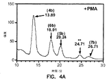


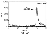


Classifications
-
- C—CHEMISTRY; METALLURGY
- C07—ORGANIC CHEMISTRY
- C07K—PEPTIDES
- C07K16/00—Immunoglobulins [IGs], e.g. monoclonal or polyclonal antibodies
- C07K16/44—Immunoglobulins [IGs], e.g. monoclonal or polyclonal antibodies against material not provided for elsewhere, e.g. haptens, metals, DNA, RNA, amino acids
-
- C—CHEMISTRY; METALLURGY
- C07—ORGANIC CHEMISTRY
- C07C—ACYCLIC OR CARBOCYCLIC COMPOUNDS
- C07C251/00—Compounds containing nitrogen atoms doubly-bound to a carbon skeleton
- C07C251/72—Hydrazones
- C07C251/74—Hydrazones having doubly-bound carbon atoms of hydrazone groups bound to hydrogen atoms or to acyclic carbon atoms
- C07C251/76—Hydrazones having doubly-bound carbon atoms of hydrazone groups bound to hydrogen atoms or to acyclic carbon atoms to carbon atoms of a saturated carbon skeleton
-
- C—CHEMISTRY; METALLURGY
- C07—ORGANIC CHEMISTRY
- C07C—ACYCLIC OR CARBOCYCLIC COMPOUNDS
- C07C251/00—Compounds containing nitrogen atoms doubly-bound to a carbon skeleton
- C07C251/72—Hydrazones
- C07C251/82—Hydrazones having doubly-bound carbon atoms of hydrazone groups bound to carbon atoms of rings other than six-membered aromatic rings
-
- C—CHEMISTRY; METALLURGY
- C07—ORGANIC CHEMISTRY
- C07C—ACYCLIC OR CARBOCYCLIC COMPOUNDS
- C07C251/00—Compounds containing nitrogen atoms doubly-bound to a carbon skeleton
- C07C251/72—Hydrazones
- C07C251/84—Hydrazones having doubly-bound carbon atoms of hydrazone groups being part of rings other than six-membered aromatic rings
-
- C—CHEMISTRY; METALLURGY
- C07—ORGANIC CHEMISTRY
- C07C—ACYCLIC OR CARBOCYCLIC COMPOUNDS
- C07C311/00—Amides of sulfonic acids, i.e. compounds having singly-bound oxygen atoms of sulfo groups replaced by nitrogen atoms, not being part of nitro or nitroso groups
- C07C311/48—Amides of sulfonic acids, i.e. compounds having singly-bound oxygen atoms of sulfo groups replaced by nitrogen atoms, not being part of nitro or nitroso groups having nitrogen atoms of sulfonamide groups further bound to another hetero atom
- C07C311/49—Amides of sulfonic acids, i.e. compounds having singly-bound oxygen atoms of sulfo groups replaced by nitrogen atoms, not being part of nitro or nitroso groups having nitrogen atoms of sulfonamide groups further bound to another hetero atom to nitrogen atoms
-
- C—CHEMISTRY; METALLURGY
- C07—ORGANIC CHEMISTRY
- C07C—ACYCLIC OR CARBOCYCLIC COMPOUNDS
- C07C35/00—Compounds having at least one hydroxy or O-metal group bound to a carbon atom of a ring other than a six-membered aromatic ring
- C07C35/22—Compounds having at least one hydroxy or O-metal group bound to a carbon atom of a ring other than a six-membered aromatic ring polycyclic, at least one hydroxy group bound to a condensed ring system
- C07C35/44—Compounds having at least one hydroxy or O-metal group bound to a carbon atom of a ring other than a six-membered aromatic ring polycyclic, at least one hydroxy group bound to a condensed ring system with a hydroxy group on a condensed ring system having more than three rings
-
- C—CHEMISTRY; METALLURGY
- C07—ORGANIC CHEMISTRY
- C07C—ACYCLIC OR CARBOCYCLIC COMPOUNDS
- C07C49/00—Ketones; Ketenes; Dimeric ketenes; Ketonic chelates
- C07C49/385—Saturated compounds containing a keto group being part of a ring
- C07C49/523—Saturated compounds containing a keto group being part of a ring containing —CHO groups
-
- C—CHEMISTRY; METALLURGY
- C07—ORGANIC CHEMISTRY
- C07C—ACYCLIC OR CARBOCYCLIC COMPOUNDS
- C07C49/00—Ketones; Ketenes; Dimeric ketenes; Ketonic chelates
- C07C49/587—Unsaturated compounds containing a keto groups being part of a ring
- C07C49/757—Unsaturated compounds containing a keto groups being part of a ring containing —CHO groups
-
- C—CHEMISTRY; METALLURGY
- C07—ORGANIC CHEMISTRY
- C07D—HETEROCYCLIC COMPOUNDS
- C07D313/00—Heterocyclic compounds containing rings of more than six members having one oxygen atom as the only ring hetero atom
- C07D313/02—Seven-membered rings
- C07D313/04—Seven-membered rings not condensed with other rings
-
- C—CHEMISTRY; METALLURGY
- C07—ORGANIC CHEMISTRY
- C07J—STEROIDS
- C07J41/00—Normal steroids containing one or more nitrogen atoms not belonging to a hetero ring
- C07J41/0005—Normal steroids containing one or more nitrogen atoms not belonging to a hetero ring the nitrogen atom being directly linked to the cyclopenta(a)hydro phenanthrene skeleton
-
- C—CHEMISTRY; METALLURGY
- C07—ORGANIC CHEMISTRY
- C07J—STEROIDS
- C07J61/00—Steroids in which the cyclopenta(a)hydrophenanthrene skeleton has been modified by contraction of only one ring by one or two atoms
-
- C—CHEMISTRY; METALLURGY
- C07—ORGANIC CHEMISTRY
- C07J—STEROIDS
- C07J9/00—Normal steroids containing carbon, hydrogen, halogen or oxygen substituted in position 17 beta by a chain of more than two carbon atoms, e.g. cholane, cholestane, coprostane
-
- C—CHEMISTRY; METALLURGY
- C07—ORGANIC CHEMISTRY
- C07K—PEPTIDES
- C07K16/00—Immunoglobulins [IGs], e.g. monoclonal or polyclonal antibodies
- C07K16/18—Immunoglobulins [IGs], e.g. monoclonal or polyclonal antibodies against material from animals or humans
-
- G—PHYSICS
- G01—MEASURING; TESTING
- G01N—INVESTIGATING OR ANALYSING MATERIALS BY DETERMINING THEIR CHEMICAL OR PHYSICAL PROPERTIES
- G01N33/00—Investigating or analysing materials by specific methods not covered by groups G01N1/00 - G01N31/00
- G01N33/48—Biological material, e.g. blood, urine; Haemocytometers
- G01N33/50—Chemical analysis of biological material, e.g. blood, urine; Testing involving biospecific ligand binding methods; Immunological testing
- G01N33/92—Chemical analysis of biological material, e.g. blood, urine; Testing involving biospecific ligand binding methods; Immunological testing involving lipids, e.g. cholesterol, lipoproteins, or their receptors
-
- C—CHEMISTRY; METALLURGY
- C07—ORGANIC CHEMISTRY
- C07C—ACYCLIC OR CARBOCYCLIC COMPOUNDS
- C07C2601/00—Systems containing only non-condensed rings
- C07C2601/12—Systems containing only non-condensed rings with a six-membered ring
- C07C2601/14—The ring being saturated
-
- C—CHEMISTRY; METALLURGY
- C07—ORGANIC CHEMISTRY
- C07C—ACYCLIC OR CARBOCYCLIC COMPOUNDS
- C07C2601/00—Systems containing only non-condensed rings
- C07C2601/12—Systems containing only non-condensed rings with a six-membered ring
- C07C2601/16—Systems containing only non-condensed rings with a six-membered ring the ring being unsaturated
-
- C—CHEMISTRY; METALLURGY
- C07—ORGANIC CHEMISTRY
- C07C—ACYCLIC OR CARBOCYCLIC COMPOUNDS
- C07C2602/00—Systems containing two condensed rings
- C07C2602/02—Systems containing two condensed rings the rings having only two atoms in common
- C07C2602/04—One of the condensed rings being a six-membered aromatic ring
- C07C2602/08—One of the condensed rings being a six-membered aromatic ring the other ring being five-membered, e.g. indane
-
- C—CHEMISTRY; METALLURGY
- C07—ORGANIC CHEMISTRY
- C07C—ACYCLIC OR CARBOCYCLIC COMPOUNDS
- C07C2602/00—Systems containing two condensed rings
- C07C2602/02—Systems containing two condensed rings the rings having only two atoms in common
- C07C2602/14—All rings being cycloaliphatic
- C07C2602/24—All rings being cycloaliphatic the ring system containing nine carbon atoms, e.g. perhydroindane
-
- C—CHEMISTRY; METALLURGY
- C07—ORGANIC CHEMISTRY
- C07C—ACYCLIC OR CARBOCYCLIC COMPOUNDS
- C07C2603/00—Systems containing at least three condensed rings
- C07C2603/02—Ortho- or ortho- and peri-condensed systems
- C07C2603/40—Ortho- or ortho- and peri-condensed systems containing four condensed rings
-
- G—PHYSICS
- G01—MEASURING; TESTING
- G01N—INVESTIGATING OR ANALYSING MATERIALS BY DETERMINING THEIR CHEMICAL OR PHYSICAL PROPERTIES
- G01N2800/00—Detection or diagnosis of diseases
- G01N2800/04—Endocrine or metabolic disorders
- G01N2800/044—Hyperlipemia or hypolipemia, e.g. dyslipidaemia, obesity
Landscapes
- Chemical & Material Sciences (AREA)
- Organic Chemistry (AREA)
- Health & Medical Sciences (AREA)
- Life Sciences & Earth Sciences (AREA)
- General Health & Medical Sciences (AREA)
- Molecular Biology (AREA)
- Immunology (AREA)
- Biophysics (AREA)
- Engineering & Computer Science (AREA)
- Biochemistry (AREA)
- Medicinal Chemistry (AREA)
- Proteomics, Peptides & Aminoacids (AREA)
- Biomedical Technology (AREA)
- Genetics & Genomics (AREA)
- Hematology (AREA)
- Urology & Nephrology (AREA)
- Pathology (AREA)
- Biotechnology (AREA)
- Physics & Mathematics (AREA)
- Analytical Chemistry (AREA)
- Food Science & Technology (AREA)
- General Physics & Mathematics (AREA)
- Microbiology (AREA)
- Cell Biology (AREA)
- Endocrinology (AREA)
- Investigating Or Analysing Biological Materials (AREA)
- Measuring Or Testing Involving Enzymes Or Micro-Organisms (AREA)
- Medicines Containing Antibodies Or Antigens For Use As Internal Diagnostic Agents (AREA)
- Preparation Of Compounds By Using Micro-Organisms (AREA)
- Pyrane Compounds (AREA)
- Steroid Compounds (AREA)
- Organic Low-Molecular-Weight Compounds And Preparation Thereof (AREA)
- Peptides Or Proteins (AREA)
- Pharmaceuticals Containing Other Organic And Inorganic Compounds (AREA)
Abstract
本発明は、アテローム硬化性プラーク物質によって生成されるコレステロールオゾン化生成物の検出ならびにコレステロールの蓄積および酸化に関する血管状態の検出方法に関する。本発明によれば、コレステロールオゾン分解生成物は、アテローム硬化性プラーク中に存在する。さらに、患者から採取した組織および体液中のコレステロールのオゾン化生成物の検出および定量は、アテローム硬化性病変が患者に実際に存在するかどうかの正確な指標である。したがって、本発明は、アテローム硬化性病変が患者中に存在するかどうかを簡潔かつ正確に検出する方法を提供する。The present invention relates to the detection of cholesterol ozonation products produced by atherosclerotic plaque material and to the detection of vascular conditions related to cholesterol accumulation and oxidation. According to the present invention, cholesterol ozonolysis products are present in atherosclerotic plaques. Furthermore, the detection and quantification of ozonation products of cholesterol in tissues and body fluids collected from patients is an accurate indicator of whether atherosclerotic lesions are actually present in the patient. Thus, the present invention provides a simple and accurate method for detecting whether an atherosclerotic lesion is present in a patient.
Description
(関連出願)
本出願は、2003年9月5日提出の米国特許仮出願番号60/500,593号および2003年11月6日提出の米国特許仮出願番号60/517,821号(その開示全体が本明細書中で参考として援用される)の優先権を主張する。
(Related application)
This application is filed with US Provisional Application No. 60 / 500,593 filed September 5, 2003 and US Provisional Patent Application No. 60 / 517,821 filed November 6, 2003 (the entire disclosure of which is hereby incorporated by reference). Claim priority) (incorporated as reference in the book).
(政府の権利に関する記述)
本明細書中に記載の発明を、国立衛生研究所によって授与された助成金番号PO1CA 27489の米国政府支援を使用して実施した。米国政府は、本発明に一定の権利を有する。
(Description of government rights)
The invention described herein was made using US government support of grant number PO1CA 27489 awarded by the National Institutes of Health. The US government has certain rights in the invention.
(発明の分野)
本発明は、コレステロールオゾン化(ozonation)生成物がアテローム硬化性病変によって生成されるという発見に関する。本発明は、アテローム性動脈硬化症および/または心血管疾患などのコレステロール関連血管病態を有する患者を診断、検出、およびモニタリングする方法を提供する。
(Field of Invention)
The present invention relates to the discovery that cholesterol ozonation products are produced by atherosclerotic lesions. The present invention provides methods for diagnosing, detecting and monitoring patients with cholesterol-related vascular conditions such as atherosclerosis and / or cardiovascular disease.
(発明の背景)
全ての人が血清コレステロールレベルを慎重にモニタリングすることを忠告され続けており、且つ自由な食事および運動不足によって動脈プラーク中にコレステロールが蓄積し、アテローム性動脈硬化症および冠状動脈性心疾患のリスクが増大し得ることが常に指摘されている。それにも関わらず、高血清コレステロールレベルがこのようなリスクの指標である一方で、実際に構築された問題のアテローム硬化性プラークが存在するという証拠はない。
(Background of the Invention)
All people continue to be advised to carefully monitor serum cholesterol levels, and cholesterol builds up in arterial plaques due to lack of free diet and exercise, risk of atherosclerosis and coronary heart disease It has always been pointed out that can increase. Nevertheless, while high serum cholesterol levels are an indicator of such risk, there is no evidence that there is actually a problem atherosclerotic plaque constructed.
血清コレステロールは、主に、低密度リポタンパク質(LDLコレステロール)、高密度リポタンパク質(HDLコレステロール)、および超低密度リポタンパク質(VLDLコレステロール)中のトリグリセリドと関連することが知られている。多数の長期臨床試験からの統計上の証拠は、HDLコレステロールの比率が高くてLDLコレステロールの比率が低い場合に相対危険度が低くなることを示す。HDLコレステロールは、そのレベルが過度に低くない(すなわち、30mg/dL未満)場合に有益である。VLDLコレステロールはいかなるリスクの決定要因に関与しないが、高トリグリセリド自体が深刻な問題であり得る。他方では、LDLコレステロールの比率が高く且つHDLコレステロールの比率が低いことは、アテローム性動脈硬化症および冠状動脈性心疾患のリスクがより高くなることの指標である。 Serum cholesterol is known to be associated primarily with triglycerides in low density lipoprotein (LDL cholesterol), high density lipoprotein (HDL cholesterol), and very low density lipoprotein (VLDL cholesterol). Statistical evidence from a number of long-term clinical trials indicates that the relative risk is low when the proportion of HDL cholesterol is high and the proportion of LDL cholesterol is low. HDL cholesterol is beneficial if its level is not too low (ie, less than 30 mg / dL). Although VLDL cholesterol does not contribute to any risk determinants, high triglycerides themselves can be a serious problem. On the other hand, a high ratio of LDL cholesterol and a low ratio of HDL cholesterol is an indicator of a higher risk of atherosclerosis and coronary heart disease.
アテローム性動脈硬化症のリスクと高LDLコレステロールレベルとの間に強い相関関係が存在する場合でさえ、いくつかの研究では、血清LDLおよびHDLコレステロールレベルの測定が不十分にしか行われておらず、しばしば信頼できない結果が得られていることが示されている。Superko,H.R.et al.High−Density Lipoprotein Cholesterol Measurements−−A Help or Hinderance in Practical Clinical Medicine,JAMA 256:2714−2717(1986);Warnick,G.R.et al.HDL Cholesterol:Results of Interlaboratory Proficiency Test,Clin.Chem.26:169−170(1980);and Grundy,S.M.et al.The Place of HDL in Cholesterol Management.A Perspective from the National Cholesterol Education Program,Arch.Inter.Med.149:505−510(1989)を参照のこと。Grundy et al.の論文は、HDLコレステロールの測定値の研究室間変動係数が38%もの高さであることを報告している。2000の研究所による同一のHDLコレステロールサンプルの測定に関するCollege of American Pathologistsによる1987年の報告は、33%を超える測定の基準値が5%を超えて異なっていたことを示した。同一の方法を使用した群の間の研究室間変動係数は16.5%に改善されたが、このような分散度は、ほとんどの試験結果が不正確すぎて臨床目的でのいかなる予測値も得られないことを依然として示す。この理由のために、総コレステロール:HDLコレステロール比はもはやリスクアセスメントで使用されない。 Even when there is a strong correlation between the risk of atherosclerosis and high LDL cholesterol levels, some studies have only poorly measured serum LDL and HDL cholesterol levels Often, it has been shown that unreliable results have been obtained. Superko, H .; R. et al. High-Density Lipoprotein Cholesterol Measurements--A Help or Hindrance in Practical Clinical Medicine, JAMA 256: 2714-2717 (1986); R. et al. HDL Cholesterol: Results of Interlaboratory Proficiency Test, Clin. Chem. 26: 169-170 (1980); and Grundy, S .; M.M. et al. The Place of HDL in Cholesterol Management. A Perspective from the National Cholesterol Education Program, Arch. Inter. Med. 149: 505-510 (1989). Grundy et al. Report that the interlaboratory coefficient of variation of HDL cholesterol measurements is as high as 38%. A 1987 report by College of American Pathologists on the measurement of the same HDL cholesterol sample by 2000 laboratories showed that the baseline value of measurement above 33% differed by more than 5%. Although the interlaboratory coefficient of variation between groups using the same method was improved to 16.5%, such a degree of dispersion is too inaccurate for most trial results and any predictive value for clinical purposes. It still shows that it cannot be obtained. For this reason, the total cholesterol: HDL cholesterol ratio is no longer used in risk assessment.
典型的な脂質プロフィール研究では、総コレステロールおよびトリグリセリドレベルを血清サンプルから直接測定する。次いで、サンプルを、LDLコレステロールおよびVLDLコレステロールを沈殿して取り出す薬剤で処理する。このようなサンプル処理後に上清中に残存したHDLコレステロールを測定する。VLDLコレステロールは、トリグリセリドの一定の比率(例えば、0.2)を取る。次いで、LDLコレステロールを、総コレステロールからHDLコレステロール値およびVLDLコレステロール値を引くことによって間接的に計算する。これら3つの独立した測定から生じるエラーの増加によってLDLコレステロールの測定値の全体的な精度および正確さが最も小さくなるにもかかわらず、心血管へのリスクの評価に最も有意であり得る。このような不正確さのため、経時的なLDLコレステロール低下療法により臨床的な進展があるかどうか意図的にモニタリングし証明することは困難である。 In a typical lipid profile study, total cholesterol and triglyceride levels are measured directly from serum samples. The sample is then treated with an agent that precipitates and removes LDL cholesterol and VLDL cholesterol. HDL cholesterol remaining in the supernatant after such sample treatment is measured. VLDL cholesterol takes a certain proportion of triglycerides (eg, 0.2). LDL cholesterol is then calculated indirectly by subtracting HDL and VLDL cholesterol values from total cholesterol. Although the increase in error resulting from these three independent measurements minimizes the overall accuracy and accuracy of LDL cholesterol measurements, it can be most significant in assessing cardiovascular risk. Because of this inaccuracy, it is difficult to intentionally monitor and prove whether clinical progress has been made with LDL cholesterol lowering therapy over time.
したがって、血清LDLコレステロールの測定値はしばしば不正確である。このような不正確さは、LDLコレステロールレベルが実際に問題のあるアテローム硬化性病変が存在することを証明しないという事実と併せて、問題のあるコレステロール負荷アテローム硬化性病変が患者に存在するかどうかを決定するための比較的簡潔であり、信頼でき、且つ再現性のある方法が必要である。 Therefore, serum LDL cholesterol measurements are often inaccurate. Such inaccuracy is combined with the fact that LDL cholesterol levels do not actually prove that there are problematic atherosclerotic lesions, and whether there are problematic cholesterol-loading atherosclerotic lesions in the patient There is a need for a relatively simple, reliable and reproducible method for determining
(発明の要旨)
本発明によれば、コレステロールオゾン分解生成物は、アテローム硬化性プラーク中に存在する。さらに、患者から採取した組織および体液中のコレステロールのオゾン化生成物の検出および定量は、アテローム硬化性病変が患者に実際に存在するかどうかの正確な指標である。したがって、本発明は、アテローム硬化性病変が患者中に存在するかどうかを簡潔かつ正確に検出する方法を提供する。本発明の方法は、コレステロールのオゾン化生成物が患者から採取した試験サンプル中に存在するかどうかを検出する工程を含む。本発明はまた、哺乳動物中のアテローム硬化性プラーク形成の程度の診断およびモニタリングの基準としての生体サンプル中に存在するコレステロールオゾン化生成物の定量を意図する。
(Summary of the Invention)
According to the present invention, cholesterol ozonolysis products are present in atherosclerotic plaques. Furthermore, the detection and quantification of ozonation products of cholesterol in tissues and body fluids collected from patients is an accurate indicator of whether atherosclerotic lesions are actually present in the patient. Thus, the present invention provides a simple and accurate method for detecting whether an atherosclerotic lesion is present in a patient. The method of the invention comprises detecting whether an ozonation product of cholesterol is present in a test sample taken from a patient. The present invention also contemplates the quantification of cholesterol ozonation products present in biological samples as a basis for diagnosis and monitoring of the extent of atherosclerotic plaque formation in mammals.
本発明の1つの態様は、アテローム硬化性プラーク中で生成されたコレステロールの単離されたオゾン化生成物である。このようなコレステロールのオゾン化生成物は、例えば、式4a〜15a、3c、または7c:
One aspect of the present invention is an isolated ozonation product of cholesterol produced in atherosclerotic plaques. Such cholesterol ozonation products are, for example,

本発明の別の態様は、亜硫酸水素付加体、イミン、オキシム、ヒドラゾン、ダンシルヒドラゾン、セミカルバゾン、またはTollins試験の生成物を含むコレステロールオゾン化生成物の検出可能な誘導体であって、前記コレステロールのオゾン化生成物がアテローム硬化性プラーク内に生成される、コレステロールオゾン化生成物の検出可能な誘導体である。 Another aspect of the present invention is a detectable derivative of a cholesterol ozonation product, including a bisulfite adduct, imine, oxime, hydrazone, dansyl hydrazone, semicarbazone, or product of Tollins test, wherein the ozone of said cholesterol A detectable derivative of a cholesterol ozonation product, wherein the oxidization product is produced in an atherosclerotic plaque.
本発明の別の態様は、式4bまたは式4c:
Another aspect of the invention is a compound of
本発明の別の態様は、式5b:
Another aspect of the invention is a compound of
本発明の別の態様は、式6b〜15bまたは10c:
Another aspect of the invention is a compound of
本発明の別の態様は、式4d:
Another aspect of the invention is a compound of
本発明の別の態様は、式5c:
Another aspect of the invention is a compound of
本発明の別の態様は、式13a、13b、14a、14b、15a、15c、または3cを有するハプテンである。 Another aspect of the invention is a hapten having the formula 13a, 13b, 14a, 14b, 15a, 15c, or 3c.
本発明の別の態様は、コレステロールのオゾン化生成物に結合し得る単離された抗体である。抗体は、モノクローナル抗体またはポリクローナル抗体であり得る。抗体が結合し得るコレステロールのオゾン化生成物は、式4a〜15a、3c、4c、7cのいずれか1つを有する化合物であり得る。いくつかの実施形態では、コレステロールのオゾン化生成物のヒドラゾン誘導体(例えば、式4b〜15b、4c、または10cのいずれか1つを有する化合物)に結合し得る単離された抗体。本発明の抗体は、例えば、式13a、13b、14a、14b、15a、15c、または3cを有するハプテンに対して惹起し得る。
Another aspect of the invention is an isolated antibody capable of binding to the ozonation product of cholesterol. The antibody can be a monoclonal antibody or a polyclonal antibody. The cholesterol ozonation product to which the antibody can bind can be a compound having any one of
本発明の別の態様は、ATCC受託番号PTA−5427またはPTA−5428を有するハイブリドーマKA1−11C5:6またはKA1−7A6:6に由来する単離された抗体である。 Another aspect of the invention is an isolated antibody derived from hybridoma KA1-11C5: 6 or KA1-7A6: 6 having ATCC accession number PTA-5427 or PTA-5428.
本発明の別の態様は、ATCC受託番号PTA−5429およびPTA−5430を有するハイブリドーマKA2−8F6:4またはKA2−1E9:4に由来する単離された抗体である。 Another aspect of the invention is an isolated antibody derived from a hybridoma KA2-8F6: 4 or KA2-1E9: 4 having ATCC accession numbers PTA-5429 and PTA-5430.
本発明の別の態様は、コレステロールのオゾン化生成物が患者から得た試験サンプル中に存在するかどうかの検出による患者のアテローム性動脈硬化症の検出方法である。オゾン化生成物は、アテローム硬化性プラークによって生成され得る。試験サンプルは、例えば、血清、血漿、血液、アテローム硬化性プラーク物質、尿、または脈管組織であり得る。アテローム性動脈硬化症の検出方法はまた、試験サンプル中に存在するコレステロールオゾン化生成物の定量を含み得る。 Another aspect of the present invention is a method for detecting atherosclerosis in a patient by detecting whether cholesterol ozonation products are present in a test sample obtained from the patient. The ozonation product can be produced by atherosclerotic plaque. The test sample can be, for example, serum, plasma, blood, atherosclerotic plaque material, urine, or vascular tissue. The method for detecting atherosclerosis can also include quantifying cholesterol ozonation products present in the test sample.
1つの実施形態では、アテローム性動脈硬化症の検出方法は、試験サンプルと、亜硫酸水素塩、アンモニア、シッフ塩基、芳香族または脂肪族ヒドラジン、ダンシルヒドラジン、ジェラード(Gerard)試薬、Tollins試験試薬とを反応させる工程と、このような反応によって形成されたコレステロールのオゾン化生成物の誘導体を検出する工程とを含み得る。 In one embodiment, the method for detecting atherosclerosis comprises a test sample and bisulfite, ammonia, Schiff base, aromatic or aliphatic hydrazine, dansyl hydrazine, Gerard reagent, Tollins test reagent. Reacting and detecting a derivative of the ozonation product of cholesterol formed by such a reaction.
別の実施形態では、アテローム性動脈硬化症の検出方法は、試験サンプルをヒドラジン化合物と反応させてコレステロールのオゾン化生成物のヒドラゾン誘導体を生成する工程を含み得る。例えば、ヒドラジン化合物は、2,4−ジニトロフェニルヒドラジンであり得る。 In another embodiment, a method for detecting atherosclerosis may comprise reacting a test sample with a hydrazine compound to produce a hydrazone derivative of a cholesterol ozonation product. For example, the hydrazine compound can be 2,4-dinitrophenylhydrazine.
別の実施形態では、アテローム性動脈硬化症の検出方法は、試験サンプルをダンシルヒドラジンと反応させてコレステロールのオゾン化生成物のダンシルヒドラゾン誘導体を生成する工程を含み得る。例えば、ダンシルヒドラゾン誘導体は、式4dまたは5cを有し得る。
In another embodiment, a method for detecting atherosclerosis may comprise reacting a test sample with dansyl hydrazine to produce a dansyl hydrazone derivative of a cholesterol ozonation product. For example, a dansyl hydrazone derivative can have the
別の実施形態では、アテローム性動脈硬化症の検出方法は、試験サンプルとコレステロールのオゾン化生成物に結合し得る抗体と接触させる工程を含み得る。本明細書中に記載の任意の抗体を、本方法で使用し得る。 In another embodiment, a method for detecting atherosclerosis may comprise contacting a test sample with an antibody that can bind to an ozonation product of cholesterol. Any antibody described herein may be used in the method.
本発明の別の態様は、コレステロールのオゾン化生成物が患者から得た試験サンプル中に存在するかどうかを検出する工程と、前記オゾン化生成物が式5aを有する化合物であることとを含む、コレステロールのオゾン化生成物が患者のアテローム硬化性プラークによって放出されるかどうかを検出する方法を含む。コレステロールのオゾン化生成物がアテローム硬化性プラークによって放出されるかどうかを検出する方法はまた、試験サンプル中に存在するコレステロールオゾン化生成物を定量する工程を含み得る。
Another aspect of the invention includes detecting whether an ozonation product of cholesterol is present in a test sample obtained from a patient, and wherein the ozonation product is a
本発明の別の態様は、患者由来の試験サンプルに2,4−ジニトロフェニルヒドラジンを添加する工程と、コレステロールのオゾン化生成物のヒドラゾン誘導体が試験サンプル中に存在するかどうかを検出する工程を含む、患者のアテローム性動脈硬化症の検出方法を含む。ヒドラゾン誘導体が、式4b、4c、5b、6b、7b、8b、9b、10b、10c、11b、12b、13b、14b、または15bのいずれか1つを有する化合物であり得る。
Another aspect of the present invention includes the steps of adding 2,4-dinitrophenylhydrazine to a patient-derived test sample and detecting whether a hydrazone derivative of a cholesterol ozonation product is present in the test sample. A method of detecting atherosclerosis in a patient. The hydrazone derivative can be a compound having any one of
本発明の別の態様は、マクロファージを試験サンプルと接触させる工程と、前記マクロファージによる脂質取り込みが増加するかどうかを決定する工程とを含む、コレステロールオゾン分解生成物が試験サンプル中に存在するかどうかを検出する方法を含む。 Another aspect of the invention is whether a cholesterol ozonolysis product is present in a test sample comprising contacting macrophages with the test sample and determining whether lipid uptake by the macrophages is increased. Including a method of detecting.
本発明の別の態様は、マクロファージを試験サンプルと接触させる工程と、前記マクロファージによる脂質取り込みが増加するかどうかを決定する工程とを含む、患者のアテローム性動脈硬化症の検出方法を含む。 Another aspect of the invention includes a method for detecting atherosclerosis in a patient comprising contacting a macrophage with a test sample and determining whether lipid uptake by the macrophage is increased.
本発明の別の態様は、低密度リポタンパク質を試験サンプルと接触させる工程と、前記低密度リポタンパク質の二次構造が変化するかどうかを観察する工程とを含む、試験サンプル中のコレステロールオゾン分解生成物の検出方法を含む。 Another aspect of the present invention comprises contacting the low density lipoprotein with a test sample and observing whether the secondary structure of the low density lipoprotein changes, cholesterol ozonolysis in the test sample Product detection methods.
本発明の別の態様は、低密度リポタンパク質を患者から得た試験サンプルと接触させる工程と、前記低密度リポタンパク質の二次構造が変化するかどうかを観察する工程とを含む、患者のアテローム性動脈硬化症の検出方法を含む。 Another aspect of the invention includes contacting a low density lipoprotein with a test sample obtained from a patient and observing whether the secondary structure of the low density lipoprotein is altered. A method for detecting atherosclerosis.
本発明の別の態様は、アポタンパク質B100を試験サンプルと接触させる工程と、前記アポタンパク質B100の二次構造が変化するかどうかを観察する工程とを含む、試験サンプル中のコレステロールオゾン分解生成物の検出方法を含む。 Another aspect of the present invention comprises contacting the apoprotein B 100 with a test sample and observing whether the secondary structure of the apoprotein B 100 changes, cholesterol ozonolysis in the test sample Product detection methods.
本発明の別の態様は、アポタンパク質B100を患者から得た試験サンプルと接触させる工程と、前記アポタンパク質B100の二次構造が変化するかどうかを観察する工程とを含む、患者のアテローム性動脈硬化症の検出方法を含む。 Another aspect of the present invention comprises the steps of contacting apoprotein B 100 with a test sample obtained from a patient and observing whether the secondary structure of said apoprotein B 100 is altered. A method for detecting atherosclerosis.
低密度リポタンパク質またはアポタンパク質B100の二次構造を円偏光二色性によって観察し得る。 It is observed by circular dichroism the secondary structure of low-density lipoprotein or apoprotein B 100.
(発明の詳細な説明)
本発明は、コレステロールのオゾン化生成物の検出方法を提供する。コレステロールのオゾン化生成物の検出のためのキットおよび試薬も提供する。これらの方法、キット、および試薬は、構築されたコレステロールに関連する血管状態の検出に有用である。例えば、方法、キット、および試薬は、アテローム性動脈硬化症などの炎症性動脈疾患の診断および予後のモニタリングに有用である。
(Detailed description of the invention)
The present invention provides a method for detecting an ozonation product of cholesterol. Kits and reagents for the detection of cholesterol ozonation products are also provided. These methods, kits, and reagents are useful for detecting vascular conditions associated with the constructed cholesterol. For example, the methods, kits, and reagents are useful for the diagnosis and prognostic monitoring of inflammatory arterial diseases such as atherosclerosis.
(コレステロールのオゾン化)
本発明によれば、コレステロールは、オゾンなどの活性酸素種によってアテローム硬化性動脈内で酸化する。この過程によって多数のコレステロールオゾン化生成物が生成され、アテローム性動脈硬化症患者から採取した組織または流動物サンプル中で検出し得る。コレステロールオゾン化生成物の検出により、アテローム性動脈硬化症などの炎症性動脈疾患が診断される。
(Cholesterol ozonization)
According to the present invention, cholesterol is oxidized in atherosclerotic arteries by reactive oxygen species such as ozone. This process produces a number of cholesterol ozonation products that can be detected in tissue or fluid samples taken from atherosclerotic patients. Detection of cholesterol ozonation products diagnoses inflammatory arterial diseases such as atherosclerosis.
コレステロールは、以下の構造(3): Cholesterol has the following structure (3):
しかし、本発明によれば、実際のアテローム硬化性プラークの存在を、コレステロールのオゾン化生成物の検出によって検出し得る。コレステロールが動脈中に蓄積された場合、アテローム硬化性プラークが形成され得る。特定の機構に限定されることを望まないが、マクロファージ、好中球、および他の免疫細胞がアテローム硬化性病変内に陥り、オゾンなどの活性酸素種を放出するようである。生成された活性酸素種は病変中のコレステロールと反応し、コレステロールが患者中で検出し得る多数の生成物に酸化される。したがって、患者から採取したサンプル中でコレステロールオゾン化のための2つの事象が起こるようである。第1に、アテローム硬化性プラーク内でコレステロールが実質的に構築されなければならない。第2に、アテローム性動脈硬化症が、活性酸素種が生成される段階に進行しなければならない。これら2つの事象が並行して起こり、コレステロールオゾン化生成物が形成される。コレステロールの構築およびオゾン生成が実質的に他の状況で起こらないため、コレステロールオゾン化生成物の検出は、アテローム性動脈硬化症などの炎症性動脈病態が患者に存在するかどうかの正確な指標である。さらに、本発明によれば、アテローム性動脈硬化症患者から採取した生体サンプル(例えば、血清)内のコレステロールオゾン化生成物の存在量は、患者のアテローム性動脈硬化症の重症度の指標である。 However, according to the present invention, the presence of actual atherosclerotic plaques can be detected by detection of the ozonation product of cholesterol. When cholesterol accumulates in the arteries, atherosclerotic plaques can be formed. While not wishing to be limited to a particular mechanism, macrophages, neutrophils, and other immune cells appear to fall into atherosclerotic lesions and release reactive oxygen species such as ozone. The generated reactive oxygen species react with cholesterol in the lesion, and the cholesterol is oxidized to a number of products that can be detected in the patient. Thus, two events for cholesterol ozonation appear to occur in samples taken from patients. First, cholesterol must be substantially built up within the atherosclerotic plaque. Second, atherosclerosis must progress to the stage where reactive oxygen species are generated. These two events occur in parallel and the cholesterol ozonation product is formed. Cholesterol ozonation product detection is an accurate indicator of whether an inflammatory arterial condition, such as atherosclerosis, is present in a patient, since cholesterol construction and ozonation do not occur in other circumstances substantially. is there. Further, according to the present invention, the abundance of cholesterol ozonation products in a biological sample (eg, serum) taken from an atherosclerotic patient is an indicator of the severity of the patient's atherosclerosis. .
本発明によれば、多数のコレステロールオゾン化生成物が同定された。例えば、コレステロール3が酸化された場合、seco−ケトアルデヒド4aおよびそのアルドール付加体5aは、形成された主な生成物である。
In accordance with the present invention, a number of cholesterol ozonation products have been identified. For example, when
したがって、本発明は、患者がアテローム硬化性病変を有するかどうかの決定および循環コレステロールがアテローム硬化性プラーク中に組み込まれるようになる範囲の決定のためのこれらのコレステロールオゾン化生成物の検出を意図する。 The present invention is therefore intended to detect these cholesterol ozonation products for the determination of whether a patient has atherosclerotic lesions and the extent to which circulating cholesterol will be incorporated into atherosclerotic plaques. To do.
(オゾンおよびコレステロール生成物の検出)
コレステロールオゾン化生成物を、当業者が利用可能な任意の手順によって検出または同定し得る。例えば、これらの生成物を、高速液体クロマトグラフィ(HPLC)、液体クロマトグラフィ質量分析(LCMS)、ガスクロマトグラフィ(GC)、ガスクロマトグラフィ質量分析(GCMS)、高速液体クロマトグラフィ質量分析(HPLC−MS)、蒸発光散乱検出(ELSD)、ガスクロマトグラフィ質量分析を使用したイオン検出(ID−GCMS)、可視、紫外線、または赤外線分光法、薄層クロマトグラフィ、電気泳動、液体クロマトグラフィ、核磁気共鳴、湿式化学アッセイ、免疫アッセイ(例えば、ELISA)、免疫組織化学、蛍光分光学、光分光学もしくは紫外線分光学、または当業者に利用可能な任意の他の手段によって検出または同定し得る。
(Detection of ozone and cholesterol products)
Cholesterol ozonation products can be detected or identified by any procedure available to those skilled in the art. For example, these products can be used for high performance liquid chromatography (HPLC), liquid chromatography mass spectrometry (LCMS), gas chromatography (GC), gas chromatography mass spectrometry (GCMS), high performance liquid chromatography mass spectrometry (HPLC-MS), evaporative light. Scattering detection (ELSD), ion detection using gas chromatography mass spectrometry (ID-GCMS), visible, ultraviolet or infrared spectroscopy, thin layer chromatography, electrophoresis, liquid chromatography, nuclear magnetic resonance, wet chemical assay, immunoassay (E.g., ELISA), immunohistochemistry, fluorescence spectroscopy, light spectroscopy or ultraviolet spectroscopy, or any other means available to those skilled in the art.
さらに、コレステロールオゾン化生成物の存在を、これらの生成物が低密度リポタンパク質(LDL)、アポタンパク質B100(アポB−100、LDLのタンパク質成分)、またはマクロファージに及ぼす影響を観察することによって検出することもできる。本明細書中に記載のように、コレステロールオゾン分解生成物4aおよび5aは、マクロファージからの泡沫細胞の形成を促進し得る。さらに、コレステロールオゾン分解生成物4aおよび5aは、LDLおよびアポB−100の二次構造を修飾する。したがって、試験サンプル中のコレステロールオゾン分解生成物の存在を、試験サンプルが泡沫細胞形成を促進するか、LDLまたはアポタンパク質B100の二次構造を変化させることができるかどうかの決定によって検出し得る。これらのアッセイを、以下により詳細に記載する。
Furthermore, by observing the presence of cholesterol ozonation products on the effects of these products on low density lipoprotein (LDL), apoprotein B 100 (apo B-100, a protein component of LDL), or macrophages. It can also be detected. As described herein,
いくつかの実施形態では、試験サンプルを、コレステロールオゾン化生成物の検出および同定を容易にする試薬と反応させる。例えば、試験サンプルを、コレステロールオゾン化生成物と反応する任意の蛍光、リン光、または着色試薬と接触させることができ、反応生成物を、蛍光、可視光、または紫外線検出器を使用して検出し得る。他の実施形態では、このような試薬を使用せず、コレステロールオゾン化生成物を、その物理的性質または化学的性質によって同定する。このような方法を、以下にさらに詳細に記載する。 In some embodiments, the test sample is reacted with a reagent that facilitates the detection and identification of cholesterol ozonation products. For example, the test sample can be contacted with any fluorescent, phosphorescent, or colored reagent that reacts with the cholesterol ozonation product, and the reaction product is detected using a fluorescent, visible light, or ultraviolet detector. Can do. In other embodiments, no such reagent is used and the cholesterol ozonation product is identified by its physical or chemical properties. Such a method is described in further detail below.
アテローム硬化性プラーク物質中のオゾンの量は、形成されたアテローム硬化性プラークの量も示す。したがって、本発明は、アテローム硬化性プラークのサイズを評価するためのアテローム硬化性プラーク物質中のオゾンの検出および/または定量を意図する。オゾンを検出し得る任意の試薬の使用によって、アテローム硬化性プラーク物質中のオゾンを検出し得る。例えば、インディゴカルミン1は、オゾンとの反応の際にその青色が脱色する有色試薬である。プロセスでは、下に示すように、イサチンスルホン酸2が形成された。
The amount of ozone in the atherosclerotic plaque material also indicates the amount of atherosclerotic plaque formed. Accordingly, the present invention contemplates the detection and / or quantification of ozone in atherosclerotic plaque material to assess the size of atherosclerotic plaque. Ozone in atherosclerotic plaque material can be detected by the use of any reagent that can detect ozone. For example,
しかし、アテローム硬化性材料中にオゾンを検出し得る一方で、実質的なアテローム硬化性プラーク材料を有する患者の血流中のコレステロールオゾン化生成物を検出し得る。したがって、アテローム硬化性プラーク物質の単離を回避するために、当業者は、血液サンプルを単離するために選択し、その後コレステロールのオゾン化生成物が存在するかどうかを検出し得る。これにより、血管内膜切除術などの高価で煩わしい手順が回避され、患者にアテローム硬化性プラーク物質がどれくらい存在するかを評価するための信頼のおける手順が得られる。 However, while ozone can be detected in atherosclerotic materials, cholesterol ozonation products in the bloodstream of patients with substantial atherosclerotic plaque material can be detected. Thus, to avoid isolation of atherosclerotic plaque material, one of ordinary skill in the art can choose to isolate a blood sample and then detect whether the ozonated product of cholesterol is present. This avoids expensive and cumbersome procedures such as endarterectomy and provides a reliable procedure for assessing how much atherosclerotic plaque material is present in the patient.
アテローム性動脈硬化症を診断するために、任意のコレステロールオゾン化生成物(例えば、seco−ケトアルデヒド4a)、そのアルドール付加体5a、および/または関連化合物6a〜15aおよび7cを検出し得る。しかし、今日まで行われている研究は、アルドール付加体5aが血清中で検出し得る主な生成物の1つであることを示す。
Any cholesterol ozonation product (eg, seco-
いくつかの実施形態では、検出を容易にするために生体サンプルから得たコレステロールオゾン化生成物を化学修飾し得る。このような化学修飾に使用し得る試薬には、亜硫酸水素塩、アンモニア、シッフ塩基(アニリンなどの脂肪族または芳香族アミンを使用する)、芳香族または脂肪族ヒドラジン、ダンシルヒドラジン、ジェラード試薬(セミカルバジド)、Tollins試験試薬(ホルムアルデヒドおよび水酸化カルシウム)などが含まれる。本発明のコレステロールオゾン化生成物と反応した場合、これらの試薬により、当業者によって容易に検出し得る亜硫酸水素付加体(ナトリウム塩として容易に結晶化される)、イミン、オキシム、ヒドラゾン、セミカルバゾン、およびTollins試験生成物などの特徴的な生成物が得られる。 In some embodiments, a cholesterol ozonation product obtained from a biological sample may be chemically modified to facilitate detection. Reagents that can be used for such chemical modifications include bisulfite, ammonia, Schiff base (using aliphatic or aromatic amines such as aniline), aromatic or aliphatic hydrazine, dansyl hydrazine, gerrard reagents (semicarbazide). ), Tollins test reagents (formaldehyde and calcium hydroxide) and the like. When reacted with the cholesterol ozonation product of the present invention, these reagents allow bisulfite adducts (easily crystallized as sodium salts), imines, oximes, hydrazones, semicarbazones, which can be easily detected by those skilled in the art. And characteristic products such as Tollins test products are obtained.
例えば、seco−ケトアルデヒド4a、そのアルドール付加体5a、および関連化合物4c、6a〜15aおよび7cのヒドラゾン誘導体を容易に形成することができ、これらは患者がアテローム硬化性病変を有するかどうかの決定のための有用なマーカーである。これらのヒドロゾン(hydrozone)誘導体には、化合物4b〜15b、おそらく4cまたは10cのような構造を有する化合物が含まれる。
For example, seco-
2,4−ジニトロフェニルヒドラジンなどのヒドラジン化合物との反応によってコレステロールオゾン化生成物をヒドロゾン誘導体に変換し得る。いくつかの実施形態では、アセトニトリルまたはアルコール(例えば、メタノールまたはエタノール)などの有機溶媒中で反応させる。酸性環境および非反応性大気を含む非酸素をしばしば使用する。 Cholesterol ozonation products can be converted to hydrozone derivatives by reaction with hydrazine compounds such as 2,4-dinitrophenylhydrazine. In some embodiments, the reaction is in an organic solvent such as acetonitrile or alcohol (eg, methanol or ethanol). Non-oxygen is often used, including acidic environments and non-reactive atmosphere.
例えば、患者から血漿を得て、EDTAに入れることができる。このサンプルをジクロロメタンで数回洗浄して、コレステロールオゾン化生成物を抽出し得る。ジクロロメタン画分を真空蒸発させ、コレステロールオゾン化生成物を含む残渣をアルコール(例えば、メタノール)に溶解し得る。次いで、2,4−ジニトロフェニルヒドラジンおよび1NHClを含むエタノール溶液を添加し得る。溶液中に窒素を短時間(例えば、5分間)注入して(bubbled)遊離酸素を除去し得る。溶液を、コレステロールオゾン化生成物のそのヒドラゾン誘導体への変換に十分な時間(例えば、2時間)撹拌し得る。この手順で検出された主産物は、アルドール付加体5aのヒドラゾン誘導体であると考えられる。さらに、予備調査により、血漿から抽出し得る5aの量が1日あたり約5%減少することが明らかとなった。したがって、新鮮な血漿サンプルにより、サンプル中のアルドール付加体5aの実際の量がより正確に測定される。
For example, plasma can be obtained from a patient and placed in EDTA. This sample can be washed several times with dichloromethane to extract the cholesterol ozonation product. The dichloromethane fraction can be evaporated in vacuo and the residue containing the cholesterol ozonation product can be dissolved in an alcohol (eg, methanol). An ethanol solution containing 2,4-dinitrophenylhydrazine and 1N HCl can then be added. Nitrogen can be bubbled into the solution (eg, 5 minutes) to remove free oxygen. The solution can be agitated for a time sufficient to convert the cholesterol ozonation product to its hydrazone derivative (eg, 2 hours). The main product detected by this procedure is considered to be a hydrazone derivative of
本発明の試薬および方法を使用して、アテローム性動脈硬化症をその進行の任意の段階で検出し得る。AHAによって採用され、本研究で使用した新規の分類によれば、アテローム性動脈硬化症の進行時に8つの病変型を区別し得る。 The reagents and methods of the present invention can be used to detect atherosclerosis at any stage of its progression. According to the new classification adopted by AHA and used in this study, eight lesion types can be distinguished during the progression of atherosclerosis.
脈管内膜中の小脂質沈着物(細胞内およびマクロファージ泡沫細胞)によってI型病変が形成され、動脈壁が最初期且つ最小に変化する。このような変化は、動脈壁を肥厚させない。 Type I lesions are formed by small lipid deposits (intracellular and macrophage foam cells) in the intima, and the arterial wall is initially and minimally altered. Such changes do not thicken the arterial wall.
II型病変は、ミリメートル未満で動脈内膜の厚さが増加する黄色の線条または斑である脂肪線条によって特徴づけられる。これらは、I型病変で認められる脂肪より蓄積された脂肪からなる。脂質含有率は、病変乾燥重量の約20〜25%である。ほとんどの脂質は、細胞内、主にマクロファージ泡沫細胞おおび平滑筋細胞に存在する。細胞外空間は、脂肪滴を含み得るが、これらは細胞内の脂肪滴および小胞粒子(smallvesicular particle)よりも小さい。化学的には、脂質は、コレステロールエステル(オレイン酸コレステリルおよびリノレン酸コレステリル)、コレステロール、およびリン脂質からなる。 Type II lesions are characterized by fat streaks, yellow streaks or plaques that increase in the thickness of the arterial intima below millimeters. These consist of fat accumulated from the fat found in type I lesions. Lipid content is about 20-25% of lesion dry weight. Most lipids are present intracellularly, mainly in macrophage foam cells and smooth muscle cells. The extracellular space may contain lipid droplets, but these are smaller than intracellular lipid droplets and small vesicle particles. Chemically, lipids consist of cholesterol esters (cholesteryl oleate and cholesteryl linolenate), cholesterol, and phospholipids.
III型病変はまた、前アテローマ(preatheroma)病変としても記載される。III型病変では、動脈内膜は、II型病変よりもわずかに肥厚するだけである。III型病変は、動脈の血流を妨害しない。細胞外脂質および小胞粒子は、II型病変で見出された小胞粒子と同一であるが、大量に存在し(乾燥重量で約25〜35%)、小貯留(smallpool)中に蓄積され始める。 Type III lesions are also described as preatheroma lesions. In type III lesions, the arterial intima is only slightly thicker than type II lesions. Type III lesions do not interfere with arterial blood flow. Extracellular lipids and vesicle particles are identical to vesicle particles found in type II lesions, but are present in large quantities (about 25-35% by dry weight) and accumulate in small pools. start.
IV型病変は、アテローマに関連する。これらは、三日月型であり、動脈の厚さを増大させる。この病変は、血漿コレステロールレベルが非常に高い患者以外は動脈内腔を非常に狭めることはないかもしれない(多くの人々において、病変を血管造影法によって視覚化することができない)。IV型病変は、内膜層中の細胞外脂質の広範な蓄積(乾燥重量で約60%)からなる。脂質コアは、無機質の小クランプを含み得る。これらの病変は、破裂および壁性血栓の形成の影響を受けやすい。 Type IV lesions are associated with atheroma. These are crescent shaped and increase the thickness of the artery. This lesion may not narrow the arterial lumen very much except in patients with very high plasma cholesterol levels (in many people the lesion cannot be visualized by angiography). Type IV lesions consist of extensive accumulation of extracellular lipids in the intimal layer (about 60% by dry weight). The lipid core may include a small inorganic clamp. These lesions are susceptible to rupture and formation of mural thrombus.
V型病変は、フィブロアテローマ(fibroatheroma)に関連する。これらは、主にI型コラーゲンからなる1つまたは複数の線維性組織の層を有する。V型病変は壁厚が増大し、アテローム性動脈硬化症の進行につれて内腔の減少が進行する。これらの病変は、さらに細分される特徴を有する。Va病変では、新規の組織は脂質コア内の損傷の一部である。Vb病変では、脂質コアおよび病変の他の部分が石灰化する(VII型病変に進行する)。Vc型病変では、脂質コアは存在せず、脂質は一般的に最小である(VIII型病変に進行する)。一般に、破壊を受けた病変は、Va型病変である。これらは比較的柔らかく、遊離コレステロール一水和物血漿よりもコレステロールエステル濃度が高い。V型病変は破裂して壁在血栓を形成し得る。 Type V lesions are associated with fibroatheroma. They have one or more layers of fibrous tissue consisting primarily of type I collagen. Type V lesions increase in wall thickness, and lumen loss progresses as atherosclerosis progresses. These lesions have features that are further subdivided. In Va lesions, the new tissue is part of the damage in the lipid core. In Vb lesions, the lipid core and other parts of the lesion are calcified (progress to type VII lesions). In type Vc lesions, there is no lipid core and lipids are generally minimal (progress to type VIII lesions). In general, lesions that have been destroyed are Va-type lesions. They are relatively soft and have higher cholesterol ester concentrations than free cholesterol monohydrate plasma. Type V lesions can rupture and form a mural thrombus.
VI型病変は、IV型病変およびV型病変に加えて、亀裂、びらん、または潰瘍形成などの病変表面の破壊(VIa型)、血腫または出血(VIb型)、および血栓性沈着物(VIc型)を有する複雑性(complicated)病変である。VI型病変は、病変の厚さが増大し、内腔がしばしば完全に遮断されている。これらの病変は、V型病変に変換し得るが、これらはより巨大且つ閉塞性を示す。 Type VI lesions include lesions such as cracks, erosion, or ulceration (type VIa), hematoma or hemorrhage (type VIb), and thrombotic deposits (type VIc) in addition to type IV and type V lesions. ) With a complicated lesion. Type VI lesions have increased lesion thickness and the lumen is often completely blocked. Although these lesions can be converted into type V lesions, they are larger and more occlusive.
VII型病変は、より進行した病変の巨大な鉱化作用によって特徴づけられる石灰化病変である。鉱化作用は、死細胞の残遺物および細胞外脂質の蓄積の代わりにリン酸カルシウムおよびアパタイトの形態を取る。 Type VII lesions are calcified lesions characterized by a huge mineralization of more advanced lesions. Mineralization takes the form of calcium phosphate and apatite instead of dead cell remnants and extracellular lipid accumulation.
VIII型病変は、主にコラーゲン層からなり、脂質をほとんど含まない線維性病変である。VIII型は、コラーゲンに変換される血栓の脂質後退または脂質病変の結果であり得る。これらの病変は、中動脈の内腔を塞ぐ。 The type VIII lesion is a fibrous lesion mainly composed of a collagen layer and hardly containing lipid. Type VIII may be the result of a lipid retraction or lipid lesion of a thrombus that is converted to collagen. These lesions block the lumen of the middle artery.
本明細書中で使用されるように、コレステロールオゾン分解生成物4aおよび5aは、マクロファージからの泡沫細胞の形成を促進し、低密度リポタンパク質(LDL)およびアポタンパク質B100(LDLのタンパク質成分)の構造を修飾し得る。LDLを、非活性化マウスマクロファージの存在下で4aまたは5aとインキュベートした。4aまたは5aへの曝露後、これらのマクロファージは、脂質を負荷し始め、泡沫細胞が反応槽中に出現し始める(図7を参照のこと)。さらに、4aおよび5a(10μM)とのヒトLDL(100μg/mL)のインキュベーションにより、円偏光二色性によって検出したところ、アポB−100の構造が時間依存性に変化した(図8B、C)。図8Aに示すように、正常なLDLのタンパク質含有量は、αヘリックス構造が大部分を占め(約40±2%)、β構造(約13±3%)、βターン(約20±3%)、およびランダムコイル(27±2%)はより少ない。しかし、LDLを4aおよび5aとインキュベートした場合、二次構造が有意に喪失する。二次構造の喪失は、主に、αヘリックス構造の喪失である(4aは約23±5%;5aは約20±2%)。相応してより高い比率のランダムコイルが認められる(4aは約39±2%;5aは32±4%)。したがって、4aおよび5aのコレステロールオゾン分解生成物は、問題のあるアテローム性動脈硬化症に関連するいくつかの生理学的変化を直接導き得る。
As used herein,
したがって、本発明は、問題のコレステロールオゾン分解生成物が試験サンプル中に存在するかどうかを診断する方法を提供する。いくつかの実施形態では、このような方法は、試験サンプルがマクロファージによる脂質取り込みを変化させることができるかどうかを決定する工程を含む。試験サンプルとマクロファージとのインキュベーション後に脂質取り込みの増加が認められた場合、試験サンプルはオゾン分解生成物を有し、試験サンプルが得られた患者は問題のアテローム性動脈硬化症を罹患している可能性が高い。別の実施形態では、本発明は、試験サンプルがLDLまたはアポタンパク質B100の二次構造を修飾し得るかどうかの決定によるコレステロールオゾン分解生成物の検出方法を提供する。LDLまたはアポタンパク質B100の二次構造を、当業者が利用可能な方法(例えば、円偏光二色性または熱量測定)を使用してモニタリングまたは観察し得る。 Accordingly, the present invention provides a method of diagnosing whether the cholesterol ozonolysis product of interest is present in a test sample. In some embodiments, such methods include determining whether a test sample can alter lipid uptake by macrophages. If increased lipid uptake is observed after incubation of the test sample with macrophages, the test sample has ozonolysis products, and the patient from whom the test sample was obtained may have the atherosclerosis in question High nature. In another embodiment, the present invention relates to a test sample provides a method for detecting LDL or whether cholesterol ozonolysis products by determining may modify the secondary structure of apoprotein B 100. The secondary structure of LDL or apoprotein B 100, one skilled in the art are available methods (e.g., circular dichroism or calorimetry) can monitor or observe using.
生体サンプル中のコレステロールオゾン化生成物の定量的測定を使用して、生体サンプルを得た動物中にどのアテローム性動脈硬化症病期および/またはどんな病変型が存在するのかを診断し得る。アテローム性動脈硬化症の別の病変型または別の病期を有することが公知の患者集団由来の生体サンプルを試験し、これらのサンプル中のコレステロールオゾン化生成物の量を集計し得る。このような集計により、アテローム性動脈硬化症の病期(または病変型)と患者サンプル中のコレステロールオゾン化生成物量との間の統計分析し、相関させる。コレステロールオゾン化生成物量の平均値および範囲を、新規の患者サンプル中のコレステロールオゾン化生成物量の知識によって新規の患者中に存在するアテローム性動脈硬化症の病期を予想することが可能なように、各患者集団について計算し得る。同様に、生体サンプルがマクロファージによって脂質負荷させることができる程度または低密度リポタンパク質および/またはアポタンパク質B100の二次構造も定量し、アテローム性動脈硬化症の病期および/またはアテローム性動脈硬化症患者中に存在する病変型と相関させることができる。 Quantitative measurement of cholesterol ozonation products in a biological sample can be used to diagnose which atherosclerotic stage and / or what lesion type is present in the animal from which the biological sample was obtained. Biological samples from a patient population known to have another pathological type or stage of atherosclerosis can be tested and the amount of cholesterol ozonation product in these samples can be aggregated. Such aggregation provides a statistical analysis and correlation between the stage (or lesion type) of atherosclerosis and the amount of cholesterol ozonation product in the patient sample. Average value and range of cholesterol ozonation product levels so that knowledge of cholesterol ozonation product levels in new patient samples can predict the stage of atherosclerosis present in new patients Can be calculated for each patient population. Similarly, the extent to which a biological sample can be lipid loaded by macrophages or the secondary structure of low density lipoprotein and / or apoprotein B 100 is also quantified to determine the stage of atherosclerosis and / or atherosclerosis Can be correlated with the type of lesion present in the patient.
患者サンプル中のコレステロールオゾン化生成物量を、任意の利用可能な方法によって定量的に測定し得る。例えば、高速液体クロマトグラフィ(HPLC)、液体クロマトグラフィ質量分析(LCMS)、可視分光法、紫外線分光法、または赤外線分光法、ガスクロマトグラフィ、液体クロマトグラフィ、または当業者に利用可能な他の手段由来の読出しのピーク下面積の決定によって定量的に測定し得る。他の実施形態では、薄層クロマトグラフィスポットまたは電気泳動バンドのサイズまたは光学密度を使用して、サンプル中のコレステロールオゾン化生成物を定量し得る。湿式化学アッセイ混合物、有色反応、または免疫アッセイ(例えば、ELISA)の光学密度を使用して、サンプル中のコレステロールオゾン化生成物を定量することもできる。試験サンプルへの曝露の際に脂質負荷を示すマクロファージの比率または数を、試験サンプル中のコレステロールオゾン分解生成物量の定量的測定として使用することもできる。同様に、試験サンプルへの曝露の際にLDLまたはアポタンパク質B100の二次構造の変化の範囲または比率を、試験サンプル中のコレステロールオゾン分解生成物量の定量的測定として使用することもできる。 The amount of cholesterol ozonation product in a patient sample can be quantitatively measured by any available method. For example, reading from high performance liquid chromatography (HPLC), liquid chromatography mass spectrometry (LCMS), visible spectroscopy, ultraviolet spectroscopy, or infrared spectroscopy, gas chromatography, liquid chromatography, or other means available to those skilled in the art It can be measured quantitatively by determining the area under the peak. In other embodiments, the size or optical density of thin layer chromatography spots or electrophoresis bands can be used to quantify cholesterol ozonation products in a sample. The optical density of wet chemical assay mixtures, colored reactions, or immunoassays (eg, ELISA) can also be used to quantify cholesterol ozonation products in a sample. The ratio or number of macrophages that exhibit a lipid load upon exposure to a test sample can also be used as a quantitative measure of the amount of cholesterol ozonolysis products in the test sample. Similarly, the extent or ratio of change in secondary structure of LDL or apoprotein B 100 upon exposure to a test sample can be used as a quantitative measure of the amount of cholesterol ozonolysis products in the test sample.
別の実施形態では、このような生成物を、免疫アッセイによって検出し得る。本発明は、式3、4a〜15a、4b〜15b、3c、4c、7c、または10cの任意の化合物に結合し得る抗体および結合構成要素を提供する。本発明は、さらに、コレステロールオゾン化生成物およびこのようなオゾン化生成物のヒドラゾン誘導体に構造的に関連するハプテンに関する。例えば、本発明は、コレステロールのオゾン化生成物およびヒドラゾン生成物と反応し得る抗体を生成するために使用し得る式3c、13a、13b、14a、14b、15a、または15bを有するハプテンを提供する。
In another embodiment, such products can be detected by immunoassay. The present invention provides antibodies and binding components that can bind to any compound of
本発明は、コレステロールオゾン化生成物の検出および同定に有用なコレステロールオゾン化生成物、ハプテン、および関連コレステロール様分子に指向する抗体調製物および結合構成要素を提供する。例えば、本発明の抗体または結合構成要素は、式3、4a〜15a、4b〜15b、3c、4c、7c、または10cの任意の1つを有する化合物を結合し得る。本明細書中で使用される、用語「結合構成要素」には、コレステロールオゾン化生成物に結合し得る抗体および他のポリペプチドが含まれる。
The present invention provides antibody preparations and binding components directed against cholesterol ozonation products, haptens, and related cholesterol-like molecules useful for the detection and identification of cholesterol ozonation products. For example, an antibody or binding component of the invention may bind a compound having any one of
1つの実施形態では、抗体または結合構成要素は、式3、4a〜15a、4b〜15b、3c、4c、7c、または10cの任意の1つを有する化合物に選択的に結合し得る。別の実施形態では、抗体または結合構成要素は、式3、4a〜15a、4b〜15b、3c、4c、7c、または10cを有する1つを超える化合物に結合し得る。抗体調製物の特定の例を、式13a、14a、13b、14b、または15aを有する抗体に対して惹起した。特に、ハイブリドーマKA1−11C5およびKA1−7A6は、式15aを有する化合物に対して惹起された抗体調製物を提供する。ハイブリドーマKA2−8F6およびKA2−1E9は、式14aを有する化合物に対して惹起された抗体調製物を提供する。
In one embodiment, the antibody or binding component may selectively bind to a compound having any one of
式15aを有する化合物から惹起されたハイブリドーマKA1−11C5およびKA1−7A6は、ブダペスト条約にしたがって、2003年8月29日にAmerican Type Culture Collection(10801 University Blvd.,Manassas,Va.,20110−2209 USA(ATCC))にATCC受託番号PTA−5427およびPTA−5428として受託された。式14aを有する化合物から惹起されたハイブリドーマKA2−8F6およびKA2−lE9も、ブダペスト条約にしたがって、2003年8月29日にATCCにATCC受託番号PTA−5429およびPTA−5430として受託された。 Hybridomas KA1-11C5 and KA1-7A6 elicited from compounds having formula 15a were obtained on August 29, 2003 from the American Type Culture Collection (10801 University City Bld., Manassas, Va., 2011-10A 2209) according to the Budapest Treaty. (ATCC)) as ATCC deposit numbers PTA-5427 and PTA-5428. Hybridomas KA2-8F6 and KA2-1E9 elicited from compounds having formula 14a were also deposited with ATCC as ATCC accession numbers PTA-5429 and PTA-5430 according to the Budapest Treaty on August 29, 2003.
本発明はまた、利用可能な手順によって作製されたコレステロールのオゾン化生成物に結合し得る抗体を提供する。このような抗体の結合ドメイン(例えば、これらの抗体のCDR領域)を、任意の従来の結合構成要素の骨格に導入するかこれと共に使用し得る。 The present invention also provides antibodies that can bind to the ozonation product of cholesterol made by available procedures. The binding domains of such antibodies (eg, the CDR regions of these antibodies) can be introduced into or used with the backbone of any conventional binding component.
免疫グロブリンと呼ばれる血漿タンパク質ファミリーに属する抗体分子、その基本的構成単位、折り畳まれた免疫グロブリン、またはドメインは、免疫系および他の生体認識系の多数の分子において種々の形態で使用される。標準的な抗体は、2つの同一の免疫グロブリン重鎖および2つの同一の軽鎖からなり、分子量が約150,000ダルトンの四量体構造である。 Antibody molecules belonging to the plasma protein family called immunoglobulins, their basic building blocks, folded immunoglobulins, or domains are used in various forms in many molecules of the immune system and other biological recognition systems. A standard antibody is a tetrameric structure consisting of two identical immunoglobulin heavy chains and two identical light chains, with a molecular weight of about 150,000 daltons.
抗体の重鎖および軽鎖は、異なるドメインからなる。各軽鎖は、1つの可変ドメイン(VL)および1つの定常ドメイン(CL)を有し、各重鎖は1つの可変ドメイン(VH)および3つまたは4つの定常ドメイン(CH)を有する。例えば、Alzari,P.N.,Lascombe,M.−B.& Poljak,R.J.(1988)Three−dimensional structure of antibodies.Annu.Rev.Immunol.6,555−580を参照のこと。約110個のアミノ酸残基からなる各ドメインは、互いに対してパッケージングされた2つのβシートから形成された特徴的なβ−サンドイッチ構造に折り畳まれる(免疫グロブリン折り畳み)。VHドメインおよびVLドメインはそれぞれループまたはターンであり、ドメインの片端でβ鎖が連結した3つの相補性決定基(CDR1−3)を有する。両軽鎖および重鎖の可変領域は、一般に、抗原特異性に寄与するにもかかわらず、各鎖の特異性への寄与は、常に等しいというわけではない。抗体分子は、6つの無作為化ループ(CDR)によって多数の分子と結合するように進化している。 Antibody heavy and light chains are composed of different domains. Each light chain has one variable domain (VL) and one constant domain (CL), and each heavy chain has one variable domain (VH) and three or four constant domains (CH). For example, Alzari, P .; N. Lascombe, M .; -B. & Poljak, R.A. J. et al. (1988) Three-dimensional structure of antigens. Annu. Rev. Immunol. See 6,555-580. Each domain of about 110 amino acid residues is folded into a characteristic β-sandwich structure formed from two β sheets packaged against each other (immunoglobulin folding). Each of the VH and VL domains is a loop or turn and has three complementarity determinants (CDR1-3) linked by a β chain at one end of the domain. Although the variable regions of both light and heavy chains generally contribute to antigen specificity, the contribution to the specificity of each chain is not always equal. Antibody molecules have evolved to bind to many molecules through six randomized loops (CDRs).
免疫グロブリンを、その重鎖の定常ドメインのアミノ酸配列に依存して異なるクラスに割り当てることができる。少なくとも以下の5つの主な免疫グロブリンクラスが存在する:IgA、IgD、IgE、IgG、IgM。これらのいくつかを、サブクラス(アイソタイプ)(例えば、IgG−1、IgG−2、IgG−3、およびIgG−4;IgA−1、IgA−2)にさらに分類し得る。免疫グロブリンのIgA、IgD、IgE、IgG、およびIgMクラスに対応する重鎖定常ドメインを、それぞれアルファ(α)、ベータ(β)、イプシロン(ε)、ガンマ(γ)、およびミュー(μ)と呼ぶ。抗体の軽鎖を、その定常ドメインのアミノ鎖配列に基づいて2つの明確に異なる型(カッパ(κ)およびラムダ(λ))の1つに割り当てることができる。異なる免疫グロブリンクラスのサブユニット構造および三次元高次構造は周知である。 Immunoglobulins can be assigned to different classes depending on the amino acid sequence of the constant domain of their heavy chains. There are at least five major immunoglobulin classes: IgA, IgD, IgE, IgG, IgM. Some of these can be further classified into subclasses (isotypes) (eg, IgG-1, IgG-2, IgG-3, and IgG-4; IgA-1, IgA-2). The heavy chain constant domains corresponding to the IgA, IgD, IgE, IgG, and IgM classes of immunoglobulins are designated as alpha (α), beta (β), epsilon (ε), gamma (γ), and mu (μ), respectively. Call. The light chain of an antibody can be assigned to one of two distinct types (kappa (κ) and lambda (λ)) based on the amino chain sequence of its constant domain. The subunit structures and three-dimensional conformations of different immunoglobulin classes are well known.
抗体の可変ドメインの文脈における、用語「可変」は、可変ドメインの一定の部分が抗体によって広範に異なるという事実をいう。可変ドメインは結合のために存在し、各特定の抗体のその特定の抗原に対する特異性を決定する。しかし、可変性は、抗体の可変ドメイン全体に均等に分布していない。代わりに、可変性は、両軽鎖可変ドメインおよび重鎖可変ドメイン中の超可変領域としても公知の相補性決定領域(CDR)と呼ばれる3つのセグメント中に集中している。 The term “variable” in the context of antibody variable domains refers to the fact that certain portions of variable domains vary widely from antibody to antibody. The variable domain is present for binding and determines the specificity of each particular antibody for that particular antigen. However, variability is not evenly distributed throughout the variable domains of antibodies. Instead, variability is concentrated in three segments called complementarity determining regions (CDRs), also known as hypervariable regions in both light and heavy chain variable domains.
可変ドメインのより高度に保存された部分は、フレームワーク(FR)領域と呼ばれる。天然の重鎖および軽鎖の可変ドメインは、それぞれ4つのFR領域を含み、大部分はβシート楮構造を取り、3つのCDRによって連結され、ループ連結を形成し、いくつかの場合、βシート構造の一部を形成する。各鎖中のCDRは、FR領域によって極めて近接して別の鎖由来のCDRと互いに保持され、抗体の抗原結合部位の形成に寄与する。定常ドメインは、抗体の抗原への結合に直接関与しないが、抗体依存性細胞傷害性における抗体の関与などの種々のエフェクター機能を示す。 The more highly conserved portions of variable domains are called the framework (FR) region. Natural heavy and light chain variable domains each contain four FR regions, most of which take a β-sheet cage structure and are linked by three CDRs to form a loop junction, in some cases β-sheets Form part of the structure. The CDRs in each chain are held in close proximity to CDRs from another chain in close proximity by the FR region and contribute to the formation of the antigen binding site of the antibody. Constant domains are not directly involved in antibody binding to antigen, but exhibit a variety of effector functions such as antibody involvement in antibody-dependent cytotoxicity.
したがって、本発明での使用が意図される抗体は、任意の種々の形態(全免疫グロブリン、抗体フラグメント(Fv、Fabなど)、類似のフラグメント、可変ドメイン相補性決定領域(CDR)を含む単鎖抗体などの形態が含まれる)であってよく、本明細書中でその全てが広範に用語「抗体」に含まれる。本発明は、抗体(ポリクローナルまたはモノクローナル)の任意の特異性の使用を意図し、特定のコレステロールオゾン化生成物またはその誘導体を認識して免疫反応する抗体に限定されない。 Thus, an antibody intended for use in the present invention can be any single form, including various immunoglobulins (total immunoglobulins, antibody fragments (Fv, Fab, etc.), similar fragments, variable domain complementarity determining regions (CDRs). All of which are broadly encompassed by the term “antibody” herein. The present invention contemplates the use of any specificity of the antibody (polyclonal or monoclonal) and is not limited to antibodies that recognize and immunoreact with a particular cholesterol ozonation product or derivative thereof.
さらに、抗体の結合領域(CDR)を、任意の従来の結合構成要素であるポリペプチドの骨格内に配置し得る。好ましい実施形態では、本明細書中に記載の文脈で、式3〜15の任意の化合物ならびにそのハプテンおよび誘導体(ヒドラゾン誘導体が含まれる)に免疫特異性を示す抗体(結合構成要素またはそのフラグメント)を使用する。 Furthermore, the binding region (CDR) of an antibody can be located within the backbone of the polypeptide, which is any conventional binding component. In a preferred embodiment, an antibody (binding component or fragment thereof) that exhibits immunospecificity for any compound of Formulas 3-15 and haptens and derivatives thereof, including hydrazone derivatives, in the context described herein. Is used.
用語「抗体フラグメント」は、全長抗体の一部、一般に、抗原結合領域または可変領域をいう。抗体フラグメントの例には、Fab、Fab’、F(ab’)2、およびFvフラグメントが含まれる。抗体のパパイン消化により、それぞれ1つの抗原結合部位および残りのFcフラグメントを有する、Fabフラグメントと呼ばれる2つの同一の抗原結合フラグメントが産生される。したがって、Fabフラグメントは、インタクトな軽鎖および1つの重鎖の一部を有する。ペプシン処理により、抗原に架橋し得る2つの抗原結合フラグメントを有するF(ab’)2フラグメントおよびpFc’フラグメントと呼ばれる残りのフラグメントが得られる。Fab’フラグメントはペプチド消化抗体の還元後に得られ、インタクトな軽鎖および重鎖の一部からなる。抗体分子あたり2つのFab’フラグメントが得られる。Fab’フラグメントは、抗体ヒンジ領域由来の1つまたは複数のシステインを含む重鎖CH1ドメインのカルボキシル末端への数個の残基の付加により、Fabフラグメントと異なる。 The term “antibody fragment” refers to a portion of a full-length antibody, generally the antigen binding or variable region. Examples of antibody fragments include Fab, Fab ′, F (ab ′) 2 , and Fv fragments. Papain digestion of the antibody produces two identical antigen binding fragments, called Fab fragments, each with one antigen binding site and the remaining Fc fragment. Thus, a Fab fragment has an intact light chain and a portion of one heavy chain. Pepsin treatment yields an F (ab ′) 2 fragment with two antigen-binding fragments that can cross-link to the antigen and the remaining fragments called pFc ′ fragments. Fab ′ fragments are obtained after reduction of peptide digested antibodies and consist of intact light chain and part of the heavy chain. Two Fab ′ fragments are obtained per antibody molecule. Fab ′ fragments differ from Fab fragments by the addition of a few residues at the carboxyl terminus of the heavy chain CH1 domain including one or more cysteines from the antibody hinge region.
Fvは、完全な抗原認識部位および結合部位を含む最小の抗体フラグメントである。この領域は、強固に非共有結合した1つの重鎖可変ドメインと軽鎖可変ドメインとの二量体(VH−VL二量体)からなる。この高次構造では、各可変ドメインの3つのCDRが相互作用してVH−VL二量体の表面上に抗原結合部位が形成される。集合的に、6つのCDRにより抗体に抗原結合特異性が付与される。しかし、1つの可変ドメイン(すなわち、抗原に特異的なたった3つのCDRしか含まないFvの半分)でさえも抗原を認識して結合する能力を有するが、全結合部位よりも親和性は低い。抗体に関して本明細書中で使用される、「機能的フラグメント」は、Fv、F(ab)、およびF(ab’)2フラグメントをいう。 Fv is the smallest antibody fragment that contains a complete antigen recognition and binding site. This region consists of one heavy chain variable domain and a light chain variable domain dimer (V H -V L dimer) that are tightly non-covalently linked. In this conformation, the three CDRs of each variable domain interact to form an antigen binding site on the surface of the V H -V L dimer. Collectively, the six CDRs confer antigen binding specificity to the antibody. However, even one variable domain (ie, a half of the Fv that contains only three CDRs specific for the antigen) has the ability to recognize and bind the antigen, but with a lower affinity than the entire binding site. As used herein with respect to antibodies, “functional fragments” refers to Fv, F (ab), and F (ab ′) 2 fragments.
さらなるフラグメントには、抗体フラグメントから形成された直鎖抗体、単鎖抗体分子、および多重特異性抗体が含まれ得る。単鎖抗体は、遺伝的に融合した単鎖分子として適切なポリペプチドリンカーによって連結された軽鎖の可変領域、重鎖の可変領域を含む遺伝子操作分子である。このような単鎖抗体を、「単鎖Fv」抗体フラグメントまたは「sFv」抗体フラグメントともいう。一般に、Fv集団は、VHドメインとVLドメインとの間にsFvが抗原結合のための望ましい構造を形成し得るポリペプチドリンカーをさらに含む。sFvの概説については、Pluckthun in The Pharmacology of Monoclonal Antibodies,vol.113,Rosenburg and Moore eds.Springer−Verlag,N.Y.,pp.269−315(1994)を参照のこと。 Additional fragments can include linear antibodies, single chain antibody molecules, and multispecific antibodies formed from antibody fragments. A single chain antibody is a genetically engineered molecule comprising a light chain variable region and a heavy chain variable region linked by a polypeptide linker suitable as a genetically fused single chain molecule. Such single chain antibodies are also referred to as “single chain Fv” antibody fragments or “sFv” antibody fragments. In general, the Fv population further comprises a polypeptide linker between the VH and VL domains where the sFv can form the desired structure for antigen binding. For a review of sFv, see Plugthun in The Pharmacology of Monoclonal Antibodies, vol. 113, Rosenburg and Moore eds. Springer-Verlag, N.M. Y. , Pp. 269-315 (1994).
用語「二重特異性抗体」は、2つの抗原結合部位を有し、前記フラグメントが同一のポリペプチド鎖中で軽鎖可変ドメイン(VL)に連結した重鎖可変ドメイン(VH)を含む(VH−VL)小抗体フラグメントをいう。短すぎて同一鎖上で2つのドメインの間で対合できないリンカーの使用によって、ドメインは、別の鎖の相補性ドメインと強制的に対合させられて2つの抗原結合部位を作製する。二重特異性抗体は、例えば、欧州特許第404,097号;WO93/11161、およびHollinger et al.,Proc.Natl.Acad Sci.USA 90:6444−6448(1993)により完全に記載されている。 The term “bispecific antibody” comprises a heavy chain variable domain (VH) having two antigen binding sites, wherein said fragment is linked to a light chain variable domain (VL) in the same polypeptide chain (VH -VL) Refers to small antibody fragments. By using a linker that is too short to allow pairing between two domains on the same chain, the domains are forced to pair with the complementary domains of another chain to create two antigen binding sites. Bispecific antibodies are described, for example, in European Patent No. 404,097; WO 93/11161, and Hollinger et al. , Proc. Natl. Acad Sci. USA 90: 6444-6448 (1993).
したがって、本発明で意図する抗体フラグメントは、全長抗体ではない。しかし、このような抗体フラグメントは、全長抗体と比較して類似しているか改善された免疫学的特性を有し得る。このような抗体フラグメントは、約4アミノ酸、5アミノ酸、6アミノ酸、7アミノ酸、9アミノ酸、約12アミノ酸、約15アミノ酸、約17アミノ酸、約18アミノ酸、約20アミノ酸、約25アミノ酸、約30アミノ酸、またはそれ以上ほどの小ささであり得る。 Accordingly, antibody fragments contemplated by the present invention are not full length antibodies. However, such antibody fragments may have similar or improved immunological properties compared to full length antibodies. Such antibody fragments are about 4 amino acids, 5 amino acids, 6 amino acids, 7 amino acids, 9 amino acids, about 12 amino acids, about 15 amino acids, about 17 amino acids, about 18 amino acids, about 20 amino acids, about 25 amino acids, about 30 amino acids. Or as small as that.
一般に、本発明の抗体フラグメントは、コレステロールのオゾン化生成物に特異的に結合する抗体と比較して類似しているか改善された免疫原性を有する限り、任意の上限サイズを有し得る。例えば、より小さな結合構成要素および軽鎖抗体フラグメントは、抗体フラグメントが軽鎖抗体サブユニットに関連する場合、約200アミノ酸未満、約175アミノ酸未満、約150アミノ酸未満、または約120アミノ酸未満であり得る。さらに、より大きな結合構成要素および重鎖抗体フラグメントは、抗体フラグメントが重鎖抗体サブユニットに関連する場合、約425アミノ酸未満、約400アミノ酸未満、約375アミノ酸未満、約350アミノ酸未満、約325アミノ酸未満、または約300アミノ酸未満であり得る。 In general, the antibody fragments of the present invention can have any upper size limit so long as they have similar or improved immunogenicity compared to antibodies that specifically bind to the ozonation product of cholesterol. For example, smaller binding components and light chain antibody fragments can be less than about 200 amino acids, less than about 175 amino acids, less than about 150 amino acids, or less than about 120 amino acids when the antibody fragment is associated with a light chain antibody subunit. . Further, the larger binding components and heavy chain antibody fragments are less than about 425 amino acids, less than about 400 amino acids, less than about 375 amino acids, less than about 350 amino acids, less than about 325 amino acids when the antibody fragment is associated with a heavy chain antibody subunit. Or less than about 300 amino acids.
本発明のコレステロールオゾン化生成物に指向する抗体を、任意の利用可能な手順によって作製し得る。ポリクローナル抗体の調製方法は、当業者に利用可能である。例えば、Green,et al.,Production of Polyclonal Antisera,in:Immunochemical Protocols(Manson,ed.),pages 1−5(Humana Press);Coligan,et al.,Production of Polyclonal Antisera in Rabbits,Rats Mice and Hamsters,in:Current Protocols in Immunology,section 2.4.1(1992)(本明細書中で参考として援用される)を参照のこと。 Antibodies directed against the cholesterol ozonation product of the present invention may be generated by any available procedure. Methods for preparing polyclonal antibodies are available to those skilled in the art. See, for example, Green, et al. , Production of Polyclonal Antisera, in: Immunochemical Protocols (Manson, ed.), Pages 1-5 (Humana Press); Coligan, et al. , Production of Polyclonal Antisera in Rabbits, Rats Mice and Hamsters, in: Current Protocols in Immunology , section 2.4.1 (1992), incorporated herein by reference.
モノクローナル抗体を本発明で使用することもできる。本明細書中で使用される、用語「モノクローナル抗体」は、実質的に均質な抗体集団から得た抗体をいう。言い換えれば、集団を含む各抗体は、いくつかの抗体で少し存在し得る偶発的な天然に存在する変異以外は同一である。モノクローナル抗体は、特異性が高く、1つの抗原部位に指向する。さらに、典型的には異なる決定基(エピトープ)に指向する異なる抗体が含まれるポリクローナル抗体調製物と対照的に、各モノクローナル抗体は、抗原上の1つの決定基に指向する。その特異性に加えて、他の免疫グロブリンによって汚染されないハイブリドーマ培養によって合成されるという点でモノクローナル抗体は有利である。修飾語句「モノクローナル」は、抗体の特徴が抗体の実質的に均質の集団から得られた抗体の特徴を示し、任意の特定の方法による抗体の産生が必要であると解釈されない。 Monoclonal antibodies can also be used in the present invention. As used herein, the term “monoclonal antibody” refers to an antibody obtained from a substantially homogeneous antibody population. In other words, each antibody, including the population, is identical except for incidental naturally occurring mutations that may be present in some antibodies. Monoclonal antibodies are highly specific and are directed to one antigenic site. Furthermore, in contrast to polyclonal antibody preparations that typically include different antibodies directed against different determinants (epitopes), each monoclonal antibody is directed against one determinant on the antigen. In addition to their specificity, monoclonal antibodies are advantageous in that they are synthesized by hybridoma cultures that are not contaminated by other immunoglobulins. The modifier “monoclonal” indicates the character of the antibody as being derived from a substantially homogeneous population of antibodies, and is not to be construed as requiring production of the antibody by any particular method.
本明細書中に記載のモノクローナル抗体には、特に、重鎖および/軽鎖の一部が特定の種由来の抗体中の対応する配列と同一または相補的であるか特定の抗体のクラスまたはサブクラスに属し、残りの鎖が別の種由来の抗体中の配列と同一または相補的であるか別の抗体のクラスまたはサブクラスに属する「キメラ」抗体が含まれる。所望の生物活性を示す限り、このような抗体のフラグメントを使用することもできる。米国特許第4,816,567号;Morrison et al.Proc.Natl.Acad Sci.81,6851−55(1984)を参照のこと。 The monoclonal antibodies described herein include, among other things, a class or subclass of a particular antibody in which a portion of the heavy and / or light chain is identical or complementary to the corresponding sequence in an antibody from a particular species And “chimeric” antibodies, wherein the remaining chains are identical or complementary to sequences in antibodies from another species or belong to another antibody class or subclass. Such antibody fragments can also be used as long as they exhibit the desired biological activity. U.S. Pat. No. 4,816,567; Morrison et al. Proc. Natl. Acad Sci. 81, 6851-55 (1984).
モノクローナル抗体の調製は同様に従来のものである。例えば、Kohler & Milstein,Nature,256:495(1975);Coligan,et al.,sections 2.5.1−2.6.7;およびHarlow,et al.,in:Antibodies:A Laboratory Manual,page 726(Cold Spring Harbor Pub.(1988))(本明細書中で参考として援用される)を参照のこと。種々の十分に確立された技術によってモノクローナル抗体をハイブリドーマ培養物から単離および精製し得る。このような単離技術には、プロテインASepharoseを使用したアフィニティクロマトグラフィ、サイズ排除クロマトグラフィ、およびイオン交換クロマトグラフィが含まれる。例えば、Coligan,et al.,sections 2.7.1−2.7.12 and sections 2.9.1−2.9.3;Barnes,et al.,Purification of Immunoglobulin G(IgG),in:Methods in Molecular Biology,Vol.10,pages 79−104(Humana Press(1992)を参照のこと。 The preparation of monoclonal antibodies is likewise conventional. See, for example, Kohler & Milstein, Nature, 256: 495 (1975); Coligan, et al. , Sections 2.5.1-2.6.7; and Harlow, et al. , In: Antibodies: A Laboratory Manual , page 726 (Cold Spring Harbor Pub. (1988)), which is incorporated herein by reference. Monoclonal antibodies can be isolated and purified from hybridoma cultures by a variety of well-established techniques. Such isolation techniques include affinity chromatography using protein AS Sepharose, size exclusion chromatography, and ion exchange chromatography. For example, Coligan, et al. , Sections 2.7.1-2.7.12 and sections 2.9.1-2.9.3; Barnes, et al. , Purification of Immunoglobulin G (IgG), in: Methods in Molecular Biology , Vol. 10, pages 79-104 (see Humana Press (1992)).
当業者は、抗体のインビトロおよびインビボ操作方法を利用可能である。例えば、本発明で使用されるべきモノクローナル抗体を、上記のハイブリドーマ法によって作製するか、例えば、米国特許第4,816,567号に記載の組換え法によって作製し得る。本発明で使用するためのモノクローナル抗体を、Clackson et al.Nature 352:624−628(1991)およびMarks et al.,J.Mol Biol.222:581−597(1991)に記載の技術を使用してファージ抗体ライブラリーから単離することもできる。 One skilled in the art can utilize methods for in vitro and in vivo manipulation of antibodies. For example, the monoclonal antibody to be used in the present invention can be produced by the hybridoma method described above, or can be produced, for example, by the recombinant method described in US Pat. No. 4,816,567. Monoclonal antibodies for use in the present invention are described in Clackson et al. Nature 352: 624-628 (1991) and Marks et al. , J .; Mol Biol. 222: 581-597 (1991) can also be used to isolate from a phage antibody library.
抗体フラグメントの作製方法はまた、当該分野で公知である(例えば、Harlow and Lane,Antibodies:A Laboratory Manual,Cold Spring Harbor Laboratory,New York,(1988)(本明細書中で参考として援用される)を参照のこと)。本発明の抗体フラグメントを、抗体のタンパク質分解性加水分解または適切な宿主中での抗体フラグメントをコードする核酸の発現によって調製し得る。従来の全抗体のペプシンまたはパパイン消化方法によって抗体フラグメントを得ることができる。例えば、抗体フラグメントを、F(ab’)2と記載される5Sフラグメントが得られるペプシンでの抗体の酵素的切断によって産生し得る。このフラグメントを、チオール還元剤および任意選択的にジスルフィド結合の切断に起因するスルフヒドリル基のための保護基を使用してさらに切断して3.5SFab’1価フラグメントを産生し得る。あるいは、ペプシンを使用した酵素切断により、2つの1価のFab’フラグメントおよびFcフラグメントが直接産生される。これらの方法は、例えば、米国特許第4,036,945号および同第4,331,647号ならびにこれらの含まれる参考文献に記載されている。これらの特許は、その全体が本明細書中で参考として援用される。 Methods for producing antibody fragments are also known in the art (eg, Harlow and Lane, Antibodies: A Laboratory Manual , Cold Spring Harbor Laboratory, New York, (1988), incorporated herein by reference). checking). Antibody fragments of the invention can be prepared by proteolytic hydrolysis of the antibody or expression of the nucleic acid encoding the antibody fragment in a suitable host. Antibody fragments can be obtained by conventional pepsin or papain digestion methods of whole antibodies. For example, antibody fragments can be produced by enzymatic cleavage of antibodies with pepsin resulting in a 5S fragment described as F (ab ′) 2 . This fragment can be further cleaved using a thiol reducing agent and optionally a protecting group for the sulfhydryl group resulting from cleavage of the disulfide bond to produce a 3.5 S Fab ′ monovalent fragment. Alternatively, enzymatic cleavage using pepsin produces two monovalent Fab ′ fragments and an Fc fragment directly. These methods are described, for example, in US Pat. Nos. 4,036,945 and 4,331,647 and references contained therein. These patents are incorporated herein by reference in their entirety.
フラグメントがインタクトな抗体によって認識される抗原に結合する限り、1価の軽鎖−重鎖フラグメントを形成するための重鎖の分離、フラグメントのさらなる切断、または他の酵素的、化学的、もしくは遺伝的技術などの他の抗体切断方法を使用することもできる。例えば、Fvフラグメントは、VH鎖とVL鎖との会合を含む。この会合は、非共有結合であり得るか、可変鎖を分子間ジスルフィド結合によって連結するか、グルタルアルデヒドなどの化学物質によって架橋し得る。好ましくは、Fvフラグメントは、ペプチドリンカーによって連結したVH鎖およびVL鎖を含む。これらの単鎖抗体結合タンパク質(sFv)を、オリゴヌクレオチドによって連結されたVHドメインおよびVLドメインをコードするDNA配列を含む構造遺伝子の構築によって調製する。構造遺伝子を発現ベクターに挿入し、その後大腸菌などの宿主細胞に移入する。組換え宿主細胞は、2つのVドメインを架橋するリンカーペプチド内に1つのポリペプチド鎖を合成する。sFvの産生方法は、例えば、Whitlow,et al.,Methods:a Companion to Methods in Enzymology,Vol.2,page 97(1991);Bird,et al.,Science 242:423−426(1988);Ladner,et al,US Patent No.4,946,778;and Pack,et al.,Bio/Technology 11:1271−77(1993)に記載されている。 As long as the fragment binds to an antigen recognized by the intact antibody, separation of the heavy chain to form a monovalent light-heavy chain fragment, further cleavage of the fragment, or other enzymatic, chemical, or genetic Other antibody cleavage methods, such as technical techniques, can also be used. For example, Fv fragments comprise an association of V H and V L chains. This association can be non-covalent, can be linked by an intermolecular disulfide bond, or can be cross-linked by chemicals such as glutaraldehyde. Preferably, the Fv fragment comprises a V H chain and a V L chain linked by a peptide linker. These single chain antibody binding proteins (sFv) are prepared by the construction of a structural gene containing DNA sequences encoding the VH and VL domains linked by oligonucleotides. The structural gene is inserted into an expression vector and then transferred into a host cell such as E. coli. A recombinant host cell synthesizes a single polypeptide chain within a linker peptide that crosslinks two V domains. The production method of sFv is described in, for example, Whitlow, et al. , Methods: a Companion to Methods in Enzymology , Vol. 2, page 97 (1991); Bird, et al. Science 242: 423-426 (1988); Ladner, et al, US Patent No. 4, 946, 778; and Pack, et al. Bio / Technology 11: 1271-77 (1993).
抗体フラグメントの別の形態は、単一の相補性決定領域(CDR)をコードするペプチドである。CDRペプチド(「最小認識単位」)は、しばしば、抗原認識および結合に関与する。CDRペプチドを、目的の抗体のCDRをコードする遺伝子のクローニングまたは構築によって得ることができる。このような遺伝子を、例えば、抗体産生細胞のRNAから可変領域を合成するためのポリメラーゼ連鎖反応の使用によって調製する。例えば、Larrick,et al.,Methods:a Companion to Methods in Enzvmology,Vol.2,page 106(1991)を参照のこと。 Another form of antibody fragment is a peptide that encodes a single complementarity determining region (CDR). CDR peptides (“minimal recognition units”) are often involved in antigen recognition and binding. CDR peptides can be obtained by cloning or construction of the gene encoding the CDR of the antibody of interest. Such genes are prepared, for example, by using the polymerase chain reaction to synthesize variable regions from the RNA of antibody producing cells. For example, Larick, et al. , Methods: a Companion to Methods in Enzvmology , Vol. 2, page 106 (1991).
本発明は、ヒト形態および非ヒト(例えば、マウス)抗体のヒト化形態を意図する。このようなヒト化抗体は、非ヒト免疫グロブリン由来の最小配列を含むキメラ免疫グロブリン、免疫グロブリン鎖、またはそのフラグメント(Fv、Fab、Fab’、F(ab’)2、または抗体の他の抗原結合サブシーケンスなど)である。ほとんどの部分について、ヒト化抗体は、レシピエントの相補性決定領域(CDR)由来の残基が、所望の特異性、親和性、および能力を有するマウス、ラット、またはウサギなどの非ヒト種(ドナー抗体)のCDR由来の残基に置換されたヒト免疫グロブリン(レシピエント抗体)である。 The present invention contemplates human forms and humanized forms of non-human (eg, murine) antibodies. Such humanized antibodies are chimeric immunoglobulins, immunoglobulin chains, or fragments thereof (Fv, Fab, Fab ′, F (ab ′) 2 ) or other antigens of the antibody that contain minimal sequence derived from non-human immunoglobulin. Combined subsequence, etc.). For the most part, humanized antibodies are non-human species such as mice, rats, or rabbits in which residues from the complementarity determining regions (CDRs) of the recipient have the desired specificity, affinity, and ability ( It is a human immunoglobulin (recipient antibody) substituted with a CDR-derived residue of (donor antibody).
いくつかの例では、ヒト免疫グロブリンのFvフレームワーク残基を、対応する非ヒト残基に沈関する。さらに、ヒト化抗体は、レシピエント抗体や移入されたCDR中に見出されない残基またはフレームワーク配列を含み得る。抗体の製法をさらに洗練および至適化するためにこれらの修飾を行う。一般に、ヒト化抗体は、実質的に全ての少なくとも1つ、典型的には2つの可変ドメインを含み、全てまたは実質的に全てのCDR領域が非ヒト免疫グロブリンのCDR領域に対応し、且つ、全てまたは実質的に全てのFR領域が非と免疫グロブリンコンセンサス配列のFR領域である。ヒト化抗体は、任意選択的に、免疫グロブリン定常領域の少なくとも一部(Fc)、典型的には、ヒト免疫グロブリンの定常領域の少なくとも一部も含む。さらなる詳細については、Jones et al.,Nature 321,522−525(1986);Reichmann et al.,Nature 332,323−329(1988);Presta,Curr.Op.Struct.Biol.2,593−596(1992);Holmes,et al.,J.Immunol.,158:2192−2201(1997)and Vaswani,et al.,Annals Allergy,Asthma & Immunol.,81:105−115(1998)を参照のこと。 In some examples, the Fv framework residues of a human immunoglobulin are deposited with the corresponding non-human residues. Further, humanized antibodies may comprise residues or framework sequences that are not found in the recipient antibody or in the imported CDR. These modifications are made to further refine and optimize antibody production. In general, a humanized antibody comprises substantially all at least one, typically two variable domains, all or substantially all of the CDR regions correspond to the CDR regions of a non-human immunoglobulin, and All or substantially all of the FR regions are non-immunoglobulin consensus sequence FR regions. The humanized antibody optionally also comprises at least a portion of an immunoglobulin constant region (Fc), typically at least a portion of a human immunoglobulin constant region. For further details, see Jones et al. , Nature 321, 522-525 (1986); Reichmann et al. , Nature 332, 323-329 (1988); Presta, Curr. Op. Struct. Biol. 2,593-596 (1992); Holmes, et al. , J .; Immunol. 158: 2192-2201 (1997) and Vaswani, et al. , Anals Allergy, Asthma & Immunol. 81: 105-115 (1998).
抗体を生成するために標準化された手順を利用可能であるが、抗体のサイズ、抗体の多鎖構造、および抗体中に存在する6つの結合ループの複雑さは、抗体の改良および大量製造の障害となっている。したがって、本発明は、さらに、コレステロールのオゾン化生成物を認識して結合し得るポリペプチドを含む結合構成要素の使用を意図する。 Although standardized procedures can be used to generate antibodies, the size of the antibody, the multi-chain structure of the antibody, and the complexity of the six binding loops present in the antibody can hinder antibody improvement and mass production. It has become. Thus, the present invention further contemplates the use of a binding component comprising a polypeptide that can recognize and bind to the ozonation product of cholesterol.
多数のタンパク質は、コレステロールオゾン化生成物の結合ドメインが結合して適切な結合構成要素を形成し得るタンパク質足場として役立ち得る。結合ドメインは、本発明のコレステロールオゾン化生成物と結合するか相互作用する一方で、タンパク質足場は結合ドメインが結合し得るように結合ドメインを保持して安定化するのみである。多数のタンパク質足場を使用し得る。例えば、ファージキャプシドタンパク質を使用し得る。Clackson & Wells,Trends Biotechnol.12:173−184(1994)の概説を参照のこと。ファージキャプシドタンパク質は、無作為なペプチド配列(ウシ膵臓トリプシンインヒビター(Roberts et al.,PNAS 89:2429−2433(1992))、ヒト成長ホルモン(Lowman et al.,Biochemistry 30:10832−10838(1991)),Venturini et al.,Protein Peptide Letters 1:70−75(1994))、およびストレプトコッカスのIgG結合ドメイン(O’Neil et al.,Techniques in Protein Chemistry V(Crabb,L,.ed.)pp.517−524,Academic Press,San Diego(1994))が含まれる)の表示のための足場として使用されている。これらの足場は、コレステロールオゾン化生成物の結合ドメインを含むように修飾し得る単一の無作為化ループまたは領域を表示した。 A number of proteins can serve as protein scaffolds that can bind the binding domains of cholesterol ozonation products to form appropriate binding components. While the binding domain binds to or interacts with the cholesterol ozonation product of the present invention, the protein scaffold only retains and stabilizes the binding domain so that the binding domain can bind. A number of protein scaffolds can be used. For example, a phage capsid protein can be used. Clackson & Wells, Trends Biotechnol. 12: 173-184 (1994). The phage capsid protein is a random peptide sequence (bovine pancreatic trypsin inhibitor (Roberts et al., PNAS 89: 2429-2433 (1992)), human growth hormone (Lowman et al., Biochemistry 30: 10832-10438 (1991)). ), Venturini et al., Protein Peptide Letters 1: 70-75 (1994)), and the IgG binding domain of Streptococcus (O'Neil et al., Techniques in Protein Chemistry V (Clabb, L, .ed.) P. 517-524, Academic Press, San Diego (1994))) It has been used Te. These scaffolds displayed a single randomized loop or region that could be modified to include the binding domain of the cholesterol ozonation product.
研究者らは、線状ファージM13上の提示足場として小さな74アミノ酸のα−アミラーゼインヒビターであるTendamistatも使用している。McConnell,S.J.,& Hoess,R.H.,J.Mol.Biol.250:460−470(1995)。Tendamistatは、ストレプトマイセス・テンダエ(Streptomycestendae)由来のβシートタンパク質である。これは、結合ペプチドの魅力的な足場となる多数の特徴(そのサイズ、安定性、高分解能NMR、およびX線結晶データが含まれる)を有する。Tendamistatの全トポロジーは、一連のループによって連結された2つのβシートを有する免疫グロブリンドメインの全トポロジーと類似する。免疫グロブリンドメインと対照的に、Tendamistatのβシートは1つよりもむしろ2つのジスルフィド結合とともに保持され、これによりタンパク質の相当な安定性が説明される。Tendamistatのループは、免疫グロブリンで見出されたCDRループに類似の機能を付与し、インビトロ変異誘発によって容易に無作為化し得る。Tendamistatは、ストレプトマイセス・テンダエに由来し、ヒトで抗原性を示し得る。したがって、Tendamistatを使用する結合構成要素を、インビトロで使用することが好ましい。 Researchers have also used Tendamistat, a small 74 amino acid α-amylase inhibitor, as a display scaffold on filamentous phage M13. McConnell, S.M. J. et al. , & Hoess, R .; H. , J .; Mol. Biol. 250: 460-470 (1995). Tendamistat is a β-sheet protein derived from Streptomyces tendae. This has a number of features that make it an attractive scaffold for binding peptides, including its size, stability, high resolution NMR, and X-ray crystal data. The overall topology of Tendamistat is similar to the overall topology of an immunoglobulin domain with two β sheets connected by a series of loops. In contrast to the immunoglobulin domain, the Tendamistat beta sheet is retained with two rather than one disulfide bonds, which explains the considerable stability of the protein. The Tendamistat loop confers similar functions to the CDR loops found in immunoglobulins and can be easily randomized by in vitro mutagenesis. Tendamistat is derived from Streptomyces tendae and may be antigenic in humans. Accordingly, it is preferred that the binding component using Tendamistat be used in vitro.
フィブロネクチンIII型を、結合構成要素が結合し得るタンパク質足場としても使用されている。フィブロネクチンIII型は、免疫グロブリンスーパーファミリーの巨大なサブファミリーの一部(Fn3ファミリーまたはs型Igファミリー)である。タンパク質足場または結合構成要素(例えば、CDRペプチド)としてのこのようなフィブロネクチンIII型ドメインの使用のための配列、ベクター、およびクローニング法は、例えば、米国特許公開公報20020019517に記載されている。Bork,P.& Doolittle,R.F.(1992)Proposed acquisition of an animal protein domain by bacteria.Proc.Natl.Acad.Sci.USA 89,8990−8994;Jones,E.Y.(1993)The immunoglobulin superfamily Curr.Opinion Struct.Biol.3,846−852;Bork,P.,Hom,L.& Sander,C.(1994)The immunoglobulin fold.Structural classification,sequence patterns and common core.J.Mol.Biol.242,309−320;Campbell,1.D.& Spitzfaden,C.(1994)Building proteins with fibronectin type III modules Structure 2,233−337;Harpez,Y.& Chothia,C.(1994)も参照のこと。
Fibronectin type III has also been used as a protein scaffold to which a binding component can bind. Fibronectin type III is part of a large subfamily of the immunoglobulin superfamily (Fn3 family or s-type Ig family). Sequences, vectors, and cloning methods for the use of such fibronectin type III domains as protein scaffolds or binding components (eg, CDR peptides) are described, for example, in US Patent Publication 20020019517. Bork, P.M. & Doolittle, R .; F. (1992) Proposed acquisition of animal protein domain by bacteria. Proc. Natl. Acad. Sci. USA 89, 8990-8994; Jones, E .; Y. (1993) The immunoglobulin superfamily Curr. Opinion Struct. Biol. 3, 844-852; Bork, P .; , Hom, L .; & Sander, C.I. (1994) The immunoglobulin fold. Structural classification, sequence patterns and common core. J. et al. Mol. Biol. 242, 309-320; Campbell, 1. D. & Spitzfaden, C.I. (1994) Building proteins with fibrectectine type III
免疫系では、特異的抗体を、巨大ライブラリー(親和性成熟)から選択および増幅する。免疫細胞で使用される組み合わせ技術を、変異誘発および結合構成要素の組み合わせライブラリーの生成によって模倣し得る。したがって、変異結合構成要素、抗体フラグメント、および抗体を、ディスプレイ型技術によって生成することもできる。このようなディスプレイ型テクノロジーには、例えば、ファージディスプレイ、レトロウイルスディスプレイ、リボソームディスプレイ、および他の技術が含まれる。当該分野で利用可能な技術を、結合構成要素ライブラリーの生成、これらのライブラリーのスクリーニングに使用することができ、選択された結合構成要素を、親和性成熟などのさらなる成熟に供し得る。Wright and Harris,supra.,Hanes and Plucthau PNAS USA 94:4937−4942(1997)(リボソームディスプレイ),Parmley and Smith Gene 73:305−318(1988)(ファージディスプレイ),Scott TIBS 17:241−245(1992),Cwirla et al.PNAS USA 87:6378−6382(1990),Russel et al.Nucl.Acids Research 21:1081−1085(1993),Hoganboom et al.Immunol.Reviews 130:43−68(1992),Chiswell and McCafferty TIBTECH 10:80−84(1992)、および米国特許第5,733,743号。 In the immune system, specific antibodies are selected and amplified from a large library (affinity maturation). Combinatorial techniques used in immune cells can be mimicked by mutagenesis and generation of combinatorial libraries of binding components. Thus, mutant binding components, antibody fragments, and antibodies can also be generated by display-type technology. Such display-type technologies include, for example, phage display, retroviral display, ribosome display, and other technologies. Techniques available in the art can be used to generate binding component libraries, screen these libraries, and the selected binding components can be subjected to further maturation, such as affinity maturation. Wright and Harris, supra. , Hanes and Plutthau PNAS USA 94: 4937-4942 (1997) (ribosome display), Parmley and Smith Gene 73: 305-318 (1988) (phage display), Scott TIBS 17: 241-245 (1992), Cwilla et al. . PNAS USA 87: 6378-6382 (1990), Russel et al. Nucl. Acids Research 21: 1081-1085 (1993), Hoganboom et al. Immunol. Reviews 130: 43-68 (1992), Chiswell and McCafferty TIBTECH 10: 80-84 (1992), and US Pat. No. 5,733,743.
したがって、本発明は、親和性、選択性、結合強度、および/または他の所望の性質を最適にするための抗体、CDR、または結合ドメインの変異方法も提供する。変異結合ドメインは、選択された結合ドメイン(例えば、CDR)のアミノ酸配列変異型をいう。一般に、変異結合ドメイン中の1つまたは複数のアミノ酸残基は、基準結合ドメイン中に存在するアミノ酸残基と異なる。このような変異抗体は、必然的に基準アミノ酸配列と100%未満配列が同一であるか類似している。一般に、変異結合ドメインは、基準結合ドメインのアミノ酸配列と少なくとも75%のアミノ酸配列が同一であるか類似している。好ましくは、変異結合ドメインは、基準結合ドメインのアミノ酸配列と少なくとも80%、より好ましくは少なくとも85%、さらにより好ましくは少なくとも90%、最も好ましくは少なくとも95%のアミノ酸配列が同一であるか類似している。 Thus, the present invention also provides methods for mutating antibodies, CDRs, or binding domains to optimize affinity, selectivity, binding strength, and / or other desired properties. A mutant binding domain refers to an amino acid sequence variant of a selected binding domain (eg, CDR). In general, one or more amino acid residues in the mutant binding domain are different from the amino acid residues present in the reference binding domain. Such a mutant antibody is necessarily identical or similar in sequence to the reference amino acid sequence by less than 100%. In general, a mutant binding domain is at least 75% identical or similar in amino acid sequence to a reference binding domain. Preferably, the variant binding domain is identical or similar to the amino acid sequence of the reference binding domain in at least 80%, more preferably at least 85%, even more preferably at least 90%, most preferably at least 95% amino acid sequence. ing.
例えば、ファージディスプレイを使用した親和性成熟を、変異結合ドメインの1つの生成方法として利用し得る。ファージディスプレイを使用した親和性成熟は、Lowman et al.,Biochemistry 30(45):10832−10838(1991)に記載の過程をいう。Hawkins et al.,J.Mol Biol.254:889−896(1992)も参照のこと。以下の記述に完全に制限されないが、この過程を、各部位で全ての可能なアミノ酸置換を行う目的で、多数の異なる部位でのいくつかの結合ドメインまたは抗体超可変領域の変異を含むと簡単に説明し得る。このようにして生成された結合ドメイン変異体を、1価様式で融合タンパク質として線状ファージ粒子から表示する。一般に、M13の遺伝子III産物に融合する。種々の変異体を発現するファージを、目的の特性(例えば、結合親和性または選択性)のためのいくつかの選択ラウンドによって循環させることができる。目的の変異体を単離し、配列決定する。このような方法は、米国特許第5,750,373号、米国特許第6,290,957号、およびCunningham,B.C.et al.,EMBO J.13(11),2508−2515(1994)にさらに詳述されている。 For example, affinity maturation using phage display can be utilized as one method of generating a mutant binding domain. Affinity maturation using phage display is described by Lowman et al. Biochemistry 30 (45): 10832-10838 (1991). Hawkins et al. , J .; Mol Biol. 254: 889-896 (1992). Although not fully limited by the description below, this process can be simplified by including several binding domain or antibody hypervariable region mutations at a number of different sites for the purpose of making all possible amino acid substitutions at each site. Can be explained. The binding domain variants thus generated are displayed from the filamentous phage particles as fusion proteins in a monovalent manner. Generally fused to the gene III product of M13. Phage expressing various variants can be cycled through several rounds of selection for the property of interest (eg, binding affinity or selectivity). The mutant of interest is isolated and sequenced. Such methods are described in US Pat. No. 5,750,373, US Pat. No. 6,290,957, and Cunningham, B. et al. C. et al. , EMBO J. et al. 13 (11), 2508-2515 (1994).
したがって、1つの実施形態では、本発明は、コレステロールオゾン化生成物を認識する改良された結合特性を有する結合構成要素、抗体、および抗体フラグメントを生成するための結合構成要素もしくは抗体ポリペプチドまたはこれらをコードする核酸の操作方法を提供する。 Accordingly, in one embodiment, the present invention provides a binding component, antibody polypeptide or antibody polypeptide or antibody polypeptide having improved binding properties that recognize cholesterol ozonation products, or antibody fragments, or these A method for manipulating a nucleic acid encoding
このような既存の結合構成要素または抗体の一部の変異方法は、コレステロールオゾン化生成物の結合ドメインをコードするポリペプチドをコードする核酸をファージの外殻タンパク質をコードする核酸に融合して融合タンパク質をコードする組換え核酸を生成する工程と、融合タンパク質をコードする組換え核酸を変異して変異融合タンパク質をコードする変異核酸を生成する工程と、ファージ表面上で組換え融合タンパク質を発現する工程と、コレステロールのオゾン化生成物に結合するファージを選択する工程とを含む。 Such methods of mutating some existing binding components or antibodies include fusing a nucleic acid encoding a polypeptide encoding a binding domain of a cholesterol ozonation product to a nucleic acid encoding a phage coat protein. Generating a recombinant nucleic acid encoding the protein; mutating the recombinant nucleic acid encoding the fusion protein to generate a mutant nucleic acid encoding the mutant fusion protein; and expressing the recombinant fusion protein on the phage surface. And selecting a phage that binds to the ozonation product of cholesterol.
したがって、本発明は、コレステロールオゾン化生成物、ハプテン、またはコレステロール誘導体を認識して結合し得る抗体、抗体フラグメント、および結合構成要素ポリペプチドを提供する。本発明は、さらに、結合特性または他の望ましい特性(例えば、安定性、サイズ、使用しやすさ)を最適にするための抗体、抗体フラグメント、および結合構成要素ポリペプチドの製造方法を提供する。 Accordingly, the present invention provides antibodies, antibody fragments, and binding component polypeptides that can recognize and bind cholesterol ozonation products, haptens, or cholesterol derivatives. The present invention further provides methods for producing antibodies, antibody fragments, and binding component polypeptides to optimize binding properties or other desirable properties (eg, stability, size, ease of use).
このような抗体、抗体フラグメント、および結合構成要素ポリペプチドを、抗体の存在の検出に有用な標識またはレポーター分子を含むように修飾し得る。本明細書中で使用される、標識またはレポーター分子は、抗体に直接または間接的に会合することができ、且つそれにより測定可能且つ検出可能なシグナルが直接または間接的に得られる任意の分子である。当業者に利用可能な多数のこのような標識を、抗体または結合構成要素に組み込むかカップリングし得る。本発明の抗体および結合構成要素との使用に適切な標識の例には、放射性同位体、蛍光分子、リン光分子、酵素、二次抗体、およびリガンドが含まれる。 Such antibodies, antibody fragments, and binding component polypeptides can be modified to include a label or reporter molecule useful for detecting the presence of the antibody. As used herein, a label or reporter molecule is any molecule that can be directly or indirectly associated with an antibody and thereby provide a measurable and detectable signal directly or indirectly. is there. A number of such labels available to those skilled in the art can be incorporated or coupled to the antibody or binding component. Examples of suitable labels for use with the antibodies and binding components of the invention include radioisotopes, fluorescent molecules, phosphorescent molecules, enzymes, secondary antibodies, and ligands.
適切な蛍光標識の例には、フルオレセイン(FITC)、5,6−カルボキシメチルフルオレセイン、テキサスレッド、ニトロベンズ−2−オキサ−1,3−ジアゾール−4−イル(NBD)、クマリン、ダンシルクロライド、ローダミン、4’−6−ジアミジノ−2−フェニリノドール(DAPI)、およびシアニン色素Cy3、Cy3.5、Cy5、Cy5.5、およびCy7が含まれる。いくつかの実施形態では、蛍光標識は、フルオレセイン(5−カルボキシフルオレセイン−N−ヒドロキシスクシンイミドエステル)またはローダミン(5,6−テトラメチルローダミン)である。いくつかの実施形態で使用した組み合わせ多色体(multicolor)には、FITCならびにシアニン色素Cy3、Cy3.5、Cy5、Cy5.5、およびCy7が含まれる。これらの蛍光色素の吸収極大および発光極大は、それぞれ、FITC(490nm;520nm)、Cy3(554nm;568nm)、Cy3.5(581nm;588nm)、Cy5(652nm:672nm)、Cy5.5(682nm;703nm)、およびCy7(755nm;778nm)であるため、同時検出が可能である。このような蛍光標識を、種々の販売者(Molecular Probes,Eugene.ORおよびResearch Organics,Cleveland,Ohio)から得ることができる。 Examples of suitable fluorescent labels include fluorescein (FITC), 5,6-carboxymethylfluorescein, Texas Red, nitrobenz-2-oxa-1,3-diazol-4-yl (NBD), coumarin, dansyl chloride, rhodamine 4'-6-diamidino-2-phenylinodol (DAPI), and the cyanine dyes Cy3, Cy3.5, Cy5, Cy5.5, and Cy7 are included. In some embodiments, the fluorescent label is fluorescein (5-carboxyfluorescein-N-hydroxysuccinimide ester) or rhodamine (5,6-tetramethylrhodamine). The combined multicolor used in some embodiments includes FITC and the cyanine dyes Cy3, Cy3.5, Cy5, Cy5.5, and Cy7. The absorption maximum and emission maximum of these fluorescent dyes are respectively FITC (490 nm; 520 nm), Cy3 (554 nm; 568 nm), Cy3.5 (581 nm; 588 nm), Cy5 (652 nm: 672 nm), Cy5.5 (682 nm; 703 nm) and Cy7 (755 nm; 778 nm), simultaneous detection is possible. Such fluorescent labels can be obtained from various vendors (Molecular Probes, Eugene. OR and Research Organics, Cleveland, Ohio).
その後、抗体またはビオチンなどの結合構成要素に組み込まれる検出標識を、当該分野で利用可能な感度の高い方法を使用して検出し得る。例えば、ビオチンを、ビオチンに結合するストレプトアビジン−アルカリホスファターゼ抱合体(Tropix.,Inc.)を使用して検出することができ、その後、適切な基質(例えば、化学発光基質CSPD:3−(4−メトキシスピロ−[1,2,−ジオキシエタン−3−2’−(5’−クロロ)トリシクロ[3.3.1.1.sup.3,7]デカン]−4−イル)フェニルリン酸二ナトリウム;Tropix,Inc.))の化学発光によって検出し得る。 Subsequently, detection labels incorporated into binding components such as antibodies or biotin can be detected using sensitive methods available in the art. For example, biotin can be detected using a streptavidin-alkaline phosphatase conjugate (Tropix., Inc.) that binds to biotin, followed by a suitable substrate (eg, chemiluminescent substrate CSPD: 3- (4 -Methoxyspiro- [1,2, -dioxyethane-3-2 '-(5'-chloro) tricyclo [3.3.1.1.sup.3,7] decan] -4-yl) diphenyl phosphate Sodium; Tropix, Inc.)).
2つまたはそれ以上のこれらのレポーター分子または検出標識と組み合わせた分子を、本発明で使用することもできる。任意の公知の検出標識を、開示の抗体、抗体フラグメント、結合構成要素、および方法と共に使用し得る。検出標識によって得られたシグナルの検出および測定方法はまた、当業者に利用可能である。例えば、シンチレーションカウンティングまたは直接視覚化によって放射性同位体を検出することができ、蛍光分光光度計を使用して蛍光分子を検出することができ、スキャナまたは分光計を使用してリン高分子を検出するか、カメラで直接視覚化することができ、酵素によって触媒された反応生成物の視覚化によって酵素を検出し得る。このような方法を、開示のコレステロールのオゾン化生成物の検出方法で直接使用し得る。 Molecules in combination with two or more of these reporter molecules or detection labels can also be used in the present invention. Any known detection label may be used with the disclosed antibodies, antibody fragments, binding components, and methods. Methods for detecting and measuring the signal obtained by the detection label are also available to those skilled in the art. For example, radioisotopes can be detected by scintillation counting or direct visualization, fluorescent molecules can be detected using a fluorescence spectrophotometer, and phosphorous macromolecules can be detected using a scanner or spectrometer Alternatively, it can be visualized directly with a camera and the enzyme can be detected by visualization of the reaction product catalyzed by the enzyme. Such methods may be used directly in the disclosed methods for detecting ozonation products of cholesterol.
(コレステロールオゾン化生成物のアッセイ)
当業者に利用可能な任意のアッセイを、コレステロールオゾン化生成物の検出のために使用することができ、このアッセイには、コレステロールオゾン化を示すコレステロールハプテンまたはコレステロール誘導体の検出アッセイが含まれる。例えば、アッセイは、質量分析、ガスもしくは液体クロマトグラフィ、核磁気共鳴、赤外分光法、紫外分光法、可視分光法、または高速液体クロマトグラフィを使用し得る。いくつかの実施形態では、任意の化合物3、4a〜15a、3c、4c、7c、10c、または4b〜15bの検出のために免疫アッセイを使用し得る。
(Cholesterol ozonation product assay)
Any assay available to those skilled in the art can be used for detection of cholesterol ozonation products, including assays for detecting cholesterol haptens or cholesterol derivatives that exhibit cholesterol ozonation. For example, the assay may use mass spectrometry, gas or liquid chromatography, nuclear magnetic resonance, infrared spectroscopy, ultraviolet spectroscopy, visible spectroscopy, or high performance liquid chromatography. In some embodiments, an immunoassay may be used for detection of any
アッセイを使用して、種々の供給源(血清、血漿、血液、リンパ、組織(例えば、プラークサンプル)、唾液、尿、便、および哺乳動物由来の他の生体サンプルが含まれる)から得た試験サンプル中のコレステロールのオゾン化生成物を検出し得る。いくつかの実施形態では、試験サンプルは組織サンプルである。しかし、他の実施形態では、試験サンプルは、尿、血液、または血漿などの体液である。哺乳動物被験体由来のこのようなサンプルの評価により、非浸襲性の血管疾患の診断が可能である。例えば、哺乳動物の体液を被験体から採取し、サンプル流動物中の放出因子または細胞上の膜結合因子のいずれかとしてコレステロールオゾン化生成物についてアッセイし得る。 Tests obtained from various sources using assays, including serum, plasma, blood, lymph, tissues (eg, plaque samples), saliva, urine, stool, and other biological samples from mammals The ozonation product of cholesterol in the sample can be detected. In some embodiments, the test sample is a tissue sample. However, in other embodiments, the test sample is a bodily fluid such as urine, blood, or plasma. Evaluation of such samples from mammalian subjects allows diagnosis of noninvasive vascular disease. For example, mammalian bodily fluids can be collected from a subject and assayed for cholesterol ozonation products as either a release factor in the sample fluid or a membrane-bound factor on the cells.
いくつかの実施形態では、免疫アッセイを使用する。このような免疫アッセイは、当業者が利用可能な任意のアッセイ方法を含み得る。免疫アッセイの例には、放射性アッセイ、競合結合アッセイ、サンドイッチアッセイ、および免疫沈降アッセイが含まれる。本発明の結合構成要素を組み合わせ、本明細書中に記載の検出可能な標識に結合し得る。使用する標識の選択は適用によって変化し、当業者が選択し得る。 In some embodiments, an immunoassay is used. Such immunoassays can include any assay method available to those of skill in the art. Examples of immunoassays include radioactive assays, competitive binding assays, sandwich assays, and immunoprecipitation assays. The binding components of the invention can be combined and bound to the detectable labels described herein. The choice of label used depends on the application and can be selected by one skilled in the art.
本発明の実施では、検出可能な標識は、西洋ワサビペルオキシダーゼもしくはアルカリホスファターゼなどの酵素、常磁性イオン、常磁性イオンのキレート、ビオチン、フルオロフォア、発色団、重金属、重金属のキレート、X線を通さない化合物もしくは元素、放射性同位体、または放射性同位体のキレートであり得る。 In the practice of the invention, the detectable label can be an enzyme such as horseradish peroxidase or alkaline phosphatase, paramagnetic ions, paramagnetic ion chelates, biotin, fluorophores, chromophores, heavy metals, heavy metal chelates, X-ray permeable. It may be a non-compound or element, a radioisotope, or a radioisotope chelate.
検出可能な標識として有用な放射性同位体には、ヨウ素−123、ヨウ素−125、ヨウ素−128、ヨウ素−131、またはクロム−51、コバルト−57、ガリウム−67、インジウム−111、インジウム−113m、水銀−197、セレン−75、タリウム−201、テクネチウム−99m、鉛−203、ストロンチウム−85、ストロンチウム−87、ガリウム−68、サマリウム−153、ユーロピウム−157、イットリウム−169、亜鉛−62、もしくはレニウム−188のキレート化金属などの同位体が含まれる。 Radioisotopes useful as detectable labels include iodine-123, iodine-125, iodine-128, iodine-131, or chromium-51, cobalt-57, gallium-67, indium-111, indium-113m, Mercury-197, selenium-75, thallium-201, technetium-99m, lead-203, strontium-85, strontium-87, gallium-68, samarium-153, europium-157, yttrium-169, zinc-62, or rhenium Isotopes such as -188 chelated metals are included.
検出可能な標識として有用な常磁性イオンには、クロム(III)、マンガン(II)、鉄(III)、鉄(II)、コバルト(II)、ニッケル(II)、銅(II)、プラセオジウム(III)、ネオジム(III)、サマリウム(III)、ガドリニウム(III)、テルビウム(III)、ジスプロシウム(III)、ホルミウム(III)、エルビウム(III)またはイッテルビウム(III)などのイオンが含まれる。 Paramagnetic ions useful as detectable labels include chromium (III), manganese (II), iron (III), iron (II), cobalt (II), nickel (II), copper (II), praseodymium ( III), neodymium (III), samarium (III), gadolinium (III), terbium (III), dysprosium (III), holmium (III), erbium (III) or ytterbium (III).
放射性免疫アッセイは、典型的には、結合構成要素(例えば、抗体)とコレステロールオゾン化生成物との間の複合体の測定における放射能を使用する。このような方法では、結合構成要素を放射性標識する。結合構成要素を、非標識コレステロールオゾン化生成物と反応させる。次いで、放射性標識複合体を、例えば、沈殿およびその後の遠心分離によって非結合物質から分離する。一旦放射性標識結合構成要素とコレステロールオゾン化生成物との間の複合体が非結合物質から分離されと、複合体を、放射線の直接的測定またはデフェニルオキサゾール(DPO)などの蛍光分子に対する放射性標識の効果の観察のいずれかによって定量する。後者のアプローチは、放射能があまり必要なく、より感度が高い。シンチレーションと呼ばれるこのアプローチは、3Hまたは32Pなどの放射性標識によって励起された色素溶液の蛍光透過を測定する。結合範囲を、蛍光粒子から放出された蛍光強度の測定によって決定する。シンチレーション近接アッセイ(SPA)と呼ばれるこの方法は、非結合放射性結合構成要素を洗浄することなくinsituで形成された結合構成要素複合体に結合し得るという利点を有する。 Radioimmunoassays typically use radioactivity in the measurement of complexes between binding components (eg, antibodies) and cholesterol ozonation products. In such a method, the binding component is radiolabeled. The binding component is reacted with unlabeled cholesterol ozonation product. The radiolabeled complex is then separated from unbound material, for example, by precipitation and subsequent centrifugation. Once the complex between the radiolabeled binding component and the cholesterol ozonation product is separated from the unbound material, the complex can be directly measured for radiation or radioactively labeled for fluorescent molecules such as dephenyloxazole (DPO). Quantify by either observation of the effect of. The latter approach requires less radioactivity and is more sensitive. This approach, called scintillation, measures the fluorescence transmission of dye solutions excited by radioactive labels such as 3 H or 32 P. The binding range is determined by measuring the fluorescence intensity emitted from the fluorescent particles. This method, called Scintillation Proximity Assay (SPA), has the advantage that unbound radioactive binding components can be bound to the binding component complex formed in situ without washing.
競合結合アッセイは、標識標準が試験サンプル分析物と一定限度の量の結合構成要素との結合について競合する能力に依存する。標識標準は、コレステロールのオゾン化生成物またはその免疫学的反応性ハプテンもしくは誘導体であり得る。試験サンプルの量は、結合構成要素に結合するようになる標準の量に反比例する。結合するようになる標準の量の決定を容易にするために、一般に、使用される結合構成要素を、競合前または競合後のいずれかで不溶性にする。結合構成要素に結合した標準および分析物を、結合しないままである標準および分析物から都合良く分離し得るようにこれを行う。 Competitive binding assays rely on the ability of a labeled standard to compete for binding between a test sample analyte and a limited amount of binding component. The labeling standard can be an ozonation product of cholesterol or an immunologically reactive hapten or derivative thereof. The amount of test sample is inversely proportional to the amount of standard that becomes bound to the binding component. In order to facilitate the determination of the amount of standard that will become bound, generally the binding component used is rendered insoluble either before or after competition. This is done so that the standards and analytes bound to the binding component can be conveniently separated from the standards and analytes that remain unbound.
サンドイッチアッセイは、それぞれ検出すべき生成物の異なる免疫学的部分(すなわち、エピトープ)に結合し得る2つの結合構成要素の使用を含む。サンドイッチアッセイでは、試験サンプル分析物を、固体支持体上に固定された第1の結合構成要素によって結合し、その後第2の結合構成要素が分析物に結合し、それにより不溶性の3つの部分を有する複合体が形成される(David& Greene、米国特許第4,376,110号)。検出可能な部分を使用して第2の結合構成要素自体を標識するか(直接サンドイッチアッセイ)、第2の結合構成要素に結合する第3の結合構成要素を使用して測定することができ、検出可能な部分で標識する(間接的サンドイッチアッセイ)。例えば、サンドイッチアッセイの1つの型は、ELISAアッセイであり、この場合、検出可能な部分は酵素である。 Sandwich assays involve the use of two binding components, each capable of binding to a different immunological part (ie, epitope) of the product to be detected. In a sandwich assay, a test sample analyte is bound by a first binding component immobilized on a solid support, after which a second binding component binds to the analyte, thereby removing the insoluble three parts. A complex is formed (David & Greene, US Pat. No. 4,376,110). The detectable moiety can be used to label the second binding component itself (direct sandwich assay) or can be measured using a third binding component that binds to the second binding component; Label with a detectable moiety (indirect sandwich assay). For example, one type of sandwich assay is an ELISA assay, in which case the detectable moiety is an enzyme.
典型的には、サンドイッチアッセイには、固定化結合構成要素とコレステロールオゾン化生成物との間の二成分(binary)固相複合体の形成によってサンプルからコレステロールオゾン化生成物を抽出するために固相に結合した結合構成要素を最初に試験されるサンプルと染色させる「順方向(forward)」アッセイが含まれる。適切なインキュベーション期間後、固体支持体を洗浄して非結合流動物サンプル(存在する場合、未反応のコレステロールオゾン化生成物)を除去する。次いで、固体支持体を、未知の量の標識した結合構成要素(標識またはレポーター分子として機能する)を含む溶液と接触させる。標識結合構成要素が固定化した結合構成要素とコレステロールオゾン化生成物との複合体と反応させるための第2のインキュベーション期間後、固体支持体を2回洗浄して未反応の標識結合構成要素を除去する。この順方向サンドイッチアッセイ型は、コレステロールオゾン化生成物が試験サンプル中に存在するかどうかを決定するための簡潔な「イエス/ノー」アッセイであり得る。 Typically, sandwich assays involve the solidification of a cholesterol ozonation product from a sample by formation of a binary solid phase complex between the immobilized binding component and the cholesterol ozonation product. A “forward” assay is included in which the binding components bound to the phase are stained with the sample to be initially tested. After an appropriate incubation period, the solid support is washed to remove unbound fluid sample (unreacted cholesterol ozonation product, if present). The solid support is then contacted with a solution containing an unknown amount of a labeled binding component (functioning as a label or reporter molecule). After a second incubation period for reacting the complex of the binding component with immobilized label binding component and the cholesterol ozonation product, the solid support is washed twice to remove unreacted label binding component. Remove. This forward sandwich assay type can be a simple “yes / no” assay to determine whether cholesterol ozonation products are present in a test sample.
使用し得る他のサンドイッチアッセイ型には、いわゆる「同時」および「逆」アッセイが含まれる。同時アッセイは、標識および非標識結合構成要素の両方が同時に試験されるサンプルに曝露される単一のインキュベーション工程を含む。非標識結合構成要素が固体支持体上に固定される一方で、標識結合構成要素は試験サンプルを含む溶液中で遊離する。インキュベーションの完了後、固体支持体を洗浄して、未反応サンプルおよび非複合体化標識結合構成要素を除去する。次いで、固体支持体に会合した標識結合構成要素の存在を、従来の「順方向」サンドイッチアッセイで行われるように決定する。 Other sandwich assay types that can be used include so-called “simultaneous” and “reverse” assays. A simultaneous assay involves a single incubation step in which both labeled and unlabeled binding components are exposed to a sample to be tested simultaneously. The unlabeled binding component is immobilized on the solid support, while the labeled binding component is released in the solution containing the test sample. After the incubation is complete, the solid support is washed to remove unreacted sample and uncomplexed labeled binding components. The presence of the label binding component associated with the solid support is then determined as done in a conventional “forward” sandwich assay.
「逆」アッセイでは、段階的添加を使用し、最初に標識結合構成要素の溶液を試験サンプルに添加し、その後に固体支持体に結合した非標識結合構成要素の溶液を添加する。第2のインキュベーション後、固相を従来の様式で洗浄して固相から試験されるサンプルの残渣および未反応の標識結合構成要素溶液を遊離する。「同時」および「順方向」アッセイと同様に、固体支持体に会合した標識結合構成要素を決定する。 In a “reverse” assay, stepwise addition is used, where a solution of labeled binding component is first added to the test sample, followed by a solution of unlabeled binding component bound to the solid support. After the second incubation, the solid phase is washed in a conventional manner to liberate sample residues and unreacted labeled binding component solution to be tested from the solid phase. As with the “simultaneous” and “forward” assays, the label binding component associated with the solid support is determined.
その診断的有用性に加えて、本発明の結合構成要素は、組織、細胞、または血清サンプル中のコレステロールオゾン化生成物レベルの長期間の試験による被験体の血管疾患の進行のモニタリングに有用である。長期間にわたるコレステロールオゾン化生成物レベルの変化は、被験体の血管疾患または心疾患のさらなる進行を示し得る。 In addition to its diagnostic utility, the binding components of the invention are useful for monitoring the progression of vascular disease in a subject by long-term testing of cholesterol ozonation product levels in a tissue, cell, or serum sample. is there. Changes in cholesterol ozonation product levels over time may indicate further progression of the subject's vascular or heart disease.
(血管疾患)
本発明によって診断される血管疾患は、哺乳動物の血管疾患である。用語「哺乳動物」は、任意の哺乳動物を意味する。哺乳動物のいくつかの例には、ブタ、ウシ、ヒツジ、およびヤギなどの家畜;マウスおよびラットなどの実験動物;サル、類人猿、およびチンパンジーなどの霊長類;ならびにヒトが含まれる。いくつかの実施形態では、本発明の方法によってヒトを診断することが好ましい。
(Vascular disease)
The vascular disease diagnosed by the present invention is a mammalian vascular disease. The term “mammal” means any mammal. Some examples of mammals include domestic animals such as pigs, cows, sheep, and goats; laboratory animals such as mice and rats; primates such as monkeys, apes, and chimpanzees; and humans. In some embodiments, it is preferred to diagnose a human by the methods of the invention.
本発明は、血管の病態、コレステロールの沈着を含む循環の病態、およびコレステロールのオゾン化を検出または診断する方法に関する。このような病態は、解剖学的部位または系内の血管の喪失、損傷、または破壊に関連する。用語「血管の病態」または「血管疾患」は、血流に障害があるか、障害があるようになり得る脈管組織の状態をいう。 The present invention relates to vascular pathologies, circulating pathologies including cholesterol deposition, and methods for detecting or diagnosing cholesterol ozonation. Such pathologies are associated with the loss, damage or destruction of blood vessels within the anatomical site or system. The term “vascular condition” or “vascular disease” refers to a condition of vascular tissue that is or can become impaired in blood flow.
多数の病的状態により、コレステロール沈着に関連する血管疾患を引き起こし得る。本発明の組成物および方法を使用して検出または診断し得る血管病態の例には、アテローム性動脈硬化症(動脈硬化)、子癇前症、末梢血管疾患、心疾患、および脳卒中が含まれる。したがって、本発明は、脳卒中、アテローム性動脈硬化症、急性冠動脈症候群(不安定狭心症、血栓症、および心筋梗塞が含まれる)、プラークの破裂、冠動脈もしくは末梢動脈の原発性および続発性(ステント内)再狭窄、移植誘導性硬化症、末梢四肢疾患、間欠性跛行症、および糖尿病性合併症(虚血性心疾患、末梢動脈疾患、鬱血性心疾患、網膜症、神経障害、および腎症が含まれる)、または血栓症などの疾患の治療方法に関する。 A number of pathological conditions can cause vascular disease associated with cholesterol deposition. Examples of vascular conditions that can be detected or diagnosed using the compositions and methods of the present invention include atherosclerosis (arteriosclerosis), pre-eclampsia, peripheral vascular disease, heart disease, and stroke. Thus, the present invention relates to stroke, atherosclerosis, acute coronary syndrome (including unstable angina, thrombosis, and myocardial infarction), plaque rupture, primary or secondary ( In-stent restenosis, transplant-induced sclerosis, peripheral limb disease, intermittent claudication, and diabetic complications (ischemic heart disease, peripheral arterial disease, congestive heart disease, retinopathy, neuropathy, and nephropathy) Or a method for treating diseases such as thrombosis.
(キット)
試験サンプル中のコレステロールオゾン化生成物の検出のためのキットも本発明に含まれる。1つの実施形態では、キットは、コレステロールのオゾン化生成物に特異的に結合する結合構成要素または抗体を含むコンテナを含む。結合構成要素または抗体は、直接結合するか間接的に会合する検出標識またはレポーター分子を有し得る。任意の従来の免疫アッセイ手順で使用するために必要に応じて、結合構成要素または抗体を液体形態で提供することもできるか、固相に結合し得る。
(kit)
A kit for the detection of cholesterol ozonation products in a test sample is also included in the present invention. In one embodiment, the kit includes a container containing a binding component or antibody that specifically binds to the ozonation product of cholesterol. The binding component or antibody can have a detection label or reporter molecule that binds directly or indirectly associates. As required for use in any conventional immunoassay procedure, the binding component or antibody can be provided in liquid form or can be bound to a solid phase.
本発明のキットは、例えば、コレステロールのオゾン化生成物のアッセイにおけるコントロールまたは標準として使用し得るコレステロールのオゾン化生成物を含む別のコンテナも含み得る。 The kits of the present invention may also include another container containing a cholesterol ozonation product that may be used, for example, as a control or standard in an assay for cholesterol ozonation products.
本発明のキットは、コレステロールと反応して任意の結合構成要素または本発明の抗体によって容易に検出し得る生成物を生成し得る試薬を含む別のコンテナをさらに含み得る。 The kit of the invention may further comprise another container that contains reagents that can react with cholesterol to produce any binding component or product that can be readily detected by the antibodies of the invention.
本発明のキットはまた、結合構成要素、抗体、または結合構成要素/抗体とコレステロールのオゾン化生成物との複合体の検出のための検出標識またはレポーター分子を含む第3のコンテナを含み得る。 The kit of the invention may also comprise a third container comprising a detection label or reporter molecule for detection of the binding component, antibody, or complex of the binding component / antibody and the ozonation product of cholesterol.
これらのキットはまた、試験サンプル(血液、血漿、血清、尿、唾液、および便など)の回収に有用なツールを含むコンテナを含み得る。このようなツールには、血液を採取および安定化するための乱切刀、チューブ、および吸湿紙または吸湿布;唾液を採取および安定化するための綿棒;尿または便サンプルの採取および安定化するためのカップが含まれる。チューブ、紙、布、綿棒、およびカップなどの採取用材料を、任意選択的に、サンプルの変性または不可逆的吸収を回避するように処理し得る。これらの採取用材料を、試料の完全性の維持を補助するために防腐剤、安定剤、または抗菌薬で処理するかこれらを含むこともできる。 These kits can also include containers that contain tools useful for the collection of test samples, such as blood, plasma, serum, urine, saliva, and stool. Such tools include swords, tubes, and absorbent paper or absorbent cloth for collecting and stabilizing blood; cotton swabs for collecting and stabilizing saliva; collecting and stabilizing urine or stool samples A cup for is included. Collection materials such as tubes, paper, cloth, swabs, and cups can optionally be treated to avoid denaturation or irreversible absorption of the sample. These collection materials can also be treated with or contain preservatives, stabilizers, or antibacterials to help maintain the integrity of the sample.
本発明は、さらに、以下の非限定的な実施例によって例示する。 The invention is further illustrated by the following non-limiting examples.
(実施例1:材料および方法)
本実施例は、本明細書中に記載のいくつかの実験のための材料と方法を提供する。
Example 1: Materials and Methods
This example provides materials and methods for some of the experiments described herein.
(アテローム硬化性動脈試料の手術による単離および取扱い。)組織サンプルを、頸動脈内膜摘除術によって得た。サンプルはアテローム硬化性プラークおよびいくつかの接着性動脈内膜および中膜を含んでいた。プラーク分析プロトコールは、Scripps Clinic Human Subjects Committeeによって承認されており、手術前に患者の承諾を得ていた。新鮮な頸動脈内膜摘除組織を、外科切除の30分以内に分析した。プラークサンプルは保存も貯蔵も行わなかったことに留意のこと。全ての分析操作を、外科切除から2時間以内に完了した。試料に固定液を添加しなかった。 (Surgery isolation and handling of atherosclerotic arterial samples.) Tissue samples were obtained by carotid endarterectomy. The sample included atherosclerotic plaques and some adherent intima and media. The plaque analysis protocol was approved by the Scripts Clinic Human Subjects Committee and had patient consent prior to surgery. Fresh carotid endarterectomy tissue was analyzed within 30 minutes of surgical resection. Note that the plaque sample was not stored or stored. All analytical operations were completed within 2 hours after surgical resection. No fixative was added to the sample.
(ヒトアテローム硬化性動脈によるインディゴカルミン1の酸化。)上記のように単離した血管内膜切除試料(n=15)を、ほぼ同湿重量(±5%)の2つの切片に分割した。各試料を、インディゴカルミン1(200μM、Aldrich)およびウシカタラーゼ(100μg)を含むリン酸緩衝化生理食塩水(PBS(pH7.4)、1.8mL)に入れた。オゾンの化学的トラップ(chemicaltrap)として作用させるためにインディゴカルミン1を添加した。Takeuchi et al.,Anal.Chim.Acta 230,183(1990);Takeuchi et al.,Anal.Chem.61,619(1989)。プロテインキナーゼCのアクチベーターとしてミリスチン酸ホルボール(PMA、40μgを含む0.2mLDMSO)またはDMSO(0.2mL)を添加した。各サンプルを、組織ホモジナイザーを使用して10分間ホモジナイズし、遠心分離した(10,000rpmで10分間)。上清をデキャントし、フィルター(0.2μm)を通過させ、濾過物を、定量的HPLCを使用してスルホン酸イサチン2の存在について分析した。
(Oxidation of
図1Bに示すように、インディゴカルミン1の可視吸光度は漂白され、反応によって新規の化学種が得られ、これを定量的HPLC(表1)を使用して検出し、スルホン酸イサチン2として同定した(図1Aも参照のこと)。
As shown in FIG. 1B, the visible absorbance of
(スルホン酸イサチン2の定量のためのHPLCアッセイ。)L−7200オートサンプラー、L−7100ポンプ、およびL−7400紫外線検出器(254nm)を具備するHitachiD−7000装置にてHPLC分析を行った。Dell GX150 PCコンピュータでHitachi−HSMソフトウェアを使用して、L−7100を制御した。LC条件は、SpherisorbRP−C18カラムおよび1.2mL/分でのアセトニトリル:水(0.1%TFA)(80:20)移動相であった。スルホン酸イサチン2の保持時間(RT)は約9.4分であった。Macintosh用のGraphPadv3.0ソフトウェアを使用して、標準サンプルの濃度に対するピーク領域と検量線に対するピーク領域との比較によって定量した。(表1)
(表1)
(活性化アテローム硬化性動脈材料によって形成されたスルホン酸イサチン2(ISA))
(HPLC assay for quantification of
(Table 1)
(
これらの実験は、プラーク物質およびH2 18O(95%超の18O)の存在下で、18O同位体がスルホン酸イサチン2のラクタムカルボニルに組み込まれたことを示す。オゾンのみがインディゴカルミン1の二重結合を酸化的に切断してH2 18O由来のスルホン酸イサチン2のラクタムカルボニルへの同位体組み込みを促進し得るため、オゾンはインディゴカルミン1を酸化する活性酸素種である可能性が高かった。したがって、アテローム硬化性病変内にオゾンが生成される。P.Wentworth Jr.et al.,Science 298,2195(2002);B.M.Babior,C.Takeuchi,J.Ruedi,A.Guitierrez,P.Wentworth Jr.,Proc.Natl.Acad.Sci.U.S.A.100,3920(2003);P.Wentworth Jr.et al.,Proc.Natl.Acad.Sci.U.S.A.100,1490(2003)も参照のこと。
These experiments show that the 18 O isotope was incorporated into the lactam carbonyl of
(アテローム硬化性動脈試料からのアルデヒドの抽出および誘導体化手順。)上記のように単離した血管内膜切除試料を、ほぼ同湿重量(±5%)の2つの切片に分割した。各試料を、ウシカタラーゼ(100μg)およびミリスチン酸ホルボール(40μgを含む0.2mLDMSO)またはDMSO(0.2mL)のいずれかを含むリン酸緩衝化生理食塩水(PBS(pH7.4)、1.8mL)に入れた。各サンプルを、組織ホモジナイザーを使用して10分間ホモジナイズした。次いで、上記のように単離したホモジナイズした血管内膜切除サンプルを、ジクロロメタン(DCM、3×5mL)で洗浄した。合わせた有機画分を、真空蒸発させた。残渣を、エタノール(0.9mL)に溶解し、2,4−ジニトロフェニルヒドラジン(100μL、2mM、および1NHCl)のエタノール溶液を添加した。溶液に窒素を5分間吹き込み、その後溶液を2時間撹拌した。得られた懸濁液を、0.22μmのフィルターで濾過し、濾過物を、HPLCアッセイ(以下を参照のこと)によって分析した。コレステロール3(1〜20μM)をこれらの条件下で処理した場合、4aや5aは形成されなかった。PMA添加の前後でのアテローム性動脈抽出物中の4bの検出量を、動脈抽出物中の4aレベルに対するPMA添加の有意性(p<0.05を有意であると見なした)を決定するために両側スチューデントt検定分析に供し、Macintosh用のGraphPadv3.0ソフトウェアを使用して決定した。 (Aldehyde extraction and derivatization procedure from atherosclerotic arterial samples.) Endometrial resection samples isolated as described above were divided into two sections of approximately the same wet weight (± 5%). Each sample was treated with bovine catalase (100 μg) and phosphate buffered saline (PBS (pH 7.4)) containing either phorbol myristate (0.2 mL DMSO containing 40 μg) or DMSO (0.2 mL). 8 mL). Each sample was homogenized for 10 minutes using a tissue homogenizer. The homogenized endometrial resection sample isolated as above was then washed with dichloromethane (DCM, 3 × 5 mL). The combined organic fractions were evaporated in vacuo. The residue was dissolved in ethanol (0.9 mL) and 2,4-dinitrophenylhydrazine (100 μL, 2 mM, and 1 N HCl) in ethanol was added. Nitrogen was bubbled through the solution for 5 minutes, after which the solution was stirred for 2 hours. The resulting suspension was filtered through a 0.22 μm filter and the filtrate was analyzed by HPLC assay (see below). When cholesterol 3 (1-20 μM) was treated under these conditions, 4a and 5a were not formed. The amount of 4b detected in the atheromatous arterial extract before and after PMA addition determines the significance of PMA addition to the 4a level in the arterial extract (p <0.05 was considered significant) For two-sided student t-test analysis and determined using GraphPad 3.0 software for Macintosh.
これらの条件下での誘導体化中に、4aの濃度範囲にわたって(5〜100μM)約20%の4aが5bに変換された。これらのデータは、5aの測定量が、同一のプラークサンプル中に存在する4aの20%を超えて、3のオゾン分解およびその後のアルドール化から得られたことを示す。使用した誘導体化条件下での4aの6bへの変換範囲は、一貫して、4aの濃度範囲にわたって2%未満(5〜100μM)であった。これらの所見は、ケトアルデヒド4a量の2%を超えるプラーク抽出物内に存在する6aの量が誘導体化前に存在し、水のβ脱離によってオゾン分解生成物から得られたことを示す。
During derivatization under these conditions, approximately 20% of 4a was converted to 5b over the 4a concentration range (5-100 μM). These data indicate that the measured amount of 5a was obtained from ozonolysis of 3 and subsequent aldolization, exceeding 20% of 4a present in the same plaque sample. The conversion range of 4a to 6b under the derivatization conditions used was consistently less than 2% (5-100 μM) over the concentration range of 4a. These findings indicate that the amount of 6a present in the plaque extract that is greater than 2% of the amount of
3つの主なヒドラゾン生成物4b〜6bに加えて、7aのヒドラゾン誘導体(7bと呼ばれる)は、いくつかのプラーク抽出物中にて微量で(5pmol/mg未満)検出された(RT約26分、[M−H]−579、SOM図2および4)。化合物7aは、5aのA環脱水生成物である。誘導体化プラーク抽出物中の7bの量は、使用したHPLCアッセイの検出限界付近であったため、全プラークサンプル中のこの化合物の完全な分析による調査を行わなかった。化合物7aおよび7bの高次構造の割り当ては、合成材料7bの1H−1HROESY実験に基づいていた。
In addition to the three
(ヒドラゾンのHPLC−MS分析。)L−7200オートサンプラー(標準注入体積10μL)、L−7100ポンプ、L−7400紫外線検出器(360nm)またはL−7455ダイオードアレイ検出器(200〜400nm)のいずれかおよびインラインM−8000イオントラップ質量分析器(陰イオンモード)を具備するHitachiD−7000装置にてHPLC−MS分析を行った。Dell GX150 PCコンピュータでHitachi−HSMソフトウェアを使用して、L−7100およびM−8000を制御した。VydecC18逆相カラムを使用してHPLCを行った。0.5mL/分で定組成移動相(75%アセトニトリル、20%メタノール、および5%水)を使用した。HitachiD7000クロマトグラフィステーションソフトウェアを使用してピークの高さおよび面積を決定し、標準物質の検量線との比較によって濃度に変換した。これらの条件下で、ヒドラゾン4b〜6bの検出限界は1〜10nMであった。このHPLCシステムを使用して、シスおよびトランスヒドラゾン異性体は解析されなかった。
(HPLC-MS analysis of hydrazone.) Any of L-7200 autosampler (
抽出および誘導体化されたアテローム硬化性物質の代表的HPLC−MSを図4に示す。重要なヒドラゾン生成物のいくつかの標準サンプルの保持時間および質量比を表2に示す。
(表2 標準ヒドラゾンのLCMS分析)
A representative HPLC-MS of the extracted and derivatized atherosclerotic material is shown in FIG. The retention times and mass ratios of several standard samples of important hydrazone products are shown in Table 2.
(Table 2 LCMS analysis of standard hydrazone)
(アルデヒド4aおよび5aについての血漿サンプルの分析。)血漿サンプルを、24時間以内に頸動脈内膜摘除術を計画している患者(n=8)から得た。全てのこのような血漿サンプルを、サンプル採取から3日後に4aおよび5aの存在について分析した。コントロール血漿サンプルを、一般的な診療所に通院している無作為な患者(n=15)から得て、採取から7日後に分析した。典型的な手順では、EDTA(1mL)中の血漿を、ジクロロメタン(DCM、3×1mL)で洗浄した。合わせた有機画分を真空蒸発させた。残渣を、メタノール(0.9mL)に溶解し、2,4−ジニトロフェニルヒドラジン(100μL、0.01M、Lancaster)および1NHClを含むエタノール溶液を添加した。溶液に窒素を5分間吹き込み、溶液を2時間撹拌した。得られた溶液を、0.22μmのフィルターで濾過し、濾過物を、HPLCアッセイ(以下を参照のこと)によって分析した。予備調査により、血漿から抽出し得る5aの量が1日あたり約5%減少することが明らかとなった。
(Analysis of plasma samples for
(標準サンプル4a、4b、5a、5b、6a、6b、7a、7b、8a、および8bの調製。)
(一般的方法。)他で記載しない限り、全ての反応を、不活性雰囲気下で、乾燥試薬、溶媒、および火炎乾燥(flame−dried)ガラス製品を使用して行った。全ての出発材料を、Aldrich、Sigma、Fisher、またはLancasterから購入し、入手したままの状態で使用した。ケトン9aを、Aldrichから得た。シリカゲル60(230〜400メッシュ)を使用して、全てのフラッシュカラムクロマトグラフィを行った。Merck(0.25、0.5、または1mm)コーティングシリカゲルKieselgel60F254プレートを使用して、分離薄層クロマトグラフィ(TLC)を行った。Bruker AMX−600(600 MHz)、AMX−500(500 MHz)、AMX−400(400 MHz)、またはAC−250(250 MHz)分光計にて1H NMRスペクトルを記録した。Bruker AMX−500(125.7 MHz)またはAMX−400(100.6 MHz)分光計にて13C NMRスペクトルを記録した。化学シフトを、内部標準からδスケールにてパーツ・パー・ミリオン(ppm)で報告する。VG ZAB−VSEにて高分解能質量スペクトルを記録した。
(Preparation of
(General method.) Unless otherwise stated, all reactions were carried out under an inert atmosphere using dry reagents, solvents, and flame-dried glassware. All starting materials were purchased from Aldrich, Sigma, Fisher, or Lancaster and used as received.
(3β−ヒドロキシ−5−オキソ−5,6−セココレスタン−6−アール(4a))。この化合物を、一般的に、K.Wang,E.Bermudez,W.A.Pryor,Steroids 58,225(1993)に記載のように合成した。コレステロール3(1g、2.6mmol)を含むクロロホルム−メタノール(9:1)溶液(100mL)を、ドライアイス温度で10分間オゾン化した。反応混合物を、蒸発させ、Zn粉末(650mg、10mmol)を含む水−酢酸(1:9、50mL)と共に室温で3時間撹拌した。還元混合物を、ジクロロメタン(100mL)で希釈し、水(3×50mL)で洗浄した。合わせた有機画分を、硫酸ナトリウムで乾燥させ、真空下で蒸発乾燥させた。残渣をシリカゲルクロマトグラフィ(酢酸エチル−ヘキサン(25:75))を使用して精製して、白色固体として表題化合物4a(820mg、76%)を得た:
(3β-Hydroxy-5-oxo-5,6-secocholestan-6-al (4a)). This compound is generally described in K. Wang, E .; Bermudez, W.M. A. Synthesized as described in Pryor, Steroids 58, 225 (1993). A chloroform-methanol (9: 1) solution (100 mL) containing cholesterol 3 (1 g, 2.6 mmol) was ozonized for 10 minutes at dry ice temperature. The reaction mixture was evaporated and stirred with water-acetic acid (1: 9, 50 mL) containing Zn powder (650 mg, 10 mmol) for 3 hours at room temperature. The reducing mixture was diluted with dichloromethane (100 mL) and washed with water (3 × 50 mL). The combined organic fractions were dried over sodium sulfate and evaporated to dryness under vacuum. The residue was purified using silica gel chromatography (ethyl acetate-hexane (25:75)) to give the
(3β−ヒドロキシ−5−オキソ−5,6−セココレスタン−6−アールの2,4−ジニトロフェニルヒドラゾン(4b))。この化合物を、一般的に、K.Wang,E.Bermudez,W.A.Pryor,Steroids 58,225(1993)に記載のように合成した。2,4−ジニトロフェニルヒドラジン(52mg、0.26mmol)およびp−トルエンスルホン酸(1mg、0.0052mmol)を、ケトアルデヒド4a(100mg、0.24mmol)のアセトニトリル溶液(10mL)に添加した。反応混合物を室温で4時間撹拌し、真空下で蒸発乾燥させた。残渣を酢酸エチル(10mL)に溶解し、水(3×20mL)で洗浄した。合わせた有機物を、硫酸ナトリウムで乾燥させ、真空下で蒸発乾燥させた。残渣を、シリカゲルクロマトグラフィ(酢酸エチル−ヘキサン(1:4))によって精製して、黄色固体(100mg、70%)であり、且つシス異性体とトランス異性体との混合物(1:4)として表題化合物4bを得た。ヘキサン−塩化メチレンからの結晶化により、黄色ニードル(30mg、21%)としてトランス4bを得た:
(2,4-Dinitrophenylhydrazone (4b) of 3β-hydroxy-5-oxo-5,6-secocholestan-6-al). This compound is generally described in K. Wang, E .; Bermudez, W.M. A. Synthesized as described in Pryor, Steroids 58, 225 (1993). 2,4-Dinitrophenylhydrazine (52 mg, 0.26 mmol) and p-toluenesulfonic acid (1 mg, 0.0052 mmol) were added to a solution of
(3β−ヒドロキシ−5β−ヒドロキシ−B−ノルコレスタン−6β−カルボキシアルデヒド(5a))。この化合物を、一般的に、T.Miyamoto,K.Kodama,Y.Aramaki,R.Higuchi,R.W.M.Van Soest,Tetrahedron Letter 42,6349(2001)に記載のように合成した。ケトアルデヒド4a(800mg、1.9mmol)のアセトニトリル−水(20:1、100mL)溶液に、L−プロリン(220mg、1.9mmol)を添加した。反応混合物を室温で2時間撹拌し、真空下で蒸発乾燥させた。残渣を、酢酸エチル(50mL)に溶解し、水(3×50mL)で洗浄した。合わせた有機画分を、硫酸ナトリウムで乾燥させ、真空蒸発させた。残渣を、シリカゲルクロマトグラフィ(酢酸エチル−ヘキサン(1:4))によって精製して、白色固体(580mg、73%)として表題化合物5aを得た:
(3β-hydroxy-5β-hydroxy-B-norcholestane-6β-carboxaldehyde (5a)). This compound is generally described in T.W. Miyamoto, K. et al. Kodama, Y .; Aramaki, R.A. Higuchi, R .; W. M.M. Synthesized as described in Van Soest, Tetrahedron Letter 42, 6349 (2001). L-proline (220 mg, 1.9 mmol) was added to a solution of
(3β−ヒドロキシ−5β−ヒドロキシ−B−ノルコレスタン−6β−カルボキシアルデヒドの2,4−ジニトロフェニルヒドラゾン(5b))。この化合物を、一般的に、K.Wang,E.Bermudez,W.A.Pryor,Steroids 58,225(1993)に記載のように合成した。2,4−ジニトロフェニルヒドラジン(52mg、0.26mmol)および塩酸(12M、2滴)を、アルデヒド5a(100mg、0.24mmol)のアセトニトリル溶液(10mL)に添加した。反応混合物を室温で4時間撹拌し、真空下で蒸発乾燥させた。残渣を酢酸エチル(10mL)に溶解し、水(3×20mL)で洗浄した。合わせた有機物を、硫酸ナトリウムで乾燥させ、真空下で蒸発乾燥させた。残渣を、シリカゲルクロマトグラフィ(酢酸エチル−ヘキサン(1:4))によって精製して、黄色固体(90mg、62%)且つトランス5bフェニルヒドラゾンとして表題化合物5bを得た:
(2,4-Dinitrophenylhydrazone (5b) of 3β-hydroxy-5β-hydroxy-B-norcholestane-6β-carboxaldehyde). This compound is generally described in K. Wang, E .; Bermudez, W.M. A. Synthesized as described in Pryor, Steroids 58, 225 (1993). 2,4-Dinitrophenylhydrazine (52 mg, 0.26 mmol) and hydrochloric acid (12M, 2 drops) were added to a solution of
(5−オキソ−5,6−セココレスト−3−エン−6−アール(6a))。この化合物を、一般的に、P.Yates,S.Stiveer,Can.J.Chem.66,1209(1988)に記載のように合成した。メタンスルホニルクロリド(400μL、2.87mmol)を、氷浴温度のケトアルデヒド4a(300mg、0.72mmol)およびトリエチルアミン(65μL、0.84mmol)を含むCH2Cl2(15mL)の撹拌溶液に滴下した。得られた溶液を、0℃のアルゴン下で30分間撹拌し、トリエチルアミン(400μL、2.87mmol)を添加し、溶液を室温に加温した。2時間後、反応混合物を、真空下で蒸発乾燥させた。残渣を塩化メチレン(15mL)に溶解し、水(3×20mL)で洗浄した。合わせた有機物を、無水硫酸ナトリウムで乾燥させ、真空下で蒸発乾燥させた。粗残渣を、シリカゲルクロマトグラフィ(酢酸エチル−ヘキサン(1:9))によって精製した。画分を蒸発させて、無色のオイルとしてアルデヒド6a(153mg、53%)を得た:
(5-Oxo-5,6-secocholest-3-en-6-al (6a)). This compound is generally described in P.I. Yates, S.M. Stiver, Can. J. et al. Chem. 66, 1209 (1988). Methanesulfonyl chloride (400 μL, 2.87 mmol) was added dropwise to a stirred solution of CH 2 Cl 2 (15 mL) containing
(5−オキソ−5,6−セココレスト−3−エン−6−アールの2,4−ジニトロフェニルヒドラゾン(6b))。2,4−ジニトロフェニルヒドラジン(45mg、0.23mmol)を、ケトアルデヒド6a(80mg、0.2mmol)およびp−トルエンスルホン酸(1mg、0.0052mmol)のアセトニトリル(10mL)溶液に添加した。反応混合物を室温で2時間撹拌し、真空下で蒸発乾燥させた。残渣を、塩化メチレン(10mL)に溶解し、水(3×20mL)で洗浄した。合わせた有機物を、硫酸ナトリウムで乾燥させ、真空下で蒸発乾燥させた。粗残渣を、シリカゲルクロマトグラフィ(酢酸エチル−ヘキサン(15:85))によって精製して、黄色固体(70mg、60%)として表題化合物6bを得た:
(2,4-Dinitrophenylhydrazone (6b) of 5-oxo-5,6-secocholest-3-en-6-al). 2,4-Dinitrophenylhydrazine (45 mg, 0.23 mmol) was added to a solution of
(5β−ヒドロキシ−B−ノルコレスト−3−エン−6β−カルボキシアルデヒド(7a))。この化合物を、一般的に、P.Yates,S.Stiveer,Can.J.Chem.66,1209(1988)に記載のように合成した。ナトリウムメトキシドを含むメタノール(0.5M、0.16mmol)を、室温のアルゴン雰囲気下でケトアルデヒド4a(50mg、0.125mmol)の無水メタノール(10mL)溶液に滴下した。30分後、メタノールを真空下で除去し、残渣をジクロロメタン(20mL)に溶解し、水(3×20mL)で洗浄した。合わせた有機画分を、硫酸ナトリウムで乾燥させ、真空蒸発させた。残渣を、シリカゲルクロマトグラフィ(酢酸エチル−ヘキサン(1:9))によって精製して、無色のオイルとしてアルデヒド7a(16mg、32%)を得た:
(5β-hydroxy-B-norcholest-3-ene-6β-carboxaldehyde (7a)). This compound is generally described in P.I. Yates, S.M. Stiver, Can. J. et al. Chem. 66, 1209 (1988). Methanol containing sodium methoxide (0.5 M, 0.16 mmol) was added dropwise to a solution of
5β−ヒドロキシ−B−ノルコレスト−3−エン−6β−カルボキシアルデヒド(7b)の2,4−ジニトロフェニルヒドラゾン:2,4−ジニトロフェニルヒドラジン(8mg、0.041mmol)およびp−トルエンスルホン酸(1mg、5.2μmol)を、アルデヒド7a(15mg、0.037mmol)のアセトニトリル(5mL)溶液に添加した。反応混合物を、室温で2時間撹拌し、真空蒸発させ、塩化メチレン(10mL)で希釈した。有機相を水(3×20mL)で洗浄し、硫酸ナトリウムで乾燥させ、蒸発乾燥させた。残渣を、シリカゲルクロマトグラフィ(酢酸エチル−ヘキサン(1:9))によって精製して、黄色固体としてヒドラゾン7b(9mg、41%)を得た:
2,4-Dinitrophenylhydrazone of 5β-hydroxy-B-norcholest-3-ene-6β-carboxaldehyde (7b): 2,4-dinitrophenylhydrazine (8 mg, 0.041 mmol) and p-toluenesulfonic acid (1 mg 5.2 μmol) was added to a solution of
(3β−ヒドロキシ−B−ノルコレスト−5−エン−6−カルボキシアルデヒド(8a))。アルデヒド5a(50mg、0.12mmol)およびリン酸(85%、5mL)のアセトニトリル−塩化メチレン(1:1、4mL)溶液を、30分間加熱還流した。反応混合物を真空蒸発させ、塩化メチレン(50mL)で希釈し、水(3×20mL)で洗浄した。有機相を硫酸ナトリウムで乾燥させ、真空蒸発させた。残渣を、シリカゲルクロマトグラフィ(酢酸エチル−ヘキサン(1:4))によって精製して、α,β不飽和アルデヒド8aの表題アルデヒド12mg(25%)を得た:
(3β-Hydroxy-B-norcholest-5-ene-6-carboxaldehyde (8a)). A solution of
白色固体としてのB−ノルコレスト−3,5−ジエン−6−カルボキシアルデヒド12a(27mg、60%)を、この反応の副生成物として得た: B-norcholest-3,5-diene-6-carboxaldehyde 12a (27 mg, 60%) as a white solid was obtained as a by-product of this reaction:
(アミノ酸でのケトアルデヒド4aのアルドール化)。典型的な手順では、NMRチューブ中で、ケトアルデヒド4a(2mg、4.8μmol)を、DMSO−d6(800μL)およびD2O(80μL)に溶解した。この溶液に、1当量の以下のいずれかを添加した:a)L−プロリン、b)グリシン、c)L−塩酸リジン、またはd)L−リジンエチルエステルジヒドロクロリド。各時点で、サンプルを、1H NMRによって分析した。反応後、日常的に多数の1H NMR(DMSO−d6)の共鳴の変化をモニタリングした。1H NMR 5aは、以下を示す:
(Aldolation of
(アテローム硬化性動脈および血液画分でのセコケトアルデヒド4aのアルドール化。)典型的な手順では、ケトアルデヒド4a(5mg、0.0012mmol)を、DMSO−d6(800μL)およびD2O(80μL)に溶解した。この溶液に、a)組織ホモジナイザーにてPBS(1mL)中にホモジナイズし、その後凍結乾燥させたアテローム硬化性動脈(2.1mg)、b)凍結乾燥ヒト血液(1mL)、c)凍結乾燥ヒト血漿(1mL)、またはd)凍結乾燥PBS(1mL)のいずれかを添加した。各時点で、サンプルを取り出し、1HNMRによって分析した(上を参照のこと)。これらの条件下では、凍結乾燥PBSの存在下で4aのアルドール化は起こらなかった。
(Aldolization of
(4aおよび5aを使用した生物学的調査。)インビボでのコレステロールの酸化によって生成されたいくつかのオキシステロールが記載されている。E.Lund,1.Bjorkhem,Acc.Chem.Res.28,241(1995)。さらに、細胞傷害性の天然産物の一般的スクリーニングの一部としてコレスタン側鎖の構造のみが異なる5aのアナログを、海綿ステレッタ・ヒワサエンシス(Stellettahiwasaensis)から単離した。T.Miyamoto,K.Kodama,Y.Aramaki,R.Higuchi,R.W.M.Van Soest,Tetrahedron Lett.42,6349(2001);B.Liu,Z.Weishan,Tetrahedron Lett.43,4187(2002)。しかし、ステロール4aおよび5aのようなステロイド核が破壊された誘導体は、ヒトでは以前に報告されていなかった。
(Biological investigation using 4a and 5a.) Several oxysterols produced by oxidation of cholesterol in vivo have been described. E. Lund, 1. Bjorchem, Acc. Chem. Res. 28, 241 (1995). Furthermore, as part of a general screening for cytotoxic natural products, an analog of 5a that differs only in the structure of the cholestane side chain was isolated from the sponge Stelletta hiwasaensis. T. T. et al. Miyamoto, K. et al. Kodama, Y .; Aramaki, R.A. Higuchi, R .; W. M.M. Van Soest, Tetrahedron Lett. 42, 6349 (2001); Liu, Z .; Weishan, Tetrahedron Lett. 43, 4187 (2002). However, derivatives with disrupted steroid nuclei such as
(細胞傷害性アッセイ。)WI−L2ヒトBリンパ球株、HAAE−1ヒト腹部大動脈内皮株、MH−Sマウス肺胞マクロファージ株、およびJ774A.1マウス組織マクロファージ株を、ATCCから得た。ヒト大動脈内皮細胞(HAEC)およびヒト血管平滑筋細胞(VSMS)を、CambrexBio Scienceから得た。Jurkat E6−1T−リンパ球は、Dr.J.Kaye(The Scripps Research Institute)から快く提供を受けた。細胞を、10%ウシ胎児血清を含むATCC推奨培地中で培養した。細胞を、5または7%CO2を使用した制御された雰囲気下にて37℃でインキュベートした。乳酸デヒドロゲナーゼ(LDH)放出アッセイのために、0.05%トリプシン/EDTAまたは掻き取りのいずれかによって接着細胞を回収した。得られた細胞を、96ウェルマイクロタイタープレートに播種し(25,000細胞/ウェル)、24〜48時間後に回収した。細胞を穏やかに洗浄し、培地を、5%ウシ胎児血清を含む新鮮な培地と交換した。2連またはそれ以上の数の細胞サンプルを、3、4a、または5a(0〜100μM)のいずれかで18時間処理した。次いで、培養物中の細胞からの乳酸デヒドロゲナーゼ(LDH)放出の測定によって、細胞傷害性を決定した。簡単に述べれば、ケトアルデヒド4a、アルドール5a、またはコレステロール3のいずれかでの処理後の96ウェルプレート中で培養された細胞のCytoTox96非放射性細胞傷害性アッセイ(Promega,USA)を使用して、細胞上清中のLDH活性を測定した。100%細胞傷害性を、トリパンブルー排除で示された死細胞によるLDHの最大放出量または0.9%TritonX−100による細胞溶解時に検出された最も高いLDH量と定義した。Macintosh用のGraphPad v3.0ソフトウェアを使用した細胞傷害性(%)に対する濃度についての2連の生データと非線形回帰分析(Hillプロット)との比較によってIC50値を決定した。
(Cytotoxicity assay.) WI-L2 human B lymphocyte line, HAAE-1 human abdominal aortic endothelial line, MH-S mouse alveolar macrophage line, and J774A. One mouse tissue macrophage line was obtained from ATCC. Human aortic endothelial cells (HAEC) and human vascular smooth muscle cells (VSMS) were obtained from CambrexBio Science. Jurkat E6-1T-lymphocytes were obtained from Dr. J. et al. Kindly provided by Kaye (The Scripts Research Institute). Cells were cultured in ATCC recommended medium containing 10% fetal calf serum. Cells were incubated at 37 ° C. under a controlled atmosphere using 5 or 7% CO 2 . Adherent cells were harvested by either 0.05% trypsin / EDTA or scraping for lactate dehydrogenase (LDH) release assay. The resulting cells were seeded in 96-well microtiter plates (25,000 cells / well) and collected after 24-48 hours. Cells were gently washed and the medium was replaced with fresh medium containing 5% fetal calf serum. Duplicate or more cell samples were treated with either 3, 4a, or 5a (0-100 μM) for 18 hours. Cytotoxicity was then determined by measuring lactate dehydrogenase (LDH) release from cells in culture. Briefly, using the CytoTox 96 non-radioactive cytotoxicity assay (Promega, USA) of cells cultured in 96 well plates after treatment with either
(脂質負荷アッセイ(泡沫細胞形成)。)J774.1マクロファージを、8ウェルチャンバースライドにおける10%ウシ胎児血清を含むATCC推奨培地中で5または7%CO2を使用した制御された雰囲気下にて37℃でインキュベートした。次いで、細胞を、抗酸化剤である2,6−ジ−tert−ブチル−4−メチルフェノールトルエン(100μM)、ジエチルトリアミン五酢酸(100μM)、ならびにLDL(100μg/mL)、LDL(100μg/mL)および4a(20μM)、またはLDL(100μg/mL)および5a(20μM)のいずれかを含む同一の培地中で72時間インキュベートした。終了時に、細胞をPBS(pH7.4)で2回洗浄した。次いで、細胞を、6%(v/v)パラホルムアルデヒドを含むPBSで30分間固定し、プロピレングリコールで2分間リンスし、脂質を5mg/mLOil Red Oで8分間染色した。細胞を、Harrishematoxylinで45秒間対比染色し、6%パラホルムアルデヒドおよびその後のPBSでの1回の洗浄および水道水での1回の洗浄によってバックグラウンド染色を除去した。グリセロールを使用してカバーガラスをスライドガラスの上に置き、スライド調製物を光学顕微鏡で試験した。各スライドの1視野中で計数された全部で少なくとも100個の細胞から脂質負荷細胞数をスコアリングし、総細胞数に対する比率として示した。100倍で写真を撮影した。
(Lipid loading assay (foam cell formation).) J774.1 macrophages were placed in a controlled atmosphere using 5 or 7% CO 2 in ATCC recommended medium containing 10% fetal calf serum in 8-well chamber slides. Incubated at 37 ° C. The cells were then divided into the
(円偏光二色性。)サーモスタットで制御した(±0.1℃)0.1cmの石英キュベットにおけるLDL(100μg/mL)、LDL(100μg/mL)および4a(10μM)、ならびにLDL(100μg/mL)および5a(10μM)を含むPBS((pH7.4)、1%イソプロパノールを含む)の円偏光二色性(CD)スペクトルを、Aviv分光偏光計にて37℃で記録した。ペプチド範囲(200〜260nm)でスペクトルを記録した。シグナル:ノイズ比を増加させるために、各測定の多スペクトル(3色)を平均した。DellPCでのCDProソフトウェアスイート(Narashima Sreerama from Colorado State University)を使用して、各測定についてのモル楕円率の解析を行った。 (Circular dichroism.) LDL (100 μg / mL), LDL (100 μg / mL) and 4a (10 μM), and LDL (100 μg / mL) in a 0.1 cm quartz cuvette controlled by thermostat (± 0.1 ° C.) The circular dichroism (CD) spectra of PBS containing (mL) and 5a (10 μM) (containing pH 7.4, 1% isopropanol) were recorded at 37 ° C. on an Aviv spectropolarimeter. Spectra were recorded in the peptide range (200-260 nm). To increase the signal: noise ratio, the multiple spectra (3 colors) of each measurement were averaged. Analysis of molar ellipticity for each measurement was performed using the CDPro software suite on DellPC (Narashima Sreerafrom Colorado State University).
(実施例2:アテローム硬化性プラークは、オゾンおよびコレステロールオゾン分解生成物を生成する)
上記方法を使用して、本実施例は、頸動脈内膜摘除術によって15人の患者(n=15)から得たアテローム硬化性組織がインディゴカルミン1との反応によって検出可能なオゾンを生成し得ることを示す。
(Example 2: Atherosclerotic plaque produces ozone and cholesterol ozonolysis products)
Using the above method, this example produces ozone detectable by reaction with
(アテローム硬化性プラークによって生成されたオゾンによるインディゴカルミンの漂白)
本発明者らは、以前に、抗体コーティング白血球をプロテインキナーゼCアクチベーターである4−β−ホルボール 12−ミリステート13−アセテート(PMA)を含むインディゴカルミン1(オゾンの化学的トラップ)溶液で処理した場合、インディゴカルミン1の可視吸収が漂白され、インディゴカルミン1がスルホン酸イサチン2に変換されることを示した。例えば、P.Wentworth Jr.et al.,Science 298,2195(2002);B.M.Babior,C.Takeuchi,J.Ruedi,A.Guitierrez,P.Wentworth Jr.,Proc.Natl.Acad.Sci.U.S.A.100,3920(2003);P.Wentworth Jr.et al.,Proc.Natl.Acad.Sci.U.S.A.100,1490(2003)を参照のこと。スルホン酸イサチン2の構造を、図1Aに示す。これらの実験をH2 18O(95%超の18O)で行う場合、スルホン酸イサチン2のラクタムカルボニルへの同位体組み込みが認められた。同上。炎症に関連すると考えられる酸化剤のうちで、オゾンのみがインディゴカルミン1の二重結合を切断してスルホン酸イサチン2のラクタムカルボニルに同位体組み込む(H2 18O由来)ため、この手順によって、オゾンとインディゴカルミン1も酸化し得る他の酸化剤由来の1O2 *とが区別された。(同上および図1A参照)
実施例1に記載するように、頸動脈内膜摘除術によって問題のあるアテローム性動脈硬化症を有すると考えられる15人の患者からプラーク物質を得た。各プラークを、2つの等しい部分(1mL PBS中に懸濁した湿重量約50mg)に分けた。各プラーク物質部分を、インディゴカルミン1(200μM)およびウシカタラーゼ(50μg/mL)のリン酸緩衝化生理食塩水(PBS(pH7.4)、10mMリン酸緩衝液、150mM NaCl)(1mL)溶液に添加した。懸濁したプラーク物質の一方または他方のアリコートへんDMSO(10μL)またはミリスチン酸ホルボール(PMA、10μL、20μg/mL)の添加によって分析を開始した。
(Bleaching of indigo carmine with ozone generated by atherosclerotic plaque)
We previously treated antibody-coated leukocytes with an indigo carmine 1 (ozone chemical trap) solution containing 4-β-phorbol 12-myristate 13-acetate (PMA), a protein kinase C activator. In this case, the visible absorption of
As described in Example 1, plaque material was obtained from 15 patients suspected of having problematic atherosclerosis by carotid endarterectomy. Each plaque was divided into two equal parts (about 50 mg wet weight suspended in 1 mL PBS). Each plaque substance portion was added to a solution of indigo carmine 1 (200 μM) and bovine catalase (50 μg / mL) in phosphate buffered saline (PBS (pH 7.4), 10 mM phosphate buffer, 150 mM NaCl) (1 mL). Added. The analysis was initiated by the addition of DMSO (10 μL) or phorbol myristate (PMA, 10 μL, 20 μg / mL) to one or the other aliquot of suspended plaque material.
PMA添加時に15個のプラークサンプルのうちの14個で1の可視吸収の漂白が認められた(図1B)。逆相HPLC分析によって測定したところ、この漂白は、スルホン酸イサチン2の形成を伴っていた(図1AおよびC)。スルホン酸イサチンの形成量は、試験したプラーク単離物に依存して、1.0〜262.1nmol/mgに変化した。異なる単離物によって生成されたスルホン酸イサチン2の平均量は、72.62±21.69nmol/mgであった。
One visible absorption bleaching was observed in 14 of 15 plaque samples upon addition of PMA (FIG. 1B). This bleaching was accompanied by the formation of
懸濁されたプラーク物質のPMA活性化を、インディゴカルミン1(200μM)を含むH2 18O含有PBS(95%超の18O)(n=2)中で行う場合、単離された切断生成物であるスルホン酸イサチン2の質量スペクトルにおける[M−H]−228および230質量フラグメントピークの相対強度によって示すように(図1D)、インディゴカルミン1の約40%のラクタムカルボニル酸素が18Oを組み込んだ。
When the PMA activation of the suspended plaque material is performed in H 2 18 O containing PBS (> 95% 18 O) (n = 2) with indigo carmine 1 (200 μM), isolated cleavage products in the mass spectra of
インディゴカルミン1を使用したこれらの研究は、活性化されたアテローム硬化性プラーク物質によってオゾンが生成されたことを示す。
These studies using
(コレステロールのオゾン分解生成物。)アテローム硬化性プラーク中に存在する主な脂質の1つは、コレステロール3である。D.M.Small,Arteriosclerosis 8,103(1988)。化学モデル研究では、研究者らは、3O2、1O2 *、・O2 −、O2 2−、ヒドロキシルラジカル、O3、および・O2 +、およびO3などの酸化剤のパネルのうち、オゾンのみがコレステロール3のΔ5,6二重結合を切断して5,6−セコステロール4aを生成することを示した(図2A)。この所見は、5,6−セコステロール4aがコレステロール3オゾン分解の主な生成物であることも示す他の化学分野の報告と一致する。Gumulka et al.J.Am.Chem.Soc.105,1972(1983);Jaworski et al.,J.Org.Chem 53,545(1988);Paryzek et al.,J.Chem.Soc.Perkin Trans.l,1222(1990);Cornforth et al.,Biochem.J.54,590(1953)。
(The ozonolysis product of cholesterol.) One of the main lipids present in atherosclerotic plaque is
したがって、さらなる実験は、5,6−セコステロール4aまたはコレステロールの他のオゾン分解生成物がアテローム硬化性プラーク中に存在するかどうかの検出および同定に関する。したがって、14人(n=14)のヒトアテローム硬化性プラークを、PMAでの活性化前後の5,6−セコステロール4aの存在について調査した。
Thus, further experiments relate to the detection and identification of whether 5,6-
Pryor and colleaguesによって開発された分析手順の修正形態を、これらの研究で使用した。K.Wang,E.Bermudez,W.A.Pryor,Steroids 58,225(1993)を参照のこと。この修正された過程は、ホモジナイズしたプラーク物質(湿重量約50mg)のPBS(1mL、pH7)の懸濁液の有機溶媒(塩化メチレン、3×5mL)での抽出およびその後の有機画分の2,4−ジニトロフェニルヒドラジンヒドロクロリド(DNPHHCl)(2mMを含むエタノール(pH6.5))のエタノール溶液での室温で2時間の処理を含んでいた。この反応混合物を、4b(オゾン分解生成物4aの2,4−ジニトロフェニルヒドラゾン誘導体)の存在についてのHPLC(直接注入、360nmでの紫外線検出)およびインライン陰イオンエレクトロスプレー質量分析によって分析した(図3)。ヒドラゾン4bは、14個の非活性化プラーク抽出物(6.8pmol/mgと61.3pmol/mgとの間)のうちの11個および全ての活性化プラーク抽出物(1.4pmol/mgと200.6pmol/mgとの間)で検出された。さらに、4bの平均量で判断したところ、プラーク物質中の4aの量は、PMAでの活性化時に有意に増加した。特に、PMAを使用しなかった場合、4bの平均量は、18.7±5.7pmol/mgであった。対照的に、PMAを添加した場合、4bの平均量は、42.5±13.6pmol/mg(n=14、p<0.05)であった(図3A〜B)。
A modified version of the analytical procedure developed by Pryor and collagues was used in these studies. K. Wang, E .; Bermudez, W.M. A. See Pryor, Steroids 58, 225 (1993). This modified process involves the extraction of a suspension of homogenized plaque material (wet weight about 50 mg) in PBS (1 mL, pH 7) with organic solvent (methylene chloride, 3 × 5 mL) and subsequent 2 fractions of organic fraction. , 4-dinitrophenylhydrazine hydrochloride (DNPHHCl) (ethanol containing 2 mM (pH 6.5)) was treated with an ethanol solution at room temperature for 2 hours. The reaction mixture was analyzed by HPLC (direct injection, UV detection at 360 nm) and in-line anion electrospray mass spectrometry for the presence of 4b (2,4-dinitrophenylhydrazone derivative of
4bに加えて、プラーク抽出物のHPLC分析時に2つの他の主なヒドラゾンピークが認められた。第1のピークは、RT約20.5分および[M−H]−=597であり、第2のピークは、RT約18.0分および[M−H]−597であった(図3A、B)。ヒドラゾン4bの保持時間は約13.8分であるため(RT約13.8分、[M−H]−597)、これらのピークと容易に区別可能であった(図3A、B)。標準サンプルとの比較によって、RT約20.8分のピークは、アルドール縮合生成物5aのヒドラゾン誘導体5bであると判断した(図2および3E)。化学モデル研究では、Pryorは、4aのヒドラジン誘導体の主な副生成物がアルドール縮合生成物5aのヒドラゾン誘導体5bであり、その相対量が酸濃度および反応時間の両方の関数であることを以前に示していた。K.Wang,E.Bermdez,W.A.Pryor,Steroids 58,225(1993)。
In addition to 4b, two other major hydrazone peaks were observed during HPLC analysis of the plaque extract. The first peak was RT about 20.5 min and [M−H] − = 597, and the second peak was RT about 18.0 min and [M−H] − 597 ( 3A, B). Since the retention time of
使用した誘導化条件下での4aの5bへの変換範囲は、試験した4a濃度範囲(5〜100μM)にわたり約20%であった。しかし、20%を超える変換もしばしば認められた。同一のプラークサンプル中に存在する4aが20%を超えた5aの測定量は、3のオゾン分解の後のアルドール化からに起因する可能性が高かった。 The conversion range of 4a to 5b under the derivatization conditions used was about 20% over the 4a concentration range tested (5-100 μM). However, conversions over 20% were often observed. The measured amount of 5a, in which 4a present in the same plaque sample exceeded 20%, was likely due to aldolization after ozonolysis of 3.
アミノ基またはカルボキシル基を含む多数の生化学構成要素は、アルドール化反応を触媒し得る。このような成分は、プラークおよび血液中に存在し、4aの5aへの変換を容易にし得る。さらなる実験により、以下のアミノ酸および材料が4aの5aへの変換を容易にすることが示された:L−Pro(2時間、完全な変換)、Gly(24時間、完全な変換)、L−Lys・HCl(24時間、完全な変換)、L−Lys(OEt)/2HCl(100時間、62%変換)、ならびにアテローム性動脈(22時間、完全な変換)、全血(15時間、完全な変換)、血漿(15時間、完全な変換)、および血清(15時間、完全な変換)由来の抽出物。バックグラウンド反応と比較して、このような薬剤は全て4aの5aへの変換を促進した。 A number of biochemical components containing amino or carboxyl groups can catalyze the aldolization reaction. Such components are present in plaque and blood and may facilitate the conversion of 4a to 5a. Further experiments have shown that the following amino acids and materials facilitate the conversion of 4a to 5a: L-Pro (2 hours, complete conversion), Gly (24 hours, complete conversion), L- Lys.HCl (24 hours, complete conversion), L-Lys (OEt) / 2HCl (100 hours, 62% conversion), and atheromatous arteries (22 hours, complete conversion), whole blood (15 hours, complete conversion) Conversion), plasma (15 hours, complete conversion), and serum (15 hours, complete conversion) extracts. All such agents promoted the conversion of 4a to 5a compared to the background reaction.
上記のように、プラーク内のケトアルデヒド4a量は、PMA活性化時に増加した。しかし、5aの形成におけるPMAの効果はあまり明確ではなかった。いくつかの場合、5aレベルは、PMA活性化後に増加する一方で(図5B、患者FおよびH)、他の場合、5aレベルはPMA活性化後に減少した(図5B、患者C、G、およびN)。
As described above, the amount of
質量スペクトル(図2B)における2,4−ジニトロフェニルヒドラゾン誘導体のピーク[M−H]−が579であった多数のカルボニル含有ステロイド誘導体6a〜9aを合成し、18分での579のピーク[M−H]−579の同定を補助するために分析した(図3A、B)。HPLC同時注入、標準サンプルの陰イオンエレクトロスプレー質量分析、および紫外線スペクトルとの比較により、約18分のピークは、6b、6aのヒドラゾン誘導体、および4aのA環脱水生成物であると判断された(図3D)。誘導体化のために選択した標準的条件下で4aの6bへの変換範囲を調査した。この変換範囲は、試験した4a濃度範囲(5〜100μM)にわたり2%未満であることが見出された。これらのデータは、抽出物内のケトアルデヒド4a量の2%を超えるプラーク抽出物内の6aの存在量が誘導体化前に存在し、水のβ脱離によるオゾン分解生成物4aに起因することを示す。
A number of carbonyl-containing
3つの主なヒドラゾン生成物4b〜6bに加えて、別の生成物7bを検出し、7aのヒドラゾン誘導体および5aのA環脱水生成物であると判断した。この生成物(7b)は、いくつかのプラーク抽出物中にて微量で(5pmol/mg未満)存在し、保持時間は、約26分であった([M−H]−579、図4)。しかし、プラーク抽出物中の7bの量は、使用したHPLCアッセイの検出限界付近であったため、全プラークサンプル中のこの化合物の有無に関する完全な分析による調査を依然として行わなかった。
In addition to the three
活性化プラーク物質がオゾンの化学的サイン(chemicalsignature)を伴ってインディゴカルミン1の二重結合を酸化的に切断し、公知の化学的性質にしたがってオゾンに固有の経路によってコレステロール3のΔ5,6二重結合が切断されるという実験的証拠により、アテローム硬化性プラークがオゾンを生成し得るという有力な証拠が得られる。さらに、プラーク活性化前にコレステロールのこれらの固有の酸化生成物も存在するため、アテローム硬化性プラークの発生時にもオゾンが生成される可能性が高い。
With a chemical signature of activated plaque material ozone (chemicalsignature) cutting the double bond of
外因的に投与したオゾンが、インターロイキン(IL)−1α、IL−8、インターフェロン(IFN)−γ、血小板凝集因子(PAF)、成長関連癌遺伝子(Gro)−α、核因子(NF)−κB、および腫瘍壊死因子(TNF)−αの活性化によってインビボで炎症性を示すことが十分に確立されている。これらの一般に公知の炎症におけるオゾンの効果に加えて、この部位で生成された場合に疾患の開始および永久化についての内因的に生成されたオゾンの病理学的役割が増大し得るアテローム硬化性プラークに固有の環境が存在する。任意の生成されたオゾンを捕捉し得る他の反応性物質の非存在下で必須の高濃度のオゾンおよびコレステロールがプラーク部位のみで生じるため、コレステロールのオゾン分解はプラークに固有であり得る。 Exogenously administered ozone is interleukin (IL) -1α, IL-8, interferon (IFN) -γ, platelet aggregation factor (PAF), growth-related oncogene (Gro) -α, nuclear factor (NF)- It is well established to be inflammatory in vivo by activation of κB and tumor necrosis factor (TNF) -α. In addition to the effects of ozone on these commonly known inflammations, atherosclerotic plaques can increase the pathological role of endogenously generated ozone for disease initiation and permanence when generated at this site Has a unique environment. Cholesterol ozonolysis may be intrinsic to the plaque because essential high concentrations of ozone and cholesterol occur only at the plaque site in the absence of any other reactive substances that can trap any generated ozone.
アテローム硬化性動脈が抗体および1O2 *生成系の両方を含む範囲で、活性化マクロファージおよびミエロペルオキシダーゼの形態で、アテローム硬化性病変が抗体触媒水酸化経路を介してO3を生成し得る可能性が高い。実際、3のΔ5,6二重結合が切断されて4aが得られるという所見は、炎症における抗体触媒によるオゾン生成のさらなる証拠である。多数のオキシステロールが、インビボでのコレステロールの酸化によって生成されることが知られており、細胞傷害性の天然産物の一般的スクリーニングの一部としてコレスタン側鎖の構造のみが異なる5aのアナログを、海綿ステレッタ・ヒワサエンシスから単離した。T.Miyamoto,K.Kodama,Y.Aramaki,R.Higuchi,R.W.M.Van Soest,Tetrahedron Letter 42,6349(2001);B.Liu,Z.Weishan,Tetrahedron Lett.43,4187(2002)。しかし、ステロール4aから6aのようなステロイド核が破壊された誘導体は、本発明者らの知る限り、ヒトでは以前に全く報告されていなかった。したがって、他のこのようなステロイドおよびその誘導体の検索を促し、その生物学的機能を調査することが重要である。
To the extent that atherosclerotic arteries contain both antibodies and 1 O 2 * production systems, it is possible that atherosclerotic lesions can produce O 3 via the antibody-catalyzed hydroxylation pathway in the form of activated macrophages and myeloperoxidase High nature. Indeed, the observation that the 3 Δ 5,6 double bond is cleaved to give 4a is further evidence of antibody-catalyzed ozone production in inflammation. Numerous oxysterols are known to be produced by oxidation of cholesterol in vivo, and as part of a general screening of cytotoxic natural products, analogs of 5a that differ only in the structure of the cholestane side chain, Isolated from sponge Stelletta chickasaensis. T.A. Miyamoto, K. et al. Kodama, Y .; Aramaki, R.A. Higuchi, R .; W. M.M. Van Soest, Tetrahedron Letter 42, 6349 (2001); Liu, Z .; Weishan, Tetrahedron Lett. 43, 4187 (2002). However, as far as the present inventors know, no derivative has been previously reported in humans, such as
(実施例3:コレステロールオゾン分解生成物は、アテローム性動脈硬化症患者の血流中に存在する)
本発明者らは、以前に、抗体触媒水酸化経路でオゾンが生成され、オゾンが強い酸化剤として炎症で役割を果たし得ることを示している。P.Wentworth Jr.et al.,Science 298,2195(2002);B.M.Babior,C.Takeuchi,J.Ruedi,A.Guitierrez,P.Wentworth Jr.,Proc.Natl.Acad.Sci.U.S.A.100,3920(2003);P.Wentworth Jr.et al.,Proc.Natl.Acad.Sci.U.S.A.100,1490(2003)
炎症は、アテローム性動脈硬化症の病原因子と考えられる。R.Ross,New Engl.J.Med.340,115(1999);G.K.Hansson,P.Libby,U.Schonbeck,Z.−Q.Yan,Circ.Res.91,281(2002)。しかし、本発明の前に、炎症性動脈疾患と他の炎症性過程とを区別し得る特異的な非浸襲性の方法は利用できなかった。アテローム硬化性プラークの固有の組成物およびアテローム硬化性プラーク物質によって血流に放出された生成物は、このような方法を提供し得る。特に、アテローム硬化性病変は、高濃度のコレステロールを含む。本明細書中に示すように、オゾンはアテローム硬化性病変によって生成され、4aなどのコレステロールオゾン分解生成物および/またはそのアルドール化生成物5aもアテローム硬化性病変によって生成される。したがって、このようなコレステロールオゾン分解生成物がアテローム性動脈硬化症などの炎症性動脈疾患のマーカーであり得るかどうかを確認するためにさらなる実験を行った。
(Example 3: Cholesterol ozonolysis products are present in the bloodstream of patients with atherosclerosis)
The inventors have previously shown that ozone is generated in the antibody-catalyzed hydroxylation pathway and that ozone can play a role in inflammation as a strong oxidant. P. Wentworth Jr. et al. Science 298, 2195 (2002); M.M. Baby, C.I. Takeuchi, J. et al. Ruedi, A .; Guiterrez, P.M. Wentworth Jr. , Proc. Natl. Acad. Sci. U. S. A. 100, 3920 (2003); Wentworth Jr. et al. , Proc. Natl. Acad. Sci. U. S. A. 100, 1490 (2003)
Inflammation is considered a pathogenic factor for atherosclerosis. R. Ross, New Engl. J. et al. Med. 340, 115 (1999); K. Hansson, P.M. Libby, U.D. Schonbeck, Z .; -Q. Yan, Circ. Res. 91,281 (2002). However, prior to the present invention, specific non-invasive methods that could distinguish between inflammatory arterial disease and other inflammatory processes were not available. The inherent composition of atherosclerotic plaque and the product released into the bloodstream by atherosclerotic plaque material may provide such a method. In particular, atherosclerotic lesions contain high concentrations of cholesterol. As shown herein, ozone is produced by atherosclerotic lesions, and cholesterol ozonolysis products such as 4a and / or its
患者の2つのコホート由来の血漿サンプルを、4aまたは5aのいずれかの存在について分析した。コホートAは、血管内膜切除術を必要とするのに十分に進行したアテローム性動脈硬化症の病態を有する患者(n=8)から構成された。コホートB患者は、一般的な診療所に通院している無作為に選択された患者であった。コホートAの8人の患者のうちの6人で、70〜1690nM(約1〜10nMがアッセイの検出限界である)の範囲の量でアルドール5aが検出された(図5A〜C)。コホートB由来の15の血漿サンプルのうちのたった1つで、検出可能な5aが存在した。ケトアルデヒド4aは、いかなる患者の血液サンプル中でも検出されなかった(約1〜10nMがアッセイの検出限界である)。これらのデータは、4aが血液中に含まれる触媒によって5aに変換されるか、血漿内の成分が4aおよび5aに対して異なる親和性を有することを示す。
Plasma samples from two cohorts of patients were analyzed for the presence of either 4a or 5a. Cohort A consisted of patients (n = 8) with atherosclerosis pathology sufficiently advanced to require endarterectomy. Cohort B patients were randomly selected patients who visited a general clinic.
以前は、「オキシステロール」の血清分析は、コレステロールの自己酸化の問題によって困難に満ちていた。H.Hietter,P.Bischoff,J.P.Beck,G.Ourisson,B.Luu,Cancer Biochem.Biophys.9,75(1986)。しかし、本明細書中に記載のように、コレステロール3の生物学的に関連する酸化によって生成されたコレステロールの全ての酸化生成物のうち、ステロイド誘導体4aおよび5aはオゾンに固有である。これらの研究は、どのDNPヒドラゾン誘導体5bとして検出された血漿中のアルドール化生成物5aの存在がアテローム性動脈硬化症のマーカーであり得ることを示す。したがって、オゾンの抗体触媒生成は、アテローム性動脈硬化症を引き起こす病理学的カスケードへのコレステロール蓄積、炎症、酸化、および細胞損傷の一見独立した別の因子と関連し得る。
Previously, serum analysis of “oxysterols” was difficult due to the problem of cholesterol autooxidation. H. Hieter, P.A. Bischoff, J. et al. P. Beck, G.M. Ourisson, B.M. Luu, Cancer Biochem. Biophys. 9, 75 (1986). However, of all the oxidation products of cholesterol produced by biologically relevant oxidation of
いくつかの研究は、コレステロール酸化生成物が細胞傷害性、アテローム発生性、および変異原性などの生物活性を有することを示す。H.Hietter,P.Bischoff,J.P.Beck,G.Ourisson,B.Luu,Cancer Biochem.Biophys.9,75(1986);J.L.Lorenso,M.Allorio,F.Bernini,A.Corsini,R.Fumagalli,FEBS Lett.218,77(1987);A.Sevanian,A.R.Peterson,Proc.Natl.Acad.Sci.U.S.A.81,4198(1984)。コレステロール酸化生成物4aおよび5aが以前にヒトで生じると見なされていなかったことから考えると、アテローム発生の重要な局面に対するこれらの化合物の効果を下記のようにさらに調査した。
Some studies indicate that cholesterol oxidation products have biological activities such as cytotoxicity, atherogenicity, and mutagenicity. H. Hieter, P.A. Bischoff, J. et al. P. Beck, G.M. Ourisson, B.M. Luu, Cancer Biochem. Biophys. 9, 75 (1986); L. Lorenso, M .; Allorio, F.M. Bernini, A.M. Corsini, R.A. Fumagali, FEBS Lett. 218, 77 (1987); Sevanian, A.M. R. Peterson, Proc. Natl. Acad. Sci. U. S. A. 81, 4198 (1984). Considering that
(実施例4:コレステロールオゾン分解生成物の細胞傷害性)
いくつかのコレステロール酸化生成物は、細胞傷害性、アテローム発生性、および変異原性などの生物活性を有する。本実施例では、種々の細胞株に対する4aおよび5aの細胞傷害効果を分析した。
(Example 4: Cytotoxicity of cholesterol ozonolysis product)
Some cholesterol oxidation products have biological activities such as cytotoxicity, atherogenicity, and mutagenicity. In this example, the cytotoxic effects of 4a and 5a on various cell lines were analyzed.
以下の細胞株を本研究で使用した:Levy et al.,Cancer 22,517(1968)に記載のヒトBリンパ球(WI−L2);Weiss et al.,J.Immunol.133,123(1984)に記載のTリンパ球細胞株(Jurkat E6.1);Folkman et al.,Proc.Natl.Acad.Sci.U.S.A.76,5217(1979)に記載の血管平滑筋細胞株(VSMC)および腹大動脈内皮(HAEC)細胞株;Ralph et al.,J.Exp.Med.143,1528(1976)に記載のマウス組織マクロファージ(J774A.1);およびMbawuike et al.,J.Leukoc.Biol.46,119(1989)に記載の肺胞マクロファージ細胞株(MH−S)。 The following cell lines were used in this study: Levy et al. , Cancer 22, 517 (1968), human B lymphocytes (WI-L2); Weiss et al. , J .; Immunol. 133, 123 (1984), a T lymphocyte cell line (Jurkat E6.1); Folkman et al. , Proc. Natl. Acad. Sci. U. S. A. 76, 5217 (1979), vascular smooth muscle cell line (VSMC) and abdominal aortic endothelial (HAEC) cell line; Ralph et al. , J .; Exp. Med. 143, 1528 (1976), mouse tissue macrophages (J774A.1); and Mbawike et al. , J .; Leukoc. Biol. 46, 119 (1989). Alveolar macrophage cell line (MH-S).
化学合成された4aおよび5aは、アテローム硬化性プラーク内に存在することが公知の一定範囲の細胞型(白血球、血管平滑筋細胞、および内皮細胞)に対して細胞傷害性を示す。結果を図6および表3に示す。 Chemically synthesized 4a and 5a are cytotoxic to a range of cell types known to be present in atherosclerotic plaques (leukocytes, vascular smooth muscle cells, and endothelial cells). The results are shown in FIG.
(表3) (Table 3)
類似の手順を使用して、化合物6a、7a、7c、10a、11a、および12aは、白血球細胞株に細胞傷害性を示すことが本発明者らによって示され、セコケトアルデヒド4aおよびそのアルドール付加体5aは、神経細胞株に細胞傷害性を示すことが示された。7c化合物は以下の構造を有する。
Using a similar procedure,
(実施例5:コレステロールオゾン分解生成物は、泡沫細胞形成を促進し、LDLおよびアポタンパク質B100構造を変化させる)
そのアテローム発生性を増大させる低密度リポタンパク質(LDL)の修飾は、心血管疾患の発症における極めて重要な事象と考えられる。D.Steinberg,J.Biol.Chem.272,20963(1997)。例えば、LDLまたはCD36および他のマクロファージスカベンジャー受容体を介したマクロファージへのLDL取り込みを増大させるアポタンパク質B100(アポB−100、LDLのタンパク質成分)に対する酸化的修飾は、アテローム性動脈硬化症の発症における重要な原因となる病理学的事象と見なされる。本実施例は、コレステロールオゾン分解生成物4aおよび5aがマクロファージからの泡沫細胞の形成を促進し、LDLおよびアポB−100の構造を修飾し得ることを示す実験を記載する。
(Example 5: cholesterol ozonolysis products promotes foam cell formation, changing the LDL and apoprotein B 100 Structure)
Modification of low density lipoprotein (LDL) that increases its atherogenicity is considered a crucial event in the development of cardiovascular disease. D. Steinberg, J.M. Biol. Chem. 272, 20963 (1997). For example, oxidative modification to apoprotein B 100 (apo B-100, a protein component of LDL) that increases LDL uptake into macrophages via LDL or CD36 and other macrophage scavenger receptors has been shown to be associated with atherosclerosis. It is considered a pathological event that is an important cause in the onset. This example describes an experiment showing that
実施例1に記載のように、LDL(100μg/mL)を、非活性化マウスマクロファージ(J774.1)の存在下で4aおよび5aとインキュベートした。4aまたは5aへの曝露後、これらのマクロファージは脂質負荷を開始し、泡沫細胞が反応容器中に出現し始める(図7)。 As described in Example 1, LDL (100 μg / mL) was incubated with 4a and 5a in the presence of non-activated mouse macrophages (J774.1). After exposure to 4a or 5a, these macrophages begin to lipid load and foam cells begin to appear in the reaction vessel (FIG. 7).
さらに、ヒトLDL(100μg/mL)の4aおよび5a(10μM)とのインキュベーションにより、円偏光二色性によって検出したところ、アポB−100の構造が時間依存性に変化した(図8B、C)。4aおよび5aを含まない総LDLの円偏光二色性分析により、LDLの二次構造が一般に実験期間(48時間)にわたり安定していることが明らかとなった(図8A)。図8Aに示すように、正常なLDLのタンパク質含有量は、αヘリックス構造が大部分を占め(約40±2%)、β構造(約13±3%)、βターン(約20±3%)、およびランダムコイル(27±2%)はより少ない。しかし、4aおよび5aとインキュベートしたLDLのスペクトルの形状が天然のLDLと幾らか類似している一方で(図8BおよびC)、二次構造が有意に喪失し、主に、αヘリックス構造が喪失し(4a、約23±5%;5a、約20±2%)、同様にランダムコイルはより高い比率で喪失する(4a、約39±2%;5a、32±4%)。したがって、4aおよび5aコレステロールオゾン分解生成物は、LDLの構造完全性を損なわせるようである。 Furthermore, incubation of human LDL (100 μg / mL) with 4a and 5a (10 μM) detected the structure of apo B-100 in a time-dependent manner as detected by circular dichroism (FIGS. 8B and C). . Circular dichroism analysis of total LDL without 4a and 5a revealed that the secondary structure of LDL was generally stable over the experimental period (48 hours) (FIG. 8A). As shown in FIG. 8A, the protein content of normal LDL is predominantly α-helical structure (about 40 ± 2%), β-structure (about 13 ± 3%), β-turn (about 20 ± 3%). ), And less random coils (27 ± 2%). However, while the spectral shape of LDL incubated with 4a and 5a is somewhat similar to that of natural LDL (FIGS. 8B and C), secondary structure is significantly lost, mainly the loss of alpha helix structure. (4a, about 23 ± 5%; 5a, about 20 ± 2%), as well as random coils are lost at a higher rate (4a, about 39 ± 2%; 5a, 32 ± 4%). Thus, the 4a and 5a cholesterol ozonolysis products appear to compromise the structural integrity of LDL.
LDL構造を修飾するために、4aおよび5aのコレステロールオゾン分解生成物のアルデヒド部分とアポB−100リジン残基のε−アミノ側鎖との間で共有結合性反応が起こりシッフ塩基またはエナンチオマー中間体が形成され得るが、これらはマロンジアルデヒドおよび4−ヒドロキシノネナールのアポB−100との反応で以前に見出された化合物に類似する。Steinbrecher et al.,Proc.Natl.Acad.Sci.U.S.A.81,3883(1984);Steinbrecher et al.,Arteriosclerosis 1,135(1987);Fong et al.,J.Lipid.Res.28,1466(1987)。このようなシッフ塩基またはエナミン中間体は、有意な寿命を有し、誘導体化LDLをマクロファージスカベンジャー受容体によって認識される形態にし得る。したがって、4aおよび5aコレステロールオゾン分解生成物とポB−100−LDLとの間の共有反応により、マクロファージスカベンジャー受容体によってより早い速度で認識および取り込まれ、それにより図7で認められる泡沫細胞を生成する誘導化アポB−100−LDL複合体を生成し得る。 To modify the LDL structure, a covalent reaction occurs between the aldehyde moiety of the cholesterol ozonolysis products of 4a and 5a and the ε-amino side chain of the apo B-100 lysine residue to give a Schiff base or enantiomer intermediate Are similar to the compounds previously found in the reaction of malondialdehyde and 4-hydroxynonenal with Apo B-100. Steinbrecher et al. , Proc. Natl. Acad. Sci. U. S. A. 81, 3883 (1984); Steinbrecher et al. , Arteriosclerosis 1,135 (1987); Fong et al. , J .; Lipid. Res. 28, 1466 (1987). Such Schiff bases or enamine intermediates have a significant lifetime and can make derivatized LDL into a form recognized by macrophage scavenger receptors. Thus, the covalent reaction between 4a and 5a cholesterol ozonolysis products and PoB-100-LDL is recognized and taken up by the macrophage scavenger receptor at a faster rate, thereby producing the foam cells seen in FIG. Derivatized apo B-100-LDL complexes can be produced.
アルデヒド成分を含むコレステロールの唯一の公知の酸化形成は、4aおよび5aオゾン分解生成物である。したがって、このようなコレステロール誘導体とLDL/アポB−100との間の反応により、これまで見逃されていたコレステロール間の結合、泡沫細胞形成、動脈プラーク形成を得ることができる。したがって、患者の血流中の高レベルの4aおよび5aのオゾン分解生成物の検出により、患者のアテローム性動脈硬化症の範囲を直接測定し得る。 The only known oxidative formation of cholesterol containing aldehyde components is the 4a and 5a ozonolysis products. Therefore, by the reaction between such a cholesterol derivative and LDL / Apo B-100, it is possible to obtain the cholesterol-binding, foam cell formation, and arterial plaque formation that have been overlooked so far. Thus, detection of high levels of 4a and 5a ozonolysis products in the patient's bloodstream can directly measure the extent of the patient's atherosclerosis.
(実施例6:コレステロールオゾン化生成物に対する抗体の精製)
本実施例は、コレステロールのオゾン化生成物およびヒドラゾン生成物と反応し得る式13a、14a、または15aを有するハプテンに対して生成された抗体を記載する。式13a、14a、または15aを有するハプテンの構造を以下に示す。
(Example 6: Purification of antibody against cholesterol ozonation product)
This example describes antibodies raised against haptens having the formula 13a, 14a, or 15a that can react with ozonation products and hydrazone products of cholesterol. The structure of a hapten having formula 13a, 14a, or 15a is shown below.
化合物13a、14a、または15aのKLH抱合体を調製した。標準的な手順によって、マウスをこれらのKLH抱合体で免疫化した。マウスから脾臓を取り出し、抗体産生細胞として脾臓細胞を得るために懸濁した。
KLH conjugates of compound 13a, 14a, or 15a were prepared. Mice were immunized with these KLH conjugates by standard procedures. The spleen was removed from the mouse and suspended to obtain spleen cells as antibody-producing cells.
脾臓細胞およびマウス骨髄腫由来のSP2/0−Ag14細胞(ATCCCRL−1581)を、予め37℃に加温した無血清RPMI−1640培地(pH7.2)に同時懸濁して、それぞれ3×104細胞/mLおよび1×104細胞/mLの細胞密度を得た。懸濁液を遠心分離して沈殿を回収した。沈殿に、50w/v%ポリエチレングリコールを含む1mLの無血清RPMI−1640培地(pH7.2)を1分間滴下し、その後得られた混合物を37℃で1分間インキュベートした。無血清RPMI−1640培地(pH7.2)を混合物にさらに滴下して最終体積を50mLとし、遠心分離によって沈殿を回収した。沈殿をHAT培地中に懸濁し、200μLアリコートを96ウェルマイクロプレートに分注した。マイクロプレートを37℃で1週間インキュベートし、約1,200型のハイブリドーマが形成された。ハイブリドーマ由来の上清を、免疫アッセイによってコレステロールオゾン化生成物への結合について分析した。 Spleen cells and mouse myeloma-derived SP2 / 0-Ag14 cells (ATCC CRL-1581) were co-suspended in serum-free RPMI-1640 medium (pH 7.2) pre-warmed to 37 ° C., and 3 × 10 4 each. Cell densities of cells / mL and 1 × 10 4 cells / mL were obtained. The suspension was centrifuged to collect the precipitate. To the precipitate, 1 mL of serum-free RPMI-1640 medium (pH 7.2) containing 50 w / v% polyethylene glycol was added dropwise for 1 minute, and then the resulting mixture was incubated at 37 ° C. for 1 minute. Serum-free RPMI-1640 medium (pH 7.2) was further added dropwise to the mixture to a final volume of 50 mL, and the precipitate was collected by centrifugation. The precipitate was suspended in HAT medium and 200 μL aliquots were dispensed into 96 well microplates. The microplate was incubated at 37 ° C. for 1 week, and about 1,200 type hybridomas were formed. Supernatants from hybridomas were analyzed for binding to cholesterol ozonation products by immunoassay.
式15aを有する化合物から惹起されたハイブリドーマKA1−11C5およびKA1−7A6は、ブダペスト条約にしたがって、2003年8月29日にAmerican Type Culture Collection(10801 University Blvd.,Manassas,Va.,20110−2209 USA(ATCC))にATCC受託番号PTA−5427およびPTA−5428として受託された。式14aを有する化合物から惹起されたハイブリドーマKA2−8F6およびKA2−lE9も、ブダペスト条約にしたがって、2003年8月29日にATCCにATCC受託番号PTA−5429およびPTA−5430として受託された。 Hybridomas KA1-11C5 and KA1-7A6 elicited from compounds having formula 15a were obtained on August 29, 2003 from the American Type Culture Collection (10801 University City Bld., Manassas, Va., 2011-10A 2209) according to the Budapest Treaty. (ATCC)) as ATCC deposit numbers PTA-5427 and PTA-5428. Hybridomas KA2-8F6 and KA2-1E9 elicited from compounds having formula 14a were also deposited with ATCC as ATCC accession numbers PTA-5429 and PTA-5430 according to the Budapest Treaty on August 29, 2003.
ハプテン15aのKLH抱合体に対して産生されたモノクローナル抗体調製物KA1−7A6:6およびKA1−11C5:6ならびにハプテン14aのKLH抱合体に対して産生されたKA2−8F6およびKA2−1E9のプールを生成した。オゾン化生成物5aおよびコレステロールハプテン3cに対する15aに対して誘発されたKA1−7A6:6およびKA1−11C5:6モノクローナル抗体の結合力価をELISAアッセイによって決定した。13b、14b、およびコレステロールハプテン3cに対する(オゾン化生成物5aに対して誘発された)KA2−8F6:4およびKA2−1E9:4抗体の結合力価を決定するためにもELISAアッセイを行った。
Monoclonal antibody preparations KA1-7A6: 6 and KA1-11C5: 6 produced against KLH conjugate of hapten 15a and a pool of KA2-8F6 and KA2-1E9 produced against KLH conjugate of hapten 14a. Generated. Binding titers of KA1-7A6: 6 and KA1-11C5: 6 monoclonal antibodies elicited against 15a against
コレステロールハプテン3cの構造を以下に提供する。 The structure of cholesterol hapten 3c is provided below.
報告した力価は、50%の最大光学密度に対応する血清希釈物である。データを、GraphpadPrism v3.0を使用して分析し、少なくとも2連の測定値の平均値として報告する。 The reported titer is the serum dilution corresponding to a maximum optical density of 50%. Data is analyzed using Graphpad Prism v3.0 and reported as an average of at least duplicate measurements.
(結果)
ELISA試験の結果を、表4および5に示す。
(result)
The results of the ELISA test are shown in Tables 4 and 5.
(表4:15a、オゾン化生成物5a、およびコレステロールハプテン3cに対する抗15a抗体KA1−7A6:6およびKA111C5:6の結合力価)
(Table 4: Binding titers of anti-15a antibodies KA1-7A6: 6 and KA111C5: 6 against 15a,
表4に示すように、上記のように測定した見かけ上の結合親和性はほとんど同一である。 As shown in Table 4, the apparent binding affinity measured as described above is almost identical.
(表5:15b、14b、およびコレステロールハプテン3cに対して5aに誘発されたKA2−8F6:4およびKA2−1E9:4の結合力価) (Table 5: Binding potencies of KA2-8F6: 4 and KA2-1E9: 4 induced in 5a against 15b, 14b, and cholesterol hapten 3c)
これらの結果は、高親和性抗体調製物をコレステロールオゾン化生成物に対して生成し得ることを示す。 These results indicate that high affinity antibody preparations can be generated against cholesterol ozonation products.
(実施例7:コレステロールオゾン化生成物のさらなる検出方法)
本実施例は、コレステロールオゾン化生成物を種々の手順(これらのオゾン化生成物上の遊離アルデヒド基の蛍光部分への抱合およびこれらのオゾン化生成物と反応性を示す抗体の使用が含まれる)によって検出し得ることを例示する。
(Example 7: Further detection method of cholesterol ozonation product)
This example involves cholesterol ozonation products in various procedures (conjugation of free aldehyde groups on these ozonation products to fluorescent moieties and the use of antibodies reactive with these ozonation products. ).
(材料および方法)
(一般的方法)
他で記載しない限り、全ての反応を、乾燥試薬、溶媒、および火炎乾燥(flame−dried)ガラス製品を使用して行った。他で記載しない限り、出発材料を、AldrichChemical Companyから購入し、入手したままの状態で使用した。コレステロール−[26,26,26,27,27,27−D6]を、MEDICAL ISOTOPES,INCから購入した。フラッシュカラムクロマトグラフィを、シリカゲル60(230−400メッシュ)を使用して行った。コレステロールオゾン化生成物4aおよび5aならびにオゾン化生成物4aおよび5aの2,4−ジニトロフェニルヒドラゾン(それぞれ4bおよび5b)を、前の実施例に記載のように合成した。Merck(0.25mm)コーティングシリカゲルKieselgel 60 F254プレートを使用して薄層クロマトグラフィ(TLC)を行い、パラアニスアルデヒド色素で視覚化した。1H NMRスペクトルを、Bruker AMX−600(600 MHz)分光計で記録した。13C NMRスペクトルを、Bruker AMX−600(150 MHz)分光計で記録した。化学シフトを、外部標準からδスケールにてパーツ・パー・ミリオン(ppm)で報告する。
(Materials and methods)
(General method)
Unless otherwise noted, all reactions were performed using dry reagents, solvents, and flame-dried glassware. Unless otherwise noted, starting materials were purchased from Aldrich Chemical Company and used as received. Cholesterol- [26,26,26,27,27,27-D 6 ] was purchased from MEDICAL ISOTOPES, INC. Flash column chromatography was performed using silica gel 60 (230-400 mesh).
(3β−ヒドロキシ−5−オキソ−5,6−セココレスタン−6−アールのダンシルヒドラゾン(4d)の合成)
ダンシルヒドラゾン(50mg、0.17mmol)およびp−トルエンスルホン酸(1mg、0.0052mmol)を、コレステロールオゾン化生成物4a(65mg、0.16mmol)のアセトニトリル(8mL)溶液に添加した。反応混合物を、アルゴン雰囲気下にて室温で2時間撹拌し、真空下で蒸発乾燥させた。残渣を塩化メチレン(10mL)に溶解し、水(2×10mL)で洗浄した。有機画分を、硫酸ナトリウムで乾燥させ、真空下で濃縮した。粗黄色オイルを、シリカゲルクロマトグラフィ(酢酸エチル−ヘキサン(1:1;7:3))によって精製して、幾何異性体の混合物(シス:トランス=8:92)として表題化合物4d(70mg、68%)を得た:
(Synthesis of dansyl hydrazone (4d) of 3β-hydroxy-5-oxo-5,6-secocholestan-6-ar)
Dansylhydrazone (50 mg, 0.17 mmol) and p-toluenesulfonic acid (1 mg, 0.0052 mmol) were added to a solution of
(3β−ヒドロキシ−5β−ヒドロキシ−B−ノルコレスタン−6β−カルボキシアルデヒドのダンシルヒドラゾン(5c)の合成)
コレステロールオゾン化生成物5a(30mg、0.072mmol)のテトラヒドロフラン(5mL)溶液に、ダンシルヒドラジン(25mg、0.08mmol)および塩酸(濃縮、0.05mL)を添加した。直後に形成された白色沈殿を、水(0.2mL)の添加によって溶解した。均質な反応混合物を、アルゴン雰囲気下にて室温で3時間撹拌し、蒸発乾燥させた。赤色残渣を、酢酸エチル(10mL)に溶解し、水(2×10mL)で洗浄した。有機画分を、硫酸ナトリウムで乾燥させ、真空蒸発させた。粗黄色オイルを、最初にシリカゲルクロマトグラフィ(酢酸エチル−塩化メチレン(1:4〜1:1))によって精製し、その後分離HPLC(C18Zorbax 21.22mmおよび25cm、100%アセトニトリル)によって精製して、幾何異性体の混合物(シス:トランス=17:83)として表題化合物5c(14.5mg、30%)を得た:
(Synthesis of dansyl hydrazone (5c) of 3β-hydroxy-5β-hydroxy-B-norcholestane-6β-carboxaldehyde)
To a solution of
(3β−ヒドロキシ−5−オキソ−5,6−セコ−[26,26,26,27,27,27−D6]−コレスタン−6−アール(D6−4a)の合成)
オゾンと酸素の気体混合物を、−78℃のD6−コレステロール(50mg、0.13mmol)の5mLクロロホルム−メタノール(9:1)溶液に1分間吹き込み、それにより溶液がわずかに青色に変わった。反応混合物を、蒸発させ、Zn粉末(40mg、0.61mmol)の2.5mL酢酸−水(9:1)と室温で3時間撹拌した。この不均一な混合物を、塩化メチレン(10mL)で希釈し、水(3×5mL)およびブライン(5mL)で洗浄した。有機画分を、硫酸ナトリウムで乾燥させ、蒸発させた。残渣を、シリカゲルクロマトグラフィ(ヘキサン−酢酸エチル(5:1、3:1、および2:1))によって精製して、白色固体(44mg、0.104mmol)として表題化合物を得た:
(Synthesis of 3β-hydroxy-5-oxo-5,6-seco- [26,26,26,27,27,27-D 6 ] -cholestan-6-al (D 6 -4a))
The gaseous mixture of ozone and oxygen was blown into a 5 mL chloroform-methanol (9: 1) solution of D 6 -cholesterol (50 mg, 0.13 mmol) at −78 ° C. for 1 minute, which turned the solution slightly blue. The reaction mixture was evaporated and stirred with Zn powder (40 mg, 0.61 mmol) in 2.5 mL acetic acid-water (9: 1) at room temperature for 3 hours. The heterogeneous mixture was diluted with methylene chloride (10 mL) and washed with water (3 × 5 mL) and brine (5 mL). The organic fraction was dried over sodium sulfate and evaporated. The residue was purified by silica gel chromatography (hexane-ethyl acetate (5: 1, 3: 1, and 2: 1)) to give the title compound as a white solid (44 mg, 0.104 mmol):
(3β−ヒドロキシ−5β−ヒドロキシ−B−ノルコレステロール−[26,26,26,27,27,27−D6]−6β−カルボキシアルデヒド(D6−5a)の合成)
D6−4a(26mg、0.061mmol)のアセトニトリル−水(20:1、5mL)溶液に、L−プロリン(11mg)を添加した。反応混合物を室温で2.5時間撹拌し、真空蒸発させた。残渣を、酢酸エチル(10mL)に溶解し、水(2×5mL)およびブラインで洗浄した。有機画分を、硫酸ナトリウムで乾燥させ、蒸発させて、NMR分析で純粋な白色固体(26mg、0.061mmol、収率:100%)を得た:
(3.beta .- hydroxy -5β- hydroxy -B- nor cholesterol - [26,26,26,27,27,27-D 6] -6β- carboxaldehyde (D 6 -5a) Synthesis of)
D 6 -4a (26mg, 0.061mmol) in acetonitrile - water (20: 1, 5 mL) to the solution was added L- proline (11 mg). The reaction mixture was stirred at room temperature for 2.5 hours and evaporated in vacuo. The residue was dissolved in ethyl acetate (10 mL) and washed with water (2 × 5 mL) and brine. The organic fraction was dried over sodium sulfate and evaporated to give a pure white solid (26 mg, 0.061 mmol, yield: 100%) by NMR analysis:
(4−(5−(4−ヒドロキシ−1−メチル−2−オキソシクロヘキシル)−7α−メチル−4−(2−オキソエチル)−オクタヒドロ−1H−インデン−1−イル)ペンタン酸15aの合成)
D6−5aに記載のように、3β−ヒドロキシコレスト−5−エン−24−イン酸3cのオゾン分解を行った。
(Synthesis of 4- (5- (4-hydroxy-1-methyl-2-oxocyclohexyl) -7α-methyl-4- (2-oxoethyl) -octahydro-1H-inden-1-yl) pentanoic acid 15a)
D 6 as described -5a, was ozonolysis of 3β- hydroxy cholecalciferol strike-5-ene-24-ynoic acid 3c.
(コレステロールオゾン化生成物の抽出)
修正Bligh and Dyer法を使用して、血液および組織サンプルの両方から総脂質を抽出した。Bligh EG,D.W.Can J Biochem Physiol 1959,37,911−17を参照のこと。抗凝固剤としてクエン酸塩およびEDTAを含むVacutainerチューブ中に採取し、4℃で保存したヒト血漿(200μL)を、リン酸二水素カリウム(KH2PO4、0.5M、300μL)を含む蓋付きガラスチューブに添加した。メタノール(500μL)を添加し、サンプルを短時間ボルテックスした。クロロホルム(1mL)を添加し、サンプルを2分間ボルテックスし、3000rpmで5分間遠心分離し、有機相を除去した。このクロロホルム添加プロセス、ボルテックス、および遠心分離を繰り返した。合わせた有機画分を合わせ、真空蒸発させた。日常的適用のために頸動脈内膜摘除術を受けた患者から血管内膜切除試料を得た。ScrippsGreen Hospital Institutional Review Boardは、ヒト被験体プロトコールを承認した。試料を凍結し、分析前に−70℃で保存した。分析のために、組織サンプルを室温に加温し、組織ホモジナイザー(Tekmar)を使用して、水性緩衝液(KH2PO4、0.5M、1〜2mL)中でホモジナイズした。ホモジネートを、メタノール:クロロホルム(1:3、6mL)溶液に添加し、3000rpmで5分間遠心分離した。有機画分を回収した。クロロホルム(6mL)を残存する水混和性画分に添加し、サンプルを遠心分離した(3000rpmで5分間)。次いで、合わせた有機画分を、真空蒸発させた。
(Extraction of cholesterol ozonation products)
Total lipid was extracted from both blood and tissue samples using the modified Bligh and Dyer method. Bligh EG, D.E. W. See Can J Biochem Physiol 1959, 37, 911-17. Human plasma (200 μL) collected in a Vacutainer tube containing citrate and EDTA as anticoagulant and stored at 4 ° C. is a lid containing potassium dihydrogen phosphate (KH 2 PO 4 , 0.5 M, 300 μL) It was added to a glass tube. Methanol (500 μL) was added and the sample was vortexed briefly. Chloroform (1 mL) was added and the sample was vortexed for 2 minutes and centrifuged at 3000 rpm for 5 minutes to remove the organic phase. This chloroform addition process, vortexing, and centrifugation were repeated. The combined organic fractions were combined and evaporated in vacuo. Endometrial resection samples were obtained from patients who underwent carotid endarterectomy for routine application. The ScriptsGreen Hospital Institutional Review Board has approved the human subject protocol. Samples were frozen and stored at -70 ° C prior to analysis. For analysis, tissue samples were warmed to room temperature and homogenized in aqueous buffer (KH 2 PO 4 , 0.5M, 1-2 mL) using a tissue homogenizer (Tekmar). The homogenate was added to a methanol: chloroform (1: 3, 6 mL) solution and centrifuged at 3000 rpm for 5 minutes. The organic fraction was collected. Chloroform (6 mL) was added to the remaining water miscible fraction and the sample was centrifuged (3000 rpm for 5 minutes). The combined organic fractions were then evaporated in vacuo.
(ダンシルヒドラジンでの誘導体化および抽出されたコレステロールオゾン化生成物のHPLC分析)
蒸発させた血液または組織抽出物(上記を参照のこと)を、ダンシルヒドラジン(200μM)およびH2SO4(100μM)を含むイソプロパノール(200μL)中に再懸濁し、37℃で48時間インキュベートした。分析方法は、蛍光検出(励起波長360nm、放射波長450nm)を使用したアセトニトリル:水(90:10、0.5mL/分)の定組成移動相を使用したVydecC−18 RPカラムに接続されたHitachi D−7000 HPLCシステムでのHPLC分析を含んでいた。オゾン化生成物5aのダンシル誘導体(5c)の保持時間(RT)は、約8.1分であった。5a(5b)のヒドラジン誘導体の保持時間は、約10.7分であった。MacintoshPCおよびPrism 4.0ソフトウェアを使用して標品を参照したピーク面積計算によって、濃度を日常的に決定した。
(HPLC analysis of derivatized dansyl hydrazine and extracted cholesterol ozonation products)
Evaporated blood or tissue extracts (see above) were resuspended in isopropanol (200 μL) containing dansyl hydrazine (200 μM) and H 2 SO 4 (100 μM) and incubated at 37 ° C. for 48 hours. The analytical method consists of a Hitachi connected to a Vydec C-18 RP column using an isocratic mobile phase of acetonitrile: water (90:10, 0.5 mL / min) using fluorescence detection (excitation wavelength 360 nm, emission wavelength 450 nm). It included HPLC analysis on a D-7000 HPLC system. The retention time (R T ) of the dansyl derivative (5c) of the
(ガスクロマトグラフィ−質量分析)
蒸発させた試料を、塩化メチレンで1mLに再構成し、濃縮プラーク抽出物への1%トリメチルクロロシランを含む100μLピリジンおよび100μLN,O−ビス(トリメチルシリル)−トリフルオロアセトアミドの添加によってシリル化した。サンプルを、37℃で2時間インキュベートし、回転蒸発によって蒸発乾燥させた。分析前に各サンプルを100μL塩化メチレンに再懸濁した。2.5μLのサンプルをスプリットレス注入(splitlessinjection)(Agilent 7673オートサンプラー)でHP−5msカラム(30m×0.25mmID×0.25μmフィルム厚、流速1.2mL/分)に注入し、インジェクター温度は290℃であり、温度プログラムは50℃から開始し、5分間保持し、その後300℃まで20℃/分でランプ制御し、12分間保持した。Agilentmodel 5973 inert(スキャン範囲50〜700m/z、その後の選択イオンモニタリング(SIM)はm/z 354および360をスキャンする)で質量分析を行った。MSの四重極(quad)温度は150℃であり、MSソース温度は280℃であった。
(Gas chromatography-mass spectrometry)
The evaporated sample was reconstituted to 1 mL with methylene chloride and silylated by addition of 100 μL pyridine containing 1% trimethylchlorosilane and 100 μL N, O-bis (trimethylsilyl) -trifluoroacetamide to the concentrated plaque extract. Samples were incubated at 37 ° C. for 2 hours and evaporated to dryness by rotary evaporation. Each sample was resuspended in 100 μL methylene chloride prior to analysis. A 2.5 μL sample was injected into a HP-5 ms column (30 m × 0.25 mm ID × 0.25 μm film thickness, flow rate 1.2 mL / min) with a splitless injection (Agilent 7673 autosampler), and the injector temperature was The temperature program started at 50 ° C. and was held for 5 minutes, then ramped to 300 ° C. at 20 ° C./min and held for 12 minutes. Mass spectrometry was performed on an Agilent model 5973 inert (scan range 50-700 m / z, followed by selected ion monitoring (SIM) scanning m /
(キャリアタンパク質KLHおよびBSAへのハプテン15aのカップリング)
1−エチル−3,3’−ジメチルアミノプロピル−カルボジイミドヒドロクロリド(EDC、1.5mg、0.008mmol)およびスルホN−ヒドロキシスクシンイミド(1.8mg、0.008mmol)を、0.01mLH2Oに溶解し、ハプテン(2.5mg、0.006mmol)の0.1mL DMF溶液に添加した。混合物をボルテックスし、室温で24時間保持し、その後BSA(5mg)を含むPBS緩衝液(0.9mL、0.05mM、pH7.5)に4℃で添加した。この最終混合物を、4℃で24時間保持し、−20℃で保存した。化合物15aのKLHまたはBSA抱合体の合成に関与する反応を以下に示す。
(Coupling of hapten 15a to carrier proteins KLH and BSA)
1-ethyl-3,3′-dimethylaminopropyl-carbodiimide hydrochloride (EDC, 1.5 mg, 0.008 mmol) and sulfo N-hydroxysuccinimide (1.8 mg, 0.008 mmol) in 0.01 mL H 2 O. Dissolved and added to a 0.1 mL DMF solution of hapten (2.5 mg, 0.006 mmol). The mixture was vortexed and kept at room temperature for 24 hours, then added to PBS buffer (0.9 mL, 0.05 mM, pH 7.5) containing BSA (5 mg) at 4 ° C. This final mixture was kept at 4 ° C. for 24 hours and stored at −20 ° C. The reactions involved in the synthesis of KLH or BSA conjugate of compound 15a are shown below.
標準的な方法によって、モノクローナル抗体を産生した。8週齢の129GIX+マウスを、同体積のRIBIアジュバントと混合したマウスあたり10μgKLH−15a抱合体を含む50μL PBSを、3日毎に全部で5回のIP注射による免疫化によって免疫化した。血清力価を、ELISAによって決定した。30日後、50μgKLH−15a抱合体を含む100μL PBSの最終の静脈内(IV)注射を外側の尾静脈に行った。動物を屠殺し、融合のために脾臓を3日後に取り出した。免疫化動物由来の脾臓細胞を、遠心分離したRPMI培地中でX63−Ag8.653骨髄腫細胞と5:1で混合し、37℃の1mLPEG 1500に再懸濁した。PEGを、9mLのRPMIで3分間にわたって希釈し、37℃で10分間インキュベートし、遠心分離し、培地に再懸濁し、15×96ウェルプレートに入れた。コレステロールオゾン化生成物4aまたは5aに結合するがコレステロールに結合しない抗体をスクリーニングするために、ELISAを行った。選択されたハイブリドーマを、2世代サブクローニングして単一クローン性を保証した。
Monoclonal antibodies were produced by standard methods. Eight week old 129GIX + mice were immunized by immunization with 50 μL PBS containing 10 μg KLH-15a conjugate per mouse mixed with the same volume of RIBI adjuvant for a total of 5 IP injections every 3 days. Serum titers were determined by ELISA. After 30 days, a final intravenous (IV) injection of 100 μL PBS containing 50 μg KLH-15a conjugate was made in the outer tail vein. The animals were sacrificed and the spleen was removed after 3 days for fusion. Spleen cells from immunized animals were mixed 5: 1 with X63-Ag8.653 myeloma cells in centrifuged RPMI medium and resuspended in 1 mL PEG 1500 at 37 ° C. PEG was diluted with 9 mL RPMI for 3 minutes, incubated at 37 ° C. for 10 minutes, centrifuged, resuspended in medium, and placed in 15 × 96 well plates. An ELISA was performed to screen for antibodies that bind to
(ApoEノックアウトマウスの上行大動脈由来の組織学的切片の調製)
試料を液体窒素中で瞬間凍結した。10ミクロンの切片を採取し、スライドガラス上に置いた。試料を、1:1エチルアルコール:ジエチルエーテル中に20分間、100%エタノール中に10分間、および95%エタノール中に10分間の連続浸漬によって固定した。PBSでの洗浄後、コレステロールオゾン化生成物に特異的の200倍希釈の抗体を適用し、組織と1時間インキュベートした。FITC標識ヤギ抗マウスIgG(Calbiochem)の40倍希釈物で二次標識を行った。オプトエレクトロニクスマイクロファイアデジタルカメラを使用して画像を得て、AdobePhotoshopを使用して処理した。
(Preparation of histological section from ascending aorta of ApoE knockout mouse)
Samples were snap frozen in liquid nitrogen. Ten micron sections were taken and placed on a glass slide. Samples were fixed by continuous immersion in 1: 1 ethyl alcohol: diethyl ether for 20 minutes, 100% ethanol for 10 minutes, and 95% ethanol for 10 minutes. After washing with PBS, a 200-fold dilution of antibody specific for the cholesterol ozonation product was applied and incubated with the tissue for 1 hour. Secondary labeling was performed with a 40-fold dilution of FITC-labeled goat anti-mouse IgG (Calbiochem). Images were acquired using an optoelectronic microfire digital camera and processed using Adobe Photoshop.
(結果)
(コレステロールオゾン化生成物のダンシルヒドラゾンの蛍光検出)
前の実施例に記載のように、コレステロールオゾン化生成物を、K.Wang,E.Bermudez,W.A.Pryor,Steroids 58,225(1993)による化学的研究で開発された分析手順の修正形態を使用してインビボで検出し得る。この修正プロセスは、ホモジナイズしたプラーク物質(湿重量約50mg)のPBS(1mL)(pH7.4)の懸濁液の有機溶媒(塩化メチレン、3×5mL)への抽出、2,4−ジニトロフェニルヒドラジンヒドロクロリド(DNPHHCl)(2mM、pH6.5)のエタノール溶液での室温で2時間の処理を含んでいた。この反応混合物を、4b(4aの2,4−ジニトロフェニルヒドラゾン(2,4−DNP)誘導体)および5b(5aの2,4−DNP誘導体)の存在についての逆相HPLC(直接注入、360nmでの紫外線検出)およびインライン陰イオンエレクトロスプレー質量分析によって分析した。この技術は迅速且つ感度が高い。しかし、生体サンプルに適用する場合、このアッセイは多数の制限事項が存在する。これらには、360nmで紫外線吸収を有する他の生物学的化合物による妨害、抱合反応時の4bの5bへの変換、および低濃度のコレステロールオゾン化生成物での抱合反応効率の減少が含まれる。
(result)
(Fluorescence detection of dansyl hydrazone, a product of cholesterol ozonation)
As described in the previous examples, the cholesterol ozonation product is purified from K.O. Wang, E .; Bermudez, W.M. A. It can be detected in vivo using a modified form of the analytical procedure developed in the chemical study by Pryor, Steroids 58, 225 (1993). This modification process involves the extraction of a suspension of homogenized plaque material (wet weight about 50 mg) in PBS (1 mL) (pH 7.4) into an organic solvent (methylene chloride, 3 × 5 mL), 2,4-dinitrophenyl Treatment with hydrazine hydrochloride (DNPHHCl) (2 mM, pH 6.5) in ethanol at room temperature for 2 hours was included. The reaction mixture was subjected to reverse phase HPLC (direct injection at 360 nm) for the presence of 4b (2,4-dinitrophenylhydrazone (2,4-DNP) derivative of 4a) and 5b (2,4-DNP derivative of 5a). UV detection) and in-line anion electrospray mass spectrometry. This technique is fast and sensitive. However, this assay has a number of limitations when applied to biological samples. These include interference by other biological compounds having UV absorption at 360 nm, conversion of 4b to 5b during the conjugation reaction, and a reduction in conjugation reaction efficiency with low concentrations of cholesterol ozonation products.
したがって、アッセイ感度を増加させることができるかどうかを確認するために新たな手順を試験した。この手順は、コレステロールオゾン化生成物の蛍光発色団を有しているヒドラジンへの抱合ならびにその後の蛍光検出およびおHPLC分析を含んでいた。選択した蛍光発色団は、ダンシル基であった。アッセイは、上記の酸性条件下でのダンシルヒドラジンでの抽出されたコレステロールオゾン化生成物の誘導体化を含んでいた。コレステロールオゾン化生成物4aとのダンシルヒドラジンの反応生成物は4dであり、以下に示す。
Therefore, a new procedure was tested to see if the assay sensitivity could be increased. This procedure involved conjugation of the cholesterol ozonation product to hydrazine having a fluorescent chromophore followed by fluorescence detection and HPLC analysis. The selected fluorescent chromophore was a dansyl group. The assay involved derivatization of the extracted cholesterol ozonation product with dansyl hydrazine under the acidic conditions described above. The reaction product of dansyl hydrazine with the
4aおよび5aコレステロールオゾン化生成物を血漿サンプルから抽出して誘導体化する効率を決定するために、ヒト血漿サンプルに5aを急上昇させ、抽出し、2,4−DNPまたはダンシルヒドラジンのいずれかと抱合した。いずれかの方法で検出された抱合ヒドラゾンの量に有意差は認められなかった;37.5±1.9%がダンシルヒドラゾン5cとして誘導体化され、31±8.9%が2,4−DNPヒドラゾン5bとして回収された。
To determine the efficiency of extracting and
(インライン質量分析を使用した同位体希釈ガスクロマトグラフィ(ID−GCMS))
現在、血液(血漿)およびアテローム硬化性動脈などのコレステロールが豊富な組織中のオキシステロールの最も分析的な測定方法は、炎イオン化検出(FID)または選択イオンモニタリング(SIM)を使用したGCに基づく。FID法を超えるSIM法の利点は、検出の特異性である。この特異性は、生物学的マトリクス中のオキシステロールの分析に必要である。SIMストラテジーの重要な態様は、内部標準の使用である。5α−コレスタンが最も一般的である。Jialil,I.;Freeman,D.A.;Grundy,S.M.Aterioscler.Thromb.1991,1l,482−488;Hodis,H.N.;Crawford,D.W.;Sevanian,A.Atherosclerosis 1991,89,117−126を参照のこと。しかし、重水素標識内部標準を使用したGC−MSは、高感度且つ特異的で、個となる分析物の異なる回収率を補正するため、好ましい方法である。Dzeletovic,S.;Brueuer,O.;Lund,E.;Diszfalusy,U.Analytical Biochem.1995,225,73−80。重水素内部標準の役割は、2つある。第1に、これらは、濃縮を使用した同位体の存在比の補正によって定量可能である。第2に、抽出手順前の既知量の重水素分子の添加により、コレステロールオゾン化生成物の抽出効率を評価可能である。Leoni,V.;Masterman,T.;Patel,P.;Meaney,S.;Diczfalusy,U.;Bjorkhelm,I.J.Lipid.Res.2003,44,793−799。
(Isotope dilution gas chromatography using in-line mass spectrometry (ID-GCMS))
Currently, the most analytical methods for measuring oxysterols in cholesterol-rich tissues such as blood (plasma) and atherosclerotic arteries are based on GC using flame ionization detection (FID) or selective ion monitoring (SIM) . The advantage of the SIM method over the FID method is the specificity of detection. This specificity is necessary for the analysis of oxysterols in biological matrices. An important aspect of the SIM strategy is the use of internal standards. 5α-cholestane is the most common. Jialil, I. et al. Freeman, D .; A. Grundy, S .; M.M. Atheroscler. Thromb. 1991, 1l, 482-488; Hodis, H .; N. Crawford, D .; W. Sevanian, A .; See Atherosclerosis 1991, 89, 117-126. However, GC-MS using a deuterium labeled internal standard is a preferred method because it is sensitive and specific and corrects for different recoveries of individual analytes. Dzeletic, S.M. Bruuer, O .; Lund, E .; Diszfallusy, U .; Analytical Biochem. 1995, 225, 73-80. The deuterium internal standard has two roles. First, they can be quantified by correction of isotope abundance using enrichment. Second, the extraction efficiency of the cholesterol ozonation product can be evaluated by the addition of a known amount of deuterium molecules prior to the extraction procedure. Leoni, V.M. Masterman, T .; Patel, P .; Meaney, S .; Diczfally, U .; Bjorkhelm, I .; J. et al. Lipid. Res. 2003, 44, 793-799.
以下に概説のように、6重水素化(hexadeuterated)コレステロールオゾン化生成物D6−4aおよびD6−5aを、[26,26,26,27,27,27−D]−コレステロール(重水素化3c)から調製した。 As outlined below, the 6 deuterated (Hexadeuterated) cholesterol ozonation products D 6 -4a and D 6 -5a, [26,26,26,27,27,27-D ] - Cholesterol (deuterium From 3c).
実験室内Agilent GC/MSでのGC/MS法の感度を試験するために、内部標準としてD6−4aおよびD6−5aを使用した。典型的な手順では、標準コレステロールサンプル4a、5a、D6−コレステロール、D6−4a、およびD6−5aを、アルゴン下での37℃で2時間のピリジンおよびBSTFAでの処理によってそのトリメチルシリルエーテルに変換した。揮発性物質の除去後(真空下)、残渣を塩化メチレンに溶解し、オートサンプラーバイアルに移した。
To test the sensitivity of the GC / MS method in the laboratory Agilent GC / MS, using D 6 -4a and D 6 -5a as an internal standard. In a typical procedure, a
次いで、5973 Inert MSDに連結したAgilantTechnologies 6890 GC(スプリット/スプリッター流入システムおよび7683オートインジェクターモジュールを備える)でGC−MSを行った。質量分析計を、フルイオンスキャンモードで操作した。認められた保持時間(RT)およびM+イオンは以下である:オゾン化生成物4aおよび5a(RT=29.6分、M+354);D6−4aおよびD6−5a(RT=29.6分、M+360);コレステロール(RT=27.2分、M+329);D6−コレステロール(RT=27.2分、M+335)。GC−MS内のコレステロールオゾン化生成物4aおよび5aの推定断片化を以下に示す。
GC-MS was then performed on an Agilent Technologies 6890 GC (comprising a split / splitter inflow system and a 7683 autoinjector module) coupled to a 5973 Inert MSD. The mass spectrometer was operated in full ion scan mode. The observed retention times (R T ) and M + ions are:
したがって、GC−MSアッセイでコレステロールオゾン化生成物4aと5aとの間に相違は認められず、おそらくシリル化工程でコレステロールオゾン化生成物4aが5aに変換したためである。したがって、M+354(または360)の量は、標準4aおよび5aコレステロールオゾン化生成物の濃度基準である。354イオンピーク面積は、濃度に比例し、したがってコレステロールオゾン化生成物の測定されたより低い感度レベルは10fg/μLである(前の実施例に記載のLC/MS由来の検出限界の評価された2log増加に等価)。
Therefore, no difference was observed between the
臨床的に切除した頸動脈プラーク物質からのコレステロールオゾン化生成物の抽出によってGCMSアッセイをさらに立証した。日常的分析のために頸動脈内膜摘除術を受けた患者から得た頸動脈内膜摘除組織(n=2)を、組織ホモジナイザーを使用して10分間ホモジナイズし(アルゴン下)、CHCl3/MeOHに抽出した。抽出物を上記のようにシリル化し、GC−MS分析に供した(図10および11)。時間に対するイオン存在率のGC−MSの軌跡は、依然として定義されていない多数のオキシステロールの存在を示す。しかし、オゾン化生成物4aおよび5aの組み合わせ(RT=22.49分)は明確に解析されていた。
The GCMS assay was further validated by extraction of cholesterol ozonation products from clinically excised carotid plaque material. Carotid endarterectomy tissue (n = 2) obtained from a patient who underwent carotid endarterectomy for routine analysis was homogenized for 10 minutes (under argon) using a tissue homogenizer and CHCl 3 / Extracted into MeOH. The extract was silylated as described above and subjected to GC-MS analysis (FIGS. 10 and 11). The GC-MS trajectory of ion abundance versus time indicates the presence of a number of oxysterols that are not yet defined. However, the combination of
これらのデータは、生体サンプル中の4aおよび5aコレステロールオゾン化生成物の分析についての全抽出およびGC−MSの実行可能性を明確に確立し、前の実施例におけるアテローム硬化性プラーク物質の分析で記載した結果を立証する。 These data clearly establish the feasibility of total extraction and GC-MS for the analysis of 4a and 5a cholesterol ozonation products in biological samples, and analysis of the atherosclerotic plaque material in the previous examples. Verify the results described.
(コレステロールオゾン化生成物4aおよび5aの免疫組織学的局在化)
上記のように、マウスを、コレステロールオゾン化生成物4aのアナログである化合物15aのKLH抱合体で免疫化した。ハイブリドーマ法によってモノクローナル抗体を生成した。2つのマウスモノクローナル抗体(11C5および7A7)は、コレステロールオゾン化生成物5aに対して良好な結合親和性(1μM未満)を有し、コレステロールを超える優れた特異性を有していた(1/1000未満の特異性)。
(Immunohistological localization of
As described above, mice were immunized with a KLH conjugate of compound 15a, an analog of
上記のように、血液へのコレステロールオゾン化生成物4aの添加によって5aに迅速に変換されるため、4aアナログであるハプテンに対する抗5a抗体の精製は、あまり驚くべきことではない。
As noted above, purification of anti-5a antibody against the hapten, a 4a analog, is less surprising since it is rapidly converted to 5a by the addition of
抗体11C5およびFITC標識抗IgGに底歌いでのApoE欠損マウス由来の大動脈の凍結固定切片の免疫組織学的染色により、非特異的マウス抗体で染色した連続切片と比較した場合に、血管の内膜下層内のアテローム性動脈硬化症領域中にコレステロールオゾン化生成物5aの局在が示された。可溶性コレステロールでの抗体の吸収は、内膜下に蛍光を発しなかった。
Endothelium of blood vessels as compared to serial sections stained with non-specific mouse antibody by immunohistological staining of frozen fixed sections of aorta from ApoE-deficient mice with bottom singing on antibody 11C5 and FITC-labeled anti-IgG The localization of
(参考文献) (References)
本明細書中に参照または記載した全ての特許および刊行物は、本発明に属する当業者の技術レベルを示し、このような各々の特許または刊行物は、その全体が個別に参考として援用されるかその全体が本明細書中に記載されているのと同一の範囲で本明細書中で参考として援用される。出願人は、任意のこのような引用した特許または刊行物からの任意のおよび全ての材料および情報を本明細書中に物理的に援用する権利を留保する。 All patents and publications referred to or described herein are indicative of the level of skill of those skilled in the art to which this invention pertains, and each such patent or publication is individually incorporated by reference in its entirety. The entirety of which is incorporated herein by reference to the same extent as described herein. Applicant reserves the right to physically incorporate any and all materials and information from any such cited patents or publications herein.
本明細書中に記載の特定の方法および組成物は、好ましい実施形態を代表および例示し、本発明の範囲を制限すると解釈することを意図しない。本明細書を考慮して、当業者は他の目的、態様、および実施形態を実施し、これらは特許請求の範囲に記載の本発明の精神の範囲内に含まれる。本発明の範囲および精神を逸脱することなく本明細書中に開示の本発明を様々に置換および修飾し得ることが当業者に容易に自明である。本明細書中に例示的に記載の本発明を、不可欠であると本明細書中で特に開示されていない任意の要素または制限の非存在下で実施し得る。本明細書中に例示的に記載されている方法およびプロセスを、工程の順序を変えて適切に実施することができ、本明細書中または特許請求の範囲に示した工程の順序に必ずしも制限されない。本明細書中および添付の特許請求の範囲で使用される、単数形「a」、「an」、および「the」には、他で明確に記載されていない限り、複数形が含まれる。したがって、例えば、「宿主細胞(ahost cell)」には、このような宿主細胞の複数形(例えば、培養物または集団)などが含まれる。いかなる場合でも、本特許を、本明細書中に開示の特定の例、実施形態、または方法に制限されると解釈されない。いかなる場合でも、本特許を、任意の審査官、特許庁の任意の他の職員によって作製された記述が具体的に留保(qualification)または保留(reservation)することなく出願人の応答書で明確に採用されない限り、このような記述に制限されると解釈されない。 The specific methods and compositions described herein are representative and exemplary of preferred embodiments and are not intended to be construed as limiting the scope of the invention. In view of this specification, those skilled in the art will implement other objects, aspects, and embodiments that are within the spirit of the invention as claimed. It will be readily apparent to those skilled in the art that various substitutions and modifications can be made to the invention disclosed herein without departing from the scope and spirit of the invention. The invention described by way of example herein may be practiced in the absence of any element or limitation not specifically disclosed herein as being essential. The methods and processes exemplarily described in this specification can be appropriately performed by changing the order of steps, and are not necessarily limited to the order of steps shown in this specification or in the claims. . As used herein and in the appended claims, the singular forms “a”, “an”, and “the” include plural referents unless the content clearly dictates otherwise. Thus, for example, reference to “a host cell” includes a plurality of such host cells (eg, a culture or population), and the like. In no way should the patent be construed as limited to the particular examples, embodiments, or methods disclosed herein. In any case, this patent must be clearly stated in the applicant's response without any specific or qualified reservation made by any examiner, any other official of the Patent Office. Unless adopted, it should not be construed as limited to such statements.
使用した用語および表現は、説明のために使用し、本発明を制限せず、表示および記載した特徴の任意の等価物またはその一部を排除するためのこのような用語および表現の使用を意図しないが、特許請求の範囲に記載の発明の範囲内で種々の修正形態が可能であると認識される。したがって、本発明を好ましい実施形態および最適な特徴によって具体的に開示しているが、当業者は本明細書中に開示の概念の修正形態および変形形態を使用することができ、このような修正形態および変形形態は添付の特許請求の範囲に定義の本発明の範囲内に含まれると見なされると理解される。 The terms and expressions used are used for illustration and are not intended to limit the invention and are intended to use such terms and expressions to exclude any equivalents or parts of the features shown and described. However, it will be appreciated that various modifications are possible within the scope of the claimed invention. Thus, although the present invention is specifically disclosed by means of preferred embodiments and optimal features, those skilled in the art can use modifications and variations of the concepts disclosed herein, and such modifications It is understood that the forms and variations are considered to fall within the scope of the present invention as defined in the appended claims.
本発明を、本明細書中に広範且つ一般的に記載している。属概念の開示内に含まれるそれぞれのより狭い種概念および下位概念も本発明の一部を形成する。これには、除外された要素が具体的に本明細書中で引用されるかどうかに関係なく、類概念から任意の主題を除く条件または消極的限定を含む本発明の属概念の記述が含まれる。 The invention has been described broadly and generically herein. Each narrower species concept and subconcept included in the genus concept disclosure also forms part of the invention. This includes a description of the genus of the invention, including conditions or passive limitations that exclude any subject matter from the concept, regardless of whether the excluded element is specifically cited herein. It is.
他の実施形態は、以下の特許請求の範囲に含まれる。さらに、本発明の特長または態様がマーカッシュ形式で記載されているが、当業者は、マーカッシュ形式により、本発明がマーカッシュ形式の任意の各選択肢(member)または選択肢の下位集団に関して記載されると認識する。 Other embodiments are within the scope of the following claims. Further, while features or aspects of the invention have been described in a Markush format, those skilled in the art will recognize by the Markush format that the present invention will be described with respect to any individual member or subset of options in the Markush format. To do.
Claims (63)
コレステロールのオゾン化生成物が患者から得た試験サンプル中に存在するかどうかを検出する工程であって、ここで、該オゾン化生成物が式5a:
を包含する、方法。 A method for detecting whether an ozonated product of cholesterol is released by a patient's atherosclerotic plaque, comprising:
Detecting whether an ozonation product of cholesterol is present in a test sample obtained from a patient, wherein the ozonation product is of formula 5a:
患者由来の試験サンプルに2,4−ジニトロフェニルヒドラジンを添加する工程、および
コレステロールのオゾン化生成物のヒドラゾン誘導体が該試験サンプル中に存在するかどうかを検出する工程
を包含する、方法。 A method for detecting atherosclerosis in a patient comprising:
A method comprising adding 2,4-dinitrophenylhydrazine to a test sample from a patient and detecting whether a hydrazone derivative of an ozonation product of cholesterol is present in the test sample.
患者由来の試験サンプルにダンシルヒドラジンを添加する工程、および
コレステロールのオゾン化生成物のダンシルヒドラゾン誘導体が前記試験サンプル中に存在するかどうかを検出する工程
を包含する、方法。 A method for detecting atherosclerosis in a patient comprising:
Adding a dansyl hydrazine to a patient-derived test sample and detecting whether a dansyl hydrazone derivative of the ozonation product of cholesterol is present in the test sample.
マクロファージを該試験サンプルと接触させる工程、および
マクロファージによる脂質取り込みが増加するかどうかを決定する工程
を包含する、方法。 A method for detecting whether cholesterol ozonolysis products are present in a test sample, comprising:
Contacting the macrophage with the test sample and determining whether lipid uptake by the macrophage is increased.
マクロファージを該患者由来の試験サンプルと接触させる工程、および
マクロファージによる脂質取り込みが増加するかどうかを決定する工程
を包含する、方法。 A method for detecting atherosclerosis in a patient comprising:
Contacting the macrophage with a test sample from the patient and determining whether lipid uptake by the macrophage is increased.
低密度リポタンパク質を該試験サンプルと接触させる工程、および
該低密度リポタンパク質の二次構造が変化するかどうかを観察する工程
を包含する、方法。 A method for detecting cholesterol ozonolysis products in a test sample, comprising:
Contacting the low density lipoprotein with the test sample and observing whether the secondary structure of the low density lipoprotein changes.
低密度リポタンパク質を該患者から得た試験サンプルと接触させる工程、および
該低密度リポタンパク質の二次構造が変化するかどうかを観察する工程
を包含する、方法。 A method for detecting atherosclerosis in a patient comprising:
Contacting the low density lipoprotein with a test sample obtained from the patient and observing whether the secondary structure of the low density lipoprotein changes.
アポタンパク質B100を該試験サンプルと接触させる工程、および
該アポタンパク質B100の二次構造が変化するかどうかを観察する工程
を包含する、方法。 A method for detecting cholesterol ozonolysis products in a test sample, comprising:
Contacting the apoprotein B 100 with the test sample and observing whether the secondary structure of the apoprotein B 100 changes.
アポタンパク質B100を該患者から得た試験サンプルと接触させる工程、および
該アポタンパク質B100の二次構造が変化するかどうかを観察する工程
を包含する、方法。 A method for detecting atherosclerosis in a patient comprising:
Contacting the apoprotein B 100 with a test sample obtained from the patient and observing whether the secondary structure of the apoprotein B 100 changes.
Applications Claiming Priority (3)
| Application Number | Priority Date | Filing Date | Title |
|---|---|---|---|
| US50059303P | 2003-09-05 | 2003-09-05 | |
| US51782103P | 2003-11-06 | 2003-11-06 | |
| PCT/US2004/029063 WO2005023831A1 (en) | 2003-09-05 | 2004-09-03 | Detection of cholesterol ozonation products |
Publications (1)
| Publication Number | Publication Date |
|---|---|
| JP2007504250A true JP2007504250A (en) | 2007-03-01 |
Family
ID=34278710
Family Applications (1)
| Application Number | Title | Priority Date | Filing Date |
|---|---|---|---|
| JP2006525517A Withdrawn JP2007504250A (en) | 2003-09-05 | 2004-09-03 | Detection of cholesterol ozonation products |
Country Status (8)
| Country | Link |
|---|---|
| US (2) | US20050085556A1 (en) |
| EP (1) | EP1668026A1 (en) |
| JP (1) | JP2007504250A (en) |
| AU (1) | AU2004270721A1 (en) |
| BR (1) | BRPI0414154A (en) |
| CA (1) | CA2537984A1 (en) |
| MX (1) | MXPA06002563A (en) |
| WO (1) | WO2005023831A1 (en) |
Families Citing this family (4)
| Publication number | Priority date | Publication date | Assignee | Title |
|---|---|---|---|---|
| JP2007504244A (en) * | 2003-09-05 | 2007-03-01 | ザ スクリプス リサーチ インスティテュート | Cholesterol ozonation products for the treatment and prevention of atherosclerosis and / or cardiovascular disease |
| US20050085556A1 (en) * | 2003-09-05 | 2005-04-21 | Paul Wentworth | Detection of cholesterol ozonation products |
| WO2006124714A2 (en) * | 2005-05-18 | 2006-11-23 | Merck & Co., Inc. | Method for detecting atherosclerosis |
| CN104447937B (en) * | 2014-12-17 | 2016-06-08 | 广西师范学院 | Cholestane benzimidazoles compound and its production and use drops in a kind of B- |
Family Cites Families (24)
| Publication number | Priority date | Publication date | Assignee | Title |
|---|---|---|---|---|
| US3561444A (en) * | 1968-05-22 | 1971-02-09 | Bio Logics Inc | Ultrasonic drug nebulizer |
| US3703173A (en) * | 1970-12-31 | 1972-11-21 | Ted A Dixon | Nebulizer and tent assembly |
| US4036945A (en) * | 1976-05-03 | 1977-07-19 | The Massachusetts General Hospital | Composition and method for determining the size and location of myocardial infarcts |
| DE2626348C3 (en) * | 1976-06-11 | 1980-01-31 | Siemens Ag, 1000 Berlin Und 8000 Muenchen | Implantable dosing device |
| US4331647A (en) * | 1980-03-03 | 1982-05-25 | Goldenberg Milton David | Tumor localization and therapy with labeled antibody fragments specific to tumor-associated markers |
| US4376110A (en) * | 1980-08-04 | 1983-03-08 | Hybritech, Incorporated | Immunometric assays using monoclonal antibodies |
| US4383529A (en) * | 1980-11-03 | 1983-05-17 | Wescor, Inc. | Iontophoretic electrode device, method and gel insert |
| US4816567A (en) * | 1983-04-08 | 1989-03-28 | Genentech, Inc. | Recombinant immunoglobin preparations |
| US4624251A (en) * | 1984-09-13 | 1986-11-25 | Riker Laboratories, Inc. | Apparatus for administering a nebulized substance |
| US4635627A (en) * | 1984-09-13 | 1987-01-13 | Riker Laboratories, Inc. | Apparatus and method |
| US4708930A (en) * | 1984-11-09 | 1987-11-24 | Coulter Corporation | Monoclonal antibody to a human carcinoma tumor associated antigen |
| US4704692A (en) * | 1986-09-02 | 1987-11-03 | Ladner Robert C | Computer based system and method for determining and displaying possible chemical structures for converting double- or multiple-chain polypeptides to single-chain polypeptides |
| US4946778A (en) * | 1987-09-21 | 1990-08-07 | Genex Corporation | Single polypeptide chain binding molecules |
| US6303121B1 (en) * | 1992-07-30 | 2001-10-16 | Advanced Research And Technology | Method of using human receptor protein 4-1BB |
| US5075219A (en) * | 1989-04-05 | 1991-12-24 | John Muir Cancer & Aging Institute | Monoclonal antibody which recognizes a specific glycoprotein of a human milk-fat globule membrane mucin antigen and said mucin antigen |
| CA2095633C (en) * | 1990-12-03 | 2003-02-04 | Lisa J. Garrard | Enrichment method for variant proteins with altered binding properties |
| US5733743A (en) * | 1992-03-24 | 1998-03-31 | Cambridge Antibody Technology Limited | Methods for producing members of specific binding pairs |
| US5759546A (en) * | 1994-02-04 | 1998-06-02 | Weinberg; Andrew D. | Treatment of CD4 T-cell mediated conditions |
| CA2239092A1 (en) * | 1995-12-05 | 1997-06-12 | Jacinta Aniagolu | Method of diagnosis and treatment of atherosclerosis using anti-cholesterol antibodies |
| AU729035B2 (en) * | 1997-06-12 | 2001-01-25 | Novartis Ag | Artificial antibody polypeptides |
| US5994511A (en) * | 1997-07-02 | 1999-11-30 | Genentech, Inc. | Anti-IgE antibodies and methods of improving polypeptides |
| US6556082B1 (en) * | 2001-10-12 | 2003-04-29 | Eic Corporation | Temperature compensated current mirror |
| US20050085556A1 (en) * | 2003-09-05 | 2005-04-21 | Paul Wentworth | Detection of cholesterol ozonation products |
| JP2007504244A (en) * | 2003-09-05 | 2007-03-01 | ザ スクリプス リサーチ インスティテュート | Cholesterol ozonation products for the treatment and prevention of atherosclerosis and / or cardiovascular disease |
-
2004
- 2004-09-03 US US10/934,319 patent/US20050085556A1/en not_active Abandoned
- 2004-09-03 JP JP2006525517A patent/JP2007504250A/en not_active Withdrawn
- 2004-09-03 MX MXPA06002563A patent/MXPA06002563A/en unknown
- 2004-09-03 WO PCT/US2004/029063 patent/WO2005023831A1/en active Application Filing
- 2004-09-03 AU AU2004270721A patent/AU2004270721A1/en not_active Abandoned
- 2004-09-03 CA CA002537984A patent/CA2537984A1/en not_active Abandoned
- 2004-09-03 BR BRPI0414154-7A patent/BRPI0414154A/en not_active IP Right Cessation
- 2004-09-03 EP EP04783348A patent/EP1668026A1/en not_active Withdrawn
-
2006
- 2006-03-06 US US11/369,226 patent/US20060210554A1/en not_active Abandoned
Also Published As
| Publication number | Publication date |
|---|---|
| EP1668026A1 (en) | 2006-06-14 |
| BRPI0414154A (en) | 2006-10-31 |
| US20060210554A1 (en) | 2006-09-21 |
| CA2537984A1 (en) | 2005-03-17 |
| MXPA06002563A (en) | 2006-06-20 |
| WO2005023831A1 (en) | 2005-03-17 |
| US20050085556A1 (en) | 2005-04-21 |
| AU2004270721A1 (en) | 2005-03-17 |
Similar Documents
| Publication | Publication Date | Title |
|---|---|---|
| AU2004270702B2 (en) | Ozonation products of cholesterol for the treatment and prevention of atherosclerosis and/or cardiovascular diseases | |
| US20090275064A1 (en) | Methods to identify therapeutic agents | |
| Praticò et al. | Localization of distinct F2-isoprostanes in human atherosclerotic lesions. | |
| JP5739157B2 (en) | Antibodies against calcified globules and uses thereof | |
| JP5174898B2 (en) | Immunoassays with reduced cross-reactivity with hydrophobic drug analyte metabolites | |
| HUE031944T2 (en) | Antibodies capable of binding specifically to amyloid beta-oligomers, and use thereof | |
| US20020052000A1 (en) | Diagnostics for and mediators of inflammatory disorders | |
| WO1998012561A9 (en) | Diagnostics for and mediators of inflammatory disorders | |
| MX2008002269A (en) | Fas binding antibodies. | |
| US20060210554A1 (en) | Detection of cholesterol ozonation products | |
| CN104937421B (en) | Predicting marker of the fasting levels of growth hormone as cardiovascular risk | |
| CN103308670B (en) | For predicting the method that object suffers from the risk of diabetes and/or metabolic syndrome | |
| WO2007072896A1 (en) | Method for prediction of prognosis of acute coronary syndrome | |
| JP5229866B2 (en) | Method for detecting colorectal cancer, arteriosclerosis, or metabolic syndrome | |
| WO2011158798A1 (en) | Method for observation and early prediction of clinical course of therapeutic effect on psoriasis, and kit for use in the method | |
| JP2003300961A (en) | New pyridinium derivative and method for utilizing the same | |
| JPWO2009020058A1 (en) | Drug delivery system for demyelinating lesions and biochemical markers of demyelinating lesions | |
| JP5429723B2 (en) | Method for detecting colorectal cancer, arteriosclerosis, or metabolic syndrome | |
| De Médina et al. | Hapten synthesis, antibody production and development of an enzyme-linked immunosorbent assay for detection of the natural steroidal alkaloid Dendrogenin A | |
| JP2013541560A (en) | Composition, antibody, asthma diagnostic method, and antibody preparation method | |
| MXPA98003985A (en) | Diagnostics for and mediators of inflammatory disorders |
Legal Events
| Date | Code | Title | Description |
|---|---|---|---|
| A621 | Written request for application examination |
Free format text: JAPANESE INTERMEDIATE CODE: A621 Effective date: 20070724 |
|
| A761 | Written withdrawal of application |
Free format text: JAPANESE INTERMEDIATE CODE: A761 Effective date: 20100823 |