CN111107866A - PDE5compositions and methods for immunotherapy - Google Patents
PDE5compositions and methods for immunotherapy Download PDFInfo
- Publication number
- CN111107866A CN111107866A CN201880051747.6A CN201880051747A CN111107866A CN 111107866 A CN111107866 A CN 111107866A CN 201880051747 A CN201880051747 A CN 201880051747A CN 111107866 A CN111107866 A CN 111107866A
- Authority
- CN
- China
- Prior art keywords
- seq
- composition
- receptor
- domain
- car
- Prior art date
- Legal status (The legal status is an assumption and is not a legal conclusion. Google has not performed a legal analysis and makes no representation as to the accuracy of the status listed.)
- Pending
Links
- 238000000034 method Methods 0.000 title claims abstract description 75
- 238000009169 immunotherapy Methods 0.000 title description 31
- 239000000203 mixture Substances 0.000 claims abstract description 84
- 230000014509 gene expression Effects 0.000 claims abstract description 71
- 230000028993 immune response Effects 0.000 claims abstract description 22
- 230000001939 inductive effect Effects 0.000 claims abstract description 19
- 108010019670 Chimeric Antigen Receptors Proteins 0.000 claims description 209
- 206010028980 Neoplasm Diseases 0.000 claims description 139
- 239000003446 ligand Substances 0.000 claims description 135
- 210000004027 cell Anatomy 0.000 claims description 132
- 239000012636 effector Substances 0.000 claims description 123
- 150000001413 amino acids Chemical class 0.000 claims description 104
- 230000035772 mutation Effects 0.000 claims description 90
- 108090000765 processed proteins & peptides Proteins 0.000 claims description 89
- 102000004196 processed proteins & peptides Human genes 0.000 claims description 73
- 125000003275 alpha amino acid group Chemical group 0.000 claims description 72
- 108091008874 T cell receptors Proteins 0.000 claims description 71
- 102000016266 T-Cell Antigen Receptors Human genes 0.000 claims description 71
- BNRNXUUZRGQAQC-UHFFFAOYSA-N sildenafil Chemical compound CCCC1=NN(C)C(C(N2)=O)=C1N=C2C(C(=CC=1)OCC)=CC=1S(=O)(=O)N1CCN(C)CC1 BNRNXUUZRGQAQC-UHFFFAOYSA-N 0.000 claims description 67
- 230000008685 targeting Effects 0.000 claims description 65
- 229920001184 polypeptide Polymers 0.000 claims description 62
- 102000005962 receptors Human genes 0.000 claims description 59
- 108020003175 receptors Proteins 0.000 claims description 59
- 201000011510 cancer Diseases 0.000 claims description 52
- 150000003384 small molecules Chemical class 0.000 claims description 44
- 230000011664 signaling Effects 0.000 claims description 41
- 230000003197 catalytic effect Effects 0.000 claims description 40
- 230000004927 fusion Effects 0.000 claims description 37
- 229960003310 sildenafil Drugs 0.000 claims description 34
- -1 CD86 Proteins 0.000 claims description 33
- 229960002381 vardenafil Drugs 0.000 claims description 33
- SECKRCOLJRRGGV-UHFFFAOYSA-N Vardenafil Chemical compound CCCC1=NC(C)=C(C(N=2)=O)N1NC=2C(C(=CC=1)OCC)=CC=1S(=O)(=O)N1CCN(CC)CC1 SECKRCOLJRRGGV-UHFFFAOYSA-N 0.000 claims description 32
- 230000004068 intracellular signaling Effects 0.000 claims description 32
- 102000040430 polynucleotide Human genes 0.000 claims description 31
- 108091033319 polynucleotide Proteins 0.000 claims description 31
- 239000002157 polynucleotide Substances 0.000 claims description 31
- 102100024222 B-lymphocyte antigen CD19 Human genes 0.000 claims description 30
- 101000980825 Homo sapiens B-lymphocyte antigen CD19 Proteins 0.000 claims description 30
- 239000002955 immunomodulating agent Substances 0.000 claims description 29
- 229960000835 tadalafil Drugs 0.000 claims description 29
- IEHKWSGCTWLXFU-IIBYNOLFSA-N tadalafil Chemical compound C1=C2OCOC2=CC([C@@H]2C3=C([C]4C=CC=CC4=N3)C[C@H]3N2C(=O)CN(C3=O)C)=C1 IEHKWSGCTWLXFU-IIBYNOLFSA-N 0.000 claims description 29
- 210000002865 immune cell Anatomy 0.000 claims description 27
- 230000004044 response Effects 0.000 claims description 26
- 230000001687 destabilization Effects 0.000 claims description 20
- 101000851370 Homo sapiens Tumor necrosis factor receptor superfamily member 9 Proteins 0.000 claims description 17
- 102100022153 Tumor necrosis factor receptor superfamily member 4 Human genes 0.000 claims description 17
- 102100036856 Tumor necrosis factor receptor superfamily member 9 Human genes 0.000 claims description 17
- 108060003951 Immunoglobulin Proteins 0.000 claims description 15
- 102000018358 immunoglobulin Human genes 0.000 claims description 15
- 108060008683 Tumor Necrosis Factor Receptor Proteins 0.000 claims description 13
- 102000003298 tumor necrosis factor receptor Human genes 0.000 claims description 13
- 101000914514 Homo sapiens T-cell-specific surface glycoprotein CD28 Proteins 0.000 claims description 12
- 102100027213 T-cell-specific surface glycoprotein CD28 Human genes 0.000 claims description 12
- 101000716102 Homo sapiens T-cell surface glycoprotein CD4 Proteins 0.000 claims description 10
- 102100036011 T-cell surface glycoprotein CD4 Human genes 0.000 claims description 10
- 239000008194 pharmaceutical composition Substances 0.000 claims description 10
- 239000013598 vector Substances 0.000 claims description 10
- 102100027207 CD27 antigen Human genes 0.000 claims description 9
- 101000914511 Homo sapiens CD27 antigen Proteins 0.000 claims description 9
- 101710165473 Tumor necrosis factor receptor superfamily member 4 Proteins 0.000 claims description 9
- 101150013553 CD40 gene Proteins 0.000 claims description 8
- 229940045513 CTLA4 antagonist Drugs 0.000 claims description 8
- 101710089372 Programmed cell death protein 1 Proteins 0.000 claims description 8
- 102100034922 T-cell surface glycoprotein CD8 alpha chain Human genes 0.000 claims description 8
- 102100040245 Tumor necrosis factor receptor superfamily member 5 Human genes 0.000 claims description 8
- 230000037396 body weight Effects 0.000 claims description 8
- 108010021064 CTLA-4 Antigen Proteins 0.000 claims description 7
- 229960000307 avanafil Drugs 0.000 claims description 7
- WEAJZXNPAWBCOA-INIZCTEOSA-N avanafil Chemical compound C1=C(Cl)C(OC)=CC=C1CNC1=NC(N2[C@@H](CCC2)CO)=NC=C1C(=O)NCC1=NC=CC=N1 WEAJZXNPAWBCOA-INIZCTEOSA-N 0.000 claims description 7
- 108010076504 Protein Sorting Signals Proteins 0.000 claims description 6
- 229960000438 udenafil Drugs 0.000 claims description 6
- IYFNEFQTYQPVOC-UHFFFAOYSA-N udenafil Chemical compound C1=C(C=2NC=3C(CCC)=NN(C)C=3C(=O)N=2)C(OCCC)=CC=C1S(=O)(=O)NCCC1CCCN1C IYFNEFQTYQPVOC-UHFFFAOYSA-N 0.000 claims description 6
- 101150029707 ERBB2 gene Proteins 0.000 claims description 5
- 108010064548 Lymphocyte Function-Associated Antigen-1 Proteins 0.000 claims description 5
- 238000012546 transfer Methods 0.000 claims description 5
- 102100038080 B-cell receptor CD22 Human genes 0.000 claims description 4
- 102100038078 CD276 antigen Human genes 0.000 claims description 4
- 102100032937 CD40 ligand Human genes 0.000 claims description 4
- 102100039498 Cytotoxic T-lymphocyte protein 4 Human genes 0.000 claims description 4
- 101000884305 Homo sapiens B-cell receptor CD22 Proteins 0.000 claims description 4
- 101000917858 Homo sapiens Low affinity immunoglobulin gamma Fc region receptor III-A Proteins 0.000 claims description 4
- 101000917839 Homo sapiens Low affinity immunoglobulin gamma Fc region receptor III-B Proteins 0.000 claims description 4
- 102000003812 Interleukin-15 Human genes 0.000 claims description 4
- 108090000172 Interleukin-15 Proteins 0.000 claims description 4
- 102000017578 LAG3 Human genes 0.000 claims description 4
- 102100029185 Low affinity immunoglobulin gamma Fc region receptor III-B Human genes 0.000 claims description 4
- 102100027208 T-cell antigen CD7 Human genes 0.000 claims description 4
- 102100025237 T-cell surface antigen CD2 Human genes 0.000 claims description 4
- 102100029175 cGMP-specific 3',5'-cyclic phosphodiesterase Human genes 0.000 claims description 4
- 239000000276 potassium ferrocyanide Substances 0.000 claims description 4
- 239000000264 sodium ferrocyanide Substances 0.000 claims description 4
- 102100029360 Hematopoietic cell signal transducer Human genes 0.000 claims description 3
- 101000889276 Homo sapiens Cytotoxic T-lymphocyte protein 4 Proteins 0.000 claims description 3
- 101000990188 Homo sapiens Hematopoietic cell signal transducer Proteins 0.000 claims description 3
- 102100025244 T-cell surface glycoprotein CD5 Human genes 0.000 claims description 3
- 101710095785 cGMP-specific 3',5'-cyclic phosphodiesterase Proteins 0.000 claims description 3
- 102100037904 CD9 antigen Human genes 0.000 claims description 2
- 102100025466 Carcinoembryonic antigen-related cell adhesion molecule 3 Human genes 0.000 claims description 2
- 101000738354 Homo sapiens CD9 antigen Proteins 0.000 claims description 2
- 101000914337 Homo sapiens Carcinoembryonic antigen-related cell adhesion molecule 3 Proteins 0.000 claims description 2
- 101001109503 Homo sapiens NKG2-C type II integral membrane protein Proteins 0.000 claims description 2
- 101000738771 Homo sapiens Receptor-type tyrosine-protein phosphatase C Proteins 0.000 claims description 2
- 101000934341 Homo sapiens T-cell surface glycoprotein CD5 Proteins 0.000 claims description 2
- 101000809875 Homo sapiens TYRO protein tyrosine kinase-binding protein Proteins 0.000 claims description 2
- 101000851376 Homo sapiens Tumor necrosis factor receptor superfamily member 8 Proteins 0.000 claims description 2
- 102100022683 NKG2-C type II integral membrane protein Human genes 0.000 claims description 2
- 102100037422 Receptor-type tyrosine-protein phosphatase C Human genes 0.000 claims description 2
- 102100038717 TYRO protein tyrosine kinase-binding protein Human genes 0.000 claims description 2
- 102100036857 Tumor necrosis factor receptor superfamily member 8 Human genes 0.000 claims description 2
- 239000003862 glucocorticoid Substances 0.000 claims description 2
- 239000000546 pharmaceutical excipient Substances 0.000 claims description 2
- 101001137987 Homo sapiens Lymphocyte activation gene 3 protein Proteins 0.000 claims 2
- 102100023990 60S ribosomal protein L17 Human genes 0.000 claims 1
- 102100026122 High affinity immunoglobulin gamma Fc receptor I Human genes 0.000 claims 1
- 101000913074 Homo sapiens High affinity immunoglobulin gamma Fc receptor I Proteins 0.000 claims 1
- 101000777628 Homo sapiens Leukocyte antigen CD37 Proteins 0.000 claims 1
- 101000934338 Homo sapiens Myeloid cell surface antigen CD33 Proteins 0.000 claims 1
- 101000914484 Homo sapiens T-lymphocyte activation antigen CD80 Proteins 0.000 claims 1
- 102100031586 Leukocyte antigen CD37 Human genes 0.000 claims 1
- 108010061593 Member 14 Tumor Necrosis Factor Receptors Proteins 0.000 claims 1
- 102100025243 Myeloid cell surface antigen CD33 Human genes 0.000 claims 1
- 102100027222 T-lymphocyte activation antigen CD80 Human genes 0.000 claims 1
- 102100028785 Tumor necrosis factor receptor superfamily member 14 Human genes 0.000 claims 1
- 108090000623 proteins and genes Proteins 0.000 abstract description 255
- 102000004169 proteins and genes Human genes 0.000 abstract description 227
- 230000001105 regulatory effect Effects 0.000 abstract description 29
- 230000001093 anti-cancer Effects 0.000 abstract description 5
- 235000018102 proteins Nutrition 0.000 description 202
- 239000000427 antigen Substances 0.000 description 164
- 108091007433 antigens Proteins 0.000 description 164
- 102000036639 antigens Human genes 0.000 description 164
- 230000027455 binding Effects 0.000 description 119
- 235000001014 amino acid Nutrition 0.000 description 90
- 229940024606 amino acid Drugs 0.000 description 89
- 239000003112 inhibitor Substances 0.000 description 62
- 210000001744 T-lymphocyte Anatomy 0.000 description 61
- 102000000311 Cytosine Deaminase Human genes 0.000 description 52
- 108010080611 Cytosine Deaminase Proteins 0.000 description 52
- 230000000368 destabilizing effect Effects 0.000 description 49
- 230000006870 function Effects 0.000 description 49
- 108010065805 Interleukin-12 Proteins 0.000 description 46
- 102000013462 Interleukin-12 Human genes 0.000 description 46
- 239000012634 fragment Substances 0.000 description 44
- 229940117681 interleukin-12 Drugs 0.000 description 40
- 125000005647 linker group Chemical group 0.000 description 38
- 230000000694 effects Effects 0.000 description 37
- 238000006731 degradation reaction Methods 0.000 description 36
- 230000015556 catabolic process Effects 0.000 description 35
- 108091008605 VEGF receptors Proteins 0.000 description 30
- 241000282414 Homo sapiens Species 0.000 description 29
- 108010002350 Interleukin-2 Proteins 0.000 description 29
- 102000000588 Interleukin-2 Human genes 0.000 description 29
- 102000009484 Vascular Endothelial Growth Factor Receptors Human genes 0.000 description 29
- 230000004913 activation Effects 0.000 description 29
- 102000004127 Cytokines Human genes 0.000 description 28
- 108090000695 Cytokines Proteins 0.000 description 28
- 230000003834 intracellular effect Effects 0.000 description 28
- 102000037865 fusion proteins Human genes 0.000 description 26
- 108020001507 fusion proteins Proteins 0.000 description 26
- 210000004881 tumor cell Anatomy 0.000 description 23
- 102000014150 Interferons Human genes 0.000 description 22
- 108010050904 Interferons Proteins 0.000 description 22
- 229940079322 interferon Drugs 0.000 description 22
- 102000017420 CD3 protein, epsilon/gamma/delta subunit Human genes 0.000 description 21
- 108050005493 CD3 protein, epsilon/gamma/delta subunit Proteins 0.000 description 21
- 102000015696 Interleukins Human genes 0.000 description 21
- 108010063738 Interleukins Proteins 0.000 description 21
- 238000003776 cleavage reaction Methods 0.000 description 21
- 230000007017 scission Effects 0.000 description 21
- ZOOGRGPOEVQQDX-UUOKFMHZSA-N 3',5'-cyclic GMP Chemical compound C([C@H]1O2)OP(O)(=O)O[C@H]1[C@@H](O)[C@@H]2N1C(N=C(NC2=O)N)=C2N=C1 ZOOGRGPOEVQQDX-UUOKFMHZSA-N 0.000 description 20
- ZOOGRGPOEVQQDX-UHFFFAOYSA-N cyclic GMP Natural products O1C2COP(O)(=O)OC2C(O)C1N1C=NC2=C1NC(N)=NC2=O ZOOGRGPOEVQQDX-UHFFFAOYSA-N 0.000 description 20
- 238000013461 design Methods 0.000 description 18
- 230000000670 limiting effect Effects 0.000 description 18
- 150000007523 nucleic acids Chemical group 0.000 description 17
- 230000001225 therapeutic effect Effects 0.000 description 17
- 230000002401 inhibitory effect Effects 0.000 description 16
- 230000003993 interaction Effects 0.000 description 16
- 125000003729 nucleotide group Chemical group 0.000 description 16
- 108010037581 Type 5 Cyclic Nucleotide Phosphodiesterases Proteins 0.000 description 15
- 102000011016 Type 5 Cyclic Nucleotide Phosphodiesterases Human genes 0.000 description 15
- 230000000139 costimulatory effect Effects 0.000 description 15
- 230000001419 dependent effect Effects 0.000 description 15
- 239000002773 nucleotide Substances 0.000 description 15
- 210000001519 tissue Anatomy 0.000 description 15
- 230000001988 toxicity Effects 0.000 description 15
- 231100000419 toxicity Toxicity 0.000 description 15
- 101100296723 Homo sapiens PDE5A gene Proteins 0.000 description 14
- 102000005789 Vascular Endothelial Growth Factors Human genes 0.000 description 14
- 108010019530 Vascular Endothelial Growth Factors Proteins 0.000 description 14
- 239000003795 chemical substances by application Substances 0.000 description 14
- 230000033228 biological regulation Effects 0.000 description 13
- 230000002209 hydrophobic effect Effects 0.000 description 13
- ROHFNLRQFUQHCH-YFKPBYRVSA-N L-leucine Chemical compound CC(C)C[C@H](N)C(O)=O ROHFNLRQFUQHCH-YFKPBYRVSA-N 0.000 description 12
- ROHFNLRQFUQHCH-UHFFFAOYSA-N Leucine Natural products CC(C)CC(N)C(O)=O ROHFNLRQFUQHCH-UHFFFAOYSA-N 0.000 description 12
- 241000700605 Viruses Species 0.000 description 12
- 230000008901 benefit Effects 0.000 description 12
- 238000006471 dimerization reaction Methods 0.000 description 12
- 102000004861 Phosphoric Diester Hydrolases Human genes 0.000 description 11
- 108090001050 Phosphoric Diester Hydrolases Proteins 0.000 description 11
- 230000001086 cytosolic effect Effects 0.000 description 11
- 230000009977 dual effect Effects 0.000 description 11
- 108020001580 protein domains Proteins 0.000 description 11
- 230000006641 stabilisation Effects 0.000 description 11
- 238000011105 stabilization Methods 0.000 description 11
- 108010047041 Complementarity Determining Regions Proteins 0.000 description 10
- 101001055144 Homo sapiens Interleukin-2 receptor subunit alpha Proteins 0.000 description 10
- 102000008394 Immunoglobulin Fragments Human genes 0.000 description 10
- 108010021625 Immunoglobulin Fragments Proteins 0.000 description 10
- 102100026878 Interleukin-2 receptor subunit alpha Human genes 0.000 description 10
- 108091005735 TGF-beta receptors Proteins 0.000 description 10
- 102000016715 Transforming Growth Factor beta Receptors Human genes 0.000 description 10
- 230000005847 immunogenicity Effects 0.000 description 10
- 210000000822 natural killer cell Anatomy 0.000 description 10
- 230000002829 reductive effect Effects 0.000 description 10
- 238000006467 substitution reaction Methods 0.000 description 10
- 239000000758 substrate Substances 0.000 description 10
- 238000002560 therapeutic procedure Methods 0.000 description 10
- 238000011282 treatment Methods 0.000 description 10
- 238000002619 cancer immunotherapy Methods 0.000 description 9
- 238000011161 development Methods 0.000 description 9
- 230000018109 developmental process Effects 0.000 description 9
- 239000003814 drug Substances 0.000 description 9
- 229910052739 hydrogen Inorganic materials 0.000 description 9
- 239000001257 hydrogen Substances 0.000 description 9
- 239000012642 immune effector Substances 0.000 description 9
- 229940121354 immunomodulator Drugs 0.000 description 9
- 230000001965 increasing effect Effects 0.000 description 9
- 229960003136 leucine Drugs 0.000 description 9
- 229960000575 trastuzumab Drugs 0.000 description 9
- 229960004441 tyrosine Drugs 0.000 description 9
- OUYCCCASQSFEME-UHFFFAOYSA-N tyrosine Natural products OC(=O)C(N)CC1=CC=C(O)C=C1 OUYCCCASQSFEME-UHFFFAOYSA-N 0.000 description 9
- BPYKTIZUTYGOLE-IFADSCNNSA-N Bilirubin Chemical compound N1C(=O)C(C)=C(C=C)\C1=C\C1=C(C)C(CCC(O)=O)=C(CC2=C(C(C)=C(\C=C/3C(=C(C=C)C(=O)N\3)C)N2)CCC(O)=O)N1 BPYKTIZUTYGOLE-IFADSCNNSA-N 0.000 description 8
- 101001002634 Homo sapiens Interleukin-1 alpha Proteins 0.000 description 8
- 102100020881 Interleukin-1 alpha Human genes 0.000 description 8
- 102100030698 Interleukin-12 subunit alpha Human genes 0.000 description 8
- ZDXPYRJPNDTMRX-VKHMYHEASA-N L-glutamine Chemical compound OC(=O)[C@@H](N)CCC(N)=O ZDXPYRJPNDTMRX-VKHMYHEASA-N 0.000 description 8
- COLNVLDHVKWLRT-QMMMGPOBSA-N L-phenylalanine Chemical compound OC(=O)[C@@H](N)CC1=CC=CC=C1 COLNVLDHVKWLRT-QMMMGPOBSA-N 0.000 description 8
- 102100040678 Programmed cell death protein 1 Human genes 0.000 description 8
- 210000003719 b-lymphocyte Anatomy 0.000 description 8
- 210000004899 c-terminal region Anatomy 0.000 description 8
- 102000003675 cytokine receptors Human genes 0.000 description 8
- 108010057085 cytokine receptors Proteins 0.000 description 8
- 210000002472 endoplasmic reticulum Anatomy 0.000 description 8
- 230000002068 genetic effect Effects 0.000 description 8
- 230000001506 immunosuppresive effect Effects 0.000 description 8
- 238000001727 in vivo Methods 0.000 description 8
- 102000039446 nucleic acids Human genes 0.000 description 8
- 108020004707 nucleic acids Proteins 0.000 description 8
- 231100000590 oncogenic Toxicity 0.000 description 8
- 230000002246 oncogenic effect Effects 0.000 description 8
- DNXIASIHZYFFRO-UHFFFAOYSA-N pyrazoline Chemical compound C1CN=NC1 DNXIASIHZYFFRO-UHFFFAOYSA-N 0.000 description 8
- 230000004936 stimulating effect Effects 0.000 description 8
- 108090000663 Annexin A1 Proteins 0.000 description 7
- ZJVFLBOZORBYFE-UHFFFAOYSA-N Ibudilast Chemical compound C1=CC=CC2=C(C(=O)C(C)C)C(C(C)C)=NN21 ZJVFLBOZORBYFE-UHFFFAOYSA-N 0.000 description 7
- 108700018351 Major Histocompatibility Complex Proteins 0.000 description 7
- 108091028043 Nucleic acid sequence Proteins 0.000 description 7
- 230000006044 T cell activation Effects 0.000 description 7
- 125000000539 amino acid group Chemical group 0.000 description 7
- 238000004458 analytical method Methods 0.000 description 7
- IMOZEMNVLZVGJZ-QGZVFWFLSA-N apremilast Chemical compound C1=C(OC)C(OCC)=CC([C@@H](CS(C)(=O)=O)N2C(C3=C(NC(C)=O)C=CC=C3C2=O)=O)=C1 IMOZEMNVLZVGJZ-QGZVFWFLSA-N 0.000 description 7
- 230000000903 blocking effect Effects 0.000 description 7
- 239000013078 crystal Substances 0.000 description 7
- 238000001415 gene therapy Methods 0.000 description 7
- ZDXPYRJPNDTMRX-UHFFFAOYSA-N glutamine Natural products OC(=O)C(N)CCC(N)=O ZDXPYRJPNDTMRX-UHFFFAOYSA-N 0.000 description 7
- 229960002743 glutamine Drugs 0.000 description 7
- 229960000598 infliximab Drugs 0.000 description 7
- 230000001404 mediated effect Effects 0.000 description 7
- 210000004379 membrane Anatomy 0.000 description 7
- 239000012528 membrane Substances 0.000 description 7
- 230000004048 modification Effects 0.000 description 7
- 238000012986 modification Methods 0.000 description 7
- COLNVLDHVKWLRT-UHFFFAOYSA-N phenylalanine Natural products OC(=O)C(N)CC1=CC=CC=C1 COLNVLDHVKWLRT-UHFFFAOYSA-N 0.000 description 7
- 229960005190 phenylalanine Drugs 0.000 description 7
- 230000035755 proliferation Effects 0.000 description 7
- 230000002441 reversible effect Effects 0.000 description 7
- 230000004083 survival effect Effects 0.000 description 7
- 230000014616 translation Effects 0.000 description 7
- 102000008203 CTLA-4 Antigen Human genes 0.000 description 6
- 108010033547 Carbonic Anhydrase I Proteins 0.000 description 6
- 102100025518 Carbonic anhydrase 1 Human genes 0.000 description 6
- 230000006782 ER associated degradation Effects 0.000 description 6
- 102000012074 GAF domains Human genes 0.000 description 6
- 108050002598 GAF domains Proteins 0.000 description 6
- DHMQDGOQFOQNFH-UHFFFAOYSA-N Glycine Chemical compound NCC(O)=O DHMQDGOQFOQNFH-UHFFFAOYSA-N 0.000 description 6
- OUYCCCASQSFEME-QMMMGPOBSA-N L-tyrosine Chemical compound OC(=O)[C@@H](N)CC1=CC=C(O)C=C1 OUYCCCASQSFEME-QMMMGPOBSA-N 0.000 description 6
- 108010027179 Tacrolimus Binding Proteins Proteins 0.000 description 6
- 102000018679 Tacrolimus Binding Proteins Human genes 0.000 description 6
- 108700019146 Transgenes Proteins 0.000 description 6
- 230000003213 activating effect Effects 0.000 description 6
- 239000000556 agonist Substances 0.000 description 6
- 230000003042 antagnostic effect Effects 0.000 description 6
- 210000000170 cell membrane Anatomy 0.000 description 6
- 230000008859 change Effects 0.000 description 6
- 231100000673 dose–response relationship Toxicity 0.000 description 6
- 206010073071 hepatocellular carcinoma Diseases 0.000 description 6
- 229940072221 immunoglobulins Drugs 0.000 description 6
- 230000001024 immunotherapeutic effect Effects 0.000 description 6
- 230000005764 inhibitory process Effects 0.000 description 6
- 230000036210 malignancy Effects 0.000 description 6
- 238000004519 manufacturing process Methods 0.000 description 6
- 229930182817 methionine Natural products 0.000 description 6
- 229960004452 methionine Drugs 0.000 description 6
- 108091070501 miRNA Proteins 0.000 description 6
- 238000013508 migration Methods 0.000 description 6
- XQYZDYMELSJDRZ-UHFFFAOYSA-N papaverine Chemical compound C1=C(OC)C(OC)=CC=C1CC1=NC=CC2=CC(OC)=C(OC)C=C12 XQYZDYMELSJDRZ-UHFFFAOYSA-N 0.000 description 6
- 230000037361 pathway Effects 0.000 description 6
- 229960003876 ranibizumab Drugs 0.000 description 6
- 210000003289 regulatory T cell Anatomy 0.000 description 6
- 238000002741 site-directed mutagenesis Methods 0.000 description 6
- 230000020382 suppression by virus of host antigen processing and presentation of peptide antigen via MHC class I Effects 0.000 description 6
- 230000009885 systemic effect Effects 0.000 description 6
- 238000013519 translation Methods 0.000 description 6
- 102100023635 Alpha-fetoprotein Human genes 0.000 description 5
- 102000007299 Amphiregulin Human genes 0.000 description 5
- 108010033760 Amphiregulin Proteins 0.000 description 5
- 239000004475 Arginine Substances 0.000 description 5
- 102000007499 CD27 Ligand Human genes 0.000 description 5
- 108010046080 CD27 Ligand Proteins 0.000 description 5
- 102000019034 Chemokines Human genes 0.000 description 5
- 108010012236 Chemokines Proteins 0.000 description 5
- 102000004190 Enzymes Human genes 0.000 description 5
- 108090000790 Enzymes Proteins 0.000 description 5
- 108700024394 Exon Proteins 0.000 description 5
- KDXKERNSBIXSRK-YFKPBYRVSA-N L-lysine Chemical compound NCCCC[C@H](N)C(O)=O KDXKERNSBIXSRK-YFKPBYRVSA-N 0.000 description 5
- KZSNJWFQEVHDMF-BYPYZUCNSA-N L-valine Chemical compound CC(C)[C@H](N)C(O)=O KZSNJWFQEVHDMF-BYPYZUCNSA-N 0.000 description 5
- KDXKERNSBIXSRK-UHFFFAOYSA-N Lysine Natural products NCCCCC(N)C(O)=O KDXKERNSBIXSRK-UHFFFAOYSA-N 0.000 description 5
- 239000004472 Lysine Substances 0.000 description 5
- 108091007491 NSP3 Papain-like protease domains Proteins 0.000 description 5
- 108010003723 Single-Domain Antibodies Proteins 0.000 description 5
- 108010022394 Threonine synthase Proteins 0.000 description 5
- QIVBCDIJIAJPQS-UHFFFAOYSA-N Tryptophan Natural products C1=CC=C2C(CC(N)C(O)=O)=CNC2=C1 QIVBCDIJIAJPQS-UHFFFAOYSA-N 0.000 description 5
- 108060008682 Tumor Necrosis Factor Proteins 0.000 description 5
- 102000000852 Tumor Necrosis Factor-alpha Human genes 0.000 description 5
- KZSNJWFQEVHDMF-UHFFFAOYSA-N Valine Natural products CC(C)C(N)C(O)=O KZSNJWFQEVHDMF-UHFFFAOYSA-N 0.000 description 5
- NMFHJNAPXOMSRX-PUPDPRJKSA-N [(1r)-3-(3,4-dimethoxyphenyl)-1-[3-(2-morpholin-4-ylethoxy)phenyl]propyl] (2s)-1-[(2s)-2-(3,4,5-trimethoxyphenyl)butanoyl]piperidine-2-carboxylate Chemical compound C([C@@H](OC(=O)[C@@H]1CCCCN1C(=O)[C@@H](CC)C=1C=C(OC)C(OC)=C(OC)C=1)C=1C=C(OCCN2CCOCC2)C=CC=1)CC1=CC=C(OC)C(OC)=C1 NMFHJNAPXOMSRX-PUPDPRJKSA-N 0.000 description 5
- 230000000259 anti-tumor effect Effects 0.000 description 5
- ODKSFYDXXFIFQN-UHFFFAOYSA-N arginine Natural products OC(=O)C(N)CCCNC(N)=N ODKSFYDXXFIFQN-UHFFFAOYSA-N 0.000 description 5
- 229960003121 arginine Drugs 0.000 description 5
- 230000000875 corresponding effect Effects 0.000 description 5
- 230000001472 cytotoxic effect Effects 0.000 description 5
- 102000004419 dihydrofolate reductase Human genes 0.000 description 5
- 229940088598 enzyme Drugs 0.000 description 5
- 239000003102 growth factor Substances 0.000 description 5
- 231100000844 hepatocellular carcinoma Toxicity 0.000 description 5
- 229960000310 isoleucine Drugs 0.000 description 5
- AGPKZVBTJJNPAG-UHFFFAOYSA-N isoleucine Natural products CCC(C)C(N)C(O)=O AGPKZVBTJJNPAG-UHFFFAOYSA-N 0.000 description 5
- 229960003646 lysine Drugs 0.000 description 5
- 230000005012 migration Effects 0.000 description 5
- 238000002703 mutagenesis Methods 0.000 description 5
- 231100000350 mutagenesis Toxicity 0.000 description 5
- 244000052769 pathogen Species 0.000 description 5
- 230000001717 pathogenic effect Effects 0.000 description 5
- 238000012545 processing Methods 0.000 description 5
- 230000004853 protein function Effects 0.000 description 5
- 230000004850 protein–protein interaction Effects 0.000 description 5
- 230000000087 stabilizing effect Effects 0.000 description 5
- 239000000126 substance Substances 0.000 description 5
- 230000035897 transcription Effects 0.000 description 5
- 238000013518 transcription Methods 0.000 description 5
- 230000005945 translocation Effects 0.000 description 5
- 229960004799 tryptophan Drugs 0.000 description 5
- 229960004295 valine Drugs 0.000 description 5
- 239000004474 valine Substances 0.000 description 5
- JLIDBLDQVAYHNE-YKALOCIXSA-N (+)-Abscisic acid Chemical compound OC(=O)/C=C(/C)\C=C\[C@@]1(O)C(C)=CC(=O)CC1(C)C JLIDBLDQVAYHNE-YKALOCIXSA-N 0.000 description 4
- MTCFGRXMJLQNBG-REOHCLBHSA-N (2S)-2-Amino-3-hydroxypropansäure Chemical compound OC[C@H](N)C(O)=O MTCFGRXMJLQNBG-REOHCLBHSA-N 0.000 description 4
- BGFTWECWAICPDG-UHFFFAOYSA-N 2-[bis(4-chlorophenyl)methyl]-4-n-[3-[bis(4-chlorophenyl)methyl]-4-(dimethylamino)phenyl]-1-n,1-n-dimethylbenzene-1,4-diamine Chemical compound C1=C(C(C=2C=CC(Cl)=CC=2)C=2C=CC(Cl)=CC=2)C(N(C)C)=CC=C1NC(C=1)=CC=C(N(C)C)C=1C(C=1C=CC(Cl)=CC=1)C1=CC=C(Cl)C=C1 BGFTWECWAICPDG-UHFFFAOYSA-N 0.000 description 4
- IOSAAWHGJUZBOG-UHFFFAOYSA-N 3-(6-amino-9h-purin-9-yl)nonan-2-ol Chemical compound N1=CN=C2N(C(C(C)O)CCCCCC)C=NC2=C1N IOSAAWHGJUZBOG-UHFFFAOYSA-N 0.000 description 4
- SQDAZGGFXASXDW-UHFFFAOYSA-N 5-bromo-2-(trifluoromethoxy)pyridine Chemical compound FC(F)(F)OC1=CC=C(Br)C=N1 SQDAZGGFXASXDW-UHFFFAOYSA-N 0.000 description 4
- 101710164309 56 kDa type-specific antigen Proteins 0.000 description 4
- 102000004149 Annexin A2 Human genes 0.000 description 4
- 108090000668 Annexin A2 Proteins 0.000 description 4
- DCXYFEDJOCDNAF-UHFFFAOYSA-N Asparagine Natural products OC(=O)C(N)CC(N)=O DCXYFEDJOCDNAF-UHFFFAOYSA-N 0.000 description 4
- 102100035526 B melanoma antigen 1 Human genes 0.000 description 4
- 108010008014 B-Cell Maturation Antigen Proteins 0.000 description 4
- 102000006942 B-Cell Maturation Antigen Human genes 0.000 description 4
- 101100296726 Caenorhabditis elegans pde-5 gene Proteins 0.000 description 4
- 241000282836 Camelus dromedarius Species 0.000 description 4
- 229920001287 Chondroitin sulfate Polymers 0.000 description 4
- 108020004705 Codon Proteins 0.000 description 4
- 102000004961 Furin Human genes 0.000 description 4
- 108090001126 Furin Proteins 0.000 description 4
- WHUUTDBJXJRKMK-UHFFFAOYSA-N Glutamic acid Natural products OC(=O)C(N)CCC(O)=O WHUUTDBJXJRKMK-UHFFFAOYSA-N 0.000 description 4
- 229920002971 Heparan sulfate Polymers 0.000 description 4
- 241000701806 Human papillomavirus Species 0.000 description 4
- 108010067060 Immunoglobulin Variable Region Proteins 0.000 description 4
- 102000017727 Immunoglobulin Variable Region Human genes 0.000 description 4
- 102000003815 Interleukin-11 Human genes 0.000 description 4
- 108090000177 Interleukin-11 Proteins 0.000 description 4
- QNAYBMKLOCPYGJ-REOHCLBHSA-N L-alanine Chemical compound C[C@H](N)C(O)=O QNAYBMKLOCPYGJ-REOHCLBHSA-N 0.000 description 4
- DCXYFEDJOCDNAF-REOHCLBHSA-N L-asparagine Chemical compound OC(=O)[C@@H](N)CC(N)=O DCXYFEDJOCDNAF-REOHCLBHSA-N 0.000 description 4
- CKLJMWTZIZZHCS-REOHCLBHSA-N L-aspartic acid Chemical compound OC(=O)[C@@H](N)CC(O)=O CKLJMWTZIZZHCS-REOHCLBHSA-N 0.000 description 4
- WHUUTDBJXJRKMK-VKHMYHEASA-N L-glutamic acid Chemical compound OC(=O)[C@@H](N)CCC(O)=O WHUUTDBJXJRKMK-VKHMYHEASA-N 0.000 description 4
- AGPKZVBTJJNPAG-WHFBIAKZSA-N L-isoleucine Chemical compound CC[C@H](C)[C@H](N)C(O)=O AGPKZVBTJJNPAG-WHFBIAKZSA-N 0.000 description 4
- FFEARJCKVFRZRR-BYPYZUCNSA-N L-methionine Chemical compound CSCC[C@H](N)C(O)=O FFEARJCKVFRZRR-BYPYZUCNSA-N 0.000 description 4
- 101710185562 Peroxiredoxin 2 Proteins 0.000 description 4
- 102100034763 Peroxiredoxin-2 Human genes 0.000 description 4
- 102100024216 Programmed cell death 1 ligand 1 Human genes 0.000 description 4
- 108010067787 Proteoglycans Proteins 0.000 description 4
- 102000016611 Proteoglycans Human genes 0.000 description 4
- MTCFGRXMJLQNBG-UHFFFAOYSA-N Serine Natural products OCC(N)C(O)=O MTCFGRXMJLQNBG-UHFFFAOYSA-N 0.000 description 4
- 229960003767 alanine Drugs 0.000 description 4
- 235000004279 alanine Nutrition 0.000 description 4
- 229960001230 asparagine Drugs 0.000 description 4
- 235000009582 asparagine Nutrition 0.000 description 4
- 229960005261 aspartic acid Drugs 0.000 description 4
- 235000003704 aspartic acid Nutrition 0.000 description 4
- 102000004441 bcr-abl Fusion Proteins Human genes 0.000 description 4
- 108010056708 bcr-abl Fusion Proteins Proteins 0.000 description 4
- OQFSQFPPLPISGP-UHFFFAOYSA-N beta-carboxyaspartic acid Natural products OC(=O)C(N)C(C(O)=O)C(O)=O OQFSQFPPLPISGP-UHFFFAOYSA-N 0.000 description 4
- 230000001851 biosynthetic effect Effects 0.000 description 4
- 108020001778 catalytic domains Proteins 0.000 description 4
- 229940059329 chondroitin sulfate Drugs 0.000 description 4
- 230000000295 complement effect Effects 0.000 description 4
- 108091008034 costimulatory receptors Proteins 0.000 description 4
- USZAGAREISWJDP-UHFFFAOYSA-N crisaborole Chemical compound C=1C=C2B(O)OCC2=CC=1OC1=CC=C(C#N)C=C1 USZAGAREISWJDP-UHFFFAOYSA-N 0.000 description 4
- 231100000433 cytotoxic Toxicity 0.000 description 4
- 210000001151 cytotoxic T lymphocyte Anatomy 0.000 description 4
- 238000012217 deletion Methods 0.000 description 4
- 230000037430 deletion Effects 0.000 description 4
- 229960002989 glutamic acid Drugs 0.000 description 4
- 235000013922 glutamic acid Nutrition 0.000 description 4
- 239000004220 glutamic acid Substances 0.000 description 4
- 238000005734 heterodimerization reaction Methods 0.000 description 4
- 230000036039 immunity Effects 0.000 description 4
- 230000001976 improved effect Effects 0.000 description 4
- 102000006495 integrins Human genes 0.000 description 4
- 108010044426 integrins Proteins 0.000 description 4
- 150000002632 lipids Chemical class 0.000 description 4
- 125000002496 methyl group Chemical group [H]C([H])([H])* 0.000 description 4
- 125000000325 methylidene group Chemical group [H]C([H])=* 0.000 description 4
- 230000002688 persistence Effects 0.000 description 4
- 238000002708 random mutagenesis Methods 0.000 description 4
- 230000009467 reduction Effects 0.000 description 4
- 238000011160 research Methods 0.000 description 4
- 229960001153 serine Drugs 0.000 description 4
- 238000001709 templated self-assembly Methods 0.000 description 4
- 238000012360 testing method Methods 0.000 description 4
- YAPQBXQYLJRXSA-UHFFFAOYSA-N theobromine Chemical compound CN1C(=O)NC(=O)C2=C1N=CN2C YAPQBXQYLJRXSA-UHFFFAOYSA-N 0.000 description 4
- ZFXYFBGIUFBOJW-UHFFFAOYSA-N theophylline Chemical compound O=C1N(C)C(=O)N(C)C2=C1NC=N2 ZFXYFBGIUFBOJW-UHFFFAOYSA-N 0.000 description 4
- 229940124597 therapeutic agent Drugs 0.000 description 4
- 230000009261 transgenic effect Effects 0.000 description 4
- 210000003171 tumor-infiltrating lymphocyte Anatomy 0.000 description 4
- 229930008281 A03AD01 - Papaverine Natural products 0.000 description 3
- 102100024085 Alpha-aminoadipic semialdehyde dehydrogenase Human genes 0.000 description 3
- 102100032187 Androgen receptor Human genes 0.000 description 3
- 102100029822 B- and T-lymphocyte attenuator Human genes 0.000 description 3
- 108010074708 B7-H1 Antigen Proteins 0.000 description 3
- 102100031650 C-X-C chemokine receptor type 4 Human genes 0.000 description 3
- 108010029697 CD40 Ligand Proteins 0.000 description 3
- 102100029968 Calreticulin Human genes 0.000 description 3
- 108090000549 Calreticulin Proteins 0.000 description 3
- 108010022366 Carcinoembryonic Antigen Proteins 0.000 description 3
- 102100025475 Carcinoembryonic antigen-related cell adhesion molecule 5 Human genes 0.000 description 3
- 102100026548 Caspase-8 Human genes 0.000 description 3
- 108090000538 Caspase-8 Proteins 0.000 description 3
- 102000009410 Chemokine receptor Human genes 0.000 description 3
- 108050000299 Chemokine receptor Proteins 0.000 description 3
- 108091007741 Chimeric antigen receptor T cells Proteins 0.000 description 3
- 241000701022 Cytomegalovirus Species 0.000 description 3
- 102000053602 DNA Human genes 0.000 description 3
- 108020004414 DNA Proteins 0.000 description 3
- 239000004471 Glycine Substances 0.000 description 3
- 108010043121 Green Fluorescent Proteins Proteins 0.000 description 3
- 102000004144 Green Fluorescent Proteins Human genes 0.000 description 3
- 102000008949 Histocompatibility Antigens Class I Human genes 0.000 description 3
- 101000864344 Homo sapiens B- and T-lymphocyte attenuator Proteins 0.000 description 3
- 101000922348 Homo sapiens C-X-C chemokine receptor type 4 Proteins 0.000 description 3
- 101000868279 Homo sapiens Leukocyte surface antigen CD47 Proteins 0.000 description 3
- 101000914496 Homo sapiens T-cell antigen CD7 Proteins 0.000 description 3
- 101000934346 Homo sapiens T-cell surface antigen CD2 Proteins 0.000 description 3
- 108090000144 Human Proteins Proteins 0.000 description 3
- 102000003839 Human Proteins Human genes 0.000 description 3
- 241001502974 Human gammaherpesvirus 8 Species 0.000 description 3
- 241000725303 Human immunodeficiency virus Species 0.000 description 3
- 206010062016 Immunosuppression Diseases 0.000 description 3
- 102000010789 Interleukin-2 Receptors Human genes 0.000 description 3
- 108010038453 Interleukin-2 Receptors Proteins 0.000 description 3
- HNDVDQJCIGZPNO-YFKPBYRVSA-N L-histidine Chemical compound OC(=O)[C@@H](N)CC1=CN=CN1 HNDVDQJCIGZPNO-YFKPBYRVSA-N 0.000 description 3
- AYFVYJQAPQTCCC-GBXIJSLDSA-N L-threonine Chemical compound C[C@@H](O)[C@H](N)C(O)=O AYFVYJQAPQTCCC-GBXIJSLDSA-N 0.000 description 3
- QIVBCDIJIAJPQS-VIFPVBQESA-N L-tryptophane Chemical compound C1=CC=C2C(C[C@H](N)C(O)=O)=CNC2=C1 QIVBCDIJIAJPQS-VIFPVBQESA-N 0.000 description 3
- 102100032913 Leukocyte surface antigen CD47 Human genes 0.000 description 3
- PCKPVGOLPKLUHR-UHFFFAOYSA-N OH-Indolxyl Natural products C1=CC=C2C(O)=CNC2=C1 PCKPVGOLPKLUHR-UHFFFAOYSA-N 0.000 description 3
- 108010079855 Peptide Aptamers Proteins 0.000 description 3
- 229940099471 Phosphodiesterase inhibitor Drugs 0.000 description 3
- 206010060862 Prostate cancer Diseases 0.000 description 3
- 208000000236 Prostatic Neoplasms Diseases 0.000 description 3
- 208000006265 Renal cell carcinoma Diseases 0.000 description 3
- 230000005867 T cell response Effects 0.000 description 3
- AYFVYJQAPQTCCC-UHFFFAOYSA-N Threonine Natural products CC(O)C(N)C(O)=O AYFVYJQAPQTCCC-UHFFFAOYSA-N 0.000 description 3
- 239000004473 Threonine Substances 0.000 description 3
- DDNCQMVWWZOMLN-IRLDBZIGSA-N Vinpocetine Chemical compound C1=CC=C2C(CCN3CCC4)=C5[C@@H]3[C@]4(CC)C=C(C(=O)OCC)N5C2=C1 DDNCQMVWWZOMLN-IRLDBZIGSA-N 0.000 description 3
- 230000009471 action Effects 0.000 description 3
- 238000007792 addition Methods 0.000 description 3
- 230000002411 adverse Effects 0.000 description 3
- 108010080146 androgen receptors Proteins 0.000 description 3
- 230000030741 antigen processing and presentation Effects 0.000 description 3
- 210000000612 antigen-presenting cell Anatomy 0.000 description 3
- 238000013459 approach Methods 0.000 description 3
- 229960001164 apremilast Drugs 0.000 description 3
- 239000002585 base Substances 0.000 description 3
- 230000015572 biosynthetic process Effects 0.000 description 3
- 230000004663 cell proliferation Effects 0.000 description 3
- 238000002659 cell therapy Methods 0.000 description 3
- 230000033077 cellular process Effects 0.000 description 3
- CFBUZOUXXHZCFB-OYOVHJISSA-N chembl511115 Chemical compound COC1=CC=C([C@@]2(CC[C@H](CC2)C(O)=O)C#N)C=C1OC1CCCC1 CFBUZOUXXHZCFB-OYOVHJISSA-N 0.000 description 3
- 208000032852 chronic lymphocytic leukemia Diseases 0.000 description 3
- 229950001653 cilomilast Drugs 0.000 description 3
- 229960004588 cilostazol Drugs 0.000 description 3
- RRGUKTPIGVIEKM-UHFFFAOYSA-N cilostazol Chemical compound C=1C=C2NC(=O)CCC2=CC=1OCCCCC1=NN=NN1C1CCCCC1 RRGUKTPIGVIEKM-UHFFFAOYSA-N 0.000 description 3
- 150000001875 compounds Chemical class 0.000 description 3
- 238000010276 construction Methods 0.000 description 3
- 125000004122 cyclic group Chemical class 0.000 description 3
- 230000003013 cytotoxicity Effects 0.000 description 3
- 231100000135 cytotoxicity Toxicity 0.000 description 3
- 230000007423 decrease Effects 0.000 description 3
- XQTWDDCIUJNLTR-CVHRZJFOSA-N doxycycline monohydrate Chemical compound O.O=C1C2=C(O)C=CC=C2[C@H](C)[C@@H]2C1=C(O)[C@]1(O)C(=O)C(C(N)=O)=C(O)[C@@H](N(C)C)[C@@H]1[C@H]2O XQTWDDCIUJNLTR-CVHRZJFOSA-N 0.000 description 3
- OMFNSKIUKYOYRG-MOSHPQCFSA-N drotaverine Chemical compound C1=C(OCC)C(OCC)=CC=C1\C=C/1C2=CC(OCC)=C(OCC)C=C2CCN\1 OMFNSKIUKYOYRG-MOSHPQCFSA-N 0.000 description 3
- 229960002065 drotaverine Drugs 0.000 description 3
- 229940079593 drug Drugs 0.000 description 3
- 230000002708 enhancing effect Effects 0.000 description 3
- 229960000972 enoximone Drugs 0.000 description 3
- ZJKNESGOIKRXQY-UHFFFAOYSA-N enoximone Chemical compound C1=CC(SC)=CC=C1C(=O)C1=C(C)NC(=O)N1 ZJKNESGOIKRXQY-UHFFFAOYSA-N 0.000 description 3
- 125000001495 ethyl group Chemical group [H]C([H])([H])C([H])([H])* 0.000 description 3
- 150000004676 glycans Chemical class 0.000 description 3
- 229960002449 glycine Drugs 0.000 description 3
- 239000005090 green fluorescent protein Substances 0.000 description 3
- 230000012010 growth Effects 0.000 description 3
- 239000000833 heterodimer Substances 0.000 description 3
- 229960002885 histidine Drugs 0.000 description 3
- HNDVDQJCIGZPNO-UHFFFAOYSA-N histidine Natural products OC(=O)C(N)CC1=CN=CN1 HNDVDQJCIGZPNO-UHFFFAOYSA-N 0.000 description 3
- 125000001165 hydrophobic group Chemical group 0.000 description 3
- 229960002491 ibudilast Drugs 0.000 description 3
- 210000000987 immune system Anatomy 0.000 description 3
- 230000002163 immunogen Effects 0.000 description 3
- 230000006872 improvement Effects 0.000 description 3
- JYGFTBXVXVMTGB-UHFFFAOYSA-N indolin-2-one Chemical compound C1=CC=C2NC(=O)CC2=C1 JYGFTBXVXVMTGB-UHFFFAOYSA-N 0.000 description 3
- 210000002540 macrophage Anatomy 0.000 description 3
- 230000003211 malignant effect Effects 0.000 description 3
- 201000001441 melanoma Diseases 0.000 description 3
- 230000002503 metabolic effect Effects 0.000 description 3
- 239000002207 metabolite Substances 0.000 description 3
- 229960003574 milrinone Drugs 0.000 description 3
- PZRHRDRVRGEVNW-UHFFFAOYSA-N milrinone Chemical compound N1C(=O)C(C#N)=CC(C=2C=CN=CC=2)=C1C PZRHRDRVRGEVNW-UHFFFAOYSA-N 0.000 description 3
- 210000004897 n-terminal region Anatomy 0.000 description 3
- 230000003472 neutralizing effect Effects 0.000 description 3
- 229950008516 olaratumab Drugs 0.000 description 3
- 238000012856 packing Methods 0.000 description 3
- 229960001789 papaverine Drugs 0.000 description 3
- 230000000144 pharmacologic effect Effects 0.000 description 3
- RRRUXBQSQLKHEL-UHFFFAOYSA-N piclamilast Chemical compound COC1=CC=C(C(=O)NC=2C(=CN=CC=2Cl)Cl)C=C1OC1CCCC1 RRRUXBQSQLKHEL-UHFFFAOYSA-N 0.000 description 3
- 229960002164 pimobendan Drugs 0.000 description 3
- GLBJJMFZWDBELO-UHFFFAOYSA-N pimobendane Chemical compound C1=CC(OC)=CC=C1C1=NC2=CC=C(C=3C(CC(=O)NN=3)C)C=C2N1 GLBJJMFZWDBELO-UHFFFAOYSA-N 0.000 description 3
- 238000002360 preparation method Methods 0.000 description 3
- 229950003700 priliximab Drugs 0.000 description 3
- 239000000047 product Substances 0.000 description 3
- 230000022532 regulation of transcription, DNA-dependent Effects 0.000 description 3
- 206010039073 rheumatoid arthritis Diseases 0.000 description 3
- 229960004641 rituximab Drugs 0.000 description 3
- 229920002477 rna polymer Polymers 0.000 description 3
- 229960002586 roflumilast Drugs 0.000 description 3
- MNDBXUUTURYVHR-UHFFFAOYSA-N roflumilast Chemical compound FC(F)OC1=CC=C(C(=O)NC=2C(=CN=CC=2Cl)Cl)C=C1OCC1CC1 MNDBXUUTURYVHR-UHFFFAOYSA-N 0.000 description 3
- HJORMJIFDVBMOB-UHFFFAOYSA-N rolipram Chemical compound COC1=CC=C(C2CC(=O)NC2)C=C1OC1CCCC1 HJORMJIFDVBMOB-UHFFFAOYSA-N 0.000 description 3
- 229950005741 rolipram Drugs 0.000 description 3
- 238000012216 screening Methods 0.000 description 3
- 210000002966 serum Anatomy 0.000 description 3
- QFJCIRLUMZQUOT-HPLJOQBZSA-N sirolimus Chemical compound C1C[C@@H](O)[C@H](OC)C[C@@H]1C[C@@H](C)[C@H]1OC(=O)[C@@H]2CCCCN2C(=O)C(=O)[C@](O)(O2)[C@H](C)CC[C@H]2C[C@H](OC)/C(C)=C/C=C/C=C/[C@@H](C)C[C@@H](C)C(=O)[C@H](OC)[C@H](O)/C(C)=C/[C@@H](C)C(=O)C1 QFJCIRLUMZQUOT-HPLJOQBZSA-N 0.000 description 3
- 125000006850 spacer group Chemical group 0.000 description 3
- 241000894007 species Species 0.000 description 3
- 230000009870 specific binding Effects 0.000 description 3
- 201000000596 systemic lupus erythematosus Diseases 0.000 description 3
- 229960002898 threonine Drugs 0.000 description 3
- 229960005267 tositumomab Drugs 0.000 description 3
- 125000001493 tyrosinyl group Chemical group [H]OC1=C([H])C([H])=C(C([H])=C1[H])C([H])([H])C([H])(N([H])[H])C(*)=O 0.000 description 3
- 238000011144 upstream manufacturing Methods 0.000 description 3
- 239000013603 viral vector Substances 0.000 description 3
- 230000003612 virological effect Effects 0.000 description 3
- YBJHBAHKTGYVGT-ZKWXMUAHSA-N (+)-Biotin Chemical compound N1C(=O)N[C@@H]2[C@H](CCCCC(=O)O)SC[C@@H]21 YBJHBAHKTGYVGT-ZKWXMUAHSA-N 0.000 description 2
- RUZIUYOSRDWYQF-HNNXBMFYSA-N (S)-glaucine Chemical compound CN1CCC2=CC(OC)=C(OC)C3=C2[C@@H]1CC1=C3C=C(OC)C(OC)=C1 RUZIUYOSRDWYQF-HNNXBMFYSA-N 0.000 description 2
- QUNWUDVFRNGTCO-UHFFFAOYSA-N 1,7-dimethylxanthine Chemical compound N1C(=O)N(C)C(=O)C2=C1N=CN2C QUNWUDVFRNGTCO-UHFFFAOYSA-N 0.000 description 2
- JPHHGFNKNXDZSD-BVDKZHGWSA-N 1-(2-chlorophenyl)-6-[(2s)-3,3,3-trifluoro-2-methylpropyl]-5,7a-dihydro-3ah-pyrazolo[3,4-d]pyrimidin-4-one Chemical compound O=C1NC(C[C@H](C)C(F)(F)F)=NC2C1C=NN2C1=CC=CC=C1Cl JPHHGFNKNXDZSD-BVDKZHGWSA-N 0.000 description 2
- WEYNBWVKOYCCQT-UHFFFAOYSA-N 1-(3-chloro-4-methylphenyl)-3-{2-[({5-[(dimethylamino)methyl]-2-furyl}methyl)thio]ethyl}urea Chemical compound O1C(CN(C)C)=CC=C1CSCCNC(=O)NC1=CC=C(C)C(Cl)=C1 WEYNBWVKOYCCQT-UHFFFAOYSA-N 0.000 description 2
- AKZZMQVISNQMRD-UHFFFAOYSA-N 1-[9-[(3-chloro-4-methoxyphenyl)methylamino]-3-ethylpyrazolo[4,5]pyrido[1,2-b]pyridazin-6-yl]-n-methylimidazole-4-carboxamide Chemical compound CCN1N=CC(C=23)=C1N=CC3=C(N1C=C(N=C1)C(=O)NC)N=NC=2NCC1=CC=C(OC)C(Cl)=C1 AKZZMQVISNQMRD-UHFFFAOYSA-N 0.000 description 2
- NFWMSIMJLJHZPQ-UHFFFAOYSA-N 1-cyclohexyl-n-[6-(4-hydroxypiperidin-1-yl)pyridin-3-yl]-3-methylthieno[2,3-c]pyrazole-5-carboxamide Chemical compound C1=2SC(C(=O)NC=3C=NC(=CC=3)N3CCC(O)CC3)=CC=2C(C)=NN1C1CCCCC1 NFWMSIMJLJHZPQ-UHFFFAOYSA-N 0.000 description 2
- DTGCLOCEXRIHBE-UHFFFAOYSA-N 2-[(4-aminophenyl)methyl]-1,3-dioxobenzo[de]isoquinoline-5-sulfonic acid Chemical compound C1=CC(N)=CC=C1CN(C1=O)C(=O)C2=C3C1=CC=CC3=CC(S(O)(=O)=O)=C2 DTGCLOCEXRIHBE-UHFFFAOYSA-N 0.000 description 2
- UQDVRVNMIJAGRK-UHFFFAOYSA-N 2-amino-6-methyl-4-propyl-[1,2,4]triazolo[1,5-a]pyrimidin-5-one Chemical compound C1=C(C)C(=O)N(CCC)C2=NC(N)=NN21 UQDVRVNMIJAGRK-UHFFFAOYSA-N 0.000 description 2
- MNHXYNNKDDXKNP-UHFFFAOYSA-N 4-(3-chlorophenyl)-1,7-diethyl-2-pyrido[2,3-d]pyrimidinone Chemical compound N=1C(=O)N(CC)C2=NC(CC)=CC=C2C=1C1=CC=CC(Cl)=C1 MNHXYNNKDDXKNP-UHFFFAOYSA-N 0.000 description 2
- ACXPOEVOYNJCHF-CZUORRHYSA-N 6-[(3s,4s)-4-methyl-1-(pyrimidin-2-ylmethyl)pyrrolidin-3-yl]-1-(oxan-4-yl)-2h-pyrazolo[3,4-d]pyrimidin-4-one Chemical compound C([C@H]([C@@H](C1)C2=NC(=O)C3=CNN(C3=N2)C2CCOCC2)C)N1CC1=NC=CC=N1 ACXPOEVOYNJCHF-CZUORRHYSA-N 0.000 description 2
- 101710137115 Adenylyl cyclase-associated protein 1 Proteins 0.000 description 2
- 108010032595 Antibody Binding Sites Proteins 0.000 description 2
- 208000023275 Autoimmune disease Diseases 0.000 description 2
- 101710131520 B melanoma antigen 1 Proteins 0.000 description 2
- 102000019260 B-Cell Antigen Receptors Human genes 0.000 description 2
- 108010012919 B-Cell Antigen Receptors Proteins 0.000 description 2
- 208000010839 B-cell chronic lymphocytic leukemia Diseases 0.000 description 2
- 208000003950 B-cell lymphoma Diseases 0.000 description 2
- IFIUFCJFLGCQPH-UHFFFAOYSA-N BRL-50481 Chemical compound CN(C)S(=O)(=O)C1=CC(N(=O)=O)=CC=C1C IFIUFCJFLGCQPH-UHFFFAOYSA-N 0.000 description 2
- 102000051485 Bcl-2 family Human genes 0.000 description 2
- 108700038897 Bcl-2 family Proteins 0.000 description 2
- 206010006187 Breast cancer Diseases 0.000 description 2
- 208000026310 Breast neoplasm Diseases 0.000 description 2
- BEPGKLOHQTXUHX-UHFFFAOYSA-N CGH 2466 Chemical compound S1C(N)=NC(C=2C=C(Cl)C(Cl)=CC=2)=C1C1=CC=NC=C1 BEPGKLOHQTXUHX-UHFFFAOYSA-N 0.000 description 2
- PNDCCASNKRUOMF-AXUFZUPDSA-N CO[C@H]1[C@H](O)[C@@H](C)O[C@H]1O[C@@H]1CO[C@@H](O[C@@H]2[C@@H](O)[C@H](O)CO[C@H]2N2[C@@H](CC(N)=O)C(=O)\C(=C(/O)\C=C\C=C\C=C\C=C\C=C(/Cl)\C=C\C=C(/Cl)[C@H]3O[C@H](C)C[C@@H]3Cl)C2=O)[C@@H](O)[C@@H]1O Chemical compound CO[C@H]1[C@H](O)[C@@H](C)O[C@H]1O[C@@H]1CO[C@@H](O[C@@H]2[C@@H](O)[C@H](O)CO[C@H]2N2[C@@H](CC(N)=O)C(=O)\C(=C(/O)\C=C\C=C\C=C\C=C\C=C(/Cl)\C=C\C=C(/Cl)[C@H]3O[C@H](C)C[C@@H]3Cl)C2=O)[C@@H](O)[C@@H]1O PNDCCASNKRUOMF-AXUFZUPDSA-N 0.000 description 2
- 238000010453 CRISPR/Cas method Methods 0.000 description 2
- 102000000584 Calmodulin Human genes 0.000 description 2
- 108010041952 Calmodulin Proteins 0.000 description 2
- 102000003846 Carbonic anhydrases Human genes 0.000 description 2
- 108090000209 Carbonic anhydrases Proteins 0.000 description 2
- 206010008342 Cervix carcinoma Diseases 0.000 description 2
- 108091026890 Coding region Proteins 0.000 description 2
- 206010009944 Colon cancer Diseases 0.000 description 2
- 102000008130 Cyclic AMP-Dependent Protein Kinases Human genes 0.000 description 2
- 102000016736 Cyclin Human genes 0.000 description 2
- 108050006400 Cyclin Proteins 0.000 description 2
- 102100037362 Fibronectin Human genes 0.000 description 2
- 108010067306 Fibronectins Proteins 0.000 description 2
- 108090000331 Firefly luciferases Proteins 0.000 description 2
- 108700028146 Genetic Enhancer Elements Proteins 0.000 description 2
- DABPOQZSGVNAAS-UHFFFAOYSA-N Glaucocalactone Natural products O=CC12C3C(C4)OC(=O)C2C(C)(C)CCC1OC(=O)C13CC4C(=C)C1OC(=O)C DABPOQZSGVNAAS-UHFFFAOYSA-N 0.000 description 2
- 102000003886 Glycoproteins Human genes 0.000 description 2
- 108090000288 Glycoproteins Proteins 0.000 description 2
- 101000874316 Homo sapiens B melanoma antigen 1 Proteins 0.000 description 2
- 101100383038 Homo sapiens CD19 gene Proteins 0.000 description 2
- 101001033312 Homo sapiens Interleukin-4 receptor subunit alpha Proteins 0.000 description 2
- 101001043809 Homo sapiens Interleukin-7 receptor subunit alpha Proteins 0.000 description 2
- 101001109501 Homo sapiens NKG2-D type II integral membrane protein Proteins 0.000 description 2
- 101001109419 Homo sapiens RNA-binding protein NOB1 Proteins 0.000 description 2
- 101000633786 Homo sapiens SLAM family member 6 Proteins 0.000 description 2
- 101100207070 Homo sapiens TNFSF8 gene Proteins 0.000 description 2
- 101000835745 Homo sapiens Teratocarcinoma-derived growth factor 1 Proteins 0.000 description 2
- 241000598436 Human T-cell lymphotropic virus Species 0.000 description 2
- TZJALUIVHRYQQB-XFDQAQKOSA-N Icariin Natural products O(C)c1ccc(C2=C(O[C@H]3[C@@H](O)[C@H](O)[C@@H](O)[C@H](C)O3)C(=O)c3c(O)cc(O[C@H]4[C@H](O)[C@@H](O)[C@H](O)[C@@H](CO)O4)c(C/C=C(\C)/C)c3O2)cc1 TZJALUIVHRYQQB-XFDQAQKOSA-N 0.000 description 2
- 108010064593 Intercellular Adhesion Molecule-1 Proteins 0.000 description 2
- 102100037877 Intercellular adhesion molecule 1 Human genes 0.000 description 2
- 108090000176 Interleukin-13 Proteins 0.000 description 2
- 102000003816 Interleukin-13 Human genes 0.000 description 2
- 102100020791 Interleukin-13 receptor subunit alpha-1 Human genes 0.000 description 2
- 102100039078 Interleukin-4 receptor subunit alpha Human genes 0.000 description 2
- 102100021593 Interleukin-7 receptor subunit alpha Human genes 0.000 description 2
- 208000007766 Kaposi sarcoma Diseases 0.000 description 2
- XUJNEKJLAYXESH-REOHCLBHSA-N L-Cysteine Chemical compound SC[C@H](N)C(O)=O XUJNEKJLAYXESH-REOHCLBHSA-N 0.000 description 2
- ONIBWKKTOPOVIA-BYPYZUCNSA-N L-Proline Chemical compound OC(=O)[C@@H]1CCCN1 ONIBWKKTOPOVIA-BYPYZUCNSA-N 0.000 description 2
- ODKSFYDXXFIFQN-BYPYZUCNSA-P L-argininium(2+) Chemical compound NC(=[NH2+])NCCC[C@H]([NH3+])C(O)=O ODKSFYDXXFIFQN-BYPYZUCNSA-P 0.000 description 2
- 101150030213 Lag3 gene Proteins 0.000 description 2
- 108060001084 Luciferase Proteins 0.000 description 2
- 239000005089 Luciferase Substances 0.000 description 2
- 208000031422 Lymphocytic Chronic B-Cell Leukemia Diseases 0.000 description 2
- 206010025323 Lymphomas Diseases 0.000 description 2
- 206010025327 Lymphopenia Diseases 0.000 description 2
- 108091054437 MHC class I family Proteins 0.000 description 2
- 102000007651 Macrophage Colony-Stimulating Factor Human genes 0.000 description 2
- 108010046938 Macrophage Colony-Stimulating Factor Proteins 0.000 description 2
- 102100028389 Melanoma antigen recognized by T-cells 1 Human genes 0.000 description 2
- 206010027476 Metastases Diseases 0.000 description 2
- 241001529936 Murinae Species 0.000 description 2
- 101100207071 Mus musculus Tnfsf8 gene Proteins 0.000 description 2
- 241000699670 Mus sp. Species 0.000 description 2
- 102100022680 NKG2-D type II integral membrane protein Human genes 0.000 description 2
- 102400000058 Neuregulin-1 Human genes 0.000 description 2
- 108090000556 Neuregulin-1 Proteins 0.000 description 2
- 208000015914 Non-Hodgkin lymphomas Diseases 0.000 description 2
- 101710163270 Nuclease Proteins 0.000 description 2
- 241000276569 Oryzias latipes Species 0.000 description 2
- 206010033128 Ovarian cancer Diseases 0.000 description 2
- 206010061535 Ovarian neoplasm Diseases 0.000 description 2
- 102000035195 Peptidases Human genes 0.000 description 2
- 108091005804 Peptidases Proteins 0.000 description 2
- 102100027913 Peptidyl-prolyl cis-trans isomerase FKBP1A Human genes 0.000 description 2
- 241000254064 Photinus pyralis Species 0.000 description 2
- ONIBWKKTOPOVIA-UHFFFAOYSA-N Proline Natural products OC(=O)C1CCCN1 ONIBWKKTOPOVIA-UHFFFAOYSA-N 0.000 description 2
- 108010072866 Prostate-Specific Antigen Proteins 0.000 description 2
- 102100038358 Prostate-specific antigen Human genes 0.000 description 2
- 239000004365 Protease Substances 0.000 description 2
- 108090000708 Proteasome Endopeptidase Complex Proteins 0.000 description 2
- 102000004245 Proteasome Endopeptidase Complex Human genes 0.000 description 2
- 102000002727 Protein Tyrosine Phosphatase Human genes 0.000 description 2
- 102100022491 RNA-binding protein NOB1 Human genes 0.000 description 2
- 102100029197 SLAM family member 6 Human genes 0.000 description 2
- 102100033082 TNF receptor-associated factor 3 Human genes 0.000 description 2
- 102000046283 TNF-Related Apoptosis-Inducing Ligand Human genes 0.000 description 2
- 108010006877 Tacrolimus Binding Protein 1A Proteins 0.000 description 2
- 102100026404 Teratocarcinoma-derived growth factor 1 Human genes 0.000 description 2
- 101710097160 Tumor necrosis factor ligand superfamily member 10 Proteins 0.000 description 2
- 102100032100 Tumor necrosis factor ligand superfamily member 8 Human genes 0.000 description 2
- 206010045170 Tumour lysis syndrome Diseases 0.000 description 2
- 102000006275 Ubiquitin-Protein Ligases Human genes 0.000 description 2
- 108010083111 Ubiquitin-Protein Ligases Proteins 0.000 description 2
- 208000006105 Uterine Cervical Neoplasms Diseases 0.000 description 2
- 108010017070 Zinc Finger Nucleases Proteins 0.000 description 2
- CHKFLBOLYREYDO-SHYZEUOFSA-N [[(2s,4r,5r)-5-(4-amino-2-oxopyrimidin-1-yl)-4-hydroxyoxolan-2-yl]methoxy-hydroxyphosphoryl] phosphono hydrogen phosphate Chemical compound O=C1N=C(N)C=CN1[C@H]1[C@H](O)C[C@@H](COP(O)(=O)OP(O)(=O)OP(O)(O)=O)O1 CHKFLBOLYREYDO-SHYZEUOFSA-N 0.000 description 2
- 238000009825 accumulation Methods 0.000 description 2
- 239000012190 activator Substances 0.000 description 2
- 229950004283 actoxumab Drugs 0.000 description 2
- 238000011467 adoptive cell therapy Methods 0.000 description 2
- 230000001270 agonistic effect Effects 0.000 description 2
- 229950001537 amatuximab Drugs 0.000 description 2
- 229960002576 amiloride Drugs 0.000 description 2
- XSDQTOBWRPYKKA-UHFFFAOYSA-N amiloride Chemical compound NC(=N)NC(=O)C1=NC(Cl)=C(N)N=C1N XSDQTOBWRPYKKA-UHFFFAOYSA-N 0.000 description 2
- 239000005557 antagonist Substances 0.000 description 2
- 230000005809 anti-tumor immunity Effects 0.000 description 2
- 230000006023 anti-tumor response Effects 0.000 description 2
- 230000010056 antibody-dependent cellular cytotoxicity Effects 0.000 description 2
- 230000006907 apoptotic process Effects 0.000 description 2
- 125000000637 arginyl group Chemical group N[C@@H](CCCNC(N)=N)C(=O)* 0.000 description 2
- 230000001580 bacterial effect Effects 0.000 description 2
- 229960004669 basiliximab Drugs 0.000 description 2
- 229950007843 bavituximab Drugs 0.000 description 2
- MYTWFJKBZGMYCS-NQIIRXRSSA-N bay 60-7550 Chemical compound C1=C(OC)C(OC)=CC=C1CC(NN12)=NC(=O)C1=C(C)N=C2[C@H]([C@@H](C)O)CCCC1=CC=CC=C1 MYTWFJKBZGMYCS-NQIIRXRSSA-N 0.000 description 2
- 239000011324 bead Substances 0.000 description 2
- 229960003270 belimumab Drugs 0.000 description 2
- 229950000321 benralizumab Drugs 0.000 description 2
- 229960000397 bevacizumab Drugs 0.000 description 2
- 229950006326 bimagrumab Drugs 0.000 description 2
- 239000011230 binding agent Substances 0.000 description 2
- 229950002903 bivatuzumab Drugs 0.000 description 2
- 229940022399 cancer vaccine Drugs 0.000 description 2
- 238000009566 cancer vaccine Methods 0.000 description 2
- 150000001720 carbohydrates Chemical class 0.000 description 2
- 235000014633 carbohydrates Nutrition 0.000 description 2
- 230000020411 cell activation Effects 0.000 description 2
- 230000030833 cell death Effects 0.000 description 2
- 230000024245 cell differentiation Effects 0.000 description 2
- 230000032823 cell division Effects 0.000 description 2
- 230000003915 cell function Effects 0.000 description 2
- 230000010261 cell growth Effects 0.000 description 2
- 230000001413 cellular effect Effects 0.000 description 2
- 230000030570 cellular localization Effects 0.000 description 2
- 201000010881 cervical cancer Diseases 0.000 description 2
- FFPXPXOAFQCNBS-MRVPVSSYSA-N chembl1513993 Chemical compound N1=CC=2C(=O)NC(C[C@@H](C)C(F)(F)F)=NC=2N1C1=CC=CC=C1Cl FFPXPXOAFQCNBS-MRVPVSSYSA-N 0.000 description 2
- 108700010039 chimeric receptor Proteins 0.000 description 2
- 238000003501 co-culture Methods 0.000 description 2
- 230000000052 comparative effect Effects 0.000 description 2
- 230000021615 conjugation Effects 0.000 description 2
- 230000001276 controlling effect Effects 0.000 description 2
- 235000018417 cysteine Nutrition 0.000 description 2
- 229960002433 cysteine Drugs 0.000 description 2
- XUJNEKJLAYXESH-UHFFFAOYSA-N cysteine Natural products SCC(N)C(O)=O XUJNEKJLAYXESH-UHFFFAOYSA-N 0.000 description 2
- 206010052015 cytokine release syndrome Diseases 0.000 description 2
- 210000000805 cytoplasm Anatomy 0.000 description 2
- 230000007402 cytotoxic response Effects 0.000 description 2
- 210000004443 dendritic cell Anatomy 0.000 description 2
- FCRACOPGPMPSHN-UHFFFAOYSA-N desoxyabscisic acid Natural products OC(=O)C=C(C)C=CC1C(C)=CC(=O)CC1(C)C FCRACOPGPMPSHN-UHFFFAOYSA-N 0.000 description 2
- 239000000539 dimer Substances 0.000 description 2
- 229960002768 dipyridamole Drugs 0.000 description 2
- IZEKFCXSFNUWAM-UHFFFAOYSA-N dipyridamole Chemical compound C=12N=C(N(CCO)CCO)N=C(N3CCCCC3)C2=NC(N(CCO)CCO)=NC=1N1CCCCC1 IZEKFCXSFNUWAM-UHFFFAOYSA-N 0.000 description 2
- 229960003722 doxycycline Drugs 0.000 description 2
- 229950004292 erlizumab Drugs 0.000 description 2
- 229950010043 fletikumab Drugs 0.000 description 2
- 229950002140 futuximab Drugs 0.000 description 2
- 229950001109 galiximab Drugs 0.000 description 2
- 229940113086 glaucine Drugs 0.000 description 2
- 229930004041 glaucine Natural products 0.000 description 2
- 208000005017 glioblastoma Diseases 0.000 description 2
- TZJALUIVHRYQQB-XLRXWWTNSA-N icariin Chemical compound C1=CC(OC)=CC=C1C1=C(O[C@H]2[C@@H]([C@H](O)[C@@H](O)[C@H](C)O2)O)C(=O)C2=C(O)C=C(O[C@H]3[C@@H]([C@@H](O)[C@H](O)[C@@H](CO)O3)O)C(CC=C(C)C)=C2O1 TZJALUIVHRYQQB-XLRXWWTNSA-N 0.000 description 2
- TZJALUIVHRYQQB-UHFFFAOYSA-N icariine Natural products C1=CC(OC)=CC=C1C1=C(OC2C(C(O)C(O)C(C)O2)O)C(=O)C2=C(O)C=C(OC3C(C(O)C(O)C(CO)O3)O)C(CC=C(C)C)=C2O1 TZJALUIVHRYQQB-UHFFFAOYSA-N 0.000 description 2
- 230000002519 immonomodulatory effect Effects 0.000 description 2
- 238000010348 incorporation Methods 0.000 description 2
- 108091008042 inhibitory receptors Proteins 0.000 description 2
- 238000003780 insertion Methods 0.000 description 2
- 230000037431 insertion Effects 0.000 description 2
- 230000002427 irreversible effect Effects 0.000 description 2
- 229950010828 keliximab Drugs 0.000 description 2
- 210000000265 leukocyte Anatomy 0.000 description 2
- 229950002950 lintuzumab Drugs 0.000 description 2
- 210000004185 liver Anatomy 0.000 description 2
- 238000011068 loading method Methods 0.000 description 2
- 230000004807 localization Effects 0.000 description 2
- 231100001023 lymphopenia Toxicity 0.000 description 2
- 239000000463 material Substances 0.000 description 2
- 230000007246 mechanism Effects 0.000 description 2
- 230000004060 metabolic process Effects 0.000 description 2
- 229910021645 metal ion Inorganic materials 0.000 description 2
- 125000001360 methionine group Chemical group N[C@@H](CCSC)C(=O)* 0.000 description 2
- 239000003607 modifier Substances 0.000 description 2
- 210000001616 monocyte Anatomy 0.000 description 2
- 229950008897 morolimumab Drugs 0.000 description 2
- DPHDSIQHVGSITN-UHFFFAOYSA-N n-(3,5-dichloropyridin-4-yl)-2-[1-[(4-fluorophenyl)methyl]-5-hydroxyindol-3-yl]-2-oxoacetamide Chemical compound C1=C(C(=O)C(=O)NC=2C(=CN=CC=2Cl)Cl)C2=CC(O)=CC=C2N1CC1=CC=C(F)C=C1 DPHDSIQHVGSITN-UHFFFAOYSA-N 0.000 description 2
- 229960003301 nivolumab Drugs 0.000 description 2
- 229960000470 omalizumab Drugs 0.000 description 2
- 230000000174 oncolytic effect Effects 0.000 description 2
- 229960002621 pembrolizumab Drugs 0.000 description 2
- 229960002087 pertuzumab Drugs 0.000 description 2
- 239000002571 phosphodiesterase inhibitor Substances 0.000 description 2
- 230000026731 phosphorylation Effects 0.000 description 2
- 238000006366 phosphorylation reaction Methods 0.000 description 2
- 229950010773 pidilizumab Drugs 0.000 description 2
- 230000004983 pleiotropic effect Effects 0.000 description 2
- 229940126621 pogalizumab Drugs 0.000 description 2
- 238000003752 polymerase chain reaction Methods 0.000 description 2
- 230000004481 post-translational protein modification Effects 0.000 description 2
- 230000003389 potentiating effect Effects 0.000 description 2
- 230000002062 proliferating effect Effects 0.000 description 2
- 229960002429 proline Drugs 0.000 description 2
- 230000009145 protein modification Effects 0.000 description 2
- 108020000494 protein-tyrosine phosphatase Proteins 0.000 description 2
- YEYHFKBVNARCNE-UHFFFAOYSA-N pyrido[2,3-b]pyrazine Chemical class N1=CC=NC2=CC=CN=C21 YEYHFKBVNARCNE-UHFFFAOYSA-N 0.000 description 2
- 230000002285 radioactive effect Effects 0.000 description 2
- 102000016914 ras Proteins Human genes 0.000 description 2
- 238000003753 real-time PCR Methods 0.000 description 2
- 108091008598 receptor tyrosine kinases Proteins 0.000 description 2
- 102000027426 receptor tyrosine kinases Human genes 0.000 description 2
- 230000004043 responsiveness Effects 0.000 description 2
- 229950000106 samalizumab Drugs 0.000 description 2
- 239000000523 sample Substances 0.000 description 2
- 230000003248 secreting effect Effects 0.000 description 2
- 230000028327 secretion Effects 0.000 description 2
- 230000019491 signal transduction Effects 0.000 description 2
- 229960002930 sirolimus Drugs 0.000 description 2
- 125000001424 substituent group Chemical group 0.000 description 2
- 239000006228 supernatant Substances 0.000 description 2
- 229950001915 suvizumab Drugs 0.000 description 2
- 229960004559 theobromine Drugs 0.000 description 2
- 229960000278 theophylline Drugs 0.000 description 2
- 238000004448 titration Methods 0.000 description 2
- 231100000331 toxic Toxicity 0.000 description 2
- 230000002588 toxic effect Effects 0.000 description 2
- 230000002103 transcriptional effect Effects 0.000 description 2
- 230000001131 transforming effect Effects 0.000 description 2
- IEDVJHCEMCRBQM-UHFFFAOYSA-N trimethoprim Chemical compound COC1=C(OC)C(OC)=CC(CC=2C(=NC(N)=NC=2)N)=C1 IEDVJHCEMCRBQM-UHFFFAOYSA-N 0.000 description 2
- 125000000430 tryptophan group Chemical group [H]N([H])C(C(=O)O*)C([H])([H])C1=C([H])N([H])C2=C([H])C([H])=C([H])C([H])=C12 0.000 description 2
- 208000010380 tumor lysis syndrome Diseases 0.000 description 2
- 210000004981 tumor-associated macrophage Anatomy 0.000 description 2
- 229950002148 vatelizumab Drugs 0.000 description 2
- 229960004914 vedolizumab Drugs 0.000 description 2
- 229960000744 vinpocetine Drugs 0.000 description 2
- 239000002023 wood Substances 0.000 description 2
- REZGGXNDEMKIQB-UHFFFAOYSA-N zaprinast Chemical compound CCCOC1=CC=CC=C1C1=NC(=O)C2=NNNC2=N1 REZGGXNDEMKIQB-UHFFFAOYSA-N 0.000 description 2
- 229950005371 zaprinast Drugs 0.000 description 2
- IECBDTGWSQNQID-JGVFFNPUSA-N (1r,5s)-4,6,6-trimethylbicyclo[3.1.1]hept-3-en-7-one Chemical compound CC1=CC[C@@H]2C(C)(C)[C@H]1C2=O IECBDTGWSQNQID-JGVFFNPUSA-N 0.000 description 1
- FOIAQXXUVRINCI-LBAQZLPGSA-N (2S)-2-amino-6-[[4-[2-[bis(carboxymethyl)amino]-3-[2-[bis(carboxymethyl)amino]ethyl-(carboxymethyl)amino]propyl]phenyl]carbamothioylamino]hexanoic acid Chemical compound N[C@@H](CCCCNC(=S)Nc1ccc(CC(CN(CCN(CC(O)=O)CC(O)=O)CC(O)=O)N(CC(O)=O)CC(O)=O)cc1)C(O)=O FOIAQXXUVRINCI-LBAQZLPGSA-N 0.000 description 1
- FDKWRPBBCBCIGA-REOHCLBHSA-N (2r)-2-azaniumyl-3-$l^{1}-selanylpropanoate Chemical compound [Se]C[C@H](N)C(O)=O FDKWRPBBCBCIGA-REOHCLBHSA-N 0.000 description 1
- WQAVPPWWLLVGFK-FRDWYVIJSA-N (2s)-3-(4-methoxyphenyl)-n-[(2s)-1-[(2s)-2-methyloxiran-2-yl]-1-oxo-3-phenylpropan-2-yl]-2-[[(2s)-2-[(2-morpholin-4-ylacetyl)amino]propanoyl]amino]propanamide Chemical compound C1=CC(OC)=CC=C1C[C@@H](C(=O)N[C@@H](CC=1C=CC=CC=1)C(=O)[C@@]1(C)OC1)NC(=O)[C@H](C)NC(=O)CN1CCOCC1 WQAVPPWWLLVGFK-FRDWYVIJSA-N 0.000 description 1
- SSOORFWOBGFTHL-OTEJMHTDSA-N (4S)-5-[[(2S)-1-[[(2S)-1-[[(2S)-1-[[(2S)-1-[[(2S)-1-[[(2S)-1-[[(2S)-1-[[(2S)-6-amino-1-[[(2S)-1-[[(2S)-1-[[(2S)-1-[[(2S)-1-[[2-[(2S)-2-[[(2S)-1-[[(2S)-1-[[(2S)-1-[[(2S)-1-[[(2S)-1-[[(2S)-1-[[(2S)-6-amino-1-[[(2S)-1-[[(2S)-1-[[(2S,3S)-1-[[(2S)-1-[[(2S)-1-[[(2S)-6-amino-1-[[(2S)-1-[[(2S)-1-[[(2S)-1-[[(2S)-1-[[(2S)-1-[[(2S)-5-amino-1-[[(2S)-1-[[(2S)-1-[[(2S)-6-amino-1-[[(2S)-6-amino-1-[[(2S)-1-[[(2S)-1-[[(2S)-5-amino-1-[[(2S)-5-carbamimidamido-1-[[(2S)-5-carbamimidamido-1-[[(1S)-4-carbamimidamido-1-carboxybutyl]amino]-1-oxopentan-2-yl]amino]-1-oxopentan-2-yl]amino]-1,5-dioxopentan-2-yl]amino]-5-carbamimidamido-1-oxopentan-2-yl]amino]-5-carbamimidamido-1-oxopentan-2-yl]amino]-1-oxohexan-2-yl]amino]-1-oxohexan-2-yl]amino]-5-carbamimidamido-1-oxopentan-2-yl]amino]-4-methyl-1-oxopentan-2-yl]amino]-1,5-dioxopentan-2-yl]amino]-4-methyl-1-oxopentan-2-yl]amino]-3-hydroxy-1-oxopropan-2-yl]amino]-3-hydroxy-1-oxopropan-2-yl]amino]-3-hydroxy-1-oxopropan-2-yl]amino]-1-oxopropan-2-yl]amino]-1-oxohexan-2-yl]amino]-3-hydroxy-1-oxopropan-2-yl]amino]-1-oxo-3-phenylpropan-2-yl]amino]-3-methyl-1-oxopentan-2-yl]amino]-3-methyl-1-oxobutan-2-yl]amino]-5-carbamimidamido-1-oxopentan-2-yl]amino]-1-oxohexan-2-yl]amino]-3-methyl-1-oxobutan-2-yl]amino]-5-carbamimidamido-1-oxopentan-2-yl]amino]-3-methyl-1-oxobutan-2-yl]amino]-4-methyl-1-oxopentan-2-yl]amino]-1-oxopropan-2-yl]amino]-5-carbamimidamido-1-oxopentan-2-yl]carbamoyl]pyrrolidin-1-yl]-2-oxoethyl]amino]-3-(1H-indol-3-yl)-1-oxopropan-2-yl]amino]-4-methyl-1-oxopentan-2-yl]amino]-1-oxo-3-phenylpropan-2-yl]amino]-5-carbamimidamido-1-oxopentan-2-yl]amino]-1-oxohexan-2-yl]amino]-3-methyl-1-oxobutan-2-yl]amino]-5-carbamimidamido-1-oxopentan-2-yl]amino]-4-methyl-1-oxopentan-2-yl]amino]-1-oxo-3-phenylpropan-2-yl]amino]-3-(1H-imidazol-4-yl)-1-oxopropan-2-yl]amino]-3-methyl-1-oxobutan-2-yl]amino]-4-methyl-1-oxopentan-2-yl]amino]-4-[[(2S)-2-[[(2S)-2-[[(2S)-2,6-diaminohexanoyl]amino]-3-methylbutanoyl]amino]propanoyl]amino]-5-oxopentanoic acid Chemical compound CC[C@H](C)[C@H](NC(=O)[C@@H](NC(=O)[C@H](CCCNC(N)=N)NC(=O)[C@H](CCCCN)NC(=O)[C@@H](NC(=O)[C@H](CCCNC(N)=N)NC(=O)[C@@H](NC(=O)[C@H](CC(C)C)NC(=O)[C@H](C)NC(=O)[C@H](CCCNC(N)=N)NC(=O)[C@@H]1CCCN1C(=O)CNC(=O)[C@H](Cc1c[nH]c2ccccc12)NC(=O)[C@H](CC(C)C)NC(=O)[C@H](Cc1ccccc1)NC(=O)[C@H](CCCNC(N)=N)NC(=O)[C@H](CCCCN)NC(=O)[C@@H](NC(=O)[C@H](CCCNC(N)=N)NC(=O)[C@H](CC(C)C)NC(=O)[C@H](Cc1ccccc1)NC(=O)[C@H](Cc1c[nH]cn1)NC(=O)[C@@H](NC(=O)[C@H](CC(C)C)NC(=O)[C@H](CCC(O)=O)NC(=O)[C@H](C)NC(=O)[C@@H](NC(=O)[C@@H](N)CCCCN)C(C)C)C(C)C)C(C)C)C(C)C)C(C)C)C(C)C)C(=O)N[C@@H](Cc1ccccc1)C(=O)N[C@@H](CO)C(=O)N[C@@H](CCCCN)C(=O)N[C@@H](C)C(=O)N[C@@H](CO)C(=O)N[C@@H](CO)C(=O)N[C@@H](CO)C(=O)N[C@@H](CC(C)C)C(=O)N[C@@H](CCC(N)=O)C(=O)N[C@@H](CC(C)C)C(=O)N[C@@H](CCCNC(N)=N)C(=O)N[C@@H](CCCCN)C(=O)N[C@@H](CCCCN)C(=O)N[C@@H](CCCNC(N)=N)C(=O)N[C@@H](CCCNC(N)=N)C(=O)N[C@@H](CCC(N)=O)C(=O)N[C@@H](CCCNC(N)=N)C(=O)N[C@@H](CCCNC(N)=N)C(=O)N[C@@H](CCCNC(N)=N)C(O)=O SSOORFWOBGFTHL-OTEJMHTDSA-N 0.000 description 1
- NFGXHKASABOEEW-UHFFFAOYSA-N 1-methylethyl 11-methoxy-3,7,11-trimethyl-2,4-dodecadienoate Chemical compound COC(C)(C)CCCC(C)CC=CC(C)=CC(=O)OC(C)C NFGXHKASABOEEW-UHFFFAOYSA-N 0.000 description 1
- ACBYRQONKZKIIY-GFCCVEGCSA-N 3-[(2s)-2-[3-(cyclopropylmethoxy)-4-methoxyphenyl]propyl]-1h-imidazol-2-one Chemical compound COC1=CC=C([C@H](C)CN2C(NC=C2)=O)C=C1OCC1CC1 ACBYRQONKZKIIY-GFCCVEGCSA-N 0.000 description 1
- 108010082808 4-1BB Ligand Proteins 0.000 description 1
- 102000002627 4-1BB Ligand Human genes 0.000 description 1
- UTUUPXBCDMQYRR-HSZRJFAPSA-N 4-[(2r)-2-(3-cyclopentyloxy-4-methoxyphenyl)-2-phenylethyl]pyridine Chemical compound COC1=CC=C([C@H](CC=2C=CN=CC=2)C=2C=CC=CC=2)C=C1OC1CCCC1 UTUUPXBCDMQYRR-HSZRJFAPSA-N 0.000 description 1
- FWMNVWWHGCHHJJ-SKKKGAJSSA-N 4-amino-1-[(2r)-6-amino-2-[[(2r)-2-[[(2r)-2-[[(2r)-2-amino-3-phenylpropanoyl]amino]-3-phenylpropanoyl]amino]-4-methylpentanoyl]amino]hexanoyl]piperidine-4-carboxylic acid Chemical compound C([C@H](C(=O)N[C@H](CC(C)C)C(=O)N[C@H](CCCCN)C(=O)N1CCC(N)(CC1)C(O)=O)NC(=O)[C@H](N)CC=1C=CC=CC=1)C1=CC=CC=C1 FWMNVWWHGCHHJJ-SKKKGAJSSA-N 0.000 description 1
- 102220525074 45 kDa calcium-binding protein_N50D_mutation Human genes 0.000 description 1
- 102100030310 5,6-dihydroxyindole-2-carboxylic acid oxidase Human genes 0.000 description 1
- 101710163881 5,6-dihydroxyindole-2-carboxylic acid oxidase Proteins 0.000 description 1
- IBSREHMXUMOFBB-JFUDTMANSA-N 5u8924t11h Chemical compound O1[C@@H](C)[C@H](O)[C@@H](OC)C[C@@H]1O[C@@H]1[C@@H](OC)C[C@H](O[C@@H]2C(=C/C[C@@H]3C[C@@H](C[C@@]4(O3)C=C[C@H](C)[C@@H](C(C)C)O4)OC(=O)[C@@H]3C=C(C)[C@@H](O)[C@H]4OC\C([C@@]34O)=C/C=C/[C@@H]2C)/C)O[C@H]1C.C1=C[C@H](C)[C@@H]([C@@H](C)CC)O[C@]11O[C@H](C\C=C(C)\[C@@H](O[C@@H]2O[C@@H](C)[C@H](O[C@@H]3O[C@@H](C)[C@H](O)[C@@H](OC)C3)[C@@H](OC)C2)[C@@H](C)\C=C\C=C/2[C@]3([C@H](C(=O)O4)C=C(C)[C@@H](O)[C@H]3OC\2)O)C[C@H]4C1 IBSREHMXUMOFBB-JFUDTMANSA-N 0.000 description 1
- GOZMBJCYMQQACI-UHFFFAOYSA-N 6,7-dimethyl-3-[[methyl-[2-[methyl-[[1-[3-(trifluoromethyl)phenyl]indol-3-yl]methyl]amino]ethyl]amino]methyl]chromen-4-one;dihydrochloride Chemical compound Cl.Cl.C=1OC2=CC(C)=C(C)C=C2C(=O)C=1CN(C)CCN(C)CC(C1=CC=CC=C11)=CN1C1=CC=CC(C(F)(F)F)=C1 GOZMBJCYMQQACI-UHFFFAOYSA-N 0.000 description 1
- ATCGGEJZONJOCL-UHFFFAOYSA-N 6-(2,5-dichlorophenyl)-1,3,5-triazine-2,4-diamine Chemical compound NC1=NC(N)=NC(C=2C(=CC=C(Cl)C=2)Cl)=N1 ATCGGEJZONJOCL-UHFFFAOYSA-N 0.000 description 1
- JFHROPTYMMSOLG-UHFFFAOYSA-N 6-[3-(dimethylcarbamoyl)phenyl]sulfonyl-4-(3-methoxyanilino)-8-methylquinoline-3-carboxamide Chemical compound COC1=CC=CC(NC=2C3=CC(=CC(C)=C3N=CC=2C(N)=O)S(=O)(=O)C=2C=C(C=CC=2)C(=O)N(C)C)=C1 JFHROPTYMMSOLG-UHFFFAOYSA-N 0.000 description 1
- KDCGOANMDULRCW-UHFFFAOYSA-N 7H-purine Chemical group N1=CNC2=NC=NC2=C1 KDCGOANMDULRCW-UHFFFAOYSA-N 0.000 description 1
- 239000013607 AAV vector Substances 0.000 description 1
- 102100021222 ATP-dependent Clp protease proteolytic subunit, mitochondrial Human genes 0.000 description 1
- 239000005660 Abamectin Substances 0.000 description 1
- 208000024893 Acute lymphoblastic leukemia Diseases 0.000 description 1
- 208000014697 Acute lymphocytic leukaemia Diseases 0.000 description 1
- 208000031261 Acute myeloid leukaemia Diseases 0.000 description 1
- 102100040069 Aldehyde dehydrogenase 1A1 Human genes 0.000 description 1
- 108700028369 Alleles Proteins 0.000 description 1
- 102100032959 Alpha-actinin-4 Human genes 0.000 description 1
- 101710115256 Alpha-actinin-4 Proteins 0.000 description 1
- 241000224489 Amoeba Species 0.000 description 1
- 101000760456 Anguilla japonica Bilirubin-inducible fluorescent protein UnaG Proteins 0.000 description 1
- 108010049777 Ankyrins Proteins 0.000 description 1
- 102000008102 Ankyrins Human genes 0.000 description 1
- 235000002198 Annona diversifolia Nutrition 0.000 description 1
- 101710145634 Antigen 1 Proteins 0.000 description 1
- 102100021569 Apoptosis regulator Bcl-2 Human genes 0.000 description 1
- 108091023037 Aptamer Proteins 0.000 description 1
- 108010031480 Artificial Receptors Proteins 0.000 description 1
- XUKUURHRXDUEBC-KAYWLYCHSA-N Atorvastatin Chemical compound C=1C=CC=CC=1C1=C(C=2C=CC(F)=CC=2)N(CC[C@@H](O)C[C@@H](O)CC(O)=O)C(C(C)C)=C1C(=O)NC1=CC=CC=C1 XUKUURHRXDUEBC-KAYWLYCHSA-N 0.000 description 1
- XUKUURHRXDUEBC-UHFFFAOYSA-N Atorvastatin Natural products C=1C=CC=CC=1C1=C(C=2C=CC(F)=CC=2)N(CCC(O)CC(O)CC(O)=O)C(C(C)C)=C1C(=O)NC1=CC=CC=C1 XUKUURHRXDUEBC-UHFFFAOYSA-N 0.000 description 1
- 108091008875 B cell receptors Proteins 0.000 description 1
- 208000004736 B-Cell Leukemia Diseases 0.000 description 1
- 208000028564 B-cell non-Hodgkin lymphoma Diseases 0.000 description 1
- 102000039506 BAGE family Human genes 0.000 description 1
- 108091067183 BAGE family Proteins 0.000 description 1
- 208000032791 BCR-ABL1 positive chronic myelogenous leukemia Diseases 0.000 description 1
- 241000894006 Bacteria Species 0.000 description 1
- 102100026596 Bcl-2-like protein 1 Human genes 0.000 description 1
- 102000015735 Beta-catenin Human genes 0.000 description 1
- 108060000903 Beta-catenin Proteins 0.000 description 1
- 206010005003 Bladder cancer Diseases 0.000 description 1
- 208000003174 Brain Neoplasms Diseases 0.000 description 1
- 108010037003 Buserelin Proteins 0.000 description 1
- 102100036008 CD48 antigen Human genes 0.000 description 1
- 101150108242 CDC27 gene Proteins 0.000 description 1
- 108091033409 CRISPR Proteins 0.000 description 1
- 241000244203 Caenorhabditis elegans Species 0.000 description 1
- 101000611262 Caenorhabditis elegans Probable protein phosphatase 2C T23F11.1 Proteins 0.000 description 1
- 108010028326 Calbindin 2 Proteins 0.000 description 1
- 102100025588 Calcitonin gene-related peptide 1 Human genes 0.000 description 1
- OYPRJOBELJOOCE-UHFFFAOYSA-N Calcium Chemical compound [Ca] OYPRJOBELJOOCE-UHFFFAOYSA-N 0.000 description 1
- 102100025338 Calcium-binding tyrosine phosphorylation-regulated protein Human genes 0.000 description 1
- 102100021849 Calretinin Human genes 0.000 description 1
- 241000282832 Camelidae Species 0.000 description 1
- 101710132601 Capsid protein Proteins 0.000 description 1
- 102100024423 Carbonic anhydrase 9 Human genes 0.000 description 1
- 102000013392 Carboxylesterase Human genes 0.000 description 1
- 108010051152 Carboxylesterase Proteins 0.000 description 1
- 102100038916 Caspase-5 Human genes 0.000 description 1
- 108091006146 Channels Proteins 0.000 description 1
- 241000251730 Chondrichthyes Species 0.000 description 1
- 208000010833 Chronic myeloid leukaemia Diseases 0.000 description 1
- IECBDTGWSQNQID-UHFFFAOYSA-N Chrysanthenon Natural products CC1=CCC2C(C)(C)C1C2=O IECBDTGWSQNQID-UHFFFAOYSA-N 0.000 description 1
- IECBDTGWSQNQID-SFYZADRCSA-N Chrysanthenone Natural products CC1=CC[C@H]2C(C)(C)[C@@H]1C2=O IECBDTGWSQNQID-SFYZADRCSA-N 0.000 description 1
- 102000011591 Cleavage And Polyadenylation Specificity Factor Human genes 0.000 description 1
- 108010076130 Cleavage And Polyadenylation Specificity Factor Proteins 0.000 description 1
- 101710094648 Coat protein Proteins 0.000 description 1
- 101710192234 Cofilin-4 Proteins 0.000 description 1
- 102100035167 Coiled-coil domain-containing protein 54 Human genes 0.000 description 1
- 108010071942 Colony-Stimulating Factors Proteins 0.000 description 1
- 208000001333 Colorectal Neoplasms Diseases 0.000 description 1
- 108010049894 Cyclic AMP-Dependent Protein Kinases Proteins 0.000 description 1
- 108010058546 Cyclin D1 Proteins 0.000 description 1
- 108010025464 Cyclin-Dependent Kinase 4 Proteins 0.000 description 1
- 108010009392 Cyclin-Dependent Kinase Inhibitor p16 Proteins 0.000 description 1
- 102100036252 Cyclin-dependent kinase 4 Human genes 0.000 description 1
- 102100024458 Cyclin-dependent kinase inhibitor 2A Human genes 0.000 description 1
- FDKWRPBBCBCIGA-UWTATZPHSA-N D-Selenocysteine Natural products [Se]C[C@@H](N)C(O)=O FDKWRPBBCBCIGA-UWTATZPHSA-N 0.000 description 1
- 241000450599 DNA viruses Species 0.000 description 1
- 206010011878 Deafness Diseases 0.000 description 1
- 229920000045 Dermatan sulfate Polymers 0.000 description 1
- 108700022150 Designed Ankyrin Repeat Proteins Proteins 0.000 description 1
- 102100030074 Dickkopf-related protein 1 Human genes 0.000 description 1
- 101100407335 Dictyostelium discoideum pde7 gene Proteins 0.000 description 1
- BWGNESOTFCXPMA-UHFFFAOYSA-N Dihydrogen disulfide Chemical compound SS BWGNESOTFCXPMA-UHFFFAOYSA-N 0.000 description 1
- 101100118093 Drosophila melanogaster eEF1alpha2 gene Proteins 0.000 description 1
- 101150084967 EPCAM gene Proteins 0.000 description 1
- 229940126626 Ektomab Drugs 0.000 description 1
- 102100031334 Elongation factor 2 Human genes 0.000 description 1
- 241000224432 Entamoeba histolytica Species 0.000 description 1
- 108010055196 EphA2 Receptor Proteins 0.000 description 1
- 102100030340 Ephrin type-A receptor 2 Human genes 0.000 description 1
- 208000010228 Erectile Dysfunction Diseases 0.000 description 1
- 102100038595 Estrogen receptor Human genes 0.000 description 1
- 229940102550 Estrogen receptor antagonist Drugs 0.000 description 1
- 208000010201 Exanthema Diseases 0.000 description 1
- 102000018700 F-Box Proteins Human genes 0.000 description 1
- 108010066805 F-Box Proteins Proteins 0.000 description 1
- 101150089023 FASLG gene Proteins 0.000 description 1
- 102100028073 Fibroblast growth factor 5 Human genes 0.000 description 1
- 102100024165 G1/S-specific cyclin-D1 Human genes 0.000 description 1
- 102000040452 GAGE family Human genes 0.000 description 1
- 108091072337 GAGE family Proteins 0.000 description 1
- 102100024405 GPI-linked NAD(P)(+)-arginine ADP-ribosyltransferase 1 Human genes 0.000 description 1
- 101710144640 GPI-linked NAD(P)(+)-arginine ADP-ribosyltransferase 1 Proteins 0.000 description 1
- 229940126656 GS-4224 Drugs 0.000 description 1
- 102100030525 Gap junction alpha-4 protein Human genes 0.000 description 1
- 229930191978 Gibberellin Natural products 0.000 description 1
- 102100041003 Glutamate carboxypeptidase 2 Human genes 0.000 description 1
- 102000009127 Glutaminase Human genes 0.000 description 1
- 108010073324 Glutaminase Proteins 0.000 description 1
- 108010051975 Glycogen Synthase Kinase 3 beta Proteins 0.000 description 1
- 102100038104 Glycogen synthase kinase-3 beta Human genes 0.000 description 1
- 229930186217 Glycolipid Natural products 0.000 description 1
- 102100021181 Golgi phosphoprotein 3 Human genes 0.000 description 1
- 102100039620 Granulocyte-macrophage colony-stimulating factor Human genes 0.000 description 1
- 102100025594 Guided entry of tail-anchored proteins factor CAMLG Human genes 0.000 description 1
- 102100028972 HLA class I histocompatibility antigen, A alpha chain Human genes 0.000 description 1
- 102100031546 HLA class II histocompatibility antigen, DO beta chain Human genes 0.000 description 1
- 108010075704 HLA-A Antigens Proteins 0.000 description 1
- 206010019233 Headaches Diseases 0.000 description 1
- HTTJABKRGRZYRN-UHFFFAOYSA-N Heparin Chemical compound OC1C(NC(=O)C)C(O)OC(COS(O)(=O)=O)C1OC1C(OS(O)(=O)=O)C(O)C(OC2C(C(OS(O)(=O)=O)C(OC3C(C(O)C(O)C(O3)C(O)=O)OS(O)(=O)=O)C(CO)O2)NS(O)(=O)=O)C(C(O)=O)O1 HTTJABKRGRZYRN-UHFFFAOYSA-N 0.000 description 1
- 102100034458 Hepatitis A virus cellular receptor 2 Human genes 0.000 description 1
- 101710083479 Hepatitis A virus cellular receptor 2 homolog Proteins 0.000 description 1
- 102000004989 Hepsin Human genes 0.000 description 1
- 108090001101 Hepsin Proteins 0.000 description 1
- 208000032672 Histiocytosis haematophagic Diseases 0.000 description 1
- 108010088652 Histocompatibility Antigens Class I Proteins 0.000 description 1
- 208000017604 Hodgkin disease Diseases 0.000 description 1
- 108010025076 Holoenzymes Proteins 0.000 description 1
- 101100118545 Holotrichia diomphalia EGF-like gene Proteins 0.000 description 1
- 101000890604 Homo sapiens A-kinase anchor protein 4 Proteins 0.000 description 1
- 101000750222 Homo sapiens ATP-dependent Clp protease proteolytic subunit, mitochondrial Proteins 0.000 description 1
- 101000890570 Homo sapiens Aldehyde dehydrogenase 1A1 Proteins 0.000 description 1
- 101000971171 Homo sapiens Apoptosis regulator Bcl-2 Proteins 0.000 description 1
- 101000765923 Homo sapiens Bcl-2-like protein 1 Proteins 0.000 description 1
- 101100165850 Homo sapiens CA9 gene Proteins 0.000 description 1
- 101000716130 Homo sapiens CD48 antigen Proteins 0.000 description 1
- 101000741445 Homo sapiens Calcitonin Proteins 0.000 description 1
- 101000932890 Homo sapiens Calcitonin gene-related peptide 1 Proteins 0.000 description 1
- 101000935132 Homo sapiens Calcium-binding tyrosine phosphorylation-regulated protein Proteins 0.000 description 1
- 101000741072 Homo sapiens Caspase-5 Proteins 0.000 description 1
- 101000737052 Homo sapiens Coiled-coil domain-containing protein 54 Proteins 0.000 description 1
- 101000864646 Homo sapiens Dickkopf-related protein 1 Proteins 0.000 description 1
- 101001060267 Homo sapiens Fibroblast growth factor 5 Proteins 0.000 description 1
- 101000892862 Homo sapiens Glutamate carboxypeptidase 2 Proteins 0.000 description 1
- 101000932902 Homo sapiens Guided entry of tail-anchored proteins factor CAMLG Proteins 0.000 description 1
- 101000866281 Homo sapiens HLA class II histocompatibility antigen, DO beta chain Proteins 0.000 description 1
- 101001068133 Homo sapiens Hepatitis A virus cellular receptor 2 Proteins 0.000 description 1
- 101100232357 Homo sapiens IL13RA1 gene Proteins 0.000 description 1
- 101100232360 Homo sapiens IL13RA2 gene Proteins 0.000 description 1
- 101001103039 Homo sapiens Inactive tyrosine-protein kinase transmembrane receptor ROR1 Proteins 0.000 description 1
- 101001037256 Homo sapiens Indoleamine 2,3-dioxygenase 1 Proteins 0.000 description 1
- 101000852968 Homo sapiens Interleukin-1 receptor-like 1 Proteins 0.000 description 1
- 101001083151 Homo sapiens Interleukin-10 receptor subunit alpha Proteins 0.000 description 1
- 101001010600 Homo sapiens Interleukin-12 subunit alpha Proteins 0.000 description 1
- 101000852992 Homo sapiens Interleukin-12 subunit beta Proteins 0.000 description 1
- 101000998137 Homo sapiens Interleukin-33 Proteins 0.000 description 1
- 101000998124 Homo sapiens Interleukin-36 gamma Proteins 0.000 description 1
- 101001027621 Homo sapiens Kinesin-like protein KIF20A Proteins 0.000 description 1
- 101001134060 Homo sapiens Melanocyte-stimulating hormone receptor Proteins 0.000 description 1
- 101000578784 Homo sapiens Melanoma antigen recognized by T-cells 1 Proteins 0.000 description 1
- 101000576802 Homo sapiens Mesothelin Proteins 0.000 description 1
- 101001103036 Homo sapiens Nuclear receptor ROR-alpha Proteins 0.000 description 1
- 101000874141 Homo sapiens Probable ATP-dependent RNA helicase DDX43 Proteins 0.000 description 1
- 101001117317 Homo sapiens Programmed cell death 1 ligand 1 Proteins 0.000 description 1
- 101000877404 Homo sapiens Protein enabled homolog Proteins 0.000 description 1
- 101000685275 Homo sapiens Protein sel-1 homolog 1 Proteins 0.000 description 1
- 101001012157 Homo sapiens Receptor tyrosine-protein kinase erbB-2 Proteins 0.000 description 1
- 101000633778 Homo sapiens SLAM family member 5 Proteins 0.000 description 1
- 101000633784 Homo sapiens SLAM family member 7 Proteins 0.000 description 1
- 101000633782 Homo sapiens SLAM family member 8 Proteins 0.000 description 1
- 101000633792 Homo sapiens SLAM family member 9 Proteins 0.000 description 1
- 101000664418 Homo sapiens Secreted Ly-6/uPAR-related protein 1 Proteins 0.000 description 1
- 101000824971 Homo sapiens Sperm surface protein Sp17 Proteins 0.000 description 1
- 101000617130 Homo sapiens Stromal cell-derived factor 1 Proteins 0.000 description 1
- 101000946843 Homo sapiens T-cell surface glycoprotein CD8 alpha chain Proteins 0.000 description 1
- 101000763579 Homo sapiens Toll-like receptor 1 Proteins 0.000 description 1
- 101000763537 Homo sapiens Toll-like receptor 10 Proteins 0.000 description 1
- 101000831567 Homo sapiens Toll-like receptor 2 Proteins 0.000 description 1
- 101000831496 Homo sapiens Toll-like receptor 3 Proteins 0.000 description 1
- 101000669447 Homo sapiens Toll-like receptor 4 Proteins 0.000 description 1
- 101000669460 Homo sapiens Toll-like receptor 5 Proteins 0.000 description 1
- 101000669406 Homo sapiens Toll-like receptor 6 Proteins 0.000 description 1
- 101000669402 Homo sapiens Toll-like receptor 7 Proteins 0.000 description 1
- 101000800483 Homo sapiens Toll-like receptor 8 Proteins 0.000 description 1
- 101000648075 Homo sapiens Trafficking protein particle complex subunit 1 Proteins 0.000 description 1
- 101000891326 Homo sapiens Treacle protein Proteins 0.000 description 1
- 101000795169 Homo sapiens Tumor necrosis factor receptor superfamily member 13C Proteins 0.000 description 1
- 101000743493 Homo sapiens V-set and immunoglobulin domain-containing protein 8 Proteins 0.000 description 1
- 101000666896 Homo sapiens V-type immunoglobulin domain-containing suppressor of T-cell activation Proteins 0.000 description 1
- 101000851018 Homo sapiens Vascular endothelial growth factor receptor 1 Proteins 0.000 description 1
- 101000988412 Homo sapiens cGMP-specific 3',5'-cyclic phosphodiesterase Proteins 0.000 description 1
- 102000004867 Hydro-Lyases Human genes 0.000 description 1
- 108090001042 Hydro-Lyases Proteins 0.000 description 1
- 206010020751 Hypersensitivity Diseases 0.000 description 1
- 206010020772 Hypertension Diseases 0.000 description 1
- 108010049171 IPSI-001 Proteins 0.000 description 1
- 102000037982 Immune checkpoint proteins Human genes 0.000 description 1
- 108091008036 Immune checkpoint proteins Proteins 0.000 description 1
- 108010054477 Immunoglobulin Fab Fragments Proteins 0.000 description 1
- 102000001706 Immunoglobulin Fab Fragments Human genes 0.000 description 1
- 102000018071 Immunoglobulin Fc Fragments Human genes 0.000 description 1
- 108010091135 Immunoglobulin Fc Fragments Proteins 0.000 description 1
- 108010043496 Immunoglobulin Idiotypes Proteins 0.000 description 1
- 102100039615 Inactive tyrosine-protein kinase transmembrane receptor ROR1 Human genes 0.000 description 1
- 102100040061 Indoleamine 2,3-dioxygenase 1 Human genes 0.000 description 1
- 206010061218 Inflammation Diseases 0.000 description 1
- 206010022004 Influenza like illness Diseases 0.000 description 1
- 108010014726 Interferon Type I Proteins 0.000 description 1
- 102000002227 Interferon Type I Human genes 0.000 description 1
- 102000000589 Interleukin-1 Human genes 0.000 description 1
- 108010002352 Interleukin-1 Proteins 0.000 description 1
- 102100036706 Interleukin-1 receptor-like 1 Human genes 0.000 description 1
- 102100030236 Interleukin-10 receptor subunit alpha Human genes 0.000 description 1
- 102100036701 Interleukin-12 subunit beta Human genes 0.000 description 1
- 108010017511 Interleukin-13 Receptors Proteins 0.000 description 1
- 102000004559 Interleukin-13 Receptors Human genes 0.000 description 1
- 101710112663 Interleukin-13 receptor subunit alpha-1 Proteins 0.000 description 1
- 102100033500 Interleukin-33 Human genes 0.000 description 1
- 102100033503 Interleukin-36 gamma Human genes 0.000 description 1
- 108020004684 Internal Ribosome Entry Sites Proteins 0.000 description 1
- 108010043610 KIR Receptors Proteins 0.000 description 1
- 102000002698 KIR Receptors Human genes 0.000 description 1
- 102100034872 Kallikrein-4 Human genes 0.000 description 1
- 102100037694 Kinesin-like protein KIF20A Human genes 0.000 description 1
- 102100023424 Kinesin-like protein KIF2C Human genes 0.000 description 1
- 101710134369 Kinesin-like protein KIF2C Proteins 0.000 description 1
- ZFOMKMMPBOQKMC-KXUCPTDWSA-N L-pyrrolysine Chemical compound C[C@@H]1CC=N[C@H]1C(=O)NCCCC[C@H]([NH3+])C([O-])=O ZFOMKMMPBOQKMC-KXUCPTDWSA-N 0.000 description 1
- 108700042652 LMP-2 Proteins 0.000 description 1
- 101150113776 LMP1 gene Proteins 0.000 description 1
- 108700026569 LMP7 Proteins 0.000 description 1
- JVTAAEKCZFNVCJ-UHFFFAOYSA-M Lactate Chemical compound CC(O)C([O-])=O JVTAAEKCZFNVCJ-UHFFFAOYSA-M 0.000 description 1
- 241000282838 Lama Species 0.000 description 1
- 108010000851 Laminin Receptors Proteins 0.000 description 1
- 102000002297 Laminin Receptors Human genes 0.000 description 1
- 101710192602 Latent membrane protein 1 Proteins 0.000 description 1
- 101710192606 Latent membrane protein 2 Proteins 0.000 description 1
- 101000688229 Leishmania chagasi Protein phosphatase 2C Proteins 0.000 description 1
- 206010058467 Lung neoplasm malignant Diseases 0.000 description 1
- 102000004083 Lymphotoxin-alpha Human genes 0.000 description 1
- 108090000542 Lymphotoxin-alpha Proteins 0.000 description 1
- 102000003959 Lymphotoxin-beta Human genes 0.000 description 1
- 108090000362 Lymphotoxin-beta Proteins 0.000 description 1
- 108010010995 MART-1 Antigen Proteins 0.000 description 1
- 208000004987 Macrophage activation syndrome Diseases 0.000 description 1
- FYYHWMGAXLPEAU-UHFFFAOYSA-N Magnesium Chemical compound [Mg] FYYHWMGAXLPEAU-UHFFFAOYSA-N 0.000 description 1
- 101710125418 Major capsid protein Proteins 0.000 description 1
- 102100034216 Melanocyte-stimulating hormone receptor Human genes 0.000 description 1
- 102000018697 Membrane Proteins Human genes 0.000 description 1
- 108010052285 Membrane Proteins Proteins 0.000 description 1
- 102000003735 Mesothelin Human genes 0.000 description 1
- 108090000015 Mesothelin Proteins 0.000 description 1
- 241001465754 Metazoa Species 0.000 description 1
- 208000034578 Multiple myelomas Diseases 0.000 description 1
- 102000007474 Multiprotein Complexes Human genes 0.000 description 1
- 108010085220 Multiprotein Complexes Proteins 0.000 description 1
- 241000699666 Mus <mouse, genus> Species 0.000 description 1
- 241000699660 Mus musculus Species 0.000 description 1
- 101000998127 Mus musculus Interleukin-36 alpha Proteins 0.000 description 1
- 101100407308 Mus musculus Pdcd1lg2 gene Proteins 0.000 description 1
- 208000000112 Myalgia Diseases 0.000 description 1
- 102100026784 Myelin proteolipid protein Human genes 0.000 description 1
- 101710094913 Myelin proteolipid protein Proteins 0.000 description 1
- 102000003505 Myosin Human genes 0.000 description 1
- 108060008487 Myosin Proteins 0.000 description 1
- 108010001657 NK Cell Lectin-Like Receptor Subfamily K Proteins 0.000 description 1
- 102000000812 NK Cell Lectin-Like Receptor Subfamily K Human genes 0.000 description 1
- 108091008877 NK cell receptors Proteins 0.000 description 1
- 208000002454 Nasopharyngeal Carcinoma Diseases 0.000 description 1
- 206010061306 Nasopharyngeal cancer Diseases 0.000 description 1
- 102000010648 Natural Killer Cell Receptors Human genes 0.000 description 1
- 101710141454 Nucleoprotein Proteins 0.000 description 1
- 102000004473 OX40 Ligand Human genes 0.000 description 1
- 108010042215 OX40 Ligand Proteins 0.000 description 1
- 102000043276 Oncogene Human genes 0.000 description 1
- 108700020796 Oncogene Proteins 0.000 description 1
- 108700026244 Open Reading Frames Proteins 0.000 description 1
- 102100021079 Ornithine decarboxylase Human genes 0.000 description 1
- 108700005126 Ornithine decarboxylases Proteins 0.000 description 1
- 241000283973 Oryctolagus cuniculus Species 0.000 description 1
- 102000004316 Oxidoreductases Human genes 0.000 description 1
- 108090000854 Oxidoreductases Proteins 0.000 description 1
- 108010058860 P.polypeptide Proteins 0.000 description 1
- 229910019142 PO4 Inorganic materials 0.000 description 1
- 102100034640 PWWP domain-containing DNA repair factor 3A Human genes 0.000 description 1
- 108050007154 PWWP domain-containing DNA repair factor 3A Proteins 0.000 description 1
- 206010061902 Pancreatic neoplasm Diseases 0.000 description 1
- 108090000526 Papain Proteins 0.000 description 1
- BYPFEZZEUUWMEJ-UHFFFAOYSA-N Pentoxifylline Chemical compound O=C1N(CCCCC(=O)C)C(=O)N(C)C2=C1N(C)C=N2 BYPFEZZEUUWMEJ-UHFFFAOYSA-N 0.000 description 1
- 108090000284 Pepsin A Proteins 0.000 description 1
- 102000057297 Pepsin A Human genes 0.000 description 1
- 108010077519 Peptide Elongation Factor 2 Proteins 0.000 description 1
- 102000017794 Perilipin-2 Human genes 0.000 description 1
- 108010067163 Perilipin-2 Proteins 0.000 description 1
- 206010057249 Phagocytosis Diseases 0.000 description 1
- 102000007982 Phosphoproteins Human genes 0.000 description 1
- 108010089430 Phosphoproteins Proteins 0.000 description 1
- 108091000080 Phosphotransferase Proteins 0.000 description 1
- 206010035226 Plasma cell myeloma Diseases 0.000 description 1
- 241000242594 Platyhelminthes Species 0.000 description 1
- 239000002202 Polyethylene glycol Substances 0.000 description 1
- 241001505332 Polyomavirus sp. Species 0.000 description 1
- 108010068086 Polyubiquitin Proteins 0.000 description 1
- 102100037935 Polyubiquitin-C Human genes 0.000 description 1
- 208000006664 Precursor Cell Lymphoblastic Leukemia-Lymphoma Diseases 0.000 description 1
- 102100035724 Probable ATP-dependent RNA helicase DDX43 Human genes 0.000 description 1
- 101710083689 Probable capsid protein Proteins 0.000 description 1
- 108700030875 Programmed Cell Death 1 Ligand 2 Proteins 0.000 description 1
- 102100024213 Programmed cell death 1 ligand 2 Human genes 0.000 description 1
- 102000001253 Protein Kinase Human genes 0.000 description 1
- 102100035093 Protein enabled homolog Human genes 0.000 description 1
- 102100023159 Protein sel-1 homolog 1 Human genes 0.000 description 1
- 102000018471 Proto-Oncogene Proteins B-raf Human genes 0.000 description 1
- 108010091528 Proto-Oncogene Proteins B-raf Proteins 0.000 description 1
- 206010037660 Pyrexia Diseases 0.000 description 1
- 238000004617 QSAR study Methods 0.000 description 1
- 238000012228 RNA interference-mediated gene silencing Methods 0.000 description 1
- 241000700159 Rattus Species 0.000 description 1
- 102100030086 Receptor tyrosine-protein kinase erbB-2 Human genes 0.000 description 1
- 101710100969 Receptor tyrosine-protein kinase erbB-3 Proteins 0.000 description 1
- 102100029986 Receptor tyrosine-protein kinase erbB-3 Human genes 0.000 description 1
- 108020004511 Recombinant DNA Proteins 0.000 description 1
- 108010052090 Renilla Luciferases Proteins 0.000 description 1
- 108091027981 Response element Proteins 0.000 description 1
- 108010084054 SBI-087 Proteins 0.000 description 1
- 102100029216 SLAM family member 5 Human genes 0.000 description 1
- 102100029198 SLAM family member 7 Human genes 0.000 description 1
- 102100029214 SLAM family member 8 Human genes 0.000 description 1
- 102100029196 SLAM family member 9 Human genes 0.000 description 1
- 102000005886 STAT4 Transcription Factor Human genes 0.000 description 1
- 108010019992 STAT4 Transcription Factor Proteins 0.000 description 1
- 240000004808 Saccharomyces cerevisiae Species 0.000 description 1
- 206010039491 Sarcoma Diseases 0.000 description 1
- 101710184528 Scaffolding protein Proteins 0.000 description 1
- 102100038583 Secreted Ly-6/uPAR-related protein 1 Human genes 0.000 description 1
- 229940124639 Selective inhibitor Drugs 0.000 description 1
- 102000012479 Serine Proteases Human genes 0.000 description 1
- 108010022999 Serine Proteases Proteins 0.000 description 1
- 101710173693 Short transient receptor potential channel 1 Proteins 0.000 description 1
- 108091081024 Start codon Proteins 0.000 description 1
- 229930182558 Sterol Natural products 0.000 description 1
- 102100021669 Stromal cell-derived factor 1 Human genes 0.000 description 1
- 108091008035 T cell costimulatory receptors Proteins 0.000 description 1
- 229940126547 T-cell immunoglobulin mucin-3 Drugs 0.000 description 1
- 208000000389 T-cell leukemia Diseases 0.000 description 1
- 208000028530 T-cell lymphoblastic leukemia/lymphoma Diseases 0.000 description 1
- 206010042971 T-cell lymphoma Diseases 0.000 description 1
- 208000027585 T-cell non-Hodgkin lymphoma Diseases 0.000 description 1
- 108700042805 TRU-015 Proteins 0.000 description 1
- 208000024313 Testicular Neoplasms Diseases 0.000 description 1
- 206010057644 Testis cancer Diseases 0.000 description 1
- 108010060818 Toll-Like Receptor 9 Proteins 0.000 description 1
- 102100027010 Toll-like receptor 1 Human genes 0.000 description 1
- 102100027009 Toll-like receptor 10 Human genes 0.000 description 1
- 102100024333 Toll-like receptor 2 Human genes 0.000 description 1
- 102100024324 Toll-like receptor 3 Human genes 0.000 description 1
- 102100039360 Toll-like receptor 4 Human genes 0.000 description 1
- 102100039357 Toll-like receptor 5 Human genes 0.000 description 1
- 102100039387 Toll-like receptor 6 Human genes 0.000 description 1
- 102100039390 Toll-like receptor 7 Human genes 0.000 description 1
- 102100033110 Toll-like receptor 8 Human genes 0.000 description 1
- 102100033117 Toll-like receptor 9 Human genes 0.000 description 1
- 102100025256 Trafficking protein particle complex subunit 1 Human genes 0.000 description 1
- 102000004357 Transferases Human genes 0.000 description 1
- 108090000992 Transferases Proteins 0.000 description 1
- 102100040421 Treacle protein Human genes 0.000 description 1
- LVTKHGUGBGNBPL-UHFFFAOYSA-N Trp-P-1 Chemical compound N1C2=CC=CC=C2C2=C1C(C)=C(N)N=C2C LVTKHGUGBGNBPL-UHFFFAOYSA-N 0.000 description 1
- 102100031988 Tumor necrosis factor ligand superfamily member 6 Human genes 0.000 description 1
- 102100029690 Tumor necrosis factor receptor superfamily member 13C Human genes 0.000 description 1
- 102000003425 Tyrosinase Human genes 0.000 description 1
- 108060008724 Tyrosinase Proteins 0.000 description 1
- 101710204865 Tyrosine-protein phosphatase 1 Proteins 0.000 description 1
- 102100021657 Tyrosine-protein phosphatase non-receptor type 6 Human genes 0.000 description 1
- 101710128901 Tyrosine-protein phosphatase non-receptor type 6 Proteins 0.000 description 1
- 108010068081 UK 101 Proteins 0.000 description 1
- 208000007097 Urinary Bladder Neoplasms Diseases 0.000 description 1
- 102100038355 V-set and immunoglobulin domain-containing protein 8 Human genes 0.000 description 1
- 102100038282 V-type immunoglobulin domain-containing suppressor of T-cell activation Human genes 0.000 description 1
- 102100033178 Vascular endothelial growth factor receptor 1 Human genes 0.000 description 1
- 102100033177 Vascular endothelial growth factor receptor 2 Human genes 0.000 description 1
- 241000251539 Vertebrata <Metazoa> Species 0.000 description 1
- 108020005202 Viral DNA Proteins 0.000 description 1
- HCHKCACWOHOZIP-UHFFFAOYSA-N Zinc Chemical compound [Zn] HCHKCACWOHOZIP-UHFFFAOYSA-N 0.000 description 1
- YPFLFUJKZDAXRA-UHFFFAOYSA-N [3-(carbamoylamino)-2-(2,4-dichlorobenzoyl)-1-benzofuran-6-yl] methanesulfonate Chemical compound O1C2=CC(OS(=O)(=O)C)=CC=C2C(NC(N)=O)=C1C(=O)C1=CC=C(Cl)C=C1Cl YPFLFUJKZDAXRA-UHFFFAOYSA-N 0.000 description 1
- IEDXPSOJFSVCKU-HOKPPMCLSA-N [4-[[(2S)-5-(carbamoylamino)-2-[[(2S)-2-[6-(2,5-dioxopyrrolidin-1-yl)hexanoylamino]-3-methylbutanoyl]amino]pentanoyl]amino]phenyl]methyl N-[(2S)-1-[[(2S)-1-[[(3R,4S,5S)-1-[(2S)-2-[(1R,2R)-3-[[(1S,2R)-1-hydroxy-1-phenylpropan-2-yl]amino]-1-methoxy-2-methyl-3-oxopropyl]pyrrolidin-1-yl]-3-methoxy-5-methyl-1-oxoheptan-4-yl]-methylamino]-3-methyl-1-oxobutan-2-yl]amino]-3-methyl-1-oxobutan-2-yl]-N-methylcarbamate Chemical compound CC[C@H](C)[C@@H]([C@@H](CC(=O)N1CCC[C@H]1[C@H](OC)[C@@H](C)C(=O)N[C@H](C)[C@@H](O)c1ccccc1)OC)N(C)C(=O)[C@@H](NC(=O)[C@H](C(C)C)N(C)C(=O)OCc1ccc(NC(=O)[C@H](CCCNC(N)=O)NC(=O)[C@@H](NC(=O)CCCCCN2C(=O)CCC2=O)C(C)C)cc1)C(C)C IEDXPSOJFSVCKU-HOKPPMCLSA-N 0.000 description 1
- XJLXINKUBYWONI-DQQFMEOOSA-N [[(2r,3r,4r,5r)-5-(6-aminopurin-9-yl)-3-hydroxy-4-phosphonooxyoxolan-2-yl]methoxy-hydroxyphosphoryl] [(2s,3r,4s,5s)-5-(3-carbamoylpyridin-1-ium-1-yl)-3,4-dihydroxyoxolan-2-yl]methyl phosphate Chemical compound NC(=O)C1=CC=C[N+]([C@@H]2[C@H]([C@@H](O)[C@H](COP([O-])(=O)OP(O)(=O)OC[C@@H]3[C@H]([C@@H](OP(O)(O)=O)[C@@H](O3)N3C4=NC=NC(N)=C4N=C3)O)O2)O)=C1 XJLXINKUBYWONI-DQQFMEOOSA-N 0.000 description 1
- 229950005186 abagovomab Drugs 0.000 description 1
- 229950008167 abamectin Drugs 0.000 description 1
- 229960000446 abciximab Drugs 0.000 description 1
- 229950005008 abituzumab Drugs 0.000 description 1
- 230000002159 abnormal effect Effects 0.000 description 1
- 230000005856 abnormality Effects 0.000 description 1
- 229950008347 abrilumab Drugs 0.000 description 1
- 238000006640 acetylation reaction Methods 0.000 description 1
- 239000002253 acid Substances 0.000 description 1
- 230000002378 acidificating effect Effects 0.000 description 1
- 239000008186 active pharmaceutical agent Substances 0.000 description 1
- 230000007059 acute toxicity Effects 0.000 description 1
- 231100000403 acute toxicity Toxicity 0.000 description 1
- 229960002964 adalimumab Drugs 0.000 description 1
- 102000035181 adaptor proteins Human genes 0.000 description 1
- 108091005764 adaptor proteins Proteins 0.000 description 1
- 239000002671 adjuvant Substances 0.000 description 1
- 239000011543 agarose gel Substances 0.000 description 1
- 208000026935 allergic disease Diseases 0.000 description 1
- 239000012637 allosteric effector Substances 0.000 description 1
- 108010026331 alpha-Fetoproteins Proteins 0.000 description 1
- AVJBPWGFOQAPRH-FWMKGIEWSA-N alpha-L-IdopA-(1->3)-beta-D-GalpNAc4S Chemical compound CC(=O)N[C@H]1[C@H](O)O[C@H](CO)[C@H](OS(O)(=O)=O)[C@@H]1O[C@H]1[C@H](O)[C@@H](O)[C@H](O)[C@H](C(O)=O)O1 AVJBPWGFOQAPRH-FWMKGIEWSA-N 0.000 description 1
- 230000004075 alteration Effects 0.000 description 1
- 229960003556 aminophylline Drugs 0.000 description 1
- FQPFAHBPWDRTLU-UHFFFAOYSA-N aminophylline Chemical compound NCCN.O=C1N(C)C(=O)N(C)C2=C1NC=N2.O=C1N(C)C(=O)N(C)C2=C1NC=N2 FQPFAHBPWDRTLU-UHFFFAOYSA-N 0.000 description 1
- 230000003321 amplification Effects 0.000 description 1
- 229960002105 amrinone Drugs 0.000 description 1
- RNLQIBCLLYYYFJ-UHFFFAOYSA-N amrinone Chemical compound N1C(=O)C(N)=CC(C=2C=CN=CC=2)=C1 RNLQIBCLLYYYFJ-UHFFFAOYSA-N 0.000 description 1
- 230000033115 angiogenesis Effects 0.000 description 1
- 238000010171 animal model Methods 0.000 description 1
- 230000002424 anti-apoptotic effect Effects 0.000 description 1
- 230000009831 antigen interaction Effects 0.000 description 1
- 230000000890 antigenic effect Effects 0.000 description 1
- 239000002246 antineoplastic agent Substances 0.000 description 1
- 230000005975 antitumor immune response Effects 0.000 description 1
- 101150024767 arnT gene Proteins 0.000 description 1
- GVTLDPJNRVMCAL-UHFFFAOYSA-N arofylline Chemical compound C1=2N=CNC=2C(=O)N(CCC)C(=O)N1C1=CC=C(Cl)C=C1 GVTLDPJNRVMCAL-UHFFFAOYSA-N 0.000 description 1
- 229950009746 arofylline Drugs 0.000 description 1
- 125000003118 aryl group Chemical group 0.000 description 1
- 238000003556 assay Methods 0.000 description 1
- 125000004429 atom Chemical group 0.000 description 1
- QVGXLLKOCUKJST-UHFFFAOYSA-N atomic oxygen Chemical compound [O] QVGXLLKOCUKJST-UHFFFAOYSA-N 0.000 description 1
- 229960005370 atorvastatin Drugs 0.000 description 1
- 230000005784 autoimmunity Effects 0.000 description 1
- 229950001863 bapineuzumab Drugs 0.000 description 1
- 230000004071 biological effect Effects 0.000 description 1
- 239000011616 biotin Substances 0.000 description 1
- 229960002685 biotin Drugs 0.000 description 1
- 235000020958 biotin Nutrition 0.000 description 1
- 229960003008 blinatumomab Drugs 0.000 description 1
- 210000004369 blood Anatomy 0.000 description 1
- 239000008280 blood Substances 0.000 description 1
- 230000008499 blood brain barrier function Effects 0.000 description 1
- 210000001218 blood-brain barrier Anatomy 0.000 description 1
- 210000001185 bone marrow Anatomy 0.000 description 1
- CUWODFFVMXJOKD-UVLQAERKSA-N buserelin Chemical compound CCNC(=O)[C@@H]1CCCN1C(=O)[C@H](CCCN=C(N)N)NC(=O)[C@H](CC(C)C)NC(=O)[C@@H](COC(C)(C)C)NC(=O)[C@@H](NC(=O)[C@H](CO)NC(=O)[C@H](CC=1C2=CC=CC=C2NC=1)NC(=O)[C@H](CC=1NC=NC=1)NC(=O)[C@H]1NC(=O)CC1)CC1=CC=C(O)C=C1 CUWODFFVMXJOKD-UVLQAERKSA-N 0.000 description 1
- 229960002719 buserelin Drugs 0.000 description 1
- 229940126608 cBR96-doxorubicin immunoconjugate Drugs 0.000 description 1
- 239000011575 calcium Substances 0.000 description 1
- 229910052791 calcium Inorganic materials 0.000 description 1
- 238000004364 calculation method Methods 0.000 description 1
- 230000004611 cancer cell death Effects 0.000 description 1
- 125000002021 canonical amino-acid side-chain group Chemical group 0.000 description 1
- 125000003178 carboxy group Chemical group [H]OC(*)=O 0.000 description 1
- 229950000771 carlumab Drugs 0.000 description 1
- 229950005629 carotuximab Drugs 0.000 description 1
- 229950010016 catramilast Drugs 0.000 description 1
- 230000008568 cell cell communication Effects 0.000 description 1
- 230000011712 cell development Effects 0.000 description 1
- 230000022534 cell killing Effects 0.000 description 1
- 230000012292 cell migration Effects 0.000 description 1
- 230000009134 cell regulation Effects 0.000 description 1
- 230000007969 cellular immunity Effects 0.000 description 1
- 230000019522 cellular metabolic process Effects 0.000 description 1
- 210000003169 central nervous system Anatomy 0.000 description 1
- 238000006243 chemical reaction Methods 0.000 description 1
- 238000002512 chemotherapy Methods 0.000 description 1
- 229950001565 clazakizumab Drugs 0.000 description 1
- 206010009887 colitis Diseases 0.000 description 1
- 208000029742 colonic neoplasm Diseases 0.000 description 1
- 238000004891 communication Methods 0.000 description 1
- 230000006854 communication Effects 0.000 description 1
- 230000024203 complement activation Effects 0.000 description 1
- 239000002299 complementary DNA Substances 0.000 description 1
- 230000009918 complex formation Effects 0.000 description 1
- 238000000205 computational method Methods 0.000 description 1
- 238000004590 computer program Methods 0.000 description 1
- 230000001143 conditioned effect Effects 0.000 description 1
- 108010015408 connexin 37 Proteins 0.000 description 1
- 239000000470 constituent Substances 0.000 description 1
- 238000004132 cross linking Methods 0.000 description 1
- 230000001351 cycling effect Effects 0.000 description 1
- 230000016396 cytokine production Effects 0.000 description 1
- 230000009089 cytolysis Effects 0.000 description 1
- 230000001461 cytolytic effect Effects 0.000 description 1
- 210000005220 cytoplasmic tail Anatomy 0.000 description 1
- 210000000172 cytosol Anatomy 0.000 description 1
- 229940127089 cytotoxic agent Drugs 0.000 description 1
- 230000006378 damage Effects 0.000 description 1
- 230000034994 death Effects 0.000 description 1
- 230000003247 decreasing effect Effects 0.000 description 1
- 229950008135 dectrekumab Drugs 0.000 description 1
- 230000002939 deleterious effect Effects 0.000 description 1
- 229950007998 demcizumab Drugs 0.000 description 1
- 229960001251 denosumab Drugs 0.000 description 1
- 229940051593 dermatan sulfate Drugs 0.000 description 1
- 229950008962 detumomab Drugs 0.000 description 1
- 206010012601 diabetes mellitus Diseases 0.000 description 1
- 238000010586 diagram Methods 0.000 description 1
- 238000002050 diffraction method Methods 0.000 description 1
- 230000029087 digestion Effects 0.000 description 1
- 230000000447 dimerizing effect Effects 0.000 description 1
- 229950011037 diridavumab Drugs 0.000 description 1
- 201000010099 disease Diseases 0.000 description 1
- 208000037265 diseases, disorders, signs and symptoms Diseases 0.000 description 1
- 125000002228 disulfide group Chemical group 0.000 description 1
- 229950000274 domagrozumab Drugs 0.000 description 1
- 230000002222 downregulating effect Effects 0.000 description 1
- 230000003828 downregulation Effects 0.000 description 1
- 230000007783 downstream signaling Effects 0.000 description 1
- 229960004679 doxorubicin Drugs 0.000 description 1
- 241001493065 dsRNA viruses Species 0.000 description 1
- 229960002224 eculizumab Drugs 0.000 description 1
- 230000008030 elimination Effects 0.000 description 1
- 238000003379 elimination reaction Methods 0.000 description 1
- 229950006925 emicizumab Drugs 0.000 description 1
- 229950003048 enavatuzumab Drugs 0.000 description 1
- 238000005516 engineering process Methods 0.000 description 1
- 229950000565 enlimomab pegol Drugs 0.000 description 1
- 229950004270 enoblituzumab Drugs 0.000 description 1
- 229950007313 enokizumab Drugs 0.000 description 1
- 229950010640 ensituximab Drugs 0.000 description 1
- 229940007078 entamoeba histolytica Drugs 0.000 description 1
- 230000007613 environmental effect Effects 0.000 description 1
- 108010087914 epidermal growth factor receptor VIII Proteins 0.000 description 1
- 102000052116 epidermal growth factor receptor activity proteins Human genes 0.000 description 1
- 108700015053 epidermal growth factor receptor activity proteins Proteins 0.000 description 1
- 229950006414 epitumomab cituxetan Drugs 0.000 description 1
- 230000008029 eradication Effects 0.000 description 1
- 229950008579 ertumaxomab Drugs 0.000 description 1
- 108010038795 estrogen receptors Proteins 0.000 description 1
- OPQRBXUBWHDHPQ-UHFFFAOYSA-N etazolate Chemical compound CCOC(=O)C1=CN=C2N(CC)N=CC2=C1NN=C(C)C OPQRBXUBWHDHPQ-UHFFFAOYSA-N 0.000 description 1
- 229950009329 etazolate Drugs 0.000 description 1
- 238000011156 evaluation Methods 0.000 description 1
- 201000005884 exanthem Diseases 0.000 description 1
- 230000001747 exhibiting effect Effects 0.000 description 1
- 206010016256 fatigue Diseases 0.000 description 1
- STTRYQAGHGJXJJ-LICLKQGHSA-N filaminast Chemical compound COC1=CC=C(C(\C)=N\OC(N)=O)C=C1OC1CCCC1 STTRYQAGHGJXJJ-LICLKQGHSA-N 0.000 description 1
- 229950004409 firivumab Drugs 0.000 description 1
- 229950010320 flanvotumab Drugs 0.000 description 1
- 238000001943 fluorescence-activated cell sorting Methods 0.000 description 1
- 102000034287 fluorescent proteins Human genes 0.000 description 1
- 108091006047 fluorescent proteins Proteins 0.000 description 1
- IJJVMEJXYNJXOJ-UHFFFAOYSA-N fluquinconazole Chemical compound C=1C=C(Cl)C=C(Cl)C=1N1C(=O)C2=CC(F)=CC=C2N=C1N1C=NC=N1 IJJVMEJXYNJXOJ-UHFFFAOYSA-N 0.000 description 1
- 230000004907 flux Effects 0.000 description 1
- 210000002683 foot Anatomy 0.000 description 1
- 229950004356 foralumab Drugs 0.000 description 1
- 229950011078 foravirumab Drugs 0.000 description 1
- 230000002538 fungal effect Effects 0.000 description 1
- 102000011778 gamma-delta T-Cell Antigen Receptors Human genes 0.000 description 1
- 108010062214 gamma-delta T-Cell Antigen Receptors Proteins 0.000 description 1
- 206010017758 gastric cancer Diseases 0.000 description 1
- 208000010749 gastric carcinoma Diseases 0.000 description 1
- 229950004792 gavilimomab Drugs 0.000 description 1
- 229960003297 gemtuzumab ozogamicin Drugs 0.000 description 1
- 238000003197 gene knockdown Methods 0.000 description 1
- 230000009368 gene silencing by RNA Effects 0.000 description 1
- 238000010353 genetic engineering Methods 0.000 description 1
- 238000010362 genome editing Methods 0.000 description 1
- IXORZMNAPKEEDV-UHFFFAOYSA-N gibberellic acid GA3 Natural products OC(=O)C1C2(C3)CC(=C)C3(O)CCC2C2(C=CC3O)C1C3(C)C(=O)O2 IXORZMNAPKEEDV-UHFFFAOYSA-N 0.000 description 1
- 239000003448 gibberellin Substances 0.000 description 1
- 230000004190 glucose uptake Effects 0.000 description 1
- 230000013595 glycosylation Effects 0.000 description 1
- 238000006206 glycosylation reaction Methods 0.000 description 1
- 230000003394 haemopoietic effect Effects 0.000 description 1
- 210000003128 head Anatomy 0.000 description 1
- 231100000869 headache Toxicity 0.000 description 1
- 230000036541 health Effects 0.000 description 1
- 230000010370 hearing loss Effects 0.000 description 1
- 231100000888 hearing loss Toxicity 0.000 description 1
- 208000016354 hearing loss disease Diseases 0.000 description 1
- 210000002443 helper t lymphocyte Anatomy 0.000 description 1
- 229920000669 heparin Polymers 0.000 description 1
- 229960002897 heparin Drugs 0.000 description 1
- 208000006454 hepatitis Diseases 0.000 description 1
- 231100000283 hepatitis Toxicity 0.000 description 1
- 125000000623 heterocyclic group Chemical group 0.000 description 1
- 210000003630 histaminocyte Anatomy 0.000 description 1
- 238000002744 homologous recombination Methods 0.000 description 1
- 230000006801 homologous recombination Effects 0.000 description 1
- 102000049109 human HAVCR2 Human genes 0.000 description 1
- 230000007062 hydrolysis Effects 0.000 description 1
- 238000006460 hydrolysis reaction Methods 0.000 description 1
- 230000005660 hydrophilic surface Effects 0.000 description 1
- 229950007354 imciromab Drugs 0.000 description 1
- 229950005646 imgatuzumab Drugs 0.000 description 1
- 230000005934 immune activation Effects 0.000 description 1
- 230000005746 immune checkpoint blockade Effects 0.000 description 1
- 230000001900 immune effect Effects 0.000 description 1
- 102000027596 immune receptors Human genes 0.000 description 1
- 108091008915 immune receptors Proteins 0.000 description 1
- 230000008629 immune suppression Effects 0.000 description 1
- 230000006058 immune tolerance Effects 0.000 description 1
- 201000001881 impotence Diseases 0.000 description 1
- 230000000415 inactivating effect Effects 0.000 description 1
- 229950009230 inclacumab Drugs 0.000 description 1
- 210000004263 induced pluripotent stem cell Anatomy 0.000 description 1
- 230000006698 induction Effects 0.000 description 1
- 210000004969 inflammatory cell Anatomy 0.000 description 1
- 230000004054 inflammatory process Effects 0.000 description 1
- 238000002347 injection Methods 0.000 description 1
- 239000007924 injection Substances 0.000 description 1
- 230000010354 integration Effects 0.000 description 1
- 230000008611 intercellular interaction Effects 0.000 description 1
- 230000000968 intestinal effect Effects 0.000 description 1
- 238000001990 intravenous administration Methods 0.000 description 1
- 238000010253 intravenous injection Methods 0.000 description 1
- 229910052740 iodine Inorganic materials 0.000 description 1
- 239000002085 irritant Substances 0.000 description 1
- 231100000021 irritant Toxicity 0.000 description 1
- 229950010984 irsogladine Drugs 0.000 description 1
- 229950007752 isatuximab Drugs 0.000 description 1
- 125000000741 isoleucyl group Chemical group [H]N([H])C(C(C([H])([H])[H])C([H])([H])C([H])([H])[H])C(=O)O* 0.000 description 1
- 108010024383 kallikrein 4 Proteins 0.000 description 1
- 210000003734 kidney Anatomy 0.000 description 1
- 230000002147 killing effect Effects 0.000 description 1
- 238000012177 large-scale sequencing Methods 0.000 description 1
- 229950007439 lenzilumab Drugs 0.000 description 1
- 229950010470 lerdelimumab Drugs 0.000 description 1
- 229940121292 leronlimab Drugs 0.000 description 1
- 231100000518 lethal Toxicity 0.000 description 1
- 230000001665 lethal effect Effects 0.000 description 1
- 229950002884 lexatumumab Drugs 0.000 description 1
- 229950005173 libivirumab Drugs 0.000 description 1
- 108020001756 ligand binding domains Proteins 0.000 description 1
- 229950009923 ligelizumab Drugs 0.000 description 1
- MVYUCRDXZXLFSB-UHFFFAOYSA-N lodenafil Chemical compound CCCC1=NN(C)C(C(N=2)=O)=C1NC=2C(C(=CC=1)OCC)=CC=1S(=O)(=O)N(CC1)CCN1CCOC(=O)OCCN(CC1)CCN1S(=O)(=O)C(C=1)=CC=C(OCC)C=1C(N1)=NC(=O)C2=C1C(CCC)=NN2C MVYUCRDXZXLFSB-UHFFFAOYSA-N 0.000 description 1
- 230000007774 longterm Effects 0.000 description 1
- 229950008140 lulizumab pegol Drugs 0.000 description 1
- 229950000128 lumiliximab Drugs 0.000 description 1
- 210000004072 lung Anatomy 0.000 description 1
- 201000005202 lung cancer Diseases 0.000 description 1
- 208000020816 lung neoplasm Diseases 0.000 description 1
- 230000000527 lymphocytic effect Effects 0.000 description 1
- 208000019420 lymphoid neoplasm Diseases 0.000 description 1
- 229910052749 magnesium Inorganic materials 0.000 description 1
- 239000011777 magnesium Substances 0.000 description 1
- 208000015486 malignant pancreatic neoplasm Diseases 0.000 description 1
- 210000004962 mammalian cell Anatomy 0.000 description 1
- 238000007726 management method Methods 0.000 description 1
- 238000004949 mass spectrometry Methods 0.000 description 1
- 239000002609 medium Substances 0.000 description 1
- 230000015654 memory Effects 0.000 description 1
- 108020004999 messenger RNA Proteins 0.000 description 1
- 229910052751 metal Inorganic materials 0.000 description 1
- 239000002184 metal Substances 0.000 description 1
- 150000002739 metals Chemical class 0.000 description 1
- 230000009401 metastasis Effects 0.000 description 1
- 230000003278 mimic effect Effects 0.000 description 1
- 210000003470 mitochondria Anatomy 0.000 description 1
- 229950007699 mogamulizumab Drugs 0.000 description 1
- 229950001907 monalizumab Drugs 0.000 description 1
- 229960001521 motavizumab Drugs 0.000 description 1
- 229940126619 mouse monoclonal antibody Drugs 0.000 description 1
- 201000006417 multiple sclerosis Diseases 0.000 description 1
- 229960003816 muromonab-cd3 Drugs 0.000 description 1
- 210000004985 myeloid-derived suppressor cell Anatomy 0.000 description 1
- DAZSWUUAFHBCGE-KRWDZBQOSA-N n-[(2s)-3-methyl-1-oxo-1-pyrrolidin-1-ylbutan-2-yl]-3-phenylpropanamide Chemical compound N([C@@H](C(C)C)C(=O)N1CCCC1)C(=O)CCC1=CC=CC=C1 DAZSWUUAFHBCGE-KRWDZBQOSA-N 0.000 description 1
- GEJPLNWGEJLXJN-UHFFFAOYSA-N n-benzyl-6-chloro-2-piperazin-1-yl-4-pyrrolidin-1-ylpteridin-7-amine Chemical compound ClC1=NC2=C(N3CCCC3)N=C(N3CCNCC3)N=C2N=C1NCC1=CC=CC=C1 GEJPLNWGEJLXJN-UHFFFAOYSA-N 0.000 description 1
- 201000011216 nasopharynx carcinoma Diseases 0.000 description 1
- 229960005027 natalizumab Drugs 0.000 description 1
- 230000001613 neoplastic effect Effects 0.000 description 1
- 238000007481 next generation sequencing Methods 0.000 description 1
- 229930027945 nicotinamide-adenine dinucleotide Natural products 0.000 description 1
- 230000009871 nonspecific binding Effects 0.000 description 1
- 238000003199 nucleic acid amplification method Methods 0.000 description 1
- 210000004940 nucleus Anatomy 0.000 description 1
- 238000006384 oligomerization reaction Methods 0.000 description 1
- 229950010006 olokizumab Drugs 0.000 description 1
- 230000000771 oncological effect Effects 0.000 description 1
- 238000011275 oncology therapy Methods 0.000 description 1
- 210000004789 organ system Anatomy 0.000 description 1
- 239000013110 organic ligand Substances 0.000 description 1
- 230000008520 organization Effects 0.000 description 1
- 239000001301 oxygen Substances 0.000 description 1
- 229910052760 oxygen Inorganic materials 0.000 description 1
- 201000002528 pancreatic cancer Diseases 0.000 description 1
- 208000008443 pancreatic carcinoma Diseases 0.000 description 1
- 229940055729 papain Drugs 0.000 description 1
- 235000019834 papain Nutrition 0.000 description 1
- 230000036961 partial effect Effects 0.000 description 1
- 230000006320 pegylation Effects 0.000 description 1
- 230000000149 penetrating effect Effects 0.000 description 1
- 230000035515 penetration Effects 0.000 description 1
- 229960001476 pentoxifylline Drugs 0.000 description 1
- 229940111202 pepsin Drugs 0.000 description 1
- 229950005079 perakizumab Drugs 0.000 description 1
- 210000003819 peripheral blood mononuclear cell Anatomy 0.000 description 1
- 210000001322 periplasm Anatomy 0.000 description 1
- 210000002824 peroxisome Anatomy 0.000 description 1
- 230000002085 persistent effect Effects 0.000 description 1
- 230000003094 perturbing effect Effects 0.000 description 1
- 238000002823 phage display Methods 0.000 description 1
- 230000005894 phagocytic removal Effects 0.000 description 1
- 230000008782 phagocytosis Effects 0.000 description 1
- NBIIXXVUZAFLBC-UHFFFAOYSA-K phosphate Chemical compound [O-]P([O-])([O-])=O NBIIXXVUZAFLBC-UHFFFAOYSA-K 0.000 description 1
- 235000021317 phosphate Nutrition 0.000 description 1
- 239000010452 phosphate Substances 0.000 description 1
- 150000003905 phosphatidylinositols Chemical class 0.000 description 1
- 102000020233 phosphotransferase Human genes 0.000 description 1
- 230000004962 physiological condition Effects 0.000 description 1
- 230000001766 physiological effect Effects 0.000 description 1
- 230000035790 physiological processes and functions Effects 0.000 description 1
- 229950005184 piclamilast Drugs 0.000 description 1
- 125000004193 piperazinyl group Chemical group 0.000 description 1
- 210000002826 placenta Anatomy 0.000 description 1
- 229950008092 placulumab Drugs 0.000 description 1
- 239000013612 plasmid Substances 0.000 description 1
- 229950004423 plozalizumab Drugs 0.000 description 1
- 229920001223 polyethylene glycol Polymers 0.000 description 1
- 102000054765 polymorphisms of proteins Human genes 0.000 description 1
- 229920001282 polysaccharide Polymers 0.000 description 1
- 239000005017 polysaccharide Substances 0.000 description 1
- 150000004804 polysaccharides Polymers 0.000 description 1
- 230000001124 posttranscriptional effect Effects 0.000 description 1
- 229940126623 prezalizumab Drugs 0.000 description 1
- 125000002924 primary amino group Chemical group [H]N([H])* 0.000 description 1
- 238000007639 printing Methods 0.000 description 1
- 230000000861 pro-apoptotic effect Effects 0.000 description 1
- 230000008569 process Effects 0.000 description 1
- 230000000750 progressive effect Effects 0.000 description 1
- 230000001737 promoting effect Effects 0.000 description 1
- 235000019833 protease Nutrition 0.000 description 1
- 235000019419 proteases Nutrition 0.000 description 1
- 238000002818 protein evolution Methods 0.000 description 1
- 230000012846 protein folding Effects 0.000 description 1
- 230000006916 protein interaction Effects 0.000 description 1
- 108060006633 protein kinase Proteins 0.000 description 1
- 238000001243 protein synthesis Methods 0.000 description 1
- 230000004844 protein turnover Effects 0.000 description 1
- 230000017854 proteolysis Effects 0.000 description 1
- 230000002797 proteolythic effect Effects 0.000 description 1
- 230000006337 proteolytic cleavage Effects 0.000 description 1
- 230000004063 proteosomal degradation Effects 0.000 description 1
- DOTPSQVYOBAWPQ-UHFFFAOYSA-N pyrazolo[4,3-d]pyrimidin-3-one Chemical group N1=CN=C2C(=O)N=NC2=C1 DOTPSQVYOBAWPQ-UHFFFAOYSA-N 0.000 description 1
- JFINOWIINSTUNY-UHFFFAOYSA-N pyrrolidin-3-ylmethanesulfonamide Chemical compound NS(=O)(=O)CC1CCNC1 JFINOWIINSTUNY-UHFFFAOYSA-N 0.000 description 1
- DXVAQZJPPDWTNY-UHFFFAOYSA-N pyrrolo[3,2-d]pyrimidin-2-one Chemical group O=C1N=CC2=NC=CC2=N1 DXVAQZJPPDWTNY-UHFFFAOYSA-N 0.000 description 1
- 229950003033 quilizumab Drugs 0.000 description 1
- ZAHRKKWIAAJSAO-UHFFFAOYSA-N rapamycin Natural products COCC(O)C(=C/C(C)C(=O)CC(OC(=O)C1CCCCN1C(=O)C(=O)C2(O)OC(CC(OC)C(=CC=CC=CC(C)CC(C)C(=O)C)C)CCC2C)C(C)CC3CCC(O)C(C3)OC)C ZAHRKKWIAAJSAO-UHFFFAOYSA-N 0.000 description 1
- 108010014186 ras Proteins Proteins 0.000 description 1
- 206010037844 rash Diseases 0.000 description 1
- 238000010188 recombinant method Methods 0.000 description 1
- 230000014493 regulation of gene expression Effects 0.000 description 1
- 230000008844 regulatory mechanism Effects 0.000 description 1
- 238000007634 remodeling Methods 0.000 description 1
- 230000001850 reproductive effect Effects 0.000 description 1
- 230000008672 reprogramming Effects 0.000 description 1
- 230000000241 respiratory effect Effects 0.000 description 1
- 230000000717 retained effect Effects 0.000 description 1
- 230000001177 retroviral effect Effects 0.000 description 1
- 238000012552 review Methods 0.000 description 1
- 229950005978 rinucumab Drugs 0.000 description 1
- 229950007943 risankizumab Drugs 0.000 description 1
- 229950010699 roledumab Drugs 0.000 description 1
- 229950010968 romosozumab Drugs 0.000 description 1
- 229950009092 rovelizumab Drugs 0.000 description 1
- 102200017779 rs10132601 Human genes 0.000 description 1
- 229950001460 sacituzumab Drugs 0.000 description 1
- 235000002020 sage Nutrition 0.000 description 1
- 238000007480 sanger sequencing Methods 0.000 description 1
- 229940055619 selenocysteine Drugs 0.000 description 1
- ZKZBPNGNEQAJSX-UHFFFAOYSA-N selenocysteine Natural products [SeH]CC(N)C(O)=O ZKZBPNGNEQAJSX-UHFFFAOYSA-N 0.000 description 1
- 235000016491 selenocysteine Nutrition 0.000 description 1
- 238000000926 separation method Methods 0.000 description 1
- 238000012163 sequencing technique Methods 0.000 description 1
- 230000007781 signaling event Effects 0.000 description 1
- 229950003804 siplizumab Drugs 0.000 description 1
- 229950006094 sirukumab Drugs 0.000 description 1
- 229950003763 sofituzumab vedotin Drugs 0.000 description 1
- 229950007874 solanezumab Drugs 0.000 description 1
- 239000007787 solid Substances 0.000 description 1
- 229950006551 sontuzumab Drugs 0.000 description 1
- 125000003003 spiro group Chemical group 0.000 description 1
- 229950002549 stamulumab Drugs 0.000 description 1
- 210000000130 stem cell Anatomy 0.000 description 1
- 150000003431 steroids Chemical class 0.000 description 1
- 150000003432 sterols Chemical class 0.000 description 1
- 235000003702 sterols Nutrition 0.000 description 1
- 239000000021 stimulant Substances 0.000 description 1
- 230000000638 stimulation Effects 0.000 description 1
- 201000000498 stomach carcinoma Diseases 0.000 description 1
- 210000002536 stromal cell Anatomy 0.000 description 1
- 238000012916 structural analysis Methods 0.000 description 1
- 235000000346 sugar Nutrition 0.000 description 1
- 230000002459 sustained effect Effects 0.000 description 1
- 206010042863 synovial sarcoma Diseases 0.000 description 1
- 108010042703 synovial sarcoma X breakpoint proteins Proteins 0.000 description 1
- 231100000057 systemic toxicity Toxicity 0.000 description 1
- 229950004218 talizumab Drugs 0.000 description 1
- 238000002626 targeted therapy Methods 0.000 description 1
- 230000002123 temporal effect Effects 0.000 description 1
- 201000003120 testicular cancer Diseases 0.000 description 1
- 210000001550 testis Anatomy 0.000 description 1
- ZRKFYGHZFMAOKI-QMGMOQQFSA-N tgfbeta Chemical compound C([C@H](NC(=O)[C@H](C(C)C)NC(=O)CNC(=O)[C@H](CCC(O)=O)NC(=O)[C@H](CCCNC(N)=N)NC(=O)[C@H](CC(N)=O)NC(=O)[C@H](CC(C)C)NC(=O)[C@H]([C@@H](C)O)NC(=O)[C@H](CCC(O)=O)NC(=O)[C@H]([C@@H](C)O)NC(=O)[C@H](CC(C)C)NC(=O)CNC(=O)[C@H](C)NC(=O)[C@H](CO)NC(=O)[C@H](CCC(N)=O)NC(=O)[C@@H](NC(=O)[C@H](C)NC(=O)[C@H](C)NC(=O)[C@@H](NC(=O)[C@H](CC(C)C)NC(=O)[C@@H](N)CCSC)C(C)C)[C@@H](C)CC)C(=O)N[C@@H]([C@@H](C)O)C(=O)N[C@@H](C(C)C)C(=O)N[C@@H](CC=1C=CC=CC=1)C(=O)N[C@@H](C)C(=O)N1[C@@H](CCC1)C(=O)N[C@@H]([C@@H](C)O)C(=O)N[C@@H](CC(N)=O)C(=O)N[C@@H](CCC(O)=O)C(=O)N[C@@H](C)C(=O)N[C@@H](CC=1C=CC=CC=1)C(=O)N[C@@H](CCCNC(N)=N)C(=O)N[C@@H](C)C(=O)N[C@@H](CC(C)C)C(=O)N1[C@@H](CCC1)C(=O)N1[C@@H](CCC1)C(=O)N[C@@H](CCCNC(N)=N)C(=O)N[C@@H](CCC(O)=O)C(=O)N[C@@H](CCCNC(N)=N)C(=O)N[C@@H](CO)C(=O)N[C@@H](CCCNC(N)=N)C(=O)N[C@@H](CC(C)C)C(=O)N[C@@H](CC(C)C)C(O)=O)C1=CC=C(O)C=C1 ZRKFYGHZFMAOKI-QMGMOQQFSA-N 0.000 description 1
- 238000011287 therapeutic dose Methods 0.000 description 1
- 229960003989 tocilizumab Drugs 0.000 description 1
- 230000003614 tolerogenic effect Effects 0.000 description 1
- 229950001802 toralizumab Drugs 0.000 description 1
- 239000003053 toxin Substances 0.000 description 1
- 231100000765 toxin Toxicity 0.000 description 1
- 108700012359 toxins Proteins 0.000 description 1
- 238000011830 transgenic mouse model Methods 0.000 description 1
- 230000010474 transient expression Effects 0.000 description 1
- 238000002054 transplantation Methods 0.000 description 1
- 229950010086 tregalizumab Drugs 0.000 description 1
- 229950007217 tremelimumab Drugs 0.000 description 1
- 230000001960 triggered effect Effects 0.000 description 1
- 229960001082 trimethoprim Drugs 0.000 description 1
- 210000002993 trophoblast Anatomy 0.000 description 1
- 230000002476 tumorcidal effect Effects 0.000 description 1
- 241001529453 unidentified herpesvirus Species 0.000 description 1
- 230000003827 upregulation Effects 0.000 description 1
- 201000005112 urinary bladder cancer Diseases 0.000 description 1
- 229950003520 utomilumab Drugs 0.000 description 1
- 229960005486 vaccine Drugs 0.000 description 1
- 229910052720 vanadium Inorganic materials 0.000 description 1
- 229950000386 vapaliximab Drugs 0.000 description 1
- 230000002792 vascular Effects 0.000 description 1
- 229950005208 vepalimomab Drugs 0.000 description 1
- 229950010789 vesencumab Drugs 0.000 description 1
- 108700026220 vif Genes Proteins 0.000 description 1
- 229950004393 visilizumab Drugs 0.000 description 1
- 229950007269 vobarilizumab Drugs 0.000 description 1
- 229950001212 volociximab Drugs 0.000 description 1
- XLYOFNOQVPJJNP-UHFFFAOYSA-N water Substances O XLYOFNOQVPJJNP-UHFFFAOYSA-N 0.000 description 1
- 238000001262 western blot Methods 0.000 description 1
- 229950008915 xentuzumab Drugs 0.000 description 1
- 229950008250 zalutumumab Drugs 0.000 description 1
- 229950009002 zanolimumab Drugs 0.000 description 1
- 229910052725 zinc Inorganic materials 0.000 description 1
- 239000011701 zinc Substances 0.000 description 1
- 229950007157 zolbetuximab Drugs 0.000 description 1
Images


Classifications
-
- C—CHEMISTRY; METALLURGY
- C07—ORGANIC CHEMISTRY
- C07K—PEPTIDES
- C07K16/00—Immunoglobulins [IGs], e.g. monoclonal or polyclonal antibodies
- C07K16/18—Immunoglobulins [IGs], e.g. monoclonal or polyclonal antibodies against material from animals or humans
- C07K16/28—Immunoglobulins [IGs], e.g. monoclonal or polyclonal antibodies against material from animals or humans against receptors, cell surface antigens or cell surface determinants
- C07K16/2803—Immunoglobulins [IGs], e.g. monoclonal or polyclonal antibodies against material from animals or humans against receptors, cell surface antigens or cell surface determinants against the immunoglobulin superfamily
- C07K16/2809—Immunoglobulins [IGs], e.g. monoclonal or polyclonal antibodies against material from animals or humans against receptors, cell surface antigens or cell surface determinants against the immunoglobulin superfamily against the T-cell receptor (TcR)-CD3 complex
-
- A—HUMAN NECESSITIES
- A61—MEDICAL OR VETERINARY SCIENCE; HYGIENE
- A61K—PREPARATIONS FOR MEDICAL, DENTAL OR TOILETRY PURPOSES
- A61K31/00—Medicinal preparations containing organic active ingredients
- A61K31/33—Heterocyclic compounds
- A61K31/395—Heterocyclic compounds having nitrogen as a ring hetero atom, e.g. guanethidine or rifamycins
- A61K31/495—Heterocyclic compounds having nitrogen as a ring hetero atom, e.g. guanethidine or rifamycins having six-membered rings with two or more nitrogen atoms as the only ring heteroatoms, e.g. piperazine or tetrazines
- A61K31/4985—Pyrazines or piperazines ortho- or peri-condensed with heterocyclic ring systems
-
- A—HUMAN NECESSITIES
- A61—MEDICAL OR VETERINARY SCIENCE; HYGIENE
- A61K—PREPARATIONS FOR MEDICAL, DENTAL OR TOILETRY PURPOSES
- A61K35/00—Medicinal preparations containing materials or reaction products thereof with undetermined constitution
- A61K35/12—Materials from mammals; Compositions comprising non-specified tissues or cells; Compositions comprising non-embryonic stem cells; Genetically modified cells
- A61K35/14—Blood; Artificial blood
- A61K35/17—Lymphocytes; B-cells; T-cells; Natural killer cells; Interferon-activated or cytokine-activated lymphocytes
-
- A—HUMAN NECESSITIES
- A61—MEDICAL OR VETERINARY SCIENCE; HYGIENE
- A61K—PREPARATIONS FOR MEDICAL, DENTAL OR TOILETRY PURPOSES
- A61K38/00—Medicinal preparations containing peptides
- A61K38/16—Peptides having more than 20 amino acids; Gastrins; Somatostatins; Melanotropins; Derivatives thereof
- A61K38/17—Peptides having more than 20 amino acids; Gastrins; Somatostatins; Melanotropins; Derivatives thereof from animals; from humans
- A61K38/177—Receptors; Cell surface antigens; Cell surface determinants
- A61K38/1774—Immunoglobulin superfamily (e.g. CD2, CD4, CD8, ICAM molecules, B7 molecules, Fc-receptors, MHC-molecules)
-
- A—HUMAN NECESSITIES
- A61—MEDICAL OR VETERINARY SCIENCE; HYGIENE
- A61K—PREPARATIONS FOR MEDICAL, DENTAL OR TOILETRY PURPOSES
- A61K38/00—Medicinal preparations containing peptides
- A61K38/16—Peptides having more than 20 amino acids; Gastrins; Somatostatins; Melanotropins; Derivatives thereof
- A61K38/17—Peptides having more than 20 amino acids; Gastrins; Somatostatins; Melanotropins; Derivatives thereof from animals; from humans
- A61K38/177—Receptors; Cell surface antigens; Cell surface determinants
- A61K38/1793—Receptors; Cell surface antigens; Cell surface determinants for cytokines; for lymphokines; for interferons
-
- A—HUMAN NECESSITIES
- A61—MEDICAL OR VETERINARY SCIENCE; HYGIENE
- A61K—PREPARATIONS FOR MEDICAL, DENTAL OR TOILETRY PURPOSES
- A61K38/00—Medicinal preparations containing peptides
- A61K38/16—Peptides having more than 20 amino acids; Gastrins; Somatostatins; Melanotropins; Derivatives thereof
- A61K38/17—Peptides having more than 20 amino acids; Gastrins; Somatostatins; Melanotropins; Derivatives thereof from animals; from humans
- A61K38/19—Cytokines; Lymphokines; Interferons
- A61K38/20—Interleukins [IL]
- A61K38/2086—IL-13 to IL-16
-
- A—HUMAN NECESSITIES
- A61—MEDICAL OR VETERINARY SCIENCE; HYGIENE
- A61K—PREPARATIONS FOR MEDICAL, DENTAL OR TOILETRY PURPOSES
- A61K38/00—Medicinal preparations containing peptides
- A61K38/16—Peptides having more than 20 amino acids; Gastrins; Somatostatins; Melanotropins; Derivatives thereof
- A61K38/43—Enzymes; Proenzymes; Derivatives thereof
- A61K38/46—Hydrolases (3)
- A61K38/465—Hydrolases (3) acting on ester bonds (3.1), e.g. lipases, ribonucleases
-
- A—HUMAN NECESSITIES
- A61—MEDICAL OR VETERINARY SCIENCE; HYGIENE
- A61K—PREPARATIONS FOR MEDICAL, DENTAL OR TOILETRY PURPOSES
- A61K39/00—Medicinal preparations containing antigens or antibodies
- A61K39/395—Antibodies; Immunoglobulins; Immune serum, e.g. antilymphocytic serum
- A61K39/39533—Antibodies; Immunoglobulins; Immune serum, e.g. antilymphocytic serum against materials from animals
- A61K39/3955—Antibodies; Immunoglobulins; Immune serum, e.g. antilymphocytic serum against materials from animals against proteinaceous materials, e.g. enzymes, hormones, lymphokines
-
- A61K39/4611—
-
- A61K39/4631—
-
- A61K39/464412—
-
- A—HUMAN NECESSITIES
- A61—MEDICAL OR VETERINARY SCIENCE; HYGIENE
- A61P—SPECIFIC THERAPEUTIC ACTIVITY OF CHEMICAL COMPOUNDS OR MEDICINAL PREPARATIONS
- A61P35/00—Antineoplastic agents
-
- A—HUMAN NECESSITIES
- A61—MEDICAL OR VETERINARY SCIENCE; HYGIENE
- A61P—SPECIFIC THERAPEUTIC ACTIVITY OF CHEMICAL COMPOUNDS OR MEDICINAL PREPARATIONS
- A61P37/00—Drugs for immunological or allergic disorders
- A61P37/02—Immunomodulators
- A61P37/04—Immunostimulants
-
- C—CHEMISTRY; METALLURGY
- C07—ORGANIC CHEMISTRY
- C07K—PEPTIDES
- C07K14/00—Peptides having more than 20 amino acids; Gastrins; Somatostatins; Melanotropins; Derivatives thereof
- C07K14/435—Peptides having more than 20 amino acids; Gastrins; Somatostatins; Melanotropins; Derivatives thereof from animals; from humans
- C07K14/52—Cytokines; Lymphokines; Interferons
- C07K14/54—Interleukins [IL]
- C07K14/5443—IL-15
-
- C—CHEMISTRY; METALLURGY
- C07—ORGANIC CHEMISTRY
- C07K—PEPTIDES
- C07K14/00—Peptides having more than 20 amino acids; Gastrins; Somatostatins; Melanotropins; Derivatives thereof
- C07K14/435—Peptides having more than 20 amino acids; Gastrins; Somatostatins; Melanotropins; Derivatives thereof from animals; from humans
- C07K14/705—Receptors; Cell surface antigens; Cell surface determinants
- C07K14/70503—Immunoglobulin superfamily
- C07K14/7051—T-cell receptor (TcR)-CD3 complex
-
- C—CHEMISTRY; METALLURGY
- C07—ORGANIC CHEMISTRY
- C07K—PEPTIDES
- C07K14/00—Peptides having more than 20 amino acids; Gastrins; Somatostatins; Melanotropins; Derivatives thereof
- C07K14/435—Peptides having more than 20 amino acids; Gastrins; Somatostatins; Melanotropins; Derivatives thereof from animals; from humans
- C07K14/705—Receptors; Cell surface antigens; Cell surface determinants
- C07K14/70503—Immunoglobulin superfamily
- C07K14/70517—CD8
-
- C—CHEMISTRY; METALLURGY
- C07—ORGANIC CHEMISTRY
- C07K—PEPTIDES
- C07K14/00—Peptides having more than 20 amino acids; Gastrins; Somatostatins; Melanotropins; Derivatives thereof
- C07K14/435—Peptides having more than 20 amino acids; Gastrins; Somatostatins; Melanotropins; Derivatives thereof from animals; from humans
- C07K14/705—Receptors; Cell surface antigens; Cell surface determinants
- C07K14/70578—NGF-receptor/TNF-receptor superfamily, e.g. CD27, CD30, CD40, CD95
-
- C—CHEMISTRY; METALLURGY
- C07—ORGANIC CHEMISTRY
- C07K—PEPTIDES
- C07K14/00—Peptides having more than 20 amino acids; Gastrins; Somatostatins; Melanotropins; Derivatives thereof
- C07K14/435—Peptides having more than 20 amino acids; Gastrins; Somatostatins; Melanotropins; Derivatives thereof from animals; from humans
- C07K14/705—Receptors; Cell surface antigens; Cell surface determinants
- C07K14/715—Receptors; Cell surface antigens; Cell surface determinants for cytokines; for lymphokines; for interferons
- C07K14/7155—Receptors; Cell surface antigens; Cell surface determinants for cytokines; for lymphokines; for interferons for interleukins [IL]
-
- C—CHEMISTRY; METALLURGY
- C07—ORGANIC CHEMISTRY
- C07K—PEPTIDES
- C07K16/00—Immunoglobulins [IGs], e.g. monoclonal or polyclonal antibodies
- C07K16/18—Immunoglobulins [IGs], e.g. monoclonal or polyclonal antibodies against material from animals or humans
- C07K16/28—Immunoglobulins [IGs], e.g. monoclonal or polyclonal antibodies against material from animals or humans against receptors, cell surface antigens or cell surface determinants
- C07K16/2803—Immunoglobulins [IGs], e.g. monoclonal or polyclonal antibodies against material from animals or humans against receptors, cell surface antigens or cell surface determinants against the immunoglobulin superfamily
-
- C—CHEMISTRY; METALLURGY
- C07—ORGANIC CHEMISTRY
- C07K—PEPTIDES
- C07K19/00—Hybrid peptides, i.e. peptides covalently bound to nucleic acids, or non-covalently bound protein-protein complexes
-
- C—CHEMISTRY; METALLURGY
- C12—BIOCHEMISTRY; BEER; SPIRITS; WINE; VINEGAR; MICROBIOLOGY; ENZYMOLOGY; MUTATION OR GENETIC ENGINEERING
- C12N—MICROORGANISMS OR ENZYMES; COMPOSITIONS THEREOF; PROPAGATING, PRESERVING, OR MAINTAINING MICROORGANISMS; MUTATION OR GENETIC ENGINEERING; CULTURE MEDIA
- C12N9/00—Enzymes; Proenzymes; Compositions thereof; Processes for preparing, activating, inhibiting, separating or purifying enzymes
- C12N9/14—Hydrolases (3)
- C12N9/16—Hydrolases (3) acting on ester bonds (3.1)
-
- C—CHEMISTRY; METALLURGY
- C12—BIOCHEMISTRY; BEER; SPIRITS; WINE; VINEGAR; MICROBIOLOGY; ENZYMOLOGY; MUTATION OR GENETIC ENGINEERING
- C12Y—ENZYMES
- C12Y301/00—Hydrolases acting on ester bonds (3.1)
- C12Y301/04—Phosphoric diester hydrolases (3.1.4)
- C12Y301/04035—3',5'-Cyclic-GMP phosphodiesterase (3.1.4.35)
-
- A—HUMAN NECESSITIES
- A61—MEDICAL OR VETERINARY SCIENCE; HYGIENE
- A61K—PREPARATIONS FOR MEDICAL, DENTAL OR TOILETRY PURPOSES
- A61K39/00—Medicinal preparations containing antigens or antibodies
- A61K2039/505—Medicinal preparations containing antigens or antibodies comprising antibodies
-
- A—HUMAN NECESSITIES
- A61—MEDICAL OR VETERINARY SCIENCE; HYGIENE
- A61K—PREPARATIONS FOR MEDICAL, DENTAL OR TOILETRY PURPOSES
- A61K2239/00—Indexing codes associated with cellular immunotherapy of group A61K40/00
- A61K2239/31—Indexing codes associated with cellular immunotherapy of group A61K40/00 characterized by the route of administration
-
- A—HUMAN NECESSITIES
- A61—MEDICAL OR VETERINARY SCIENCE; HYGIENE
- A61K—PREPARATIONS FOR MEDICAL, DENTAL OR TOILETRY PURPOSES
- A61K2239/00—Indexing codes associated with cellular immunotherapy of group A61K40/00
- A61K2239/38—Indexing codes associated with cellular immunotherapy of group A61K40/00 characterised by the dose, timing or administration schedule
-
- C—CHEMISTRY; METALLURGY
- C07—ORGANIC CHEMISTRY
- C07K—PEPTIDES
- C07K2317/00—Immunoglobulins specific features
- C07K2317/50—Immunoglobulins specific features characterized by immunoglobulin fragments
- C07K2317/52—Constant or Fc region; Isotype
- C07K2317/53—Hinge
-
- C—CHEMISTRY; METALLURGY
- C07—ORGANIC CHEMISTRY
- C07K—PEPTIDES
- C07K2317/00—Immunoglobulins specific features
- C07K2317/60—Immunoglobulins specific features characterized by non-natural combinations of immunoglobulin fragments
- C07K2317/62—Immunoglobulins specific features characterized by non-natural combinations of immunoglobulin fragments comprising only variable region components
- C07K2317/622—Single chain antibody (scFv)
-
- C—CHEMISTRY; METALLURGY
- C07—ORGANIC CHEMISTRY
- C07K—PEPTIDES
- C07K2319/00—Fusion polypeptide
- C07K2319/01—Fusion polypeptide containing a localisation/targetting motif
- C07K2319/02—Fusion polypeptide containing a localisation/targetting motif containing a signal sequence
-
- C—CHEMISTRY; METALLURGY
- C07—ORGANIC CHEMISTRY
- C07K—PEPTIDES
- C07K2319/00—Fusion polypeptide
- C07K2319/01—Fusion polypeptide containing a localisation/targetting motif
- C07K2319/03—Fusion polypeptide containing a localisation/targetting motif containing a transmembrane segment
-
- C—CHEMISTRY; METALLURGY
- C07—ORGANIC CHEMISTRY
- C07K—PEPTIDES
- C07K2319/00—Fusion polypeptide
- C07K2319/30—Non-immunoglobulin-derived peptide or protein having an immunoglobulin constant or Fc region, or a fragment thereof, attached thereto
-
- C—CHEMISTRY; METALLURGY
- C07—ORGANIC CHEMISTRY
- C07K—PEPTIDES
- C07K2319/00—Fusion polypeptide
- C07K2319/33—Fusion polypeptide fusions for targeting to specific cell types, e.g. tissue specific targeting, targeting of a bacterial subspecies
-
- C—CHEMISTRY; METALLURGY
- C07—ORGANIC CHEMISTRY
- C07K—PEPTIDES
- C07K2319/00—Fusion polypeptide
- C07K2319/95—Fusion polypeptide containing a motif/fusion for degradation (ubiquitin fusions, PEST sequence)
-
- Y—GENERAL TAGGING OF NEW TECHNOLOGICAL DEVELOPMENTS; GENERAL TAGGING OF CROSS-SECTIONAL TECHNOLOGIES SPANNING OVER SEVERAL SECTIONS OF THE IPC; TECHNICAL SUBJECTS COVERED BY FORMER USPC CROSS-REFERENCE ART COLLECTIONS [XRACs] AND DIGESTS
- Y02—TECHNOLOGIES OR APPLICATIONS FOR MITIGATION OR ADAPTATION AGAINST CLIMATE CHANGE
- Y02A—TECHNOLOGIES FOR ADAPTATION TO CLIMATE CHANGE
- Y02A50/00—TECHNOLOGIES FOR ADAPTATION TO CLIMATE CHANGE in human health protection, e.g. against extreme weather
- Y02A50/30—Against vector-borne diseases, e.g. mosquito-borne, fly-borne, tick-borne or waterborne diseases whose impact is exacerbated by climate change
Landscapes
- Health & Medical Sciences (AREA)
- Life Sciences & Earth Sciences (AREA)
- Chemical & Material Sciences (AREA)
- Immunology (AREA)
- Organic Chemistry (AREA)
- General Health & Medical Sciences (AREA)
- Medicinal Chemistry (AREA)
- Proteomics, Peptides & Aminoacids (AREA)
- Zoology (AREA)
- Engineering & Computer Science (AREA)
- Pharmacology & Pharmacy (AREA)
- Animal Behavior & Ethology (AREA)
- Public Health (AREA)
- Veterinary Medicine (AREA)
- Gastroenterology & Hepatology (AREA)
- Genetics & Genomics (AREA)
- Bioinformatics & Cheminformatics (AREA)
- Biochemistry (AREA)
- Cell Biology (AREA)
- Epidemiology (AREA)
- Molecular Biology (AREA)
- Biophysics (AREA)
- Toxicology (AREA)
- Wood Science & Technology (AREA)
- Chemical Kinetics & Catalysis (AREA)
- Nuclear Medicine, Radiotherapy & Molecular Imaging (AREA)
- General Chemical & Material Sciences (AREA)
- Microbiology (AREA)
- General Engineering & Computer Science (AREA)
- Biomedical Technology (AREA)
- Biotechnology (AREA)
- Endocrinology (AREA)
- Mycology (AREA)
- Hematology (AREA)
- Developmental Biology & Embryology (AREA)
- Virology (AREA)
- Medicines That Contain Protein Lipid Enzymes And Other Medicines (AREA)
- Medicines Containing Material From Animals Or Micro-Organisms (AREA)
- Peptides Or Proteins (AREA)
- Pharmaceuticals Containing Other Organic And Inorganic Compounds (AREA)
Abstract
The present invention relates to compositions and methods for the regulated and controlled expression of proteins. Methods for inducing an anti-cancer immune response in a subject are also provided.
Description
Cross Reference to Related Applications
The present application claims 62/518,078 entitled PDE5 Tunable Protein Regulation, filed on 12.6.2017, 62/523,850 entitled PDE5 Tunable Protein Regulation, filed on 23.6.2017, 62/523,862 entitled PDE5Compositions and Methods for immunology, filed on 23.9.7, 62/555,313 entitled PDE5 Tunable Protein Regulation, filed on 23.2017, the contents of each of which are incorporated herein by reference in their entirety.
Reference to sequence listing
This application is filed in conjunction with a sequence listing in electronic format. The sequence listing is provided in the form of a file entitled 2095_1003PCT _ sl.txt, created on 12.6.2018, the size of which is 18,074,972 bytes. The information in the sequence listing in electronic format is incorporated by reference herein in its entirety.
Technical Field
The present invention relates to tunable bio-circuit systems for the development of controlled and/or regulated therapeutic systems. In particular, a controllable biological circuit comprising a Destabilizing Domain (DD) derived from a mutant human cGMP-specific phosphodiesterase type 5 (PDE5) is disclosed. The invention also relates to compositions and methods for immunotherapy.
Polypeptides provided in the invention including biological circuit systems, effector modules, Stimulus Response Elements (SREs), and immunotherapeutic agents; a polynucleotide encoding the polypeptide; vectors and cells comprising the polypeptides and/or polynucleotides are useful in cancer immunotherapy. In one embodiment, the composition comprises a Destabilizing Domain (DD) that modulates protein stability.
Background
Safe and effective gene therapy requires that the expression of therapeutic transgene products (e.g., protein products) be tightly regulated. Similarly, analysis of gene function in development, cell differentiation and other physiological activities requires that the expression of the protein of interest be controllable. However, current techniques do not allow for titration of levels of induced target proteins or induction kinetics. Inadequate exogenous and/or endogenous gene control is a key issue in numerous settings, including ex vivo and in vivo gene therapy. This lack of tunability also makes it difficult to safely express proteins with narrow or uncertain therapeutic windows or those that require more titration, control or transient expression.
One approach to achieve regulated protein expression or function is to use a Destabilizing Domain (DD). Destabilization domains are small protein domains that can be attached to a payload such as a protein of interest (POI). DD destabilizes the associated protein of interest (POI) in the absence of DD binding ligands to rapidly degrade the protein from the ubiquitin-proteasome system of cells (Stankunas, K. et al, (2003) mol. Cell 12, 1615-. In some cases, the protein of interest is not fully processed and may not be secreted or presented on the membrane in the absence of a DD binding ligand (Sellmeyer et al, (2012), doi. org/10.1371/journal. pane.0043297; the contents of which are incorporated herein by reference in their entirety). However, when a specific small molecule ligand binds to its predetermined DD as a ligand binding partner, the instability is reversed and protein function is restored, or in some cases processing is restored and the target protein is presented on the membrane or secreted. Such systems are referred to herein as biological circuits, wherein the typical DD-containing biological circuit described above is a prototype model biological circuit.
It is believed that improvements in biological circuits, including those containing DD, may form the basis for a new class of cell and gene therapies that employ tunable and time-sequential control of gene expression and function. Such novel moieties are described by the inventors as Stimulus Responsive Elements (SREs) which, in the case of effector modules, function to complete the biological circuit that is generated by the stimulus and ultimately produces a signal or result. When properly formatted with a polypeptide payload, and when activated by a particular stimulus, such as a small molecule, the biological circuit system can be used to up or down regulate transgene and/or protein levels by perpetuating the destabilizing or destabilizing signal. This approach has many advantages over existing methods of regulating protein function and/or expression, which currently focus on top-level transcriptional regulation by inducible promoters.
The present invention provides novel protein domains, in particular Destabilizing Domains (DDs) derived from mutated human cGMP-specific phosphodiesterase type 5 (PDE5), in particular the catalytic domain of human PDE5, exhibiting small molecule-dependent stability, as well as biological circuit systems and effector modules comprising such DDs. Methods for using each of the above to tune transgene function are also provided.
Cancer immunotherapy aims at eradicating cancer cells by restoring the tumoricidal function of tumor-reactive immune cells, mainly T cells. Cancer immunotherapy strategies that can increase anti-tumor immune effector cells, including the development of recent checkpoint blockade, Adoptive Cell Transfer (ACT), and cancer vaccines, have produced significant results in the case of several tumors.
The impact of host anti-tumor immunity and cancer immunotherapy is hampered by three major obstacles: 1) the number of tumor antigen-specific T cells is low due to clonal deletion; 2) poor activation of innate immune cells and accumulation of tolerogenic antigen presenting cells in the tumor microenvironment; and 3) forming an immunosuppressive tumor microenvironment. In particular, in the case of solid tumors, the therapeutic efficacy of the immunotherapeutic regimen remains unsatisfactory due to the lack of an effective anti-tumor response in the immunosuppressive tumor microenvironment. Tumor cells often induce immune tolerance or suppression, and the tolerance is acquired, because even true foreign tumor antigens will become tolerant. Such tolerance is also active and dominant, as cancer vaccines and adoptive metastasis preactivate immune effector cells (e.g., T cells) undergo inhibition by suppressors in the Tumor Microenvironment (TME).
In addition, administration of engineered T cells can produce on-target/off-target toxicity as well as cytokine release syndromes (reviewed by Tey clin. trans. immunol.,2014,3: e 1710.1038).
There is a need to develop a tunable switch that can turn expression of a transgenic immunotherapeutic on or off in the event of an adverse event. For example, adoptive cell therapies can have extremely long and variable half-lives. Because toxicity can be progressive, a safety switch is required to eliminate infused cells. Systems and methods that allow a high degree of flexibility in tuning the level and expression window of transgenic proteins can provide enhanced therapeutic benefits and reduced potential side effects.
In an attempt to develop a regulatable therapeutic agent for disease therapy, particularly cancer immunotherapy, the present invention provides a biological circuit system to control the expression of immunotherapeutic agents. A biological circuit system includes a stimulus and at least one effector module responsive to the stimulus. The effector moiety may include a stimulus-responsive element (SRE) that binds to and responds to a stimulus and an immunotherapeutic agent operably linked to the SRE. In one example, an SRE is a Destabilizing Domain (DD) that is destabilized in the absence of its specific ligand and is stabilized by binding to its specific ligand.
Disclosure of Invention
The present invention provides compositions and methods for immunotherapy. Compositions relate to tunable systems and agents that induce an anti-cancer immune response in a cell or in a subject. The tunable system and the agent may be a biological circuit system comprising at least one effector module responsive to at least one stimulus. The biological circuit system may be, but is not limited to, a Destabilizing Domain (DD) biological circuit system, a dimerization biological circuit system, a receptor biological circuit system, and a cell biological circuit system. These systems are further taught in commonly owned U.S. provisional patent application nos. 62/320,864 (filed 2016,4, 11), 62/466,596 (filed 2017, 3), and international publication WO2017/180587 (the contents of each of which are incorporated herein by reference in their entirety).
The present invention provides compositions for inducing an immune response in a cell or subject. In some embodiments, a composition may include a Stimulus Responsive Element (SRE) and at least one payload. In some embodiments, the payload may be associated, attached or associated with the SRE. The SRE may include a Destabilizing Domain (DD). In some embodiments, the DD may comprise, in whole or in part, a cGMP-specific 3',5' -cyclic phosphodiesterase (hPDE 5; SEQ ID NO. 1). In some implementations, the payload may be attached to the SRE.
In some embodiments, the DD may comprise the catalytic domain of hPDE5(SEQ ID No. 3). In some aspects, the catalytic domain can comprise amino acids 535-860 of hPDE5(SEQ ID NO. 1). In some embodiments, the DD may comprise a mutation of the amino acid at position 732 in SEQ ID No.1 (R732). Mutations at position R732 may include, but are not limited to, R732L, R732A, R732G, R732V, R732I, R732P, R732F, R732W, R732Y, R732H, R732S, R732T, R732D, R732E, R732Q, R732N, R732M, R732C, and R732K.
In some aspects, the DD may be selected from the group including, but not limited to: hPD 5(WT amino acids 535-860, R732L) (SEQ ID NO. 12); hPD 5(WT amino acids 535-860, R732A) (SEQ ID NO. 384); hPD 5(WT amino acids 535-860, R732G) (SEQ ID NO. 383); hPD 5(WT amino acids 535-860, R732V) (SEQ ID NO. 385); hPD 5(WT amino acids 535-860, R732I) (SEQ ID NO. 386); hPD 5(WT amino acids 535-860, R732P) (SEQ ID NO. 387); hPDE5 (amino acids 535-860, R732F of WT) (SEQ ID NO. 388); hPD 5(WT amino acids 535-860, R732W) (SEQ ID NO. 389); hPD 5(WT amino acids 535-860, R732Y) (SEQ ID NO. 390); hPDE5(WT amino acids 535-860, R732H) (SEQ ID NO. 391); hPD 5(WT amino acids 535-860, R732S) (SEQ ID NO. 392); hPD 5(WT amino acids 535-860, R732T) (SEQ ID NO. 393); hPD 5(WT amino acids 535-860, R732D) (SEQ ID NO. 394); hPD 5(WT amino acids 535-860, R732E) (SEQ ID NO. 395); hPDE5 (amino acids 535-860, R732Q of WT) (SEQ ID NO. 396); hPD 5(WT amino acids 535-860, R732N) (SEQ ID NO. 397); hPD 5(WT amino acids 535-860, R732M) (SEQ ID NO. 398); hPDE5(WT amino acids 535-860, R732C) (SEQ ID NO. 399); and hPD 5(WT amino acids 535-860, R732K) (SEQ ID NO. 400).
In some embodiments, the mutation of the amino acid at position R732 may be R732L. In one embodiment, the DD may comprise the amino acid sequence of SEQ ID NO. 12. In some embodiments, the mutation of the amino acid at position R732 may be R732A. In one embodiment, the DD may comprise the amino acid sequence of SEQ ID NO. 384. In some embodiments, the mutation of the amino acid at position R732 may be R732G. In one embodiment, the DD may comprise the amino acid sequence SEQ id No. 383.
The DD of the present invention may further comprise one or more mutations independently selected from the group consisting of: h653, F736, D764, Y612, W853, I821, Y829, F787, D656, Y728, M625, E535, E536, Q541, K555, F559, F561, F564, K591, N587, K604, K608, N609, K630, K633, N636, N661, Y676, C677, H678, D687, T712, D724, L738, N742, a762, D764, S766, K795, L797, I799, T802, S815, M816, I824, C839, K852, S560, V585, I599, I648, S663, L744, T711, F755, L746, F755, L804, M816 and F840.
In one embodiment, the DD can include the mutation H653A. In one embodiment, the DD may comprise the amino acid sequence SEQ ID NO. 509. In one embodiment, the DD may comprise the mutation F736A. In one embodiment, the DD may comprise the amino acid sequence of SEQ ID NO. 227. In one embodiment, the DD may comprise the mutation D764A. In one embodiment, the DD comprises the amino acid sequence SEQ ID No. 510. In one embodiment, the DD may comprise the mutation Y612F. In one aspect, the DD can comprise the amino acid sequence SEQ ID No. 506. In one embodiment, the DD may comprise the Y612W mutation. In one aspect, the DD can include the amino acid sequence SEQ ID No. 507. In one embodiment, the DD may comprise the mutation Y612A. In one embodiment, the DD may comprise the amino acid sequence SEQ ID No. 508. In one embodiment, the DD may comprise the mutation D64N. In some aspects, the DD can comprise the amino acid sequence SEQ ID No. 505.
In some embodiments, the SREs of the present invention may be attached to a payload. In some aspects, the payload can be an immunotherapeutic agent. In some aspects, the immunotherapeutic agent may be selected from a Chimeric Antigen Receptor (CAR) and a cytokine-cytokine receptor fusion polypeptide.
In some embodiments, the CAR can include an extracellular target portion, a hinge and transmembrane domain, an intracellular signaling domain; and optionally, one or more co-stimulatory domains. In some embodiments, the CAR can be a standard CAR, a split CAR, a close switch CAR, an open switch CAR, a first generation CAR, a second generation CAR, a third generation CAR, or a fourth generation CAR.
The extracellular target portion of the CAR may have affinity for or bind to a target molecule on the surface of a cancer cell. in some aspects, the extracellular target portion of the CAR may be an scFv. in one aspect, the target molecule may be a CD 19. in some embodiments, the extracellular target portion of the CAR is a CD19 scFv (SEQ ID No. 8233). in some embodiments, wherein the hinge and transmembrane domains of the CAR may be paired, the paired hinge and transmembrane domains of the CAR may be derived from CD8a, CD5, CD4, CD 36148, DAP10, EpoRI, GITR, LAG 4, ICOS, Her 4, SEQ 4 (CD134), 4-1BB (CD137), CD152, CD154, PD-1 or CTLA CD-receptor binding domain of the invention may comprise a CD4, CD 3675, CD-B, CD-h, CD-g, CD-g, CD-g-.
In some embodiments, the immunotherapeutic agent may be a cytokine-cytokine receptor fusion polypeptide. The cytokine-cytokine receptor fusion polypeptide may comprise a whole or a portion of IL15 fused to a whole or a portion of IL15Ra to produce an IL15-IL15Ra fusion polypeptide. In one embodiment, the cytokine-cytokine receptor fusion polypeptide may comprise the amino acid sequence SEQ ID No.8324 fused to the amino acid sequence SEQ ID No.8324 to produce a fusion polypeptide.
In some embodiments, the effector moiety of the invention may be a DD-CD19 CAR fusion polypeptide. The fusion polypeptide may comprise the amino acid sequence SEQ ID No. 8283; 8271-8282 or 8284. In some embodiments, the effector moiety of the invention may be a DD-IL15-IL15Ra fusion polypeptide (SEQ ID NO. 8338; 8334-8337 or 8339-8343).
In some embodiments, the SRE may respond to one or more stimuli. The stimulus may be a small molecule such as, but not limited to, Tadalafil (Tadalafil), Vardenafil (Vardenafil), Sildenafil (Sildenafil), Avanafil (Avanafil), lotdenafil (Lodenafil), milonafil (mirodeafil), Udenafil (Udenafil), benzanafil (benzamidafil), dactadalafil (dasatafil), and bemidafil (beninafil). In one aspect, the small molecule can be tadalafil.
The present invention also provides a pharmaceutical composition that can include a composition described herein and a pharmaceutically acceptable excipient.
The invention also provides polynucleotides encoding the compositions and pharmaceutical compositions described herein.
Also provided herein are immune cells for adoptive transfer. The immune cell can express a composition, pharmaceutical composition, polynucleotide, or vector described herein.
The invention also provides methods of inducing an immune response in a cell. The method can include administering to the cell a therapeutically effective amount of any of the pharmaceutical compositions and administering to the cell a therapeutically effective amount of a stimulus to modulate the expression of the immunotherapeutic agent. In some embodiments, the stimulus can be a ligand. In some aspects, the immunotherapeutic agent may be capable of inducing an immune response in a cell in response to a stimulus.
Also provided are methods of using the compositions described herein to reduce tumor burden. The method can comprise administering to a subject a therapeutically effective amount of an immune cell described herein and administering to the subject a therapeutically effective amount of a stimulus. In some embodiments, the stimulus can be a ligand. In some embodiments, the stimulus may be capable of modulating the expression of an immunotherapeutic agent, thereby causing a reduction in tumor burden. In some embodiments, the ligand is tadalafil. In some aspects, tadalafil can be administered to the subject at a dose in the range of about 0.1mg to about 100mg per kg of body weight of the subject. In some aspects, tadalafil can be administered to a subject at a dose of 10mg per kg of body weight of the subject. In some aspects, tadalafil can be administered to a subject at a dose of 30mg per kg of body weight of the subject. In some aspects, tadalafil can be administered to a subject at a dose of 100mg per kg of body weight of the subject.
Drawings
The foregoing and other objects, features and advantages will be apparent from the following description of particular embodiments of the invention, as illustrated in the accompanying drawings. The figures are not necessarily to scale; emphasis instead being placed upon illustrating the principles of various embodiments of the invention.
FIG. 1 shows a schematic diagram of a biological circuit system of the present invention. The biological circuit includes a stimulus and at least one effector module responsive to the stimulus, wherein the response to the stimulus produces a signal or a result. The effector module includes at least one Stimulus Responsive Element (SRE) and a payload.
Figure 2 shows a representative effector module carrying one payload. The Signal Sequences (SS), SRE and payload can be positioned or placed in various arrangements without (A to F) or with (G to Z, and AA to DD) cleavage sites. Optionally, linkers may be inserted between the components of the effector moiety.
Figure 3 shows a representative effector module carrying two payloads without cleavage sites. The two payloads may be directly connected to each other or separated.
Figure 4 shows a representative effector module carrying two payloads with cleavage sites. In one embodiment, SS is located at the N-terminus of the construct, while the other components: the SRE, the two payloads, and the cleavage site may be located at different positions (a to L). In another embodiment, the cleavage site is located at the N-terminus (M to X) of the construct. Optionally, linkers may be inserted between the components of the effector moiety.
FIG. 5 shows an effector moiety of the present invention carrying two payloads, where the SRE is located at the N-terminus (A to L) of the construct, while the SS, the two payloads, and the cleavage site can be in any configuration. Optionally, linkers may be inserted between the components of the effector moiety.
Figure 6 shows an effector moiety of the invention carrying two payloads, where the two payloads (a to F) or one of the two payloads (G to X) is at the N-terminus of the construct (a to L), while the SS, SRE and cleavage site can be in any configuration. Optionally, linkers may be inserted between the components of the effector moiety.
Fig. 7 depicts a representative configuration of stimulator and effector modules within a biological circuit system. The transmembrane effector module is activated by either free (a) or membrane-bound (B) stimuli that bind to SREs. The response to the stimulus results in the lysis of the intracellular signal/payload which activates the downstream effector/payload.
Fig. 8 depicts a dual stimulus-dual presenter bio-circuit system in which two bound stimuli (a and B) from two different presenters (e.g., different cells) simultaneously bind two different effector modules in a single recipient (e.g., another single cell) and generate dual signals toward a downstream payload.
Fig. 9 depicts a dual stimulus-single presenter bio-circuit system in which two bound stimuli (a and B) from the same presenter (e.g., a single cell) simultaneously bind to two different effector modules in another single cell and generate a dual signal.
Fig. 10 depicts a single stimulus-bridging recipient biological circuit system. In this configuration, the bound stimulus (a) binds to an effector moiety in the bridging cell and generates a signal to activate the payload that is the stimulus (B) of another effector moiety in the ultimate recipient (e.g., another cell).
Fig. 11 depicts a single stimulant-single recipient biological circuit system, wherein a single recipient contains two effector modules that are sequentially activated by a single stimulant.
Fig. 12 depicts a biological circuit system requiring dual activation. In this embodiment, one stimulus must first bind to the transmembrane effector module to prime the recipient cell that is being activated by the other stimulus. The recipient is activated only when it senses both stimuli (B).
Figure 13 depicts the standard effector modules of a Chimeric Antigen Receptor (CAR) system comprising an antigen binding domain as an SRE and a signaling domain as a payload.
Figure 14 depicts the structural design of a regulatable CAR system, wherein the transmembrane effector module comprises an antigen binding domain that senses an antigen and a first switch domain, and the intracellular module comprises a second switch domain and a signaling domain. A stimulus (e.g., a dimerizing small molecule) can dimerize the first and second switch domains and assemble the activated CAR system.
FIG. 15 shows a schematic representation of the CAR system with one (A) or two (B and C) SREs incorporated into the effector moiety.
Figure 16 depicts a split CAR design to control T cell activation by dual stimuli (e.g., antigen and small molecule). (A) Normal T cell activation is shown, which entails dual activation of the TCR and costimulatory receptors. Conventional CAR design (B) combines the antigen recognition domain with the TCR signaling motif and the co-stimulatory motif in a single molecule. The split CAR system separates the components of a conventional CAR into two separate effector modules that can be reassembled in the presence of a heterodimerizing small molecule (stimulator).
Figure 17 depicts positive and negative regulation of CAR-engineered T cell activation. The absence or presence of the second stimulus can negatively (a) or positively (B) control T cell activation.
Figure 18 shows a schematic representation of gated activation of CAR engineered T cells. A recipient T cell remains inactive if the normal cell has no stimulus (e.g., antigen) (a), or has an antigen that is unable to bind to the transmembrane effector moiety (B), or has only an antigen that activates the transmembrane effector moiety, and the recipient T cell is primed to express the second effector (C). T cells are activated when two stimuli (e.g., two antigens) that bind to the transmembrane effector module and the elicited effector are present on a presenting cell (e.g., a cancer cell) (D).
Figure 19A shows the percentage of CAR positive cells obtained with cells transduced with different volumes of virus associated with the CAR construct. Figure 19B shows the percentage of CAR positive cells in the case of vardenafil treatment. Figure 19C shows the expression of CAR in the case of restimulation with CD3/CD28 beads. Figure 19D shows the percentage of CAR-positive cells at different concentrations of virus and in the presence or absence of CD3/CD28 bead restimulation. Figure 19E shows the dose response of the CD19CAR construct to sildenafil, vardenafil and tadalafil. Figure 19F shows the response of the CD19CAR construct to different ligands and with varying durations of ligand treatment. Figure 19G shows IL2 and IFN γ levels obtained from supernatants of co-cultures of effector and target cells in the presence of vardenafil. Figure 19H shows the percentage of CAR-positive T cells obtained with tadalafil treatment. Figure 19I shows IL2 and IFN γ levels obtained from supernatants of co-cultures of effector and target cells in the presence of tadalafil. Figure 19J shows target cell killing achieved by CAR-expressing T cells in the presence of vardenafil. Figure 19K shows proliferation of target cells co-cultured with CAR-expressing T cells in the presence of vardenafil.
Fig. 20A shows the total flux of Nalm6 luc cells in mice in the presence of tadalafil. Fig. 20B and 20C show the pharmacokinetics of vardenafil and tadalafil in the plasma of mice after injection of the ligand at the indicated doses.
Detailed Description
The details of one or more embodiments of the invention are set forth in the accompanying description below. Although any materials and methods similar or equivalent to those described herein can be used in the practice or testing of the present invention, the preferred materials and methods are now described. Other features, objects, and advantages of the invention will be apparent from the description. In the description, the singular forms also include the plural forms unless the context clearly dictates otherwise. Unless defined otherwise, all technical and scientific terms used herein have the same meaning as commonly understood by one of ordinary skill in the art to which this invention belongs. In case of conflict, the present description will control.
I. Introduction to the design reside in
Protein regulation
The ability to conditionally control protein levels is a powerful tool in gene and cell therapies. Techniques to control protein expression at the genetic level have been extensively studied. The Cre-Lox technique provides a method suitable for activating or inactivating genes. Tissue-or cell-specific promoters can be used to control the spatial and temporal expression of genes of interest. However, this system is limited in application due to the irreversible nature of the disturbance. Transcription of a target gene can be conditionally regulated using tools such as Doxycycline (Dox) inducible systems. Alternatively, the stability of mRNA can be modulated using RNA interference techniques. However, methods of targeting DNA or RNA are slow acting, irreversible, and have low efficiency.
Direct manipulation of activity at the protein level offers significant advantages in terms of flexibility, reversibility and speed. Strategies have been developed to directly trigger the natural degradation system of cells. Szostak and colleagues showed that small peptide sequences can be fused to the N-terminus of the protein of interest to modulate protein stability (Park, E-C. et al, Proc. Natl. Acad. Sci. U.S.A.1992,89: 1249-. Varshavsky and co-workers isolated a temperature sensitive peptide sequence that greatly reduced the half-life of dihydrofolate reductase (DHFR) at non-permissive temperatures (Dohmen et al Science 1994,263: 1273-. These mutants have been widely used for studying protein function in yeast (Labib et al Science 2000,288: 1643-1646; and Kanemaki et al Nature 2003,423: 720-724).
Subsequently, reversible systems were developed using rapamycin (rapamycins) derivatives that were used to modulate GSK-3 β kinase fused to labile triple mutant (FRB) domains of FRB rapamycin derivatives induced dimerization of FRB-GSK-3 β and endogenous FKBP12 and stabilized FRB fusions, thereby restoring the function of the fused kinase (Stankunas et al, mol.2003; 12: 1615-cell 1624 and Liu et al, Nature.2007; 446: 79-82).
Established on the FRB-domain system, Banaszynski et al developed a cell-permeable ligand system using a mutant of the FKBP12 protein engineered to be unstable in the absence of the high affinity ligand Shield-1. (Banaszynski et al, cell.2006; 126: 995-1004). They refer to these unstable domains as Destabilizing Domains (DDs).
The FKBP/shield-1 tuned system has been worked in several studies to control target proteins. For example, Detwier et al fused with FKBP to tune the expression of NADPH P450 Oxidoreductase (POR) (Detwier et al, PLoS One,2014,9(11): e 113540).
The FKBP DD-shield system has been used to probe the activity of target proteins in cell lines, transgenic mice, protozoan tissue-soluble amoeba (Entamoeba histolytica), the flatworm C.elegans (Caenorhabditis elegans), medaka (medaka) and transgenic xenografts (Maynard-Smith et al, J Biol chem.2007,282(34): 8662424872; Liu et al, Int J Parasitiol.2014, 44(10): 729-; banaszynski et al Nat Med.2008,14(10): 1123-1127; rodriguez and Wolfgang, Chem biol.2011,19(3): 391-398; and Froschauer et al, PLoS One,2015,10(7): e0131252) for iPSC reprogramming (Sui et al, Stem cell reports, 2014,2(5): 721-.
In addition, destabilizing domains have been used to conditionally knock down or knock out target genes fused to the destabilizing domain. Park et al achieved this genome engineering by CRISPR/Cas 9-mediated homologous recombination and a donor template encoding a resistance cassette and a DD-tagged TCOF1 sequence (Park et al, PLoS one.2014,9(4): e 95101).
More recently, Protein switches and novel chimeric antigen receptors and other small molecule stabilizing frameworks have been disclosed that are suitable for use as biosensors (An W et al PLoS ONE,2015,10(12): e0145783.doi:10.1371/journal. po. 0145783; Nichols et al, Protein Engineering, Design & Selection,2016, Vol. 29, No.2, pp. 77-85; Nath et al, Biochemical and Biophysical research communications,2016,470:411e 416); stevers et al, PNAS,2016, Vol 119, p 9, pp E112-1161; juillerat, a. et al, sci. rep.2016,6: 18950; roybal, Cell,2016, volume 164, pages 1-10; and morput, Cell,2016, volume 164, pages 1-12).
One disadvantage of FKBP/Shield-1 is that Shield-1 is a novel drug whose biodistribution is not fully characterized and it is not known to what extent Shield-1 crosses the blood brain barrier.
Other pairs of DD ligands include the estrogen receptor domain regulated by several estrogen receptor antagonists (Miyazaki et al, J Am chem. Soc.,2012,134(9):3942-3945) and the Fluorescence Destabilization Domain (FDD) derived from the bilirubin (bilirubin) inducible fluorescent protein UnaG. FDD and its cognate ligand Bilirubin (BR) induce degradation of proteins fused to FDD (Navarro et al, ACS Chem biol.,2016, 6.6.2016, electronically published). Other known DDs and their use in protein stability include those described in U.S. patent No.8,173,792 and U.S. patent No.8,530,636, the contents of each of which are incorporated herein by reference in their entirety.
In an orthogonal approach, the destabilizing domain of the bacterial dihydrofolate reductase (ecDHFR) was explored. (Iwamoto et al, Chem biol.2010,17(9): 981-. Numerous inhibitors of DHFR have been developed as drugs and one such inhibitor, Trimethoprim (TMP), inhibits ecDHFR more potently than mammalian DHFR which provides specificity of the interaction. In addition, TMP is commercially available and has desirable pharmacological properties, making this pair of protein-ligands ideal for development for use as a biological circuit (Iwamoto et al, Chem Biol. (2010) 24.9; 17(9): 981-.
The present invention provides novel protein domains that exhibit small molecule-dependent stability. The protein domain is called a Destabilization Domain (DD). In the absence of its binding partner the DD has destabilization and leads to degradation of the payload (e.g. protein of interest (POI)) fused to the DD, whereas in the presence of its binding partner the fused DD and payload can be stabilized and its stability is dose dependent. Also provided are methods for tuning the expression level and activity of a protein of interest using the DDs, effector modules, biological circuit systems, and compositions of the invention. In some embodiments, the SRE may be a Destabilizing Domain (DD). In some examples, the DD may be a portion or region of the human protein PDE 5. In this case, the bio-circuit system is a DD bio-circuit system.
The present invention details techniques for tuning protein stability using novel destabilizing domains derived from the human PDE5 protein. Destabilization and stabilization of a protein of interest, such as a transgene for gene therapy, can be controlled by the PDE5 mutant DD with destabilizing or stabilizing properties and their ligands that specifically bind to the protein domains, such as sildenafil and vardenafil. The presence and/or absence of small molecule ligands can tune the activity of a payload (e.g., a protein of interest) operably linked to a destabilizing domain.
Immunotherapy
Cancer immunotherapy aims at inducing or restoring the responsiveness of the immune system to cancer. Significant advances in immunotherapy research have led to the development of various strategies that can be broadly classified into active immunotherapy and passive immunotherapy. In general, these strategies can be used to kill cancer cells directly or to combat immunosuppressive tumor microenvironments. Active immunotherapy aims at inducing endogenous persistent tumor antigen-specific immune responses. The response may be further enhanced by non-specifically stimulating immune response modifiers such as cytokines. In contrast, passive immunotherapy includes methods in which immune effector molecules, such as tumor antigen-specific cytotoxic T cells or antibodies, are administered to a host. This method is short lived and requires multiple applications.
Despite significant advances, the efficacy of current immunotherapy strategies is limited by associated toxicity. These toxicities are often associated with the narrow therapeutic window associated with immunotherapy, which manifests itself in part from the need to push therapeutic doses to the margins of potentially lethal toxicity for clinically meaningful therapeutic effects. Furthermore, the dose will expand in vivo as adoptively transferred immune cells continue to proliferate in the patient, often in an unpredictable manner.
One of the major risks involved in immunotherapy is the on-target but off-tumor side effects caused by activation of T cells expressed by normal tissues in response to Tumor Associated Antigens (TAAs). Clinical trials with T cells expressing T cell receptors for specific TAAs reported rash, colitis, and hearing loss in response to immunotherapy.
Immunotherapy can also produce in-target tumor toxicity that manifests when tumor cells are killed in response to immunotherapy. Adverse effects include tumor lysis syndrome, cytokine release syndrome, and associated macrophage activation syndrome. Importantly, these adverse effects can occur during the destruction of tumors, and thus, even successful moderate tumor immunotherapy may produce toxicity. Thus, methods to controllably control immunotherapy are highly desirable because they have the potential to reduce toxicity and maximize efficacy.
The present invention provides systems, compositions, immunotherapeutic agents and methods for cancer immunotherapy. These compositions provide tunable regulation of gene expression and function in immunotherapy. The invention also provides biological circuit systems, effector modules, stimulus-responsive elements (SREs) and payloads, and polynucleotides encoding any of the foregoing. In one aspect, the systems, compositions, immunotherapeutics, and other components of the invention can be controlled by separately added stimulants, which provides significant flexibility to modulate cancer immunotherapy. In addition, the systems, compositions, and methods of the invention can also be combined with therapeutic agents such as chemotherapeutic agents, small molecules, gene therapy, and antibodies.
The tunable nature of the systems and compositions of the present invention potentially allows for improved efficacy and duration of efficacy of immunotherapy. The use of the compositions of the present invention to silence the biological activity of adoptively transferred cells in a reversible manner allows for maximizing the potential of cell therapy without killing and terminating therapy in an irretrievable manner.
In particular, the invention provides methods for fine tuning immunotherapy after administration to a patient. This in turn leads to improved safety and efficacy of immunotherapy and an increased population of subjects who may benefit from immunotherapy.
Compositions of the invention
A variety of strategies are available that can directly control protein, e.g., transgene expression and function. The present invention provides novel protein domains that exhibit small molecule-dependent stability. The protein domain is called a Destabilization Domain (DD). In the absence of its binding ligand, the DD causes degradation of a payload, such as a protein of interest (POI), operably linked to the DD, whereas in the presence of its binding ligand, the fused DD and payload can be stabilized, and its stability is dose-dependent.
According to the present invention, novel destabilizing domains derived from human hPD 5 (cGMP-specific phosphodiesterase type 5; also known as cGMP-specific 3',5' -cyclic phosphodiesterases) proteins are provided. The Destabilizing Domain (DD) mutant is derived from the human PDE5 protein comprising the amino acid sequence SEQ ID NO.1 (encoded by the nucleic acid sequence SEQ ID NO. 2). The hPDE5 DD mutant may also comprise more than one mutation, for example two, three, four, five or more mutations, in the catalytic domain of human PDE5 encoded by the nucleic acid sequence SEQ ID No.339 (SEQ ID No. 3). These hPDE5 DDs can bind to sildenafil and/or vardenafil and are stabilized. In some embodiments, the hPDE5 DD may include a methionine attached to the N-terminus of the catalytic domain (SEQ ID No.237), encoded by SEQ ID No. 4.
According to the invention, a biological circuit system is provided which comprises at least one effector module system at their core. The effector module system comprises at least one effector module having one or more Stimulus Responsive Elements (SREs) associated or integrated therewith. The general construction of the biological circuit system of the present invention is illustrated in fig. 1. In general, a Stimulus Responsive Element (SRE) can be operably linked to a payload construct, which can be any protein of interest (POI), such as an immunotherapeutic agent, to form an effector moiety. SREs can produce a signal or result when activated by a particular stimulus, such as a small molecule, to up or down regulate the transcription and/or protein level of an attached payload by perpetuating a stabilizing signal or destabilizing signal, or any other type of regulation. A more detailed description of a bio-circuit system can be found in commonly owned U.S. provisional patent application nos. 62/320,864 (filed 4/11/2016), 62/466,596 (filed 3/2017), and international publication WO2017/180587 (the contents of each of which are incorporated herein by reference in their entirety). According to the present invention, biological circuit systems, effector modules, SREs and components are provided that tune the expression levels and activity of any agent used in immunotherapy. In particular, biological circuit systems and effector modules comprising the novel hPDE5 destabilizing domains discussed herein are provided.
As used herein, a "biological circuit" or "biological circuit system" is defined as a circuit within or suitable for use in a biological system comprising a stimulus and at least one effector moiety that is responsive to the stimulus, wherein the response to the stimulus is produced within the biological system, between biological systems, as an indication of the biological system, or at least one signal or result on the biological system. A biological system is generally understood to be any cell, tissue, organ system or organism, whether animal, vegetal, fungal, bacterial or viral. It is also understood that the biological circuit can be an artificial circuit that employs a stimulus or effector module taught by the present invention and that achieves a signal or result in a cell-free environment, such as employing a diagnostic reporter subsystem, device, assay, or kit. The artificial circuit may be associated with one or more electronic, magnetic or radioactive components or parts.
According to the present invention, the biological circuit system may be a Destabilizing Domain (DD) biological circuit system, a dimerization biological circuit system, a receptor biological circuit system, and a cell biological circuit system. Any of these systems may act as a signal to any other of these biological circuit systems. In some embodiments, the invention provides biological circuit systems, effector modules and compositions comprising a DD of the invention. In one aspect, the biological circuit system is a DD biological circuit system.
In one aspect of the invention, the biological circuit system is a DD biological circuit system. DD is hPDE5 derived DD.
Effector modules and SREs for immunotherapy
According to the present invention, biological circuit systems, effector modules, SREs and components are provided that tune the expression levels and activity of any agent used in immunotherapy. As non-limiting examples, the immunotherapeutic agent may be an antibody and fragments and variants thereof, a cancer specific T Cell Receptor (TCR) and variants thereof, an anti-tumor specific Chimeric Antigen Receptor (CAR), an inhibitor of a chimeric switch receptor, a co-inhibitory receptor or ligand, an agonist of a co-stimulatory receptor and ligand, a cytokine, a chemokine, a cytokine receptor, a chemokine receptor, a soluble growth factor, a metabolic factor, a suicide gene, a homing receptor, or any agent that induces an immune response in cells and subjects.
As stated, the biological circuit of the present invention comprises at least one effector module as a component of an effector module system. As used herein, an "effector moiety" is a monocomponent or multicomponent construct or complex comprising at least (a) one or more Stimulus Responsive Elements (SREs) and (b) one or more payloads, i.e., proteins of interest (POIs). In the context of the present invention, an SRE is a DD derived from the human PDE5 protein.
As used herein, a "Stimulus Responsive Element (SRE)" is a component of an effector module that is joined, linked, or associated with one or more payloads of the effector module and, in some cases, results in responsive properties of the effector module to one or more stimuli. As used herein, the "responsive" nature of an SRE to a stimulus can be characterized by covalent or non-covalent interaction, direct or indirect association, or structural or chemical reaction with the stimulus. Further, the response of any SRE to a stimulus may be a question as to degree or kind. The response may be a partial response. The response may be a reversible response. The response may ultimately result in a regulated signal or output. The output signal may have a property related to the stimulus, for example producing a modulation between 1% and 100%, or a fold increase or decrease, such as 2-fold, 3-fold, 4-fold, 5-fold, 10-fold or more.
In some embodiments, a biological circuit system of the invention comprises a stimulus and an effector moiety of the invention. The DD of the effector moiety binds to the stimulus and modulates the stability of the attached payload. DD may destabilize a target protein at a destabilization ratio between 0 and 0.09, wherein the destabilization ratio comprises a ratio of expression, function or level of the target protein in the absence of a stimulus specific to DD to expression, function or level of the target protein expressed in a constitutive manner and in the absence of a stimulus specific to DD. In some embodiments, the DD may stabilize the target protein with a stabilization ratio of1 or greater, wherein the stabilization ratio comprises a ratio of expression, function, or level of the target protein in the presence of the stimulus to expression, function, or level of the target protein in the absence of the stimulus.
In some embodiments, the invention provides methods for modulating protein expression, function or level. In some aspects, modulating protein expression, function, or level refers to at least about 20%, such as at least about 30%, 40%, 50%, 60%, 70%, 80%, 85%, 90%, 95%, and 100%, or at least 20-30%, 20-40%, 20-50%, 20-60%, 20-70%, 20-80%, 20-90%, 20-95%, 20-100%, 30-40%, 30-50%, 30-60%, 30-70%, 30-80%, 30-90%, 30-95%, 30-100%, 40-50%, 40-60%, 40-70%, 40-80%, 40-90%, 40-95%, 40-100%, 50-60%, 50-70%, 50-80%, 50-90%, (all inclusive), 50-95%, 50-100%, 60-70%, 60-80%, 60-90%, 60-95%, 60-100%, 70-80%, 70-90%, 70-95%, 70-100%, 80-90%, 80-95%, 80-100%, 90-95%, 90-100%, or 95-100% to modulate expression, function, or level.
According to the invention, the biological circuit systems and effector modules of the invention can be used to modulate the expression and activity of a payload in response to the presence or absence of a ligand that specifically binds to a DD integrated within the biological circuit systems and effector modules.
In some aspects, the DDs, effector modules, and biological circuit systems of the invention can be used to modulate the expression, function, and activity of a payload in a cell or subject. Regulation means regulation by at least about 10%, or at least about 20%, or at least about 30%, or at least about 40%, or at least about 50%, or at least about 60%, or at least about 70%, or at least about 80%, or at least about 85%, or at least about 90%, or at least about 95%, or at least about 100%, or at least 20-30%, 20-40%, 20-50%, 20-60%, 20-70%, 20-80%, 20-90%, 20-95%, 20-100%, 30-40%, 30-50%, 30-60%, 30-70%, 30-80%, 30-90%, 30-95%, 30-100%, 40-50%, 40-60%, 40-70%, 40-80%, 40-90%, 40-95%, 40-100%, (i.e., at least about 10%, or at least about 20%, or at least about 70%, or at least about 80%, or at least about 85%, or at least, 50-60%, 50-70%, 50-80%, 50-90%, 50-95%, 50-100%, 60-70%, 60-80%, 60-90%, 60-95%, 60-100%, 70-80%, 70-90%, 70-95%, 70-100%, 80-90%, 80-95%, 80-100%, 90-95%, 90-100%, or 95-100% to alter its expression, function, and activity.
In some embodiments, the mutation coefficients of the biological circuits of the invention may also be measured as a means of assessing the ability of a DD to modulate protein expression, function or level. As used herein, "mutation coefficient" refers to the expression, function or level of a protein of interest attached to a particular DD mutant in the absence of a stimulus specific for SRE relative to the protein expression, function or level of said protein of interest attached to the corresponding wild-type sequence from which said particular DD mutant is derived and in the absence of a stimulus. The mutation coefficient indicates the effect of a destabilizing mutation on the basal expression of the protein, irrespective of whether the corresponding wild-type protein can be destabilized without any mutation. In some aspects, the mutation coefficient ratio is at least 0, such as at least 0.1, 0.2, 0.3, 0.4, 0.5, 0.6, 0.7, 0.8, 0.9, or at least 0-0.1, 0-0.2, 0-0.3, 0-0.4, 0-0.5, 0-0.6, 0-0.7, 0-0.8, 0-0.9, 0.1-0.2, 0.1-0.3, 0.1-0.4, 0.1-0.5, 0.1-0.6, 0.1-0.7, 0.1-0.8, 0.1-0.9, 0.2-0.3, 0.2-0.4, 0.2-0.5, 0.2-0.6, 0.2-0.7, 0.2-0.8, 0.2-0.9, 0.2-0.3, 0.2-0.4, 0.5, 0.2-0.6, 0.2-0.7, 0.8, 0.9, 0.0.9, 0.5-0.3, 0.5, 0.0.6, 0.5-0.0.6, 0.6, 0.5-0.7, 0.6, 0.0.5-0.0.0.7, 0.6, 0.5-0.6, 0.9, 0.0.9, 0.0., 0.5-0.7, 0.5-0.8, 0.5-0.9, 0.6-0.7, 0.6-0.8, 0.6-0.9,0.7-0.8, 0.7-0.9 or 0.8-0.9.
In some embodiments, the present invention provides methods for modulating protein expression, function or level by measuring the stabilization ratio, destabilization ratio and/or destabilization mutation coefficient. As used herein, a "stabilization ratio" is the ratio of the expression, function, or level of a protein of interest in response to a stimulus to the expression, function, or level of the protein of interest in the absence of a stimulus specific for an SRE. In some aspects, the stabilization ratio is at least 1, such as at least 1-10, 1-20, 1-30, 1-40, 1-50, 1-60, 1-70, 1-80, 1-90, 1-100, 20-30, 20-40, 20-50, 20-60, 20-70, 20-80, 20-90, 20-95, 20-100, 30-40, 30-50, 30-60, 30-70, 30-80, 30-90, 30-95, 30-100, 40-50, 40-60, 40-70, 40-80, 40-90, 40-95, 40-100, 50-60, 50-70, 50-80, 50-90, 50-95, 50-100, 60-70, 60-80, 60-90, 60-95, 60-100, 70-80, 70-90, 70-95, 70-100, 80-90, 80-95, 80-100, 90-95, 90-100, or 95-100. As used herein, a "destabilization ratio" is the ratio of the expression, function, or level of a protein of interest in the absence of a stimulus specific for an effector moiety to the expression, function, or level of said protein of interest expressed in a constitutive manner and in the absence of a stimulus specific for an SRE. As used herein, "constitutive" refers to the expression, function, or level of a protein of interest that is not linked to an SRE, and thus is expressed both in the presence and absence of a stimulus for an SRE. As used herein, a "destabilizing mutation coefficient" can be defined as the ratio of the expression or level of a target protein attached to a DD in the absence of a stimulus specific for an effector moiety to the expression, function, or level of the protein attached to the wild-type protein from which the DD is derived. In some aspects, the destabilization ratio and/or destabilizing mutation coefficient is at least 0, such as at least 0.1, 0.2, 0.3, 0.4, 0.5, 0.6, 0.7, 0.8, 0.9, or at least 0-0.1, 0-0.2, 0-0.3, 0-0.4, 0-0.5, 0-0.6, 0-0.7, 0-0.8, 0-0.9, 0.1-0.2, 0.1-0.3, 0.1-0.4, 0.1-0.5, 0.1-0.6, 0.1-0.7, 0.1-0.8, 0.1-0.9, 0.2-0.3, 0.2-0.4, 0.2-0.5, 0.2-0.6, 0.2-0.7, 0.8, 0.1-0.9, 0.2-0.3, 0.2-0.4, 0.5, 0.2-0.6, 0.7, 0.8, 0.7, 0.0.0.0.3-0.5, 0.3, 0.6, 0.0.7, 0.8-0.0.0.0.7, 0.0.3-0.3, 0.3-0.3, 0.3.3, 0.4, 0.5, 0.4-0.9, 0.5-0.6, 0.5-0.7, 0.5-0.8, 0.5-0.9, 0.6-0.7, 0.6-0.8, 0.6-0.9,0.7-0.8, 0.7-0.9 or 0.8-0.9.
The SRE of the effector moiety may be selected from, but is not limited to, a peptide complex, a peptide-protein complex, a protein, a fusion protein, a protein complex, a protein-protein complex. An SRE may comprise one or more regions derived from any native or mutated protein or antibody. In this regard, SREs are elements that can tune the intracellular localization, intramolecular activation, and/or degradation of the payload in response to a stimulus.
In some embodiments, the effector modules of the present invention may comprise additional features that facilitate expression and regulation of the effector module, such as one or more Signal Sequences (SS), one or more cleavage and/or processing sites, one or more targeting and/or penetrating peptides, one or more tags, and/or one or more linkers. In addition, the effector moiety of the present invention may further comprise other regulatory moieties such as inducible promoters, enhancer sequences, microrna sites, and/or microrna targeting sites. The various aspects or tuned modes can bring features to the effector module or biological circuit that are tuned in a differential manner. For example, an SRE may represent a destabilizing domain, while a mutation in the protein payload may alter its cleavage site or dimerization properties or half-life, and the inclusion of one or more micrornas or microrna binding sites may confer cell-retargeting or migration characteristics. Thus, the present invention includes biological circuits that are multifactorial in their rationality. The biological circuit can be engineered to contain one, two, three, four, or more tuned features.
As shown in fig. 2, representative effector module embodiments comprising one payload, i.e., one immunotherapeutic, are illustrated. The components of the effector moiety may be positioned or positioned in various arrangements without (a to F) or with (G to Z, and AA to DD) cleavage sites. Optionally, linkers may be inserted between the components of the effector moiety.
Fig. 3-6 illustrate representative effector module embodiments comprising two payloads, i.e., two immunotherapeutic agents. In some aspects, more than two immunotherapeutic agents (payloads) can be included in an effector moiety, regulated by the same SRE (e.g., the same DD). Two or more agents may be directly connected to each other or separated (fig. 3). The SRE may be placed in the N-terminus of the construct, or the C-terminus of the construct, or in an internal position.
In some aspects, the two or more immunotherapeutic agents may be of the same type, such as two antibodies, or of different types, such as CAR constructs and cytokine IL 12. The biological circuits and components using the effector molecules are given in fig. 7-12.
As used herein, a "payload" or "target payload" is defined as any protein or nucleic acid whose function is to be altered. The payload may comprise any coding or non-coding gene or any protein or fragment thereof, or a fusion construct, or an antibody.
The payload is often associated with one or more SREs (e.g., DDs) and may be encoded in the polynucleotides of the invention, alone or in combination with one or more DDs. The payload itself may be altered (at the protein or nucleic acid level), thereby providing an additional layer of rationality for the effector moiety. For example, a payload can be engineered or designed to contain a single or multiple mutations that affect the stability of the payload or its susceptibility to degradation, cleavage, or migration. The combination of a DD that can have a range of responses to a stimulus with a payload that is altered to exhibit multiple responses or multiple levels of output signal, e.g., expression levels, results in a biological circuit that is superior to those in the art. For example, mutant or substituted designs, such as those created for IL12 in WO2016048903 (specifically in example 1 thereof), the contents of which are incorporated herein by reference in their entirety, can be used in conjunction with the DD of the present invention in any protein payload to create a dual tunable bio-circuit. The ability to independently tune both the DD and the payload greatly increases the range of use of the effector module of the present invention.
The effector module may be designed to include one or more payloads, one or more DDs, one or more cleavage sites, one or more signal sequences, one or more tags, one or more targeting peptides, and one or more additional features, including the presence or absence of one or more linkers. Representative effector module embodiments of the present invention are illustrated in fig. 2-6. In some aspects, the DD can be disposed N-terminal, or C-terminal, or internal to the effector moiety construct. The different components of the effector modules, such as DD, payload and additional features, are organized in a linear fashion in one construct, or are constructed separately in separate constructs.
In addition, the effector modules of the present invention may further comprise other regulatory moieties such as inducible promoters, enhancer sequences, microrna sites, and/or microrna targeting sites that provide flexibility with respect to controlling the activity of the payload. The payload of the present invention can be any native protein and variants thereof, or fusion polypeptides, antibodies and variants thereof, transgenes, and therapeutic agents.
The stimulus of the biological circuit system can be, but is not limited to, a ligand, a small molecule, an environmental signal (e.g., pH, temperature, light, and subcellular location), a peptide, or a metabolite. In one aspect of the invention, the stimulus is an hPDE5 DD binding ligand, including sildenafil and vardenafil.
Provided in the present invention are DD, biological circuit systems and effector modules comprising said DD and a payload construct, polypeptides of other components; polynucleotides encoding these polypeptides and variants thereof; vectors comprising these polynucleotides. The vector may be a plasmid or a viral vector, including but not limited to lentiviral vectors, retroviral vectors, recombinant AAV vectors, and oncolytic viral vectors.
The position of the payload relative to the DD within the SRE can be varied to achieve optimal DD regulation. In some embodiments, the payload may be fused to the N-terminus of the DD. In another embodiment, the payload may be fused to the C-terminus of the DD. An optional start codon nucleotide sequence encoding methionine may be added to the DD and/or payload.
In some embodiments, more than one biological circuit system, each using a different pair of DD and ligand, and which can be individually regulated, can be combined in the same cell or organism for controlling various protein functions.
In some embodiments, the biological circuits of the invention may be modified to reduce their immunogenicity. Immunogenicity is the result of a complex series of responses to substances considered foreign and may include the production of neutralizing and non-neutralizing antibodies, the formation of immune complexes, complement activation, mast cell activation, inflammation, allergic responses and allergic reactions. Several factors may contribute to protein immunogenicity, including but not limited to protein sequence, route and frequency of administration, and patient population. In a preferred embodiment, protein engineering can be used to reduce the immunogenicity of the compositions of the invention. In some embodiments, modifications to reduce immunogenicity may include those that reduce binding of processed peptides derived from sequences of the compositions of the invention to MHC proteins. For example, amino acids may be modified such that virtually no immune epitopes or a minimal number of immune epitopes are present in the compositions of the invention, which are predicted to bind to any of the prevalent MHC alleles. Several methods to identify MHC binding epitopes of known protein sequences are known in the art and can be used to score epitopes in the compositions of the invention. The methods are disclosed in U.S. patent publication nos. US20020119492, US20040230380, and US 20060148009; the contents of each of these patents are incorporated herein by reference in their entirety.
Epitope identification and subsequent sequence modification can be applied to reduce immunogenicity. The identification of immunogenic epitopes can be achieved in a practical or computational manner. Practical methods of epitope identification may include, for example, mass spectrometry and tissue culture/cell techniques. Computational methods can also be utilized that utilize information related to antigen processing, loading and display, structural and/or proteomic data to identify peptides that can result from antigen processing and that may have characteristics that achieve good binding in the groove of the MHC. One or more mutations can be introduced into the biological circuits of the invention to render the identified epitopes less or non-immunogenic while maintaining functionality.
In some embodiments, protein modifications such as glycosylation and pegylation engineered into the structure of the compositions of the invention to interfere with antigen processing and peptide loading may also be suitable for use in the invention. The compositions of the invention can also be engineered to include non-canonical amino acid side chains. Any of the methods discussed in international patent publication No. WO2005051975 for reducing immunogenicity may be suitable for use in the present invention (the contents of which are incorporated herein by reference in their entirety).
In one embodiment, patients may also be stratified according to immunogenic peptides presented by their immune cells, and may be used as a parameter to determine a group of patients who may benefit therapeutically from the compositions of the invention.
In some embodiments, the reduction in immunogenicity can be achieved by limiting immunoproteasome processing. The proteasome is an important cellular protease found in two forms: a constitutive proteasome, which is expressed in all cell types and which contains a catalytic subunit; and immunoproteasome, which is expressed in cells of hematopoietic lineage and which contains different active subunits, referred to as low molecular weight proteins (LMPs), LMP-2, LMP-7 and LMP-10. Immunoproteasome exhibits altered peptidase activity and cleavage site preference, which results in more efficient release of many class I MHC epitopes. A well-described function of immunoproteasomes is to produce peptides with hydrophobic C-termini that can be processed to fit within the grooves of MHC class I molecules. Deol P et al have shown that immunoproteasome can lead to frequent cleavage of specific peptide bonds and thus to faster appearance of a certain peptide on the surface of antigen presenting cells; and the amount of peptide was increased (Deol P et al (2007) J Immunol 178(12) 7557-7562; the contents of which are incorporated herein by reference in their entirety). This study indicates that reduced immunoproteasome processing may be accompanied by reduced immunogenicity. In some embodiments, the immunogenicity of the compositions of the invention may be reduced by modifying the sequences encoding the compositions of the invention to prevent immunoproteasome processing. The biological circuits of the invention can also be combined with immunoproteasome selective inhibitors to achieve the same effect. Examples of inhibitors suitable for use in the present invention include UK-101 (a B1i selective compound), IPSI-001, ONX 0914(PR-957), and PR-924 (IPSI).
In some embodiments, the effector moiety of the present invention may comprise one or more degradation determinants to tune expression. As used herein, "degradation determinant" refers to the smallest sequence within a protein sufficient to achieve recognition and degradation by a proteolytic system. An important property of degradation determinants is that they are transferable, i.e., attaching a degradation determinant to a sequence confers degradation to the sequence. In some embodiments, the degron may be attached to a destabilizing domain, a payload, or both. The incorporation of degradation determinants within the effector modules of the present invention confers additional protein instability to the effector module and can be used to reduce basal expression. In some embodiments, the degradation determinant may be an N-degradation determinant, a phosphate degradation determinant, a thermally induced degradation determinant, a photosensitive degradation determinant, an oxygen dependent degradation determinant. As a non-limiting example, the degradation determinant may be an ornithine decarboxylase degradation determinant, as described by Takeuchi et al (Takeuchi J et al (2008). Biochem J.2008. 3.1.2008; 410(2): 401-7; the contents of which are incorporated herein by reference in their entirety). Other examples of degradation determinants suitable for use in the present invention include those described in international patent publication nos. WO2017004022, WO2016210343 and WO2011062962, the contents of each of which are herein incorporated by reference in their entirety.
In some embodiments, the effector moieties of the present invention may comprise a degradation determinant at their C-terminus. The degron may comprise-GG, -RG, -KG, -QG, -WG, -PG and-AG as the penultimate and final amino acids of the SRE. In addition, some-2 amino acids (D, E, V, I and L) may be more abundant at the C-terminus of the effector moiety. Other degradation determinants include, but are not limited to, the RxxG motif, where x is any amino acid; a C-terminal diglutamic acid (EE) motif; and a motif comprising arginine at the-3 position. The degron may also be selected from the group consisting of the R-3 motif, the G-terminus, R at the-3 position, the A-terminus, A at the-2 position, V at the-2 position. Any of the degradation determinants described in Koren et al, 2018, Cell 173, 1-14 may be suitable for use in the present invention (the contents of which are incorporated herein by reference in their entirety). In some aspects, the expression of an effector moiety can be tuned by changing its overall amino acid composition. In some aspects, the amino acid composition of the effector moiety can be tuned such that the basal expression is reduced. In some embodiments, basal expression can be tuned by increasing the number of bulky aromatic residues such as tryptophan (W), phenylalanine (F), and tyrosine (Y) in the effector moiety. Such bulky amino acids are known to decrease protein stability. In some embodiments, if increased basal expression of an SRE is desired, the amino acid composition of the SRE may be enriched with acidic residues such as, but not limited to, aspartic acid (D) and glutamic acid (E), as well as positively charged lysine (K).
In some embodiments, endoplasmic reticulum-associated degradation (ERAD) pathways may be used to optimize the degradation of payloads, e.g., secretory and membrane payloads, described herein. In one embodiment, the effector module of the present invention can be directed to an ER E3 ligase by using an adapter protein or protein domain. The endoplasmic reticulum has specialized mechanisms to ensure proper folding and assembly of proteins deployed to the distal secretory pathway into native oligomeric complexes. Proteins that fail to meet this conformational criterion are degraded by the ERAD pathway, a process by which folding-defective proteins are selected and ultimately degraded by the ubiquitin-proteasome system. ERAD proceeds through four major steps involving substrate selection, translocation across the ER membrane, covalent conjugation with polyubiquitin, and proteasomal degradation. Any of these steps can be adjusted to optimize degradation of the payload and effector modules described herein. Protein adaptors within the ER membrane associate substrate recognition with the ERAD machinery (referred to herein as "translocation channels"), which results in translocation of the protein from the ER. Non-limiting examples of protein adaptors that can be used to optimize ERAD pathway degradation include, but are not limited to, SEL1L (an adaptor that allows glycans to recognize associated translocation pathways), Erlins (transmembrane substrate adaptors), Insigs (customer specific adaptors), F-Box proteins (adaptors that serve as translocated glycoproteins in the cytoplasm), and virus-encoded adaptors.
According to the present invention, novel destabilizing domains derived from the human hPDE5 (cGMP-specific phosphodiesterase type 5) protein are provided. The destabilizing mutant is derived from the human PDE5 protein comprising the amino acid sequence SEQ ID NO.1 (encoded by the nucleic acid sequence SEQ ID NO: 2). The hPDE5 DD mutant may also comprise more than one mutation, for example two, three, four, five or more mutations, in the catalytic domain of human PDE5 having SEQ ID No.3 (encoded by the nucleic acid sequence SEQ ID No. 339). These hPDE5 DDs can bind to sildenafil and/or vardenafil and are stabilized.
Destabilization Domain (DD)
As used herein, the term "Destabilizing Domain (DD)" refers to a protein domain that is unstable and degraded in the absence of ligand, but whose stability is restored by binding to high affinity, cell permeable ligands. The Destabilizing Domain (DD) may be attached to a target protein of interest (POI) and may impart its destabilizing properties to the protein of interest, thereby causing protein degradation. The presence, absence, or amount of small molecule ligands that bind to or interact with the DD can modulate the stability of the payload, and thus the function of the payload, upon such binding or interaction. When any protein of interest is fused to a destabilizing domain, the protein domain with destabilizing properties (e.g., DD) is used in conjunction with a cell-permeable ligand to regulate the any protein of interest. DD renders the linked target protein unstable in the absence of DD binding ligands to allow rapid degradation of the protein by the cell's ubiquitin-proteasome system. However, when a specific small molecule ligand binds to its predetermined DD as a ligand binding partner, the instability is reversed and protein function is restored. The conditional nature of DD stability allows for a rapid and non-perturbing switch from a stable protein to an unstable substrate to achieve degradation. In addition, its dependence on the concentration of its ligand further provides tunable control of the degradation rate. Depending on the degree of integration and/or interaction, the functional changes of the payload may vary, thus providing "tuning" of the payload function.
Due to its reversibility, specificity and rapid and easy regulation of protein levels, the post-transcriptional tuning system provides a system suitable for gene regulation. Furthermore, modulation can be dose-dependent, whereby the protein turnover rate is altered to convert short-lived or undetectable proteins into proteins that function for a precisely controlled period (Iwamoto et al, chem. biol.2010,17: 981-.
In some embodiments, desirable characteristics of DD can include, but are not limited to, low protein levels (i.e., low basal stability), large dynamic range, robust and predictable dose-response characteristics, and rapid degradation kinetics in the absence of a ligand for DD. Candidate DDs that bind the desired ligand rather than the endogenous molecule may be preferred.
Candidate destabilizing domain sequences identified from the protein domain of a known wild-type protein (as a template) can be mutated to generate a library of mutants based on the template candidate domain sequence. The mutagenesis strategy used to generate the DD library may comprise site-directed mutagenesis, e.g. achieved by using structure-guided information, or random mutagenesis, e.g. achieved using error-prone PCR, or a combination of both. In some embodiments, the destabilizing domains identified using random mutagenesis can be used to identify structural properties of the candidate DD that may be desirable for destabilization, which can then be used to further generate a library of mutations using site-directed mutagenesis.
In some embodiments, novel DDs can be identified by mutating one or more amino acids in a candidate destabilizing domain to amino acids adjacent to the mutation site. As used herein, adjacent amino acids refer to amino acids that are 1, 2,3, 4,5 or more amino acids upstream or downstream of the mutation site in the linear sequence and/or crystal structure of the candidate destabilizing domain. In some embodiments, the neighboring amino acids can be conserved amino acids (having similar physicochemical properties as the amino acid at the mutation site), semi-conserved amino acids (e.g., changing from a negatively charged amino acid to a positively charged amino acid), or non-conserved amino acids (having different physicochemical properties as compared to the amino acid at the mutation site).
In some embodiments, the DD mutant library may be screened for mutations that have an altered, preferably higher, binding affinity for the ligand compared to the wild-type protein. DD libraries may also be screened using two or more ligands, and DD mutations stabilized by some ligands but not others may be preferentially selected. DD mutations can also be selected that preferentially bind ligands compared to naturally occurring proteins. The method can be used to optimize ligand selection and ligand binding affinity of DD. In addition, the methods can be used to minimize deleterious effects caused by off-target ligand binding.
In some embodiments, a suitable DD can be identified by screening a library of mutants using a barcode. The methods can be used to detect, identify and quantify individual mutant clones within a heterogeneous mutant library. Each DD mutant within the library may have a unique barcode sequence (relative to each other). In other cases, the polynucleotide can also have barcode sequences that differ with respect to 2,3, 4,5, 6,7, 8, 9, 10, or more nucleic acid bases. Each DD mutant within the library may also comprise multiple barcode sequences. When used, multiple barcodes may be used such that each barcode is unique from any other barcode. Alternatively, each barcode used may not be unique, but the combination of barcodes used may create a unique sequence that can be individually tracked. The barcode sequences may be placed upstream of the SREs, downstream of the SREs, or in some cases, may be placed within the SREs. DD mutants can be identified by barcode using sequencing methods such as Sanger sequencing and next generation sequencing, but also using polymerase chain reaction and quantitative polymerase chain reaction. In some embodiments, polymerase chain reaction primers that amplify different sized products of each barcode can be used to identify each barcode on an agarose gel. In other cases, each barcode may have a unique quantitative polymerase chain reaction probe sequence that enables targeted amplification of each barcode.
The inventors of the present invention explored several human proteins and identified a novel human DD that can confer its instability characteristics to the fused payload and that facilitates rapid degradation of the fusion polypeptide in the absence of its ligand, but stabilizes the fused payload in response to binding to its ligand. In particular, the novel DD is derived from the human PDE5 protein.
Human PDE5 mutant
In some embodiments, the DD of the invention may be derived from human cGMP-specific phosphodiesterase type 5 (PDE 5); the gene name: PDE5A (referred to herein as "hPDE 5"). hPDE5 (phosphodiesterase 5) is a member of the 3, 5' -cyclic nucleotide phosphodiesterase family, which degrades/hydrolyzes cyclic GMP (cGMP) to its inactive form, GMP, and regulates cGMP signaling. cGMP/PDE 5 signaling promotes the development of hypertension in vascular structures, the central nervous system and the kidneys. hPD 5 is known to bind small molecules such as sildenafil, vardenafil, tadalafil, avanafil, raldenafil, milnafil, udenafil, bundanafil, dactinofil, bemidafil, SLx-2101, LAS34179, UK-343,664, UK-357903, UK-371800 and BMS-341400. Sildenafil, vardenafil, avanafil and tadalafil are hPDE5 inhibitors that are clinically approved for the treatment of erectile dysfunction. In particular, the binding of these small molecules to hPDE5 occurs within the catalytic domain. In some embodiments, a DD of the present invention may be derived from the catalytic domain of hPD 5(SEQ ID NO.3) comprising residues 535 to 860 of hPD 5(SEQ ID NO. 1; GenBank accession No. 076074.2), said hPD 5 being stabilized by a ligand such as a small molecule inhibitor of hPD 5, e.g., sildenafil and vardenafil. As used herein, the term "PDE 5 WT" or "hPDE 5 WT" refers to the human wild-type PDE5 protein sequence, defined as SEQ ID No.1, having GenBank accession No. 076074.2. In some embodiments, the DD of the present invention can be identified by using a cocktail of hPDE5 inhibitors. In other cases, a suitable DD may be identified by first screening with one hPDE5 inhibitor and then screening with a second hPDE5 inhibitor.
In one embodiment, the hPDE5 derived DD may comprise UniProt ID: o76074(SEQ ID NO.1) or a part or fragment thereof. In another embodiment, the hPDE5 derived DD may comprise UniProt ID: the catalytic domain spanning from amino acid position 535 to position 860 of O76074(SEQ ID No. 3); encoded by SEQ ID No. 339. In addition to the catalytic domain, the hPDE5 derived DD may also comprise one or more GAF domains and/or a C-terminal portion extending beyond the catalytic domain. In a certain embodiment, the DD derived from hPDE5 can be identified by testing constructs resulting from truncation of the 5 'and/or 3' end of hPDE 5. In one embodiment, hPDE5 derived DD can be truncated and a minimal DD based on hPDE5 can be identified. In another embodiment, the hPDE5 derived DD may comprise amino acids from positions 535 to 836 of SEQ ID No.1, this removing the C-terminal helix. In another embodiment, hPDE5 derived DD can be derived from a nucleic acid sequence derived from UniProt ID: positions 567 to 860 of O76074(SEQ ID No.1), or from UniProt ID: o76074(SEQ ID NO.1) amino acid composition at position 590 to 860, which removes a portion of the N-terminal domain. In another embodiment, hPDE5 derived DD can be derived from a nucleic acid sequence derived from UniProt ID: amino acid composition of positions 590 to 836 of O76074(SEQ ID No.1), which removes a portion of the N-terminal domain and the C-terminal helix. The DD may include the data from the UniProt ID: an amino acid at position 535 to position 875 of O76074(SEQ ID NO. 1). In another embodiment, hPDE5 derived DD can be derived from a nucleic acid sequence derived from UniProt ID: positions 466 to 875 of O76074(SEQ ID No.1), or positions derived from UniProt ID: amino acid composition from positions 420 to 875 of O76074(SEQ ID NO. 1).
According to the present invention, several hPDE5 destabilizing mutations were discovered by site-directed mutagenesis of the catalytic domain of wild-type human PDE5 achieved using site-directed mutagenesis. The mutants were tested for destabilization in the absence of its binding partner. Testing for binding to the hPDE5 ligands of human PDE5, sildenafil, tadalafil and vardenafil, and characterizing ligand-dependent stabilization. Based on structural analysis of hPDE5 bound to sildenafil, several residues were selected for mutagenesis. In some embodiments, one or more of the residues described herein can be subjected to mutagenesis to obtain DD derived from hPDE 5. The tryptophan at position 853 of SEQ ID No.1 can be mutated to phenylalanine to induce hydrophobic stacking near the binding site while maintaining a pi bond with the adjacent tryptophan at position 772. Isoleucine at position 821 can be mutated to valine or alanine, which results in hydrophobic packing near the binding site. The tyrosine at position 829 may be mutated to isoleucine, valine or alanine, which results in a hydrophobic packing near the binding site. The aspartic acid at position 656 may be mutated to asparagine or leucine to break the charge and salt bridges on the loop/helix. The tyrosine at position 728 can be mutated to phenylalanine or leucine to disrupt the salt bridge at position 732 and affect hydrophobic packing. Alternatively, arginine at position 732 may be mutated to lysine or leucine to produce the opposite effect of mutating tyrosine at position 728. The methionine at position 625 can be mutated to leucine or isoleucine to alter stacking away from the binding site. In some embodiments, destabilizing mutations that do not affect ligand binding may be preferentially selected.
In some embodiments, the novel destabilization domains of the present invention are derived from the catalytic domain of the human PDE5 protein comprising the amino acid sequence SEQ ID No. 3. In some aspects, the destabilizing mutant domain may comprise one, two, three, four, five or more mutations, such as, but not limited to, M625I, D656L, Y728L, R732L, F736A, F787A, I821A, Y829A, and W853F. The DD of the invention may further comprise additional mutations, such as E535, E536, Q541, K555, S560, F559, F561, F564, S766, V585, N587, K591, I599, K604, K608, N609, K630, K633, N636, I648, N661, S663, L675, Y676, C677, H678, D687, T711, T712, D724, L738, N742, F744, L746, F755, a762, D764, K795, L797, I799, L804, T802, S815, M816, I824, C839, F840 and K852. In some embodiments, any of the mutation sites disclosed herein can be mutated to any known amino acid, such as histidine, alanine, isoleucine, arginine, leucine, aspartic acid, lysine, cysteine, methionine, glutamic acid, phenylalanine, glutamine, threonine, glycine, tryptophan, proline, valine, serine, tyrosine, asparagine, selenocysteine, pyrrolysine. In some embodiments, the DD of the present invention may be stabilized by ligands such as sildenafil, vardenafil, tadalafil, avanafil, ledenafil, milonafil, udenafil, bunzanafil, dactadalafil and bemidafil.
The amino acid sequence of a destabilizing domain encompassed by the invention is at least about 40%, 50, or 60% identical to the amino acid sequence set forth therein, further at least about 70% identical, preferably at least about 75% or 80% identical, more preferably at least about 85%, 86%, 87%, 88%, 89%, or 90% identical, and further preferably at least about 91%, 92%, 93%, 94%, 95%, 96%, 97%, 98%, or 99% identical. Percent identity can be determined, for example, by: the sequence information was compared using the advanced BLAST computer program available from the National Institutes of Health, including version 1.2.0 of Magic-BLAST. The BLAST programs are based on the alignment methods discussed in Karl and Altschul (1990) Proc. Natl. Acad. Sci USA,87:2264-68, the contents of which are incorporated herein by reference in their entirety.
Several hPDE5 destabilizing mutants were identified and are provided in table 1. The positions of the mutated amino acids listed in Table 1 relate to the full length hPDE5 having SEQ ID NO. 1. The domains described in Table 1 include the catalytic domain of hPDE5 (e.g., 535-860 of SEQ ID NO. 1). In some embodiments, the hPDE5 DD described in table 1 may include a methionine at the N-terminus of the catalytic domain of hPDE5, i.e., amino acid 535-860 of PDE5 WT. In table 1, the mutated amino acids are indicated in bold and underlined.
Table 1: hPD 5
DD


In some embodiments, the DD derived from hPDE5 may include one, two, three, four, five or more mutations described in the previous table (table 1).
In some embodiments, the DD derived from hPDE may further comprise one, two, three, four, five or more mutations to the catalytic domain of hPDE, and the mutations may be selected from E535, E536, Q541, K555, S560, F559, F561, F564, S766, V585, N587, K591, I599, K604, K608, N609, K630, K633, N636, I648, N661, S663, L675, Y676, C677, H678, D687, T711, T712, D724, L738, N742, F744, L746, F755, a762, D764, D795, L797, I799, L804, T802, S815, M816, I839, F840, and K852 (as listed in table 2). The position of the mutated amino acid is related to hPDE5 having SEQ ID NO. 1. In table 2, the mutated amino acids are underlined and indicated in bold. The positions of the mutated amino acids listed in Table 2 relate to the full length hPDE5 having SEQ ID NO. 1.
Table 2: additional hPD 5
DD


In some embodiments, a DD derived from hPDE5 may include at least one, two, three, four, or five mutations listed in table 1 in combination with at least 0,1, 2,3, 4,5, or more mutations listed in table 2.
In some embodiments, the DD may be obtained from hPD 5(SEQ ID NO.3) by mutating one or more amino acid residues between positions 530-. In some embodiments, the mutation can be conservative (having similar physicochemical properties as the amino acid at the mutation site), semi-conservative (e.g., changing from a negatively charged amino acid to a positively charged amino acid), or non-conservative (an amino acid having different physicochemical properties than the amino acid at the mutation site). In some embodiments, the amino acid lysine may be mutated to glutamic acid or arginine; the amino acid phenylalanine can be changed into leucine; the amino acid leucine can be mutated to phenylalanine; or the amino acid asparagine may be mutated to serine. A region or part or domain of the wild-type protein may be used wholly or partly as SRE/DD. They can be combined or rearranged to create new peptides, proteins, regions or domains, any of which can be used as an SRE/DD or as a starting point for designing other SREs and/or DDs.
In some embodiments, DD may be obtained from hPDE5 by mutating amino acid residues that are conserved within all members of the PDE family. Exemplary conserved residues include, but are not limited to, the E682, H613, H617, H653, D654 and H657 residues of full-length human PDE5(SEQ ID NO.1) and are taught in Sung et al Nature (2003)425,98-102, the contents of which are incorporated herein by reference in their entirety. In other embodiments, residues critical for binding to metals such as zinc and magnesium may be mutated to identify novel hPDE5 DD. In some embodiments, hPDE 5-derived DD can be identified from a library of mutants of hPDE5 catalytic domain generated using a combination of error-prone PCR and nucleotide analogue mutagenesis achieved by random mutagenesis. Any mutation identified by site-directed mutagenesis can be combined with a mutation identified by random mutagenesis. In some embodiments, the DD described herein may be obtained by mutating amino acid Y612 of hPDE5(SEQ ID No. 1). In some embodiments, mutations to the Y612 amino acid can be combined with any of the mutations described herein. Separate cocrystals of hPDE5 with vardenafil and hPDE5 with sildenafil have shown that one ring of both ligands interacts with Y612 of hPDE5, said Y612 being an amino acid located within the catalytic site of hPDE 5. The interaction occurs through hydrogen bonds with water molecules as well as through hydrophobic bonds. The Y612F mutation, which abrogates hydrogen bonding potential, resulted in increased inhibition of hPDE5 activity by both ligands. It has been shown that the Y612A mutation, which results in the elimination of both hydrogen bonding potential and hydrophobic bonding potential, results in a reduction in the inhibition of hPDE5 catalytic activity by vardenafil and to a lesser extent in the inhibition of hPDE5 catalytic activity by sildenafil. These studies indicate that hydrophobic bonding involving Y612 is more intense for vardenafil than for sildenafil (Corbin et al 2006, International Journal of Impulse Research 18, 251-.
The destabilization domains described herein can also include amino acid and nucleotide substitutions that do not affect stability, including conservative, non-conservative substitutions and/or polymorphisms.
In some embodiments, the hPDE5 DD described herein can also be a fragment of the above destabilizing domains, including fragments containing variant amino acid sequences. Preferably the fragments are not stable in the absence of the stimulus and are stable upon addition of the stimulus. Preferably the fragments retain the ability to interact with a stimulus with a similar efficiency as the DD's described herein.
In one embodiment, the hPDE5 mutant is fused to AcGFP at the N-terminus or C-terminus of the fusion construct by a linker sequence. AcGFP (SEQ ID NO.363) with Uniprot ID BAE93141 can be used as the GFP template and is referred to as the wild-type or "WT" form of AcGFP. In some embodiments, the hPDE5 mutants described herein may also be operably linked to a luciferase (luc) gene, such as firefly luciferase (Fluc) or renilla luciferase (Rluc). The position of the mutation in the Fluc protein sequence described herein is based on a comparison of SEQ ID NO.223 with the North American firefly (Photinus pyralis) wild-type luciferase sequence (Uniprot ID: P08659.1) or "Fluc WT" comprising the amino acid sequence SEQ ID NO. 364. Destabilization and ligand-dependent stabilization properties of fusion proteins can be assessed by methods such as western blotting and FACS. The hPDE5 mutants fused to the N-terminus of GFP are provided in table 3. All constructs can be cloned into any vector known in the art and/or described herein such as, but not limited to, plvx. OT-hPDE5C-036(OT-001232) was placed under the transcriptional control of the EF1a promoter, while the other constructs described in Table 3 were placed under the transcriptional control of the CMV promoter. In table 3, asterisks indicate translation of stop codons. Table 3 also provides alternative aliases for a given construct identifier. These aliases are identified by the prefix OT.
Table 3: hPD 5-AcGFP construct


In addition to human PDE5, other phosphodiesterases can be used to generate novel destabilizing domains. The human PDE superfamily comprises 11 structurally related, but functionally distinct gene families (hPDE1 to hPDE 11). These gene families differ in their cellular function, primary structure, affinity for cAMP and cGMP, catalytic properties, and response to specific activators, inhibitors, effectors, and regulatory mechanisms. As a modular protein, hPDE exhibits a common structural organization with distinct amino-terminal regulatory regions and a conserved carboxy-terminal catalytic core. The hPDE family of proteins contain an N-terminal regulatory region and a C-terminal catalytic region. The N-terminal regulatory region contains structural determinants that target individual hpdes to different subcellular locations and allow individual hpdes to respond specifically to different post-translational modifications. The structural elements include dimerization domains, self-inhibitory modules, binding sites for ligands and allosteric effectors. In contrast, the catalytic domains of nine hPDE families (hPDE 1-hPDE 5 and hPDE 7-hPDE 10) that are separated by X-ray crystal structure have shown that the catalytic domains of hPDE share a similar topographical pattern consisting of folding of about 350 amino acids into 16 helices. Throughout the hPDE family, the active sites form a deep hydrophobic pocket that contains the histidine-containing characteristic motif HD (X) characteristic of hPDE2)H(X4) N (SEQ ID NO:8377) and binding sites for two divalent metal ions necessary for catalytic function. Within a family, hPDE differs in affinity for specific cyclic nucleotides-some hPDE specifically hydrolyzes cAMP (hPDE4, hPDE7, and hPDE8), while others hydrolyze cGMP (hPDE5, hPDE6, and hPDE9), and some hPDE hydrolyzes both cAMP and cGMP (hPDE1, hPDE2, hPDE3, hPDE10, and hPDE11)。
Similar to hPDE5, the catalytic domain or other functional domain of any hPDE family member can be mutagenized and screened for destabilizing mutations. Known inhibitors of the respective hPDE proteins can also be tested with respect to ligand-dependent stabilization.
In some embodiments, known mutations in phosphodiesterases that affect protein stability can be used to identify novel hPDE derived DD. Previously identified mutations include, but are not limited to, hPD 5(I778T), or hPD 6C (H602L), hPD 6C (E790K), hPD 6C (R104W), hPD 6C (Y323N) and hPD 6C (P391L), or hPD 4D (S752A), hPD 4D (S754A), hPD 4D (S752A, S754A) and hPD 4D (E75 757A, E758A, D759A) (Zhu et al (2010) Mol CellBiol.4379-4390; Alexandre et al (2015).
In some embodiments, novel DD can be produced from hPDE1, also known as calcium and calmodulin (calmodulin) -dependent phosphodiesterase. It has three subtypes, namely hPDE1A, hPDE1B and hPDE 1C. The enzyme contains three functional domains; conserved catalytic core, regulatory N-terminus, and C-terminus. The catalytic domain of hPDE1 has three spiro subdomains; an N-terminal cyclin (cyclin) fold region, a linker region, and a C-terminal helix bundle. Vinpocetine (vinpocetin) is a known inhibitor of hPDE 1.
In some embodiments, novel DD can be produced from hPDE2 as a dual substrate enzyme that hydrolyzes both cAMP and cGMP. The distinguishing feature of this hPDE is that it is allosterically stimulated by cGMP binding to a GAF domain thereof. The crystal structure of the hPD 2 GAF-B domain reveals that the GAF-B domain binds cGMP with high affinity and selectivity. Exemplary hPDE2 inhibitors include EHNA (erythro-9- (2-hydroxy-3-nonyl) adenine), Oxindole (Oxindole), PDP, and BAY 60-7550. Inhibitors that selectively inhibit the hPDE2 subtype, such as substituted pyrido (2,3-b) pyrazines having selective inhibition of hPDE2A, may also be used (see, e.g., U.S. Pat. No.9,527,841; the contents of which are incorporated herein by reference in their entirety).
In some embodiments, novel DD can be produced from hPDE3 that preferentially hydrolyzes cAMP. Like other hPDE family members, it contains three functional domains, a conserved catalytic core, regulatory termini, and a C-terminus. The catalytic core of hPDE is characterized by a unique insert of 44 amino acids. Amiloride (Amrinone), Cilostazol (Cilostazol), Milrinone (Milrinone), Enoximone (Enoximone) and Pimobendan (Pimobendan) are inhibitors of the hPDE3 enzyme.
In some embodiments, novel DDs can be generated from hPDE4 as the primary second messenger for immune response modulation and responsible for hydrolysis of cAMP. There are four subtypes of hPDE4, each of which has a unique N-terminal region that specifies cellular localization by mediating interactions with the scaffolding protein, which may further comprise an Upstream Conserved Region (UCR). All subtypes share an invariant catalytic domain. The catalytic pocket is lined with highly conserved and invariant residues, including invariant glutamine (Q369), which form key hydrogen bonds with the substrate. Analysis of the crystal structure of the hPDE-inhibitor complex indicates that two conserved residues are essential for inhibitor binding. The formation of hydrogen bonds with invariant glutamine determines the orientation of the inhibitor, and the conserved hydrophobic residues (I336 and F340) form hydrophobic jaws that anchor the inhibitor in the pocket. A number of small molecules can inhibit hPDE4 activity, some of which are FDA approved, such as AN2728(4- [ (1-hydroxy-1, 3-dihydro-2, 1-benzoxaborole-5-yl) oxy ] benzonitrile), Apremilast (Apremilast)/CC10004(N- {2- [ (1S) -1- (3-ethoxy-4-methoxyphenyl) -2- (methylsulfonyl) ethyl ] -1, 3-dioxo-2, 3-dihydro-1H-isoindol-4-yl } acetamide), and Roflumilast (Roflumilast). Other small molecules that inhibit hPD 4 also include E6005/RVT501, Cilomilast (Cilomilast)/SB-207,499, Ibudilast (Ibudilast) (AV-411 or MN-166), buserelin base (Mesemprenone), piracetamide (Piclamilast)/RP 73401, Rolipram (Rolipram), altizolam (Atizoralane)/CP-80633, arophylline (Arofylline), CC-1088, katrimast (Catramilast), CGH-2466, sipelastine (Cipamylline), Drotaverine (Drotaverine), felast (Filamilast)/WAY-1720, HT-0712, DNS-001, ICI-63197, Enimilsilast (Indilast), isordadindine (Irsogladine)/MN 95, milnacipristal (Wolst)/WAY-17219, Rotmilast (1697-8004), Roxemlastist (Rogost)/Rogost (16K), Roxemlastist (Rogoast)/Palmast (Irsoglastrigiline)/Wal-2546), Roxemlast (Rogomasast/L-46), Roxemlast (Roxmilast), Roxmilast-51, Roxmilast (Roxmilast), and so-60, AWD 12-281 and YM-976.
In some embodiments, novel DD may be produced from hPD 6 this holoenzyme includes hPD 6 α, hPD 6 β and/or two identical inhibitory subunits hPD 6 γ. the forms hPD 6 α and β contain three domains, two N-terminal GAF domains and one C-terminal catalytic domain.
In some embodiments, novel DD can be generated from hPDE7 as cAMP specific hPDE consisting of two genes, hPDE7A and hPDE 7B. As determined for most other hPDE families, there is no known regulatory domain on the N-terminus, but a consensus PKA phosphorylation site is present in this region. Several small molecules can inhibit PDE7, including BRL-50481(N, 2-trimethyl-5-nitrobenzenesulfonamide) and ASB16165 (1-cyclohexyl-N- (6- (4-hydroxy-1-piperidinyl) -3-pyridinyl) -3-methyl-1H-thieno (2,3-C) pyrazole-5-carboxamide).
In some embodiments, novel DD can be generated from hPDE 8. There are two subfamilies of hPDE8, and they both have a very high affinity for the substrate cAMP and are insensitive to the non-specific PDE inhibitor IBMX. Each protein contains a catalytic core, PAS (Per, Arnt and Sim) and REC (acceptor) domains. The crystal structure of the catalytic core of hPDE8 identified the Tyr748 residue as a unique residue that distinguished hPDE8 inhibitor binding from binding of other hPDE proteins to inhibitors. PF-04957325(Pfizer) is a small molecule inhibitor of hPDE 8.
In some embodiments, novel DDs can be generated from hPDE9 with the highest affinity for cGMP. The primary structure of hPDE9A is simple in that it does not appear to contain any GAF domains or other N-terminal regulatory sequences found in other hpdes. The catalytic pocket is lined with highly conserved and invariant residues, including invariant glutamine Gln 453 of hPDE9a2, which forms critical hydrogen bonds with the substrate. The formation of hydrogen bonds with invariant glutamine determines the orientation of the inhibitor, and the conserved hydrophobic residues form hydrophobic jaws that anchor the inhibitor in the pocket and wedge their loop structure against F456 in hPDE 9A. Tested hPDE9 inhibitors may include BAY73-6691(1- (2-chlorophenyl) -6- [ (2R) -3,3, 3-trifluoro-2-methylpropyl ] -1, 5-dihydro-4H-pyrazolo [3,4-d ] pyrimidin-4-one), PF-04447943(6- [ (3S,4S) -4-methyl-1- (pyrimidin-2-ylmethyl) pyrrolidin-3-yl ] -1- (oxan-4-yl) -2H-pyrazolo [3,4-d ] pyrimidin-4-one) and WYQ-C28L.
In some embodiments, novel DD can be generated from hPDE10, which can hydrolyze both cAMP and cGMP. Like some other hPDE family proteins, hPDE10 contains 2 GAF domains in the N-terminal region, a protein kinase a phosphorylation site, and a catalytic domain. The catalytic pocket is lined with highly conserved and invariant residues, including the invariant glutamine Q726 of hPDE10a2, which forms critical hydrogen bonds with the substrate. Several PDE10 inhibitors are in clinical trials including OMS 824, Papaverine (Papaverine), and PF-2545920(2- (4- (1-methyl-4-pyridin-4-yl-1H-pyrazol-3-yl) phenoxymethyl) quinolone).
In some embodiments, novel DD can be generated from hPDE 11. As with hPDE10, the newly discovered hPDE11 can hydrolyze both cAMP and cGMP. It contains only one gene, with four subtype variants. The longest variant, hPDE11a4, has two N-terminal GAF domains, while the other variants are truncated forms of this variant with different lengths.
In some embodiments, any of the destabilizing mutations associated with hPDE5 described herein can be structurally positioned on other phosphodiesterases to create a destabilizing domain. In one embodiment, mutations that destabilize hPDE5, and subsequently lead to stabilization in the presence of sildenafil and/or vardenafil, can be engineered on hPDE 6. In one embodiment, mutations that destabilize hPDE5, and subsequently lead to stabilization in the presence of tadalafil, can be engineered on hPDE 11.
The full-length hpdes and their catalytic domains that can be used to obtain the novel DD are listed in table 4.
Table 4: sequences of human PDE proteins and their catalytic domains


In some embodiments, the hPDE 5-derived destabilizing domain can be derived from a variant and/or subtype of hPDE 5. The three subtypes, hPDE5 subtype 1, hPDE5 subtype 2 and hPDE5 subtype 3, differ at their N-terminal region and all 3 have a unique first exon followed by a common sequence of 823 amino acids. Thus, the hPDE 5-derived DD may be derived from hPDE5 subtype 1(SEQ ID NO. 1; encoded by the nucleotide sequence SEQ ID NO. 2), hPDE5 subtype 2(SEQ ID NO. 147; encoded by nucleotide 113-2611 of the nucleotide sequence SEQ ID NO.148 or the nucleotide sequence SEQ ID NO. 380) and/or hPDE5 subtype 3(SEQ ID NO. 149; encoded by nucleotide 95-2563 of the nucleotide sequence SEQ ID NO.150 or SEQ ID NO. 381). In some embodiments, the hPDE5 DD may be derived from hPDE5 subtype X1(SEQ ID No. 382).
In some embodiments, the DD mutations identified herein can be mapped back to the hPDE5 sequence to identify DD hotspots. DD hotspot as used herein refers to an amino acid having the "response" property that a mutation within hPDE5 of SEQ ID No.1 results in a stimulus-responsive element arising from hPDE 5. DD characteristics can be improved by saturation mutagenesis, which involves mutating the amino acid at the hotspot position to any known amino acid, including but not limited to lysine, aspartic acid, glutamic acid, glutamine, asparagine, histidine, serine, threonine, tyrosine, cysteine, methionine, tryptophan, alanine, isoleucine, leucine, phenylalanine, valine, proline and glycine. In some cases, a library of hotspot mutations can be generated by site-directed mutagenesis, and each mutant in the library is fused to a reporter protein, e.g., AcGFP (SEQ ID No.79), by a linker, such as SG. The DD can be analyzed for its properties in the presence and absence of ligands such as sildenafil, vardenafil and tadalafil. In some embodiments, the arginine at position 732 of hPDE5 having SEQ ID No.1 (also referred to as R732) may be mutated to any known amino acid and is provided in table 5A. Mutations of amino acid R732 may include, but are not limited to, R732L, R732A, R732G, R732V, R732I, R732P, R732F, R732W, R732Y, R732H, R732S, R732T, R732D, R732E, R732Q, R732N, R732M, R732C, and R732K. hPDE5 mutants fused to linker and GFP at the N-or C-terminus are provided in Table 6. In Table 5A, mutations are shown in bold. Table 5B provides additional components that can be combined with the mutants listed in table 5A to produce the constructs described in table 6. In table 6, asterisks indicate translation of stop codons. Table 6 also provides alternative aliases for a given construct identifier. These aliases are identified by the prefix OT.
Table 5A: hPD 5
R732 mutant


Table 5B: additional hPD 5
R732 construct Components

Table 6: hPD 5R 732 constructs


In some embodiments, any of the mutations taught in table 1, table 2, and table 5A may be combined. In one embodiment, the combinatorial mutations are provided in table 7. The combinatorial mutation may be linked to AcGFP (amino acids 2-239 of WT) (SEQ ID No.79) encoded by the nucleotide sequence SEQ ID No. 372; AcGFP (amino acids 1-239 of WT) encoded by the nucleotide sequence SEQ ID NO.94 (SEQ ID NO. 365); or the firefly luciferase Fluc (N50D, N119G, S548I, K549A, L550V) encoded by the nucleotide sequence of SEQ ID No.224 (SEQ ID No. 223). In some embodiments, the portions of the construct may be linked by a linker, such as an SG linker encoded by a nucleotide sequence (AGTGGT). In some embodiments, the construct described in table 7 may comprise an Xba I restriction site SR encoded by the nucleotide sequence TCTAGA. Table 7 provides constructs comprising combinatorial mutations. In table 7, translation of the stop codon is indicated by an asterisk. Alternative aliases for a given construct identifier are also provided in table 7. These aliases are identified by the prefix OT.
Table 7: hPDE5 combinatorial mutations


In some embodiments, any of the mutations described herein can be tested in the context of a truncated hPDE5 domain. As used herein, a truncated hPDE5 domain refers to a region and/or portion of hPDE5 having SEQ ID No.1 and is exemplified in table 8. Also provided in table 8 are constructs utilizing hPDE5 fused to regions or portions of AcGFP by linkers. In table 8, translation of the stop codon is indicated by an asterisk. Table 8 also provides alternative aliases for a given construct identifier. These aliases are identified by the prefix OT.
Table 8: hPDE5 truncated constructs and components thereof


Irritant substance
The biological circuit of the present invention is triggered by one or more stimuli. The stimulus may be selected from the group consisting of a ligand, an externally added or endogenous metabolite, determining the presence or absence of a ligand, pH, temperature, light, ionic strength, radioactivity, cellular location, subject site, microenvironment, presence or concentration of one or more metal ions.
In some embodiments, the stimulus is a ligand. The ligand may be a nucleic acid-based ligand, a protein-based ligand, a lipid-based ligand, an organic ligand, an inorganic ligand, or any combination of the foregoing. In some embodiments, the ligand is selected from the group consisting of: proteins, peptides, nucleic acids, lipids, lipid derivatives, sterols, steroids, metabolite derivatives, and small molecules. In some embodiments, the stimulus is a small molecule. In some embodiments, the small molecule is cell permeable. Ligands suitable for use in the present invention include any of those taught in table 2, not limited to commonly owned U.S. provisional patent application No. 62/320,864 (filed 2016,4, 11, d.), 62/466,596 (filed 2017, 3, d.), and international publication WO2017/180587, the contents of each of which are incorporated herein by reference in their entirety. In some embodiments, the small molecule is an FDA-approved safe and oral administration small molecule.
In some embodiments, the ligand binds phosphodiesterase. In some embodiments, the ligand binds to phosphodiesterase and inhibits phosphodiesterase function, and is referred to herein as a phosphodiesterase inhibitor.
In some embodiments, the ligand is a small molecule that binds phosphodiesterase 5. In one embodiment, the small molecule is an hPDE5 inhibitor. Examples of hPD 5 inhibitors include, but are not limited to, sildenafil, vardenafil, tadalafil, avanafil, ralfinal, milnafil, udenafil, bundanafil, dactafel, bemidafil, SLx-2101, LAS34179, UK-343,664, UK-357903, UK-371800, and BMS-341400.
In some embodiments, the ligand comprises a sildenafil-derived ligand comprising a portion of the ligand known to mediate binding to hPDE 5. Ligands can also be modified to reduce off-target binding to phosphodiesterase and increase specific binding to hPDE 5.
The hPDE5 inhibitors cover a broad pharmacokinetic space with respect to approved doses and their duration of action, and are described in table 9. In table 9, PO represents oral (i.e., peroral); QD stands for daily (i.e., daily); IV represents intravenous; TID represents three times daily (i.e., three times a day); and Cmax represents the peak serum concentration reached by the drug after its administration.
Table 9: pharmacokinetics of hPDE5 inhibitors

In some embodiments, ligand selection is determined according to the magnitude and duration of expression of the effector moiety of the present invention using the PK parameters described in table 9. In some embodiments, high levels of expression may require a payload to last for a short duration. In such cases, vardenafil may be selected as a ligand. In some embodiments, high levels of expression may be required for a payload to last for a long duration. In such cases, the ligand tadalafil may be selected. In some embodiments, a payload may be required to last a long duration for low level expression. In such cases, sildenafil may be selected as a ligand.
In some embodiments, additional hPDE5 inhibitors may be developed to show selectivity for a particular phosphodiesterase protein; decreasing or increasing the duration of treatment with the ligand; and improved rates of action of the ligand.
The ligands can also be selected by Structural Activity Relationship (SAR) studies based on analysis of the dependence of the activity of known hPDE5 ligands on its molecular/chemical structure, any SAR-related method known in the art can be used to identify the stabilizing ligands of the present invention, SAR can be used to improve the properties of the ligand, such as specificity, potency, pharmacokinetics, bioavailability and safety, SAR analysis of known hPDE5 inhibitors can also be combined with the high resolution X-ray structure of hPDE5 complexed with ligands, hPD 5 has been studied with the X-ray structure of sildenafil, tadalafil and vardenafil co-crystals, and the binding mode of the inhibitors has been identified (Zhang, K.Y.J. et al (2004) mol.cell.,15,279; Card, G.L. et al (2004) Structure.12, 2233; the contents of each of which are incorporated herein by reference in their entirety.) there are several classes of inhibitors described that include the various classes of arylpyrazoline, and further substituted with various arylpyrazoline, pyrazoline, and pyrazoline fused pyrazoline, and pyrazoline fused, pyrazoline, fused, pyrazoline, and fused, pyrazoline, fused, pyrazoline, fused, and fused.
Many hPDE inhibitors act by competing with the substrate cGMP for the catalytic site of the enzyme. Sildenafil and vardenafil differ in the heterocyclic ring system that mimics the purine ring of cGMP. They also differ in the substituents of the piperazine side chain (ethyl/methyl). Although these are the only structural differences, vardenafil is more potent than sildenafil. In some embodiments, structural differences between different known inhibitors of hPDE can be used to design better hPDE inhibitor-based ligands. For example, Corbin et al synthesized an analog of sildenafil containing the sildenafil ring system, but with an attached ethyl group found in vardenafil; and also produces the vardenafil-containing ring system of vardenafil, but with the attached methyl group found in sildenafil (dimethyl-vardenafil). These studies identify that the ring system plays a critical role in the efficacy of vardenafil over sildenafil (Corbin JD et al (2004) neurohem Int; 45(6): 859-63; the contents of which are incorporated herein by reference in their entirety). Based on the X-ray crystal structure of hPDE complexed with sildenafil, the active site region was identified as a sphere within 6.5 angstroms of the reference ligand sildenafil. A surface hydrophobicity (lipophilicity) potentiometric physicochemical profile of the active site of hPDE5 can be generated based on this information. The ethoxyphenyl group of sildenafil is fitted into a hydrophobic pocket formed by Phe 786, Ala783, Leu804 and Val782, and the pyrazolopyrimidinone ring also forms a hydrophobic interaction with the side chains of Val782, Tyr612 and Phe820 in the binding pocket.
A series of analogs of known hPDE5 inhibitors may also be used to develop novel hPDE5 inhibitors. 3D quantitative structure activity analysis (QSAR), CoMFA (comparative molecular field analysis), and CoMSIA (comparative molecular similarity index analysis) can be performed for this purpose. In one embodiment, the structure of the hPDE5 inhibitor may preferably incorporate an N-ethyl group in the pyrrolopyrimidinone ring at position N5; and a propoxyphenyl group bearing a substituent; compared with the corresponding position of sildenafil, the compound is respectively lengthened by one methylene unit. Any modification to sildenafil structure taught by Koo J et al may be suitable for use in the present invention (Koo J et al (2007) Bioorg. & Med. chem Lett 17; 4271-.
In some embodiments, the stimulus may be a ligand that binds more than one phosphodiesterase. In one embodiment, the stimulant is a whole phosphodiesterase inhibitor that binds two or more hpdes, such as aminophylline (aminotheoline), para-xanthine (paraxanthin), Pentoxifylline (pentaoxifylline), Theobromine (Theobromine), Dipyridamole (Dipyridamole), theophylline (theophylline), Zaprinast (Zaprinast), Icariin (Icariin), CDP-840, edazolate (Etazolate), and Glaucine (Glaucine).
In some embodiments, the ligand is an hPDE1 inhibitor. In some cases, the exemplary hPDE1 inhibitor vinpocetine may be used as a ligand in the present invention. In some embodiments, the ligand is an hPDE2 inhibitor. Exemplary hPDE2 inhibitors include EHNA (erythro-9- (2-hydroxy-3-nonyl) adenine), oxindole, PDP, and BAY 60-7550. Inhibitors that selectively inhibit the hPDE2 subtype, such as substituted pyrido (2,3-b) pyrazines having selective inhibition of hPDE2A, may also be used (see, e.g., U.S. Pat. No.9,527,841; the contents of which are incorporated herein by reference in their entirety). In some embodiments, the ligand is an hPDE3 inhibitor. hPDE3 inhibitors suitable for use in the present invention include, but are not limited to, amiloride, cilostazol, milrinone, enoximone and pimobendan. In some embodiments, the ligand is an hPDE4 inhibitor. Exemplary hPDE4 inhibitors include FDA-approved small molecules such as, but not limited to, AN2728(4- [ (1-hydroxy-1, 3-dihydro-2, 1-benzoxaborole-5-yl) oxy ] benzonitrile), apremilast/CC 10004(N- {2- [ (1S) -1- (3-ethoxy-4-methoxyphenyl) -2- (methylsulfonyl) ethyl ] -1, 3-dioxo-2, 3-dihydro-1H-isoindol-4-yl } acetamide), and roflumilast. Other exemplary hPDE4 inhibitors include other small molecules that inhibit hPDE4, which also include E6005/RVT501, cilomilast/SB-207,499, ibudilast (AV-411 or MN-166), chrysanthenone base, piracetamide/RP 73401, rolipram, altizolam/CP-80633, arophylline, CC-1088, catomilast, CGH-2466, cilomiline, drotaverine, lamilast/WAY-PDA 641, HT-0712, DNS-001, ICI-63197, Enmidilast, isradine/MN 1695, lamilast/BAY 19-8004, Omilast, remilast, Ro 20-1724, Rosemilast, GSK256066, DC-TA-46, AWD 12-281, and YM-976. Any of the hPDE4 inhibitors described in international patent publication No. WO2014078220a1 and U.S. patent publication No. US20170129887a1 may be suitable for use in the present invention (the contents of each of which are incorporated herein by reference in their entirety). In some embodiments, the ligand is an hPDE6 inhibitor. In some embodiments, the ligand is an hPDE7 inhibitor. Exemplary hPDE7 inhibitors include BRL-50481(N, 2-trimethyl-5-nitrobenzenesulfonamide) and ASB16165 (1-cyclohexyl-N- (6- (4-hydroxy-1-piperidinyl) -3-pyridinyl) -3-methyl-1H-thieno (2,3-C) pyrazole-5-carboxamide). In some embodiments, the ligand is an hPD 8 inhibitor such as PF-04957325 (Pfizer). In some embodiments, the ligand is an hPDE9 inhibitor. Exemplary hPDE9 inhibitors include, but are not limited to, BAY73-6691(1- (2-chlorophenyl) -6- [ (2R) -3,3, 3-trifluoro-2-methylpropyl ] -1, 5-dihydro-4H-pyrazolo [3,4-d ] pyrimidin-4-one), PF-04447943(6- [ (3S,4S) -4-methyl-1- (pyrimidin-2-ylmethyl) pyrrolidin-3-yl ] -1- (oxan-4-yl) -2H-pyrazolo [3,4-d ] pyrimidin-4-one), and WYQ-C28L. In some embodiments, the ligand is an hPDE10 inhibitor. Exemplary PDE10 inhibitors include, but are not limited to, OMS 824, papaverine, and PF-2545920(2- (4- (1-methyl-4-pyridin-4-yl-1H-pyrazol-3-yl) phenoxymethyl) quinolone) and GS-5759 (Gilead).
In one embodiment, the stimulus of the invention may be an FDA-approved ligand capable of binding to a specific DD or targeting a region within the DD. In other embodiments, FDA-approved ligands may be used to screen for potential binding agents in human proteins. DD can be designed based on positive hits from the screen using the ligand-binding portion of the protein. In one embodiment, proteins that bind FDA-approved ligands as off-target interactions may be used to design the DDs of the invention.
In some embodiments, it may be preferable to select ligands that do not affect the activity of immune cells and/or chimeric antigen receptors in the absence of SREs.
Stabilization-causing domain
In some embodiments, the stimulus-responsive element can be stabilized in the absence of the stimulus, but destabilized by the stimulus. In some embodiments, the SRE may be derived from a protein complex comprising at least one protein-protein interaction. In other aspects, the SRE may form protein-protein interactions with native proteins within the cell. Protein complexes reduce exposure of constituent proteins to the risk of undesired oligomerization by reducing the concentration of the free monomeric state. The payload attached to the SRE may be stable in the absence of a stimulus. In some aspects, the stimulus can be a small molecule capable of disrupting or destroying protein-protein interactions associated with the SRE. In such cases, addition of the stimulus results in a decrease in the expression and/or function of the payload. In some embodiments, a stimulus that induces a conformational change in an SRE can be utilized. In one aspect, the SRE can be stabilized by conformational changes. In another aspect, the SRE may be destabilized by a conformational change. The stimulus may also be a small molecule that disrupts post-translational modifications to the SRE, which disruption may result in disruption of protein-protein interactions associated with the SRE. In some embodiments, SREs can be identified using protein interactomic techniques known in the art. The methods may enable identification of therapeutically relevant protein interactions. Any of the large-scale quantitative proteomics methods described in international patent publication nos. WO2017210427a1, WO2016196994a9, and WO2014200987A3 may be applicable to the present invention (the contents of each of these patents are incorporated herein by reference in their entirety).
Payload
In some embodiments, the payload may be any native protein, fusion polypeptide, antibody or variant, mutant and derivative thereof in the genome of the organism. In some embodiments, the effector moiety of the invention is a fusion construct comprising a DD of the invention operably linked to at least one payload. In one aspect, the payload can be any native protein of interest (POI) or variant thereof, an antibody or fragment thereof, a therapeutic agent, or any artificial peptide or polypeptide.
In some embodiments, the payload of the invention can be an immunotherapeutic agent. As used herein, an immunotherapeutic agent is any agent that induces an immune response in an organism. The immunotherapeutic agent may be a native protein in the organism, or it may be an artificial protein such as a fusion protein or an antibody. Immunotherapeutics can be, but are not limited to, antibodies and fragments and variants thereof, MHC molecules, antigens and fragments thereof, T Cell Receptors (TCRs) such as tumor specific TCRs and variants thereof, Chimeric Antigen Receptors (CARs), chimeric switch receptors, costimulatory molecules, co-inhibitory molecules, inhibitors of co-inhibitory receptors or ligands, agonists of co-stimulatory receptors and ligands, cytokines, chemokines, cytokine receptors, chemokine receptors, soluble growth factors, metabolic factors, homing receptors, safety switches (e.g., suicide genes), or any agent that induces an immune response. In one embodiment, the immunotherapeutic agent induces an anti-cancer immune response in a cell or in a subject. In some aspects, the immunotherapeutic agent reduces tumor burden in the subject.
The payload can be any immunotherapeutic agent used in cancer immunotherapy, such as a T Cell Receptor (TCR), a Chimeric Agent Receptor (CAR) of any molecule that targets tumor cells, such as a CD19CAR, an antibody, an antigen binding domain or a combination of antigen binding domains, a cytokine, such as IL2, IL12, IL15, or an IL15/IL15Ra fusion, an antagonist of an immune checkpoint, an agonist of a co-stimulatory molecule, a chimeric switch receptor, a safety switch, a metabolic factor, a growth factor, a chemokine receptor, a homing receptor, or any agent that can induce an immune response. The SRE and payload may be operably connected by one or more linkers, and the position of each component within the effector module may vary.
1. Target protein
In some embodiments, the payload of the present invention may be a native protein in the genome of the organism, or a variant, mutant, derivative thereof. The native protein can be derived from, for example, mammalian organisms, bacteria, and viruses.
In one example, the payload can be a protein of interest or a polypeptide from a human genome.
2. Antibodies
In some embodiments, the antibodies, fragments and variants thereof are payloads of the present invention. The antibody may be a whole antibody, an antibody light chain, an antibody heavy chain, an antibody fragment, an antibody variant, or an antibody derivative.
In some embodiments, the payload of the invention may be an antibody or fragment thereof. Antibodies suitable for use in this method include, without limitation, any of those taught in commonly owned U.S. provisional patent application nos. 62/320,864 (filed 2016,4, 11, d.), 62/466,596 (filed 2017, 3, d.), and international publication WO2017/180587 (the contents of each of which are incorporated herein by reference in their entirety).
Antibody fragments and variants
In some embodiments, antibody fragments and variants may comprise an antigen binding region from a whole antibody. Examples of antibody fragments and variants can include, but are not limited to, Fab ', F (ab')2, and Fv fragments; a minibifunctional antibody; a linear antibody; single chain antibody molecules such as single chain variable fragments (scFv); and multispecific antibodies formed from antibody fragments. Digestion of an antibody with papain produces two identical antigen binding fragments, called "Fab" fragments, each having a single antigen binding site. Residual "Fc" fragments are also produced, the name of which reflects its ability to crystallize readily. Pepsin treatment produces F (ab')2 fragments that have two antigen binding sites and are still capable of cross-linking with antigen. The pharmaceutical compositions, biological circuits, biological circuit components, effector modules of SREs comprising them, or payloads of the invention may comprise one or more of these fragments.
For purposes herein, an "antibody" may comprise heavy and light variable domains and an Fc region. As used herein, the term "natural antibody" generally refers to an heterotetrameric glycan protein having about 150,000 daltons (dalton) and consisting of two identical light (L) chains and two identical heavy (H) chains. The genes encoding The heavy and light chains of antibodies are known, and The segments that make up each have been fully characterized and described (Matsuda et al, The Journal of Experimental medicine 1998,188(11):2151-62 and Li et al, Blood,2004,103(12): 4602-4609; The contents of each of which are incorporated herein by reference in their entirety). Each light chain is linked to a heavy chain by one covalent disulfide bond, while the number of disulfide bonds varies between heavy chains of different immunoglobulin isotypes. Each heavy and light chain also has regularly spaced intrachain disulfide bridges. Each heavy chain has a variable domain (VH) at one end, followed by a number of constant domains. Each light chain has a variable domain (VL) at one end and a constant domain at its other end; the constant domain of the light chain is aligned with the first constant domain of the heavy chain, and the light chain variable domain is aligned with the variable domain of the heavy chain.
As used herein, the term "variable domain" refers to a specific antibody domain found on both the heavy and light chains of an antibody that differs widely in sequence between antibodies and is used to achieve the binding and specificity of each specific antibody for its specific antigen. The variable domain comprises a hypervariable region. As used herein, the term "hypervariable region" refers to a region within a variable domain which comprises the amino acid residues responsible for antigen binding. Amino acids present within a hypervariable region determine the structure of a Complementarity Determining Region (CDR) that becomes part of the antigen-binding site of an antibody. As used herein, the term "CDR" refers to the region of an antibody that comprises a structure complementary to its target antigen or epitope. The other parts of the variable domains that do not interact with the antigen are called Framework (FW) regions. The antigen-binding site (also known as the antigen-binding site) or paratope comprises the amino acid residues necessary for interaction with a particular antigen. The exact residues that make up the antigen binding site are typically elucidated by performing a co-crystallography with the bound antigen, however, computational evaluations based on comparisons with other antibodies may also be used (Strohl, w.r. therapeutic antibody engineering. woodhead Publishing, philiadelphia pa.2012. chapter 3, pages 47-54, the contents of which are incorporated herein in their entirety by reference). Determination of the residues that make up the CDRs may include the use of numbering schemes including, but not limited to, those taught by Kabat (Wu et Al, JEM,1970,132(2):211-250 and Johnson et Al, Nucleic Acids Res.2000,28(1):214-218, the contents of each of which are incorporated herein by reference in their entirety), Chothia (Chothia and Lesk, J.mol. biol.1987,196,901, Chothia et Al, Nature,1989,342,877, and Al-Lazikani et Al, J.mol. biol.1997,273(4):927-948, the contents of each of which are incorporated herein by reference in their entirety), Lefranc (Lefanrc et Al, ImmunoRes.2001, 1:3) and Honegger (Honegger and Pluckthun, J.biol. 309, 6570, the contents of which are incorporated herein by reference in their entirety.
The VH and VL domains each have three CDRs. The VL CDRs are referred to herein as CDR-L1, CDR-L2 and CDR-L3, in the order of occurrence when moving along the variable domain polypeptide from the N-terminus to the C-terminus. The VH CDRs are referred to herein as CDR-H1, CDR-H2 and CDR-H3, in the order of occurrence when moving along the variable domain polypeptide from the N-terminus to the C-terminus. Each CDR has an advantageous typical structure, with the exception of CDR-H3, which contains an amino acid sequence that can be highly variable in sequence and length between antibodies, resulting in a variety of three-dimensional structures in the antigen-binding domain (Nikolouudins et al, PeerJ.2014,2: e 456). In some cases, CDR-H3 among a group of related antibodies can be analyzed to assess antibody diversity. Various methods of determining CDR sequences are known in the art and can be applied to known Antibody sequences (Strohl, w.r. therapeutic Antibody engineering. wood head Publishing, philiadelphia pa.2012, pages 47-54, the contents of which are incorporated herein by reference in their entirety).
As used herein, the term "Fv" refers to antibody fragments that comprise the smallest fragment of an antibody necessary to form a complete antigen-binding site. These regions consist of dimers of a heavy chain variable domain and a light chain variable domain in tight non-covalent association. Fv fragments can be produced by proteolytic cleavage, but are essentially unstable. Recombinant methods for producing stable Fv fragments are known in the art, typically by inserting a flexible linker between the light and heavy chain variable domains (to form a single chain Fv (scfv)) or by introducing a disulfide bridge between the heavy and light chain variable domains (Strohl, w.r. therapeutic Antibody engineering, woodhead Publishing, Philadelphia pa.2012, chapter 3, pages 46-47, the contents of which are incorporated herein in their entirety by reference).
As used herein, the term "light chain" refers to a component of an antibody from any vertebrate species that is designated as one of two distinct types, designated κ (kappa) and λ (lambda), based on the amino acid sequence of the constant domain. Depending on the amino acid sequence of the constant domains of their heavy chains, antibodies can be assigned to different classes. There are five main classes of intact antibodies: IgA, IgD, IgE, IgG and IgM, and several of these classes can be further divided into subclasses (isotypes), such as IgG1, IgG2, IgG3, IgG4, IgA and IgA 2.
As used herein, the term "single chain Fv" or "scFv" refers to a fusion protein of a VH antibody domain and a VL antibody domain, wherein these domains are linked together by a flexible peptide linker as a single polypeptide chain. In some embodiments, the Fv polypeptide linker enables the scFv to form the structure required for antigen binding. In some embodiments, scfvs are utilized in conjunction with phage display, yeast display, or other display methods, where they can be expressed in conjunction with surface members (e.g., phage coat proteins), and used to identify peptides with high affinity for a given antigen.
Using molecular genetics, two scfvs can be engineered in tandem into a single polypeptide separated by a linker domain, referred to as a "tandem scFv" (tasv). Construction of a tascFv with the genes for two different scFvs will result in a "bispecific single chain variable fragment" (bi-scFv). Only two tasvs have been developed clinically by commercial companies; both are bispecific agents for oncological indications in active early stage development by Micromet and are described as "bispecific T cell engagers (BiTE)". Bornatemab (Blinatumomab) is an anti-CD 19/anti-CD 3 bispecific tascFv that enhances T cell responses to B cell non-hodgkin lymphoma in phase 2. MT110 is an anti-EP-CAM/anti-CD 3 bispecific tascFv that enhances T cell responses to solid tumors in stage 1. Bispecific tetravalent "TandAb" is also being studied by Affimed (Nelson, a.l., mabs., 1-2 months 2010; 2(1): 77-83). Large antibodies (maxibodies) (bivalent scFv fused to the amino terminus of the Fc (CH2-CH3 domain) of IgG) can also be included.
The term "bispecific antibody" as used herein refers to an antibody capable of binding two different antigens. The antibodies typically comprise regions from at least two different antibodies. Bispecific antibodies can include any of those described in Riethmuller, G.cancer Immunity.2012,12:12-18, Marvin et al, 2005.Acta Pharmacological Sinica.2005,26(6): 649-. In some aspects, the bispecific antibody can be a trifunctional antibody (3funct) and a BiTE (bispecific T cell engager).
As used herein, the term "minibifunctional antibody" refers to a small antibody fragment having two antigen binding sites. The minibifunctional antibody is a functional bispecific single chain antibody (bscAb). The minibifunctional antibody comprises a heavy chain variable domain VH linked to a light chain variable domain VL in the same polypeptide chain. By using a linker that is too short to allow pairing between two domains on the same strand, the domains are forced to pair with the complementary domains of the other strand and two antigen binding sites are created. Minibifunctional antibodies are more fully described in, for example, EP404,097; WO 93/11161; and Hollinger et al (Hollinger, P. et al, "diabetes": Small bivalve and biological antibodies. PNAS,1993.90: 6444-; the contents of each of the patents and documents are incorporated by reference herein in their entirety.
The term "intrabody" refers to a form of an antibody that is not secreted from the cell in which it is produced, but which targets one or more intracellular proteins. Intrabodies can be used to affect numerous cellular processes including, but not limited to, intracellular migration, transcription, translation, metabolic processes, proliferative signaling, and cell division. In some embodiments, the methods of the invention may comprise intrabody-based therapies. In some such embodiments, the variable domain sequences and/or CDR sequences disclosed herein may be incorporated into one or more constructs to achieve intrabody-based therapy.
As used herein, the term "monoclonal antibody" refers to an antibody obtained from a population of substantially homogeneous cells (or clones), i.e., the individual antibodies comprising the population are identical and/or bind the same epitope, except for possible variants that may be produced during production of the monoclonal antibody (which variants are typically present in minor amounts). In contrast to polyclonal antibody preparations, which typically include different antibodies directed against different determinants (epitopes), each monoclonal antibody is directed against a single determinant on the antigen.
The modifier "monoclonal" indicates the character of the antibody as being obtained from a substantially homogeneous population of antibodies, and is not to be construed as requiring production of the antibody by any particular method. Monoclonal antibodies herein include "chimeric" antibodies (immunoglobulins) in which a portion of the heavy and/or light chain is identical or homologous to corresponding sequences in antibodies derived from a particular species or belonging to a particular antibody class or subclass, while the remainder of one or more chains is identical or homologous to corresponding sequences in antibodies derived from another species or belonging to another antibody class or subclass; and fragments of said antibodies.
As used herein, the term "humanized antibody" refers to a chimeric antibody that comprises a minimal portion derived from one or more non-human (e.g., murine) antibodies, while the remainder is derived from one or more human immunoglobulin sources. For the most part, humanized antibodies are human immunoglobulins (recipient antibody) in which residues from a hypervariable region of the recipient antibody are replaced by residues from a hypervariable region of an antibody (donor antibody) of a non-human species, such as mouse, rat, rabbit or non-human primate, having the desired specificity, affinity and/or capacity. In one embodiment, the antibody can be a humanized full length antibody. As a non-limiting example, the antibody may have been humanized using the methods taught in U.S. patent publication No. US20130303399, the contents of which are incorporated by reference herein in their entirety.
As used herein, the term "antibody variant" refers to a modified antibody (as opposed to a native or starting antibody) or a biomolecule (e.g., an antibody mimetic) that is similar in structure and/or function to a native or starting antibody. Antibody variants may vary in their amino acid sequence, composition, or structure as compared to the native antibody. Antibody variants may include, but are not limited to, antibodies with altered isotypes (e.g., IgA, IgD, IgE, IgG1, IgG2, IgG3, IgG4, or IgM), humanized variants, optimized variants, multispecific antibody variants (e.g., bispecific variants), and antibody fragments.
In some embodiments, the pharmaceutical compositions, biological circuits, biological circuit components, effector moieties or payloads of SREs comprising the same of the invention can be antibody mimetics. As used herein, the term "antibody mimetic" refers to any molecule that mimics the function or action of an antibody and binds their molecular targets in a specific manner and with high affinity. In some embodiments, the antibody mimetic can be a single domain antibody (monobody) designed to incorporate a type III fibronectin domain (Fn3) as the protein backbone (US 6,673,901; US6,348,584). In some embodiments, antibody mimetics can be those known in the art, including but not limited to affibody molecules, affilins, affitins, anti-transportins (anticalins), affibodimers, centryrins, DARPINSTM, fynomers, and Kunitz (Kunitz) and domain peptides. In other embodiments, the antibody mimetic can include one or more non-peptide regions.
In one embodiment, the antibody may comprise a modified Fc region. As a non-limiting example, the modified Fc region can be prepared by the methods described in U.S. patent publication No. US20150065690, or can be any region described in U.S. patent publication No. US20150065690, the contents of which are incorporated herein by reference in their entirety.
In some embodiments, the payloads of the present invention can encode multispecific antibodies that bind more than one epitope. As used herein, the term "multi-antibody" or "multispecific antibody" refers to an antibody in which two or more variable regions bind different epitopes. The epitopes may be on the same or different targets. In one embodiment, multispecific antibodies can be produced and optimized by the methods described in international patent publication No. WO2011109726 and U.S. patent publication No. US20150252119, the contents of each of which are incorporated herein by reference in their entirety. These antibodies are capable of binding multiple antigens with high specificity and high affinity.
In certain embodiments, a multispecific antibody is a "bispecific antibody" that recognizes two different epitopes on the same or different antigens. In one aspect, a bispecific antibody is capable of binding two different antigens. The antibodies typically comprise antigen binding regions from at least two different antibodies. For example, bispecific monoclonal antibodies (BsMAb, BsAb) are artificial proteins consisting of fragments of two different monoclonal antibodies, thereby allowing BsAb to bind to two different types of antigens. A bispecific antibody framework can include Riethmuller, g.,2012.Cancer Immunity,2012,12: 12-18; marvin et al, Acta Pharmacological Sinica.2005,26(6): 649-; and any of those bispecific antibody frameworks described in Schaefer et al, PNAS.2011,108(27): 11187-. New generations of BsMAb, termed "trifunctional bispecific" antibodies, have been developed. These antibodies consist of two heavy chains and two light chains, each from two different antibodies, wherein the two Fab regions (arms) are directed against two antigens and the Fc region (foot) comprises two heavy chains and forms a third binding site.
In some embodiments, the payload can encode an antibody comprising a single antigen binding domain. These molecules are extremely small, having molecular weights that are about one-tenth those observed for full-size mabs. Other antibodies may include "nanobodies" derived from the antigen-binding variable heavy chain region (VHH) of heavy chain antibodies lacking a light chain as found in camels and llamas (Nelson, a.l., mabs.2010, months 1-2; 2(1): 77-83).
In some embodiments, an antibody can be "miniaturized. Among the best examples of mAb miniaturization are Small Modular Immunopharmaceuticals (SMIPs) from trubiun Pharmaceuticals. These molecules, which may be monovalent or divalent, are recombinant single-chain molecules containing one VL, one VH antigen-binding domain, and one or two constant "effector" domains, all of which are joined by a linker domain. It is speculated that such molecules may provide an increase in tissue or tumor penetration achieved by the fragments, while retaining the advantage of the immune effector function conferred by the constant domains. At least three "miniaturized" SMIPs have entered clinical development. TRU-015, an anti-CD 20 SMIP developed in concert with Wyeth, is the most advanced project, having progressed to stage 2 for Rheumatoid Arthritis (RA). Earlier attempts in the case of Systemic Lupus Erythematosus (SLE) and B cell lymphoma were eventually discontinued. Trubiun and Facet Biotechnology are working to develop TRU-016 as an anti-CD 37 SMIP for the treatment of CLL and other lymphocytic neoplasms, a project that has reached stage 2. Wyeth has licensed anti-CD 20 SMIP SBI-087 for the treatment of autoimmune diseases including RA, SLE and possibly multiple sclerosis, but these items are still at the earliest stages of clinical testing. (Nelson, A.L., MAbs, 1-2 months 2010; 2(1): 77-83).
One example of a miniaturized antibody is known as a "monoclonal antibody", in which the hinge region has been removed from an IgG4 molecule. Although IgG4 molecules are unstable and can exchange light chain-heavy chain heterodimers with each other, deletion of the hinge region completely prevents heavy chain-heavy chain pairing, leaving a highly specific monovalent light/heavy heterodimer, while retaining the Fc region to ensure in vivo stability and half-life. This configuration may minimize the risk of immune activation or oncogenic growth because IgG4 interacts poorly with fcrs and monovalent monoclonal antibodies fail to promote intracellular signaling complex formation (see, e.g., Nelson, a.l., MAbs, months 1-2 2010; 2(1): 77-83).
In some embodiments, the payloads of the invention can encode single domain antibodies (sdabs or nanobodies), which are antibody fragments consisting of a single monomeric variable antibody domain. Like an intact antibody, it is capable of selectively binding to a particular antigen. In one aspect, the sdAb may be a "camel Ig" or a "camelid VHH". As used herein, the term "camel Ig" refers to the smallest known antigen binding unit of a heavy chain antibody (Koch-No lte et al, FASEB J.,2007,21: 3490-. "heavy chain antibody" or "camelid antibody" refers to an antibody that contains two VH domains and does not contain a light chain (Riechmann L. et al, J.Immunol. methods,1999,231: 25-38; International patent publication Nos. WO1994/04678 and WO 1994/025591; and U.S. Pat. No. 6,005,079). In another aspect, the sdAb can be an "immunoglobulin neoantigen receptor" (IgNAR). As used herein, the term "immunoglobulin neoantigen receptor" refers to the class of antibodies from the shark immune lineage that consist of homodimers of one variable neoantigen receptor (VNAR) domain and five constant neoantigen receptor (CNAR) domains. IgNAR represents the smallest known protein backbone of some immunoglobulins and is highly stable and has high binding characteristics. Intrinsic stability can be attributed to both: (i) an underlying Ig scaffold that presents an appreciable number of charged and hydrophilic surface-exposed residues compared to the VH and VL domains of conventional antibodies found in murine antibodies; and (ii) stabilizing structural features in Complementarity Determining Region (CDR) loops, including inter-ring disulfide bridges and various patterns of intra-ring hydrogen bonds.
In some embodiments, the payload of the invention can encode an intrabody. An intrabody is a form of antibody that is not secreted from the cell in which it is produced, but which targets one or more intracellular proteins. Intrabodies are expressed and function within cells and can be used to affect numerous cellular processes including, but not limited to, intracellular migration, transcription, translation, metabolic processes, proliferative signaling, and cell division. In some embodiments, the methods described herein comprise intrabody-based therapies. In some such embodiments, the variable domain sequences and/or CDR sequences disclosed herein are incorporated into one or more constructs to achieve intrabody-based therapy. For example, an intrabody may target one or more glycated intracellular proteins, or may modulate the interaction between one or more glycated intracellular proteins and an alternative protein.
Intracellular expression of intrabodies in different compartments of mammalian cells allows the blocking or modulation of the function of endogenous molecules (Biocca et al, EMBO J.1990,9: 101-108; Colby et al, Proc. Natl. Acad. Sci. U.S.A.2004,101: 17616-17621). Intrabodies can alter protein folding, protein-protein interactions, protein-DNA interactions, protein-RNA interactions, and protein modifications. They can induce phenotypic knockout by binding directly to the target antigen, by diverting intracellular migration of the target antigen, or by inhibiting the association of the target antigen with the binding partner and act as neutralizing agents. Because of their high specificity and affinity for target antigens, intrabodies have the advantage of blocking certain binding interactions of specific target molecules while sparing other molecules from the effects.
Sequences from donor antibodies can be used to develop intrabodies. Intrabodies are often recombinantly expressed intracellularly as single domain fragments, such as isolated VH and VL domains, or as single chain variable fragment (scFv) antibodies. For example, intrabodies are often expressed as a single polypeptide to form a single chain antibody comprising the variable domain of a heavy chain and the variable domain of a light chain joined by a flexible linker polypeptide. Intrabodies generally lack disulfide bonds and are capable of modulating the expression or activity of target genes by their specific binding activity. Single chain intrabodies are often expressed from recombinant nucleic acid molecules and engineered to be retained intracellularly (e.g., in the cytoplasm, endoplasmic reticulum, or periplasm). Intrabodies can be produced using methods known in the art, such as those disclosed and reviewed in: (Marasco et al, PNAS,1993,90: 7889-7893; Chen et al, hum. Gene ther.1994,5: 595-601; Chen et al, 1994, PNAS,91: 5932-5936; Maciejewski et al, 1995, Nature Med.,1: 667-673; Marasco,1995, Immunotech,1: 1-19; Mhashirar et al, 1995, EMBO J.14: 2-51; Chen et al, 1996, hum. Gene therapy, 7: 1515-5; Maraso, Gene ther.4:11-15,1997; Rondon and Marasco,1997, Annu.Rebiol.51: 257-283; Cohen et al, 1998, onco 17: 2445-56; Proton et al, 1998, 90: 7889-275: 51-52; Mortohal et al, 1998, Biotech et al, 1999: 52, Gecko et al, 11-15,1997; Gecko et al, 1998, Gecko et al, Geckol.11-1999, Gecko et al, Gekt et al, 1998, Gekt et al, 1998, Gekt et al, Gekt, 2000, Cancer GeneTher.7: 1250-6; der Maur et al, 2002, J.biol.chem.277: 45075-85; mhashilkar et al, 2002, GeneTher.9: 307-19; and Wheeler et al, 2003, FASEB J.17: 1733-5; and references cited therein).
In some embodiments, the payloads of the present invention can encode biosynthetic antibodies as described in U.S. patent No.5,091,513, the contents of which are incorporated by reference herein in their entirety. The antibody may comprise one or more amino acid sequences that constitute a region that serves as a Biosynthetic Antibody Binding Site (BABS). The sites comprise 1) synthetic VH and VL dimers associated in a non-covalent manner or disulfide bonded, 2) VH-VL or VL-VH single chains in which VH and VL are linked by a polypeptide linker, or 3) individual VH or VL domains. The binding domain comprises linked CDR and FR regions derivable from separate immunoglobulins. Biosynthetic antibodies can also include other polypeptide sequences that serve, for example, as enzymes, toxins, binding sites, or attachment sites to an immobilization medium or radioactive atom. Methods for producing biosynthetic antibodies, for designing BABS with any specificity that can be elicited by producing antibodies in vivo, and for producing analogs thereof are disclosed.
In some embodiments, the payload can encode an antibody having an antibody acceptor framework taught in U.S. patent No.8,399,625. The antibody acceptor framework may be particularly well suited for accepting CDRs from an antibody of interest.
In one embodiment, the antibody can be a conditionally active biological protein. Antibodies are useful for producing conditionally active biological proteins that are reversibly or irreversibly inactivated under wild-type normal physiological conditions, and the use of the conditionally active biological proteins and the conditionally active biological proteins is provided. The methods and conditionally active proteins are taught, for example, in international publications nos. WO2015175375 and WO2016036916 and U.S. patent publication No. US20140378660, the contents of each of which are incorporated herein by reference in their entirety.
Antibody preparation
The preparation of antibodies, whether monoclonal or polyclonal, is known in the art. Techniques for generating Antibodies are well known in the art and are described, for example, in Harlow and Lane "Antibodies, A Laboratory Manual", Cold Spring Harbor Laboratory Press, 1988; harlow and Lane "usangantibodies A Laboratory Manual" Cold Spring Harbor Laboratory Press,1999 and "Therapeutic Antibody Engineering" Current and Future Advances Driving the Strongest Growth Area in the Pharmaceutical Industry "Wood Publishing, 2012.
Antibodies and fragments and variants thereof as described herein can be produced using recombinant polynucleotides. In one embodiment, the polynucleotide has a modular design to encode at least one of an antibody, fragment or variant thereof. As a non-limiting example, the polynucleotide construct may encode any of the following designs: (1) the heavy chain of an antibody, (2) the light chain of an antibody, (3) the heavy and light chains of an antibody, (4) the heavy and light chains separated by a linker, (5) the VH1, CH1, CH2, CH3 domains, linker, and light chain, or (6) the VH1, CH1, CH2, CH3 domains, VL region, and light chain. Any of these designs may also include optional linkers between any domains and/or regions. The polynucleotides of the present invention may be engineered to produce any standard class of immunoglobulins using the antibodies described herein or any component thereof as a starting molecule.
Antibodies for immunotherapy
In some embodiments, the payloads of the present invention can be antibodies, fragments and variants thereof, specific for Tumor Specific Antigens (TSA) and Tumor Associated Antigens (TAA). The antibodies circulate throughout the body until they are found and attached to the TSA/TAA. Once attached, they recruit other parts of the immune system, thereby increasing ADCC (antibody-dependent cell-mediated cytotoxicity) and ADCP (antibody-dependent cell-mediated phagocytosis) to destroy tumor cells. As used herein, the term "Tumor Specific Antigen (TSA)" means an antigenic substance produced in tumor cells that can trigger an anti-tumor immune response in a host organism. In one embodiment, the TSA may be a tumor neoantigen. Tumor antigen-specific antibodies mediate complement-dependent cytotoxic responses against tumor cells expressing the same antigen.
Of particular interest are TSAs as the following antigens: a breast cancer antigen, an ovarian cancer antigen, a prostate cancer antigen, a cervical cancer antigen, a pancreatic cancer antigen, a lung cancer antigen, a bladder cancer antigen, a colon cancer antigen, a testicular cancer antigen, a glioblastoma cancer antigen, an antigen associated with a B cell malignancy, an antigen associated with multiple myeloma, an antigen associated with non-hodgkin's lymphoma, or an antigen associated with chronic lymphocytic leukemia.
Suitable antibodies that can bind TSA in an immunologically active manner include those specific for 5T, 707-AP, A, AFP (-fetoprotein), AKAP-4(A kinase-anchoring protein 4), ALK, 15 integrin, androgen receptor, annexin II (annexin II), cofilin-4 (alpha-actin-4), BCMA, BCR-ABL fusion protein, 2-catenin (2-catenin), BKT antigen, BTAA, CA-I (carbonic anhydrase I), CA (cancer antigen 50), CA125, CA-3, CA195, CA242, calreticulin (calreticulin), VEGF-receptor-related (CD-receptor), VEGF-2-receptor-CD-receptor-1-CD-receptor-related protein (CD-2-receptor-related protein, VEGF-receptor-CD-receptor-CD-receptor-CD-receptor (CD-receptor-induced protein (VEGF-induced protein-induced protein, CD-induced protein, CD-protein (CD-induced protein, CD-induced protein-protein, CD-protein-receptor-2-protein, CD-protein-induced protein (1-CD-protein-CD-receptor-protein, CD-protein-CD-protein-CD-induced protein, CD-receptor-CD-induced protein, CD-protein, CD-receptor-protein-induced protein-receptor-induced protein-CD-induced protein-CD-receptor-protein-CD-protein, CD-induced protein-CD-protein, CD-protein-receptor-induced protein-CD-induced protein-CD-protein, CD-induced protein, CD-protein-receptor-CD-induced protein, CD-receptor-induced protein, CD-receptor-protein-receptor-CD-receptor-CD-.
In one embodiment, the payload of the invention may be an anti-CD 47 antibody. CD47 is an ubiquitously expressed immunomodulatory protein that prevents phagocytic removal of healthy cells by the immune system. CD47 is expressed on the surface of many types of cancer cells, thereby disrupting the anti-cancer immune response. CD47 is also involved in a variety of other important cellular processes, such as angiogenesis, cancer cell death, and regulation of T cell immunity. anti-CD 47 antibodies have been shown in several preclinical studies to have therapeutic benefit in the context of solid cancers and most particularly in B cell malignancies.
In one embodiment, the payload of the invention may be an anti-CD 22 antibody. As a non-limiting example, an anti-CD 22 antibody is any antibody, fragment or variant thereof described in U.S. patent publication No. US 20150086562. The anti-CD 22 antibody may comprise the heavy chain variable region of US20150086562 having the amino acid sequence of SEQ ID NOs 49-64 and/or the light chain variable region of US20150086562 having the amino acid sequence of SEQ ID NOs 17-32; the contents of said patent are incorporated herein by reference in their entirety.
In some embodiments, the payloads of the present invention can be antibodies, fragments and variants thereof that can specifically block immunosuppressive signals. These blocking antibodies (also known as antagonists) bind to co-inhibitory receptors, thus blocking their signal transduction. As a non-limiting example, blocking antibodies may be specific for CTLA-4, PD-1, and PD-L1/L2. In one embodiment, the anti-CTLA-4 antibody is YILIPUMAb (Iplilimumab). In another embodiment, the anti-PD-1 antibody is Nivolumab (Nivolumab). Antibodies that bind PD-L1 and enhance T cell immune responses may include U.S. patent publication nos: 2016/0108123; the contents of said patent are incorporated herein by reference in their entirety. Other inhibitory immunomodulatory targets may include B7-H3, which may increase Cancer cell metabolism such as glucose uptake and lactate production (Lim et al, Cancer Res.,2016,76(8): 1-12). Antibodies that block B7-H3 are disclosed in U.S. patent nos.: 9,150,656, respectively; the contents of said patent are incorporated herein by reference in their entirety.
In one aspect, the payload of the present invention may be U.S. patent publication nos.: an antagonistic antibody specific for VSIG8 (group v and immunoglobulin domain containing protein 8) comprising amino acid sequences SEQ ID NOs 1, 2 and 3 in US 2016/0159927; the contents of said patent are incorporated herein by reference in their entirety. Antagonistic antibodies can also include chimeric IL2 receptor (CD25) antibodies (Basiliximab) (U.S. patent publication No.: 20080171017; the contents of which are incorporated by reference in their entirety), as well as antagonistic antibodies that bind human TIM-3 (U.S. patent publication No.: US 2015/0218274; the contents of which are incorporated by reference in their entirety), BTLA, VISTA, and LAG-3 (see, e.g., U.S. patent publication No.: US 2015/0259420; the contents of which are incorporated by reference in their entirety).
In one aspect, the payload of the invention may be an anti-CSF-IR antibody characterized by dimerization domains D4 to D5 that bind the extracellular domain of human CSF-IR. This antibody inhibitor can inhibit cell proliferation and survival of CSF-IR ligand-dependent and CSF-1 ligand-independent CSF-IR expressing tumor cells, monocytes, and infiltrating macrophages (see, e.g., international patent publication No.: WO 2013/132044; the contents of which are incorporated herein by reference in their entirety).
In another aspect, the payload of the invention can be an antagonistic antibody to CXCL 12. anti-CXCL 12 antibodies block the interaction of CXCL12 with its receptor CXCR4, thereby inhibiting CXCR4 signaling. Inhibitors of CXCR4 signaling increase the proximity or frequency of T cells among cancer cells in tumor tissue (see international patent publication No.: WO 2015/019284; the contents of which are incorporated herein by reference in their entirety).
In some embodiments, the payloads of the present invention can be agonist antibodies, fragments and variants thereof that trigger an immune response, including antibodies specific for costimulatory molecules including, but not limited to, 4-1BB (CD137), OX40(CD134), CD40, GITR, and CD 27.
In one embodiment, the payload of the invention may be an agonistic CD40 antibody. In another embodiment, the agonistic antibody specific for 4-1BB (CD137) may be eculizumab (Uremab), a fully human IgG4 monoclonal antibody that specifically binds to and activates 4-1BB (CD137) expressing immune cells, thereby stimulating an immune response, particularly a cytotoxic T cell response, against tumor cells; or umyluzumab (Utomilumab), which is a fully human IgG2 monoclonal antibody; or international patent publication No.: anti-CD 137 antibodies described in WO 2006/088447; the contents of said patent are incorporated herein by reference in their entirety.
In some embodiments, the payload of the invention may be antagonistic to Amphiregulin (ampheiglulin)An antibody. Amphiregulin (AREG) is an EGF-like growth factor that binds to the EGFR receptor and enhances T-regulatory cell function. AREG is phenotypically and functionally distinct CD4 with a distinct T Cell Receptor (TCR) lineage and expressing IL33R+Regulated T cell (Treg) subtypes. AREG promotes immunosuppression in the tumor environment. The anti-amphiregulin antibody may comprise a heavy chain variable region having the amino acid sequence SEQ ID No.2, 4 and 12 in U.S. patent No.7,223,393 and/or a light chain variable region having the amino acid sequence SEQ ID No.3, 5 and 14 in U.S. patent No.7,223,393; the contents of said patent are incorporated herein by reference in their entirety.
In some embodiments, the antibody specific for the co-inhibitory and co-stimulatory molecules may be a secreted scFv antibody.
In some embodiments, an antibody payload of the invention can be a T cell bispecific antibody (e.g., a T cell engaging BiTE)TMAntibodies CDS-CD 19, CD3-EpCam and CD3-EGFR other bispecific antibodies for immunotherapy can also be included as payloads in the present invention, e.g., bispecific anti-TNF- α and anti-IL 6 antibodies (EP3062818), bispecific antibodies against immune cell antigens and TAG-72 (WO2016/089610), anti-ovarian and D3 bispecific antibodies in U.S. patent No.7,262,276, bispecific antibodies against CD133 and CD3 in WO2014/128185, bispecific antibodies against CTLA-4 and PD-1 discussed in US 0145355, WO2015/006749, and U.S. patent No.7,635,472, bispecific antibodies against CD3 and CD19 disclosed in 7,112,324, bispecific antibodies against Her2 and CD3 in US2014/0170149, bispecific antibodies against CD19 and CD16 in US2005/0089519, the contents of each of which are incorporated herein in their entirety.
In some embodiments, antibodies with reduced affinity relative to antibodies with high affinity for the same antigen may be selected. The low affinity antibodies more effectively distinguish tumors expressing high levels of the antigen from normal tissues expressing the same antigen at lower levels while maintaining a similar anti-tumor response.
In some embodiments, targeting moieties of the invention may comprise a variable heavy chain and a variable light chain comprising amino acid sequences selected from those in table 10.
Table 10: variable heavy and light chain sequences


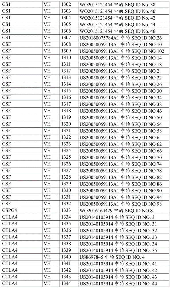


Intrabody antibodies
In some embodiments, the payload of the invention may be an intrabody directed to a participant that modulates an immune cell. Intrabodies are antibodies designed to be expressed intracellularly and can be directed to a specific target antigen present in a variety of subcellular locations including cytosol, nucleus, Endoplasmic Reticulum (ER), mitochondria, peroxisomes, plasma membrane and the reverse golgi network (TGN) by in-frame fusion with an intracellular translocation/localization peptide sequence. The most commonly used format is a single chain antibody (scFv) created by: the antigen-binding variable domain of the heavy chain and the antigen-binding variable domain of the light chain are joined with a linker between the Variable Heavy (VH) chain and the Variable Light (VL) chain, which is most often a15 amino acid linker (e.g., (GGGGS)3(SEQ ID NO: 307)). Intracellular intrabodies are being developed to bind, neutralize, or modulate the function or localization of cancer-associated targets and thereby affect malignant phenotypes.
A participant in the modulation of an immune cell (e.g., a T cell) can be any intracellular signaling checkpoint. Exemplary participants may include co-inhibitory ligands, castias B lineage lymphomatogen oncogene-B (Cbl-B), an E3 ligase, Protein Tyrosine Phosphatases (PTPs) such as protein tyrosine phosphatase 1(SHP-1) containing the Src homology region 2 domain, and Ras. For example, the intrabody can be PCT patent publication No.: intrabodies to oncogenic forms of RAS disclosed in WO 2004/046186; the contents of said patent are incorporated herein by reference in their entirety.
Therapeutic antibodies
In some embodiments, an antibody payload of the invention can be a therapeutic antibody. As non-limiting examples, antibodies, fragments and variants thereof can be specific for a tumor-associated antigen or a tumor-specific antigen or a pathogen antigen. In some aspects, the antibody can be a blocking antibody (also referred to as an antagonistic antibody), such as blocking antibodies against PD-1, PD-L1, PD-L2, CTLA-4, and other inhibitory molecules. In other aspects, the antibody can be an agonist antibody such as an agonist antibody specific for a stimulatory molecule, e.g., 4-1BB (CD137), OX40(CD134), CD40, GITR, and CD 27.
Other exemplary therapeutic antibodies may include, but are not limited to, abamectin (Abagovomab), abciximab (Abcxmab), trastuzumab (Abituzumab), arritumumab (Abrilumab), Actoxumab (Actoxumab), Adalimumab (Adalilimumab), Addimizumab (Adecatuzumab), Africancizumab (Afacivuzumab), Afacivuzumab (Afacivikuzumab), Afaciumumab (Afaciumumab), Afacituzumab (Afacizumab), Afacizumab (Alacizumab), Afacizumab (Altuzumab), Amatuximab (Amatuximab), Aditumumab (Anfuitumumab), Aditumumab (Anifozumab), Adeniumumab (Anifozumab), Afacizumab (Afacizumab), Afacizumab (Avicizumab), Ab (Avicizumab), Atacilizumab (Avicizumab), Atuizumab (Avicib), Atuizumab (Avicinobab (Atuizumab), Atuizumab (Atuizumab), Atuimuramicab (Atuizumab), Atuimuratezomib (Atuizumab), Atuimura (Avuizumab), Atuimura (Avuimura (Avenub), Ab (Avenub), Abuizumab), Abeli (Avenub), Abuimura (Atuimura (Avenub), Abuizumab), Abeli (, Bapiduzumab (Bapineuzumab), basiliximab, Bavituximab (Bavituximab), bematuzumab (bectmomab), begolombumab (bezelomab), Belimumab (Belimumab), Benralizumab (benimumab) (Benralizumab), patimumab (bertimab), bessemuzumab (besilsomab), Bevacizumab (Bevacizumab), bezitratuzumab (beziximab), bicirzumab (becmituzumab), Bimagrumab (Bimagrumab), bimekuzumab (bimekikuzumab), Bivatuzumab (Bivatuzumab), breluzumab (bleelumab), bonatinumab, banuzumab (blosomamab), brontuzumab (brosuzumab), brosuzumab (brolizumab), brazizumab (brolizumab), brazilizumab (brolizumab), brazilizumab), braziliz, Carrocumab (Caprozumab), Carluumab (Carlumab), Carludox (Carotuximab), Cartuzumab (Catuxomab), cBR96-doxorubicin immunoconjugate (cBR96-doxorubicin immuconate), Celizumab (Cedellizumab), Cetuzumab (Cergituzumab), Centulizumab (Certoluzumab), Centilizumab (Clitumumab), Cetuzumab (Citatuzumab), Cetuzumab (Cixtuzumab), Clazatuzumab (Clazakizumab), Crelizumab (Clienlixiximab), Clivalizumab (Clivatuzumab), Corituzumab (Corituzumab), Cotuzumab (Corituzumab), Coltuximab (Coltuximab), Co (Cattuzumab), Centulizumab (Clivalizumab), Centilizumab (Clivalizumab), Cetuzumab (Critulizumab), Cetuzumab (Critumumab (CR (Clitumumab), Cetuzumab (Critumumab), Cetuzumab (Critumumab), Countuzumab (Clitumumab), Countuzumab), Count, Trastuzumab (Dect rekumab), bimirlizumab (Demcizumab), dinituzumab (Denintuzumab), dinozumab (Dentuzumab), biotin delotuzumab (Denosumab), Deltoimab (Derluximab bbioti), dimuzumab (Detumomab), dinituximab (Dinutuzumab), didatuzumab (Diridavumab), Domazumab (Domagrozumab), atorvastatin (Dorguzumab), Zeuzumab (Dumaruzumab), Durivaluzumab (Durvaluzumab), Dusiguzumab (Durivaluzumab), Dusiguzumab (Dusiguzumab), Eimeliximab (Ermuzumab), Esomemazumab (Elsigitumomab), Esomemazumab (Egenuzumab), Emetuzumab (Egenuzumab), Esomemazumab (Egentuzumab), Egytuzumab (Egytuzumab), Egytuzumab (Egytuzumab), Egylco-mebrutuzumab), Egylco (Egylco-mex, Emifeprituzumab (Emicizumab), Envantuzumab (Enavatuzumab), vedotin Envuzumab (Enfortuzumab), Endotti Envuzumab (Enfortuzumab), Endomab pembrolizumab (Enlimomab pegol), Entuzumab (Enoblituzumab), Enkuzumab (Enokizumab), Entituzumab (Enotizumab), Enastuximab (Ensituximab), Ceitumomab (Epitumomab cituxetan), Epipezumab, Erlizumab (Erlizumab), Ervatuzumab (Ertumaxomab), Eduzumab (Etarazezumab), Ultrazumab (Etarazezumab), Evelumab (Etroluzumab), Evinazuuzumab (Evinuzumab), Evovolumumab (Evozemazumab), Evozumab (Evomazumab), Evomazumab (Exviruzumab), Exvinumab (Exvinuzumab), Favinlizumab (Faveluzumab), Faveluzumab (Favelutvazumab), Faveltuzumab (Fevinatuzumab), Favelutuzumab (Feratuzumab), Favelutuzumab (Feratuzumab), Faveluttatuzumab), Faltuzumab (Feratuzumab), Faltuzumab (Feratuzumab), Falt, Feireuzumab (Firivumab), Frantuzumab (Flanv otumab), Florekumamab (Fletikumab), Farituzumab (Fletikumab), Artuzumab (Fontalizumab), Flaluzumab (Foralumab), Furazumab (Foravirumab), Fusappan (Freulimumab), Fragranumab (Fulramumab), Furazumab (Fulranimab), Futuximab (Futuximab), Gakalizumab (Galcanizumab), Galiximab (Galiximab), Ganituzumab (Ganituzerumab), Gantemab such as mAb (Gantenerumamab), Gavitexin (Gavilimomab), Ozomizuzumab (Gemtuzumab ozogamicin), Gevojiuzumab (Getuzumab gejiuzumab), Girtumazumab (Girtumab), Giruituximab (Girentuzumab), Gotuzumab (Glrentzikumakumab), Gotukumuramikumakumakumakumab (Glitumomab), Gotuzumab (Gotuzumab), Gotuzumab ozuguentuckii), Gotuzumab (Gotuzumab), Gotuzumab ozotai (Gotuvu (Gotukuvietuzumab), Gotuvu, Gotukuvietuzumab), Gotuzumab), Gotuvu (Gotukuvietuzumab), Gotuzumab), Gotuvu (Gotuvu, Gotuvu (Gotuzumab), Go, IMAB362, imazeumab (I malumamab), infliximab (Imciromab), engatuzumab (Imgatuzumab), inchurumab (Inclacumab), infliximab (Indatuximab), infliximab (indatuzumab), infliximab (inebrizumab), infliximab (infliximab), infliximab (intetuzumab), inolimumab (inomomab), inolimumab (inolimumab), inolimumab (inozumab), rituximab (iramab), isatuzumab (Isatuximab), itozumab (itatuzumab), ibritumumab (isatuzumab), eltamimab (iximab), laulizumab (laxumab), laizumab (laxatizumab), lailizumab (Ix ekizumab), Keliximab (Keliximab), trastuzumab (labuzumab), lagucizumab (lailizumab), laguizumab (lailizumab), laguilizumab (lai-griffuzumab), lapuizumab (lai-griffusal (lai-L), lapuizumab (laguilizumab), lapuilizumab (laguilizumab), lattuzumab), lapuilizumab (lat-la-leukalimeli-leu-griffia (lat-mab), lat-la-leu-mab), lat-leu-mab (lat-, Rizeluzumab (Lenzilumab), ledumumab (Lerdelimumab), lexamumab (Lexatumumab), livinmab (Libivirumab), Livatuzumab (Lifastumab), Rigelizumab (ligelizumab), Lilotuzumab (Lilotumab), Lintuzumab (Lintuzumab), Rituzumab (Rituzumab), Lodellizumab (Lodeluzumab), Lokvepizumab (Lokivatumab), Lotuzumab (Lortuzumab), Lorteuzumab (Lucatazumab), Polyglycollizumab (Lulizumab pegol), Lumilipolar (Lumiliximab pegol), Lumilizumab (Lumilizumab), Lumatuzumab (Lumfitumumab), Mamrtuzumab (Mapateumab), Mapatumab), Magnetizumab (Masgetuzumab (Margefituzumab), Martimumab (Mermetizumab), Merituzumab (Merituximab (Merituzumab), Merituximab (Merituximab), Merituximab (Merituximab) and the like), Momurizumab (Mogamulizumab), monelizumab (Monalizumab), molulizumab (Morolimumab), Moloremumab (Morolimumab), Movizumab (Motavizumab), Pradelimumab (Moxeumumab), Molutumona-CD 3(Muromonab-CD3), Tanakamazumab (nacolomomab tafenatox), namifuzumab (Naummilab), natuzumab (natuzumab), Natuximab (Naratumumab), Natuzumab (Narmanataumab), Natalizumab (Natalilizumab), Naxizumab (Navixizumab), Navizumab (Naviuzumab), Navizumab (Navituzumab), Navituzumab (Navivuzumab), Navikumab (Nevituzumab), Netuzumab (Neciumumab), Netuzumab (Nemaculizumab), Novazumab (Novazumab), Novituzumab (Novituzumab), Novazumab (Novituzumab), Novituzumab (Novituzumab), and Novituzumab (Novituzumab), Novituzumab (Novietnavietnavitu, Oxalizumab, olaratumab (Olaratumab), olaratumab, olobizumab (Olokizumab), Omalizumab (Omalizumab), Onauzumab (Onaruzumab), Otuximab (Ontuzumab), Otuximab (Otuximab), Opimelizumab (Opicinsumab), Moorezumab (Oportuzumab monatx), Ogovzumab (Oregomab), Ovatuzumab (Orticuzumab), Otetuzumab (Oteluzumab), Oteuximab (Oteluzumab), Ottuzumab (Otituzumab), Otexizumab (Oxexetuzumab), Ozanuzumab (Ozanuzumab), Ozolizumab (Ozolizumab), Pagiliximab (Pagibasimab), Parivumab (Paliviazumab), Palifumab), Pacifumab (Pacifuzumab), Pazolizumab (Parzanuzumab), Pazouzumab (Pazouzumab), Pagilixuzumab), Pagizumab (Pagizumab), Pachiuzumab (Pazouzumab), Pazortuzumab (Pazortuzumab), Pacifumab (Pacifumab), Pazortuzumab (Pazortuzumab), Pacifb (Pacifb), Pacifb (Pacifb), Pa, Pembrolizumab (pemtumumab), perlizumab (Perakizumab), Pertuzumab (Pertuzumab), pelizumab (pexluzumab), Pidilizumab (Pidilizumab), pinatuzumab (pindolizumab), pinguzumab (pintumumab), prakuuzumab (Placulumab), palulizumab (Plozalizumab), Pogalizumab (Pogalizumab), perlatizumab (Polatuzumab), panlizumab (Prez alizumab), Priliximab (Priliximab), Priliximab (prituximab), prilizumab (prituzumab), PRO 140, quinlizumab (Quilizumab), Ranibizumab (racuzumab), Ranibizumab (raclizumab), Ranibizumab (rexinumab), Ranibizumab (rex), Ranibizumab (rezumab), Ranibizumab (rex), ranibix (rex), ranibix (ranibix), ranibix (ranibix) and ranibix (ranibix) antibody (ranibix) and ranibix (ranibix) are included in, Rituximab (Rilutumumab), linnouzumab (Rinucumab), lissazumab (Risankizumab), rituximab, polyethylene glycol Ribazumab (Rivabazum ab pegol), Rituzumab (Robatuzumab), roducumab (Roledumab), romovazumab (Romosozumab), Rilizumab (Rotalumab), Rovellizumab (Rovalpiruzumab), Rovellizumab (Rovelizumab), Lulizumab (R uplizumab), Saxituzumab (Sacituzumab), Samalizumab (Samalizumab), Sapinzumab (Sapelizumab), Sapellizumab (Sapellizumab), Sepellizumab (Sepezimab), Serilizumab (Sariluzumab), pentoxizumab (Tumumab) (Tumopentamab), Semilizumab (Satruzumab), Sepeluzumab (Sepeluzumab), Sepellizumab (Sepeluzumab), Sepeluzumab (Sepeluzumab), Sepeluzumab (Sepelvituzumab), Sepellucin (Sepelvituzumab), Sepexi, Hiprilinzumab (Siplizumab), stechikumab (Sirukumab), vedoline Sofituzumab (Sofituzumab vedotin), Sulan (Solanezumab), Soritotuzumab (Solitumab), Sonupuzumab (Solitumab), Sonepuzumab (Sonepuzumab), Sontuzumab (S ontuzumab), Setariuzumab (Stamulumab), Thiozumab (Sulesumab), Suvizumab (Suvizumab), Tabaluzumab (Tabaluzumab), Takamazumab (Tacateuzumab), Tadu mab (Tadocuzumab), Tallizumab (Talizumab), Tamtuzumab (Tamtuzumab), Tenuizumab (Tatenuimab), Tapleuzumab), Tamtuzumab (Tamtuzumab), Tezumab (Tatenuizumab), Tatenuizumab (Tapleuzumab), Noveltexiluzumab (Tamtuzumab), Tamtuzumab (Tepleuzumab), Tamtuzumab), Tapleuzumab (Tapleuzumab), Tapletuximab (Tapletix), Tapleuzumab), Tapletix (Tapleuzumab), Tapletichituximab (Tepletix, Tapletix (Tepletix), Tapletix (Tepleuzumab), Tapletix (Tepletix, Tamtuzumab), Tamtte, Tiximumab (ticlizumab), tiruzumab (tildarkizumab), tegafuzumab, temoluumab (timoluumab), vedotti tiuxotuzumab (tisotuzumab), TNX-650, toluzumab (Tocilizumab), tollizumab (Toralizumab), tosatuzumab (tosatoxuumab), tositumomab (tositumomab), tositumomab (tovirtumab), teraluotuzumab (Trastuzumab), Trastuzumab (Trastuzumab), TRBS07, Trastuzumab (Tregalizumab), Tremelimumab (tremelizumab), Trastuzumab (trevelumab), trefluzumab (trefluvituzumab), tuzumab (tuzumab), tuzumab (valtuzumab), valtuzumab (valtuvurtuzumab), and valtuzumab (valtuvurtuzumab), valtuzumab (valtuvurtux (valtuvu), valtuzumab), valtuvu (valtuvu), valtuvurtune (valtuvurtuzumab), valtuvu (valtuvu), valtuvu, Valliximab (Vapaliximab), tilimumab (varluumab), vatuzumab (vatelizumab ab), veltezumab (vatelizumab ab), Vedolizumab (Vedolizumab), vetuzumab, vepamitumab (Vepalimo mab), vessekumab (Vesencumab), velizumab (Visilizumab), vebailizumab (Vobarilizumab), vollocycloximab (Volociximab), volotuzumab (V oretuzumab), volutumumab (zetuzumab), zetuzumab (Xentuzumab), Zalutumumab (zumab), Zanolimumab (zumab), zantuximab (ztitumumab), zilarumab (zaolimumab), zituximab (ztituximab), zilaramumab (zilarumab), and adozolimumab (zolimumab ari).
Bicistronic and/or pseudobicistronic antibody payloads
According to the present invention, a dicistronic payload is a polynucleotide that encodes a double protein chain antibody on a single polynucleotide chain. A pseudo-dicistronic payload is a polynucleotide that intermittently encodes a single-chain antibody on a single polynucleotide strand. For a dicistronic payload, the two strands or two portions/regions and/or domains encoded (as in the case of a pseudo-dicistronic) are separated by at least one nucleotide that does not encode a strand or domain. More often, the separation portion comprises a cleavage signal or site or a non-coding nucleotide region. Such cleavage sites include, for example, a furin (furin) cleavage site encoded as an "RKR" site in the resulting polypeptide, or a modified furin cleavage site, or any of the cleavage sites taught herein.
According to the invention, a single domain payload comprises one or two polynucleotides encoding a single monomeric variable antibody domain. Typically, a single domain antibody comprises one variable domain of a heavy chain antibody (VH).
According to the present invention, a single chain Fv payload is a polynucleotide that encodes at least two coding regions, as well as a linker region. The scFv payload can encode a fusion protein of the variable region of the heavy chain (VH) and the variable region of the light chain (VL) of an immunoglobulin, which are linked with a short linker peptide of 10 to about 25 amino acids. The linker is typically rich in glycine for flexibility and serine or threonine for solubility, and may link the N-terminus of VH with the C-terminus of VL, or vice versa. Other linkers include those known in the art and disclosed herein.
According to the present invention, a bispecific payload is a polynucleotide encoding portions or regions of two different antibodies. The bispecific payload encodes a polypeptide that can bind to two different antigens. The polynucleotides of the invention may also encode trispecific antibodies having affinity for three antigens.
3. Tumor and pathogen specific antigens
In some embodiments, the payload of the invention can be a Tumor Specific Antigen (TSA), a Tumor Associated Antigen (TAA), a pathogen associated antigen, or a fragment thereof. The antigen may be expressed in peptide form or in the form of a complete protein or a portion thereof. The entire protein or a portion thereof may be native or mutagenized. Antigens associated with cancer or virus-induced cancer as described herein are well known in the art. Such TSA or TAA may have been previously associated with cancer, or may be identified by any method known in the art.
The Tumor Specific Antigen (TSA) may be a tumor neoantigen. A neoantigen is a mutant antigen that is expressed only by tumor cells due to genetic or transcriptional changes that alter the protein coding sequence, thus generating a novel foreign antigen. The genetic change is caused by a genetic substitution, insertion, deletion or any other genetic change of the native homologous protein (i.e., the molecule expressed in normal cells).
As non-limiting examples, the novel antigen may include mutant novel peptides derived from α -actinin-4, ARTC1, BCR-ABL fusion protein (B3a2), B-RAF, CASP-5, CASP-8, β -catenin, Cdc27, CDK4, CDKN2A, CLPP, CML-66, COA-1, connexin 37, A-can fusion protein, EFTUD A, elongation factor 2, ETV A-AML A fusion protein, fibronectin, FLT A-ITD, FN A, GPNM A, LDLR-trehalose transferase AS fusion protein, HLA-A A, Hsp-70-1A-2, HLA A, MUM-1, MUM-2, MUM-3, RARIC-like myosin, NFO-acetylation, neo-OGT, NEOGT, NEOST-9, MUOS-72, PSN-72, MAPTMX-A, MAPTX A, MAPTMYX A-A, MAPTMYX A-A, TFX A, MAPTMYX A, TFX A, MAPTDE A, NFX A, MAPTX A, and the like, AS disclosed in the disclosures of the patent publications.
Novel antigens identified by large scale sequencing and algorithmic calculations may also be included. See, for example, international patent publication nos.: WO 2014/168874; nishimura et al, Cancer Sci.2015,106(5): 505-511; and Linnemann et al, nat. Med.,2015,21(1): 81-85; the contents of each are incorporated herein by reference in their entirety.
In tumor tissues, numerous proteins (e.g., oncogenes) are upregulated, including but not limited to lipophilin (adipophilin), AIM-2, ALDH1A1, BCLX (L), BING-4, CALCA, CD45, CD274, CPSF, cyclin D1, DKK1, ENAH, epCAM, ephA3, EZHH 2, FGF5, G250, HER-2/neu, HLA-DOB, serine protease (Hepsin), IDO1, IGFB3, IL13R α, intestinal carboxyesterase, kallikrein4 (kallikkrein 4), KIF20A, glutaminase-containing domain protein (crystalline CSF), M-CSF, MCmBG 2, MClBG 2, MMG 3, MMG 2, MMG 3, MMG 7, VEGF-RNG 7, MMG 5, MMG 7, MMG 5, VEGF-RNG 3, VEGF 11, VEGF 3, rG 7, rG 3, rG 3, rG 7, rG.
Examples of carcinoembryonic antigens may include, but are not limited to, CEA (carcinoembryonic antigen) in colorectal cancer, iLRP/OFA (immature laminin receptor protein/carcinoembryonic antigen) in Renal Cell Carcinoma (RCC), TAG-72 (tumor-associated glycoprotein-72) in prostate cancer, AFP (α -fetoprotein) in hepatocellular carcinoma (HCC), ROR1 (a receptor tyrosine kinase) in many malignant cells such as brain tumors, sperm protein 17, HMGA2 (high mobility family 39A 2), carcinoembryonic H19, CR-1(Cripto-1, growth factor-s) in HCC, and the prolipoprotein of heparan (a-s of the HCC-EGF receptor tyrosine kinase), the prolease of ovarian cancer, and the phosphatidylinositol 3 family of heparan.
The TAA may be a cancer-testis antigen expressed solely by cancer cells and adult reproductive tissues such as testis and placenta, including but not limited to antigens from the BAGE family, the CAGE family, the HAGE family, the GAGE family, the MAGE family (e.g., MAGE-A1, MAGE-A2, MAGE-A3, MAGE-A6 and MAGE-A13), the SAGE family, the XAGE family, MCAK, NA88-A (cancer/testis antigen 88), PSAD1, SSX-2 and SLLP-1.
TAA can be a lineage-defined antigen expressed predominantly by a single cancer tissue type, such as Melan-A/MART-1, Gp100/pmel17, tyrosinase, TRP-1/-2, P. polypeptide, MC1R in melanoma; and Prostate Specific Antigen (PSA) in prostate cancer.
TAAs can be oncogenic viral antigens encoded by oncogenic transforming viruses (also known as oncogenic viruses). Oncogenic viruses can insert their own DNA (or RNA) into the DNA of a host cell when they infect the host cell. When viral DNA or RNA affects the genes of a host cell, it can push the cell toward becoming cancerous. Oncogenic viruses include, but are not limited to, RNA viruses and DNA viruses. Some examples of commonly known oncogenic viruses include Human Papilloma Virus (HPV), which is the main etiology of cervical cancer, ebstein-Barr virus (EBV) that can cause nasopharyngeal carcinoma, certain types of rapidly growing lymphomas such as burkittlymoma and gastric carcinoma, hepatitis b, c and d viruses (HBV, HCV and HDV) in the case of hepatocellular carcinoma (HCC), Human Immunodeficiency Virus (HIV) that increases the risk of acquiring many types of cancer such as liver, anal and hodgkin's cancer, Kaposi's sarcoma (Kaposi sarcoma) herpes virus (KSHV; also known as human herpes virus 8(HHV8)), human T-lymphotropic virus (HTLV-1) and merkel (merkel) cell polyomavirus (MCV) associated with lymphoma. The viral antigen may be any defined antigen of the virus that is associated with human cancer. For example, antigens from EBV may include, but are not limited to, ebbs-bal nuclear antigen-1 (EBNA1), latent membrane protein 1(LMP1), or latent membrane protein 2(LMP 2).
TAAs may be idiotypic antigens generated from highly polymorphic genes, where tumor cells express a particular "clonotype", i.e. such as immunoglobulins and T Cell Receptors (TCRs), as in the case of B cell lymphoma/leukemia, T cell lymphoma/leukemia resulting from clonal abnormalities. Idiotypic antigens are a class of non-pathogen associated neoantigens. For example, malignant B cells express rearranged and multiply mutated surface immunoglobulins (igs). Tumor-specific idiotypes (e.g., immunoglobulin idiotypes) are considered to be particularly attractive tumor-specific antigens that can be successfully targeted by immunotherapy (e.g., Alejandro et al, Front oncol, 2012,2: 159).
T Cell Receptor (TCR)
In some embodiments, the payloads of the present invention can be T Cell Receptors (TCRs), TCRs can be specific for cancer antigens, have a specific α chain and β chain that together form a TCR αβ heterodimer, or have a specific γ chain and δ chain that together form a TCR γ δ heterodimer.
In some embodiments, a biological circuit or effector module comprising a tumor-specific TCR may be transduced into T cells, and TCR-engineered T cells will be infused into cancer patients suffering from lymphopenia or lymphopenia due to chemotherapy or irradiation, allowing for efficient engraftment, but at the same time inhibiting immunosuppression.
The sequence encoding the tumor antigen-recognizing TCR chain can be obtained from tumor-reactive T cells (e.g., tumor-infiltrating lymphocytes isolated from a tumor of a patient).
In accordance with the present invention, TCRs specific for tumor cells can be produced by the methods described in International patent publication No.: WO2014/083173, the contents of which are incorporated herein by reference in their entirety, a transgenic host organism expressing a certain Human Leukocyte Antigen (HLA) type known or presumed to be capable of presenting a mutant tumor-specific antigen (TSA) is transduced to express unrearranged human TCR loci, preferably these loci encode TCR α and β chains, and preferably comprise multiple, desirably all, of human TCR V, D, J and/or C genes.
In some embodiments, the payload of the invention may be a TCR that specifically recognizes a TSA, TAA, or epitope thereof complexed to an MHC molecule.
Exemplary tumor antigens recognized by TCR include tumor antigens 5T, 707-AP, A, AFP (-fetoprotein), AKAP-4 (kinase-anchored protein 4), ALK, 5 integrin, androgen receptor, annexin II, 0-actin-4, ART-4, B7H, BAGE (B melanoma antigen), BCMA, BCR-ABL fusion protein, -catenin, BKT antigen, BTA, CA-I (carbonic anhydrase I), CA (cancer antigen 50), CA125, CA-3, CA195, CA242, calreticulin, CAIX (carbonic anhydrase), CAMEL (cytotoxic T lymphocyte-recognized antigen), CEMELP), CD, CAP-1, caspase-8/M, CD, TNF-M, CD, TNF-related receptor, VEGF-receptor, VEGF-related protein (VEGF-2-receptor-related protein), VEGF-receptor-related protein (VEGF-2-receptor-related protein), VEGF-receptor-related protein (VEGF-2), VEGF-receptor-related protein (VEGF-receptor-related protein (VEGF-2), VEGF-receptor-related protein (VEGF-TNF-receptor-related protein), VEGF-receptor-related protein (VEGF-receptor-related protein), VEGF-receptor-related protein (VEGF-receptor-related protein (VEGF-receptor-related protein), VEGF-related protein (VEGF-1-180), VEGF-TNF-VEGF-TNF-2-150), VEGF-TNF-7, VEGF-TNF-receptor-TNF-7, VEGF-TNF-7, VEGF-TNF-receptor (VEGF-TNF-induced, VEGF-TNF-receptor (VEGF-TNF-receptor-7, VEGF-receptor-7, VEGF-150), VEGF-receptor-7-receptor (VEGF-7-receptor-150), VEGF-receptor-7-150, VEGF-receptor-7, VEGF-150, VEGF-receptor-TNF-receptor-TNF-receptor-150, VEGF-receptor-7-150, VEGF-receptor-TNF-receptor-7, VEGF-receptor-TNF-receptor-induced, VEGF-induced cytokine, VEGF-TNF-receptor-TNF-receptor-TNF-7-150, VEGF-7, VEGF-TNF-7, VEGF-receptor-TNF-150, VEGF-TNF-receptor-TNF-7, VEGF-receptor-7, VEGF-TNF-150, VEGF-TNF-7-TNF-150, VEGF-related protein (VEGF-TNF-150, VEGF-TNF-receptor fusion protein (VEGF-receptor-TNF-receptor-TNF-receptor-7, VEGF-TNF-receptor-TNF-receptor-TNF-receptor-TNF-150, VEGF-TNF-7, VEGF-TNF-150, VEGF-TNF-receptor (VEGF-7, VEGF-TNF-7, VEGF-150, VEGF-receptor-TNF-receptor, TNF-150, VEGF-TNF-receptor, TNF-receptor, VEGF-receptor fusion protein (VEGF-7, VEGF-TNF-7, TNF-receptor-TNF-150, VEGF-TNF-receptor, VEGF-TNF-150, TNF-receptor fusion protein (VEGF-receptor, TNF-receptor, VEGF-150, TNF-receptor, VEGF-receptor fusion protein (VEGF-receptor-TNF-receptor fusion protein (VEGF-receptor, VEGF-receptor, VEGF-TNF-150-receptor-TNF-receptor, VEGF-TNF-receptor, VEGF-150, TNF-receptor, VEGF-TNF-receptor-TNF-receptor, VEGF-receptor, TNF-receptor, VEGF-receptor, VEGF-150, TNF-receptor, VEGF-receptor, or VEGF-150, TNF-receptor fusion protein (VEGF-receptor-150, VEGF-receptor fusion protein (VEGF-receptor-TNF-receptor, VEGF-150, VEGF-receptor-150, or VEGF-TNF-receptor fusion protein (VEGF-7-receptor fusion protein (VEGF-receptor, TNF-receptor.
In one aspect, the payload of the invention can be a TCR that specifically recognizes the Her2/neu epitope with the nucleic acid sequences of the α and β chains disclosed in U.S. patent publication No. US20110280894 and International patent publication No. WO2016133779A1, the contents of each of which are incorporated herein in their entirety by reference.
In other aspects, the payloads of the present invention can be TCRs having a polypeptide sequence specific for synovial sarcoma X breakpoint (SSX) -2 antigen (U.S. Pat. No.9,345,748), Human Papilloma Virus (HPV)16E6 antigen (International patent publication No. WO2015/009606), Cytomegalovirus (CMV) phosphoprotein pp65 (U.S. Pat. No.8,722,048), and WT-1 specific TCRs as disclosed in U.S. Pat. No. US2016/0083449, the WT-1 specific TCRs comprising a TCR α chain having an amino acid sequence as set forth in any one of SEQ ID nos. 5-8 and comprising a TCR β chain having an amino acid sequence as set forth in SEQ ID nos. 12 or 13, the contents of each of which are incorporated herein by reference in their entirety.
In some embodiments, a TCR specific for a TSA may be modified to have a sequence encoding an affinity-attenuating motif that confers a reduction in non-specific binding to the TSA. In some embodiments, affinity-reducing sequences having modifications to the TCRCDR1 or CDR2 regions can be used to attenuate the interaction between a TCR and an HLA protein (see International patent publication No.: WO 2016/014725; the contents of which are incorporated herein by reference in their entirety).
In some embodiments, the high affinity TCR may be produced by using a TCR α chain to select a regenerated TCR β chain that pairs with an antigen-specific TCR α chain during T cell development to form an enhanced TCR (see International patent publication No: WO 2013/166321; the contents of which are incorporated herein by reference in their entirety). in other embodiments, the modified TCR may also have a sequence encoding an affinity enhancing modification to a CDR3 region that enhances the interaction between the TCR and TSA.
In one embodiment, TCRs specific for TSAs can be modified using Zinc Finger Nucleases (ZFNs), TALENs, or CRISPR/Cas systems.TCR α and β chains can contain target sites for nucleases the nucleases can cleave the TCR sequence, resulting in some degree of disruption of the TCR (see U.S. patent publication No.: US 2014/0301990; the contents of which are incorporated herein by reference in their entirety).
In one embodiment, the TCR specific for TSA can be a soluble single chain TCR having the structure V α 2-L-V β or V β -L-V α 2, wherein L is a linker peptide linking the TCR variable β region (V β) to the TCR variable α region of family 2 (V α 2) as discussed in International patent publication No. WO2011/044186, the contents of which are incorporated herein by reference in their entirety.
In one embodiment, the compositions may be prepared according to U.S. patent publication nos.: the method described in US2014/0065111 to mature a TCR specific for TSA so that its affinity for TSA is increased; the contents of said patent are incorporated herein by reference in their entirety.
In some embodiments, the TCR may be specific for the Fc domain of an antibody (e.g., FcgRla) and used to enhance the efficacy of antibody-mediated therapy, as described in international patent publication no: as discussed in WO 2015/179833; the contents of said patent are incorporated herein by reference in their entirety.
In some embodiments, the payloads of the present invention may be recombinant constructs that act as trifunctional T-cell signaling conjugates (tritacs) that mimic native signaling through T-cell receptors. TriTAC enhances chimeric receptor activity while retaining MHC undefined targeting. In some aspects, the recombinant construct may comprise a target-specific binding ligand such as a scFV or designed ankyrin repeat (DARPin) specific for TSA, a ligand that binds a protein associated with the TCR complex, and a TCR signaling domain polypeptide, e.g., as described in international patent application No.: as described in WO 2015/117229; the contents of said patent are incorporated herein by reference in their entirety. The TCR-associated protein may be selected from CD3, ZAP70 (zeta chain associated protein kinase 70), TYN and CD 247. In some aspects, the ligand of TriTAC that binds to a TCR-associated protein can be an antibody or antibody fragment (e.g., scFv). In other aspects, the TCR signaling domain polypeptide of TriTAC may comprise the transmembrane and cytoplasmic domains of CD 4.
In other embodiments, the α and β chains of the inventive TCR may be included in a single effector moiety as two payloads of the same effector moiety, e.g., as illustrated in fig. 3-6.
5. Chimeric Antigen Receptor (CAR)
In some embodiments, the payloads of the invention can be Chimeric Antigen Receptors (CARs) that, when transduced into immune cells (e.g., T cells and NK cells), can redirect the immune cells against a target (e.g., a tumor cell) that expresses a molecule recognized by the extracellular targeting portion of the CAR.
As used herein, the term "Chimeric Antigen Receptor (CAR)" refers to a synthetic receptor that mimics a TCR on the surface of a T cell. Generally, a CAR consists of an extracellular targeting domain, a transmembrane domain/region, and an intracellular signaling/activation domain. In the standard CAR receptor, the components: the extracellular targeting domain, transmembrane domain and intracellular signaling/activation domain are constructed linearly into a single fusion protein. The extracellular region comprises a targeting domain/moiety (e.g., scFv) that recognizes a particular tumor antigen or other tumor cell surface molecule. The intracellular domain may comprise a signaling domain of the TCR complex (e.g., the signaling domain of CD3 ζ), and/or one or more costimulatory signaling domains, such as those from CD28, 4-1BB (CD137), and OX-40(CD 134). For example, a "first generation CAR" has only a CD3 zeta signaling domain, while in an attempt to potentiate T cell persistence and proliferation, a costimulatory intracellular domain is added, resulting in a second generation CAR with a CD3 zeta signaling domain plus one costimulatory signaling domain, and a third generation CAR with a CD3 zeta signaling domain plus two or more costimulatory signaling domains. When expressed by a T cell, the CAR confers antigen specificity on the T cell that is determined by the extracellular targeting portion of the CAR. Recently, it may also be desirable to add one or more elements such as homing and suicide genes to develop a more competent and safer construct of CARs, the so-called fourth generation CARs.
Cells engineered to express the CAR, such as T cells, can be redirected to attack target cells expressing molecules recognizable by the targeting portion of the CAR.
In some embodiments, the extracellular targeting domain is joined to the intracellular signaling domain by a hinge (also referred to as a spacer domain or spacer) and a transmembrane region. The hinge connects the extracellular targeting domain to a transmembrane domain that traverses the cell membrane and is connected to an intracellular signaling domain. Due to the size of the target protein to which the targeting moiety binds and the size and affinity of the targeting domain itself, it may be desirable to alter the hinge to optimize the efficacy of the CAR-transformed cell on the cancer cell. Upon recognition and binding of the target cell by the targeting moiety, the intracellular signaling domain generates an activation signal for the CAR T cell that is further amplified by a "second signal" from one or more intracellular co-stimulatory domains. CAR T cells, once activated, can destroy the target cell.
In some embodiments, the CARs of the invention can be divided into two parts, each part having attached thereto a dimerization domain, such that an input that triggers dimerization promotes the assembly of a fully functional receptor. Wu and Lim recently reported resolved CARs in which the extracellular CD19 binding domain and the intracellular signaling element are separated and linked to an FKBP domain and an FRB (T2089L mutant of FKBP-rapamycin binding domain) domain that heterodimerize in the presence of the rapamycin analogue AP 21967. The split receptors assemble in the presence of AP21967 and, along with specific antigen binding, activate T cells (Wu et al, Science,2015,625(6258): aab 4077).
In some embodiments, the CAR of the invention can be designed as an inducible CAR. Sakemura et al have recently reported the incorporation of the Tet-On inducible system into the CD19CAR construct. CD19CAR was activated only in the presence of doxycycline (Dox). Sakemura reports that Tet-CD19CAR T cells target CD19 in the presence of Dox compared to conventional CD19CAR T cells+Cell lines were equally cytotoxic and had equal cytokine production and proliferation following CD19 stimulation (Sakemura et al, Cancer immunity. res.,2016, 6/21 days, electronic publication prior to printing). In one example, this Tet-CAR can be the payload of an effector module under the control of an SRE (e.g., DD) of the present invention. The dual system provides greater flexibility to turn CAR expression on and off in transduced T cells.
According to the invention, the payload of the invention can be a first generation CAR, or a second generation CAR, or a third generation CAR, or a fourth generation CAR. Representative effector module embodiments comprising CAR constructs are illustrated in figures 13-18. In some embodiments, the payload of the invention may be an intact CAR construct consisting of an extracellular domain, a hinge and transmembrane domain, and an intracellular signaling region. In other embodiments, the payload of the invention may be a component of an entire CAR construct, including an extracellular targeting portion, a hinge region, a transmembrane domain, an intracellular signaling domain, one or more costimulatory domains, and other additional elements that improve CAR construction and functionality, including but not limited to a leader sequence, a homing element, and a safety switch, or a combination of said components.
The CARs regulated by the biological circuits and compositions of the invention are tunable and thus provide several advantages. A reversible switching mechanism allows management of acute toxicity caused by excessive CAR-T cell expansion. Pulsed CAR expression using the SREs of the invention can be achieved by cycling the ligand levels. The ligand-conferred modulation of the CAR can effectively counteract tumor escape induced by antigen loss, avoid functional depletion by trophoblast signaling due to long-term antigen exposure, and allow for sustained improvement of CAR-expressing cells in vivo.
In some embodiments, the biological circuits and compositions of the invention can be used to down-regulate CAR expression to limit tissue toxicity in the target caused by tumor lysis syndrome. Downregulating the expression of the CARs of the invention following anti-tumor efficacy can prevent (1) off-target tumor toxicity caused by antigen expression in normal tissues. (2) Antigen-independent activation in vivo.
Extracellular targeting domain/moiety
According to the invention, the extracellular targeting moiety of the CAR can be any agent that recognizes and binds with high specificity and affinity to a new antigen on a given target molecule, e.g., a tumor cell. The targeting moiety may be an antibody and variants thereof that specifically bind to a target molecule on a tumor cell, or a peptide aptamer selected from random sequence pools based on its ability to bind to a target molecule on a tumor cell, or variants or fragments thereof that can bind to a target molecule on a tumor cell, or an antigen recognition domain from a native T Cell Receptor (TCR) (e.g., the CD4 extracellular domain to recognize HIV infected cells), or a foreign recognition component such as an attached cytokine that results in recognition of a target cell bearing a cytokine receptor, or a native ligand of the receptor.
In some embodiments, the targeting domain of the CAR can be an Ig NAR, Fab fragment, Fab ' fragment, f (ab) '2 fragment, f (ab) '3 fragment, Fv, single chain variable fragment (scFv), bis-scFv, (scFv)2, minibody, minidiabody, minitrifunctional antibody, minitetrafunctional antibody, disulfide stabilized Fv protein (dsFv), monoclonal antibody, nanobody, or an antigen binding region derived from an antibody that specifically recognizes a target molecule, such as a Tumor Specific Antigen (TSA). In one embodiment, the targeting moiety is a scFv antibody. When the scFv domain is expressed on the surface of the CAR T cell, and subsequently binds to a target protein on a cancer cell, it is able to maintain the CAR T cell in proximity to the cancer cell and trigger activation of the T cell. scFv can be produced using conventional recombinant DNA technology techniques and are discussed herein.
In one embodiment, the targeting moiety of the CAR construct may be an aptamer, such as a peptide aptamer, that specifically binds to a target molecule of interest. Peptide aptamers can be selected from a pool of random sequences based on their ability to bind to a target molecule of interest.
In some embodiments, the targeting moiety of the CAR construct may be a natural ligand of the target molecule or a variant and/or fragment thereof capable of binding the target molecule. In some aspects, the targeting moiety of the CAR can be a receptor for a target molecule, e.g., full length human CD27, which is a CD70 receptor, can be fused in-frame with the signaling domain of CD3 ζ, thereby forming a CD27 chimeric receptor as an immunotherapeutic agent against CD70 positive malignancies (see, e.g., U.S. patent publication No.: US 20130323214; the contents of which are incorporated herein by reference in their entirety).
In some embodiments, the targeting portion of the CAR can recognize a Tumor Specific Antigen (TSA), such as a cancer neoantigen expressed on tumor cells in a defined manner.
As non-limiting examples, the CAR of the invention may comprise an extracellular targeting domain capable of binding to a tumor-specific antigen selected from the group consisting of 5T, 707-AP, A, AFP (-fetoprotein), AKAP-4(A kinase-anchored protein 4), ALK, 5 integrin, androgen receptor, annexin II, 0-coacilin-4, ART-4, B7H, BAGE (B melanoma antigen), BCMA, BCR-ABL fusion protein, -catenin, BKT antigen, BTA, CA-I (carbonic anhydrase I), CA (cancer antigen 50), CA125, CA-3, CA195, CA242, calretinin, CAIX (carbonic anhydrase), CAMEL (cytotoxic T lymphocyte receptor recognition antigen), MDM, CAP-1, caspase-8/M, CD, TNF-receptor specific receptor tyrosine, VEGF-receptor (MAG), VEGF-receptor-related protein (MAG), VEGF-7), VEGF-7, VEGF-2, VEGF-receptor-related protein (MAG-150), VEGF-receptor-related protein (MAG-150), VEGF-150), VEGF-2-VEGF-related protein (MAG-related protein-VEGF-receptor-VEGF-150, VEGF-150), VEGF-receptor-related protein (VEGF-receptor-VEGF-150), VEGF-related protein (MAG-related protein-receptor-150), VEGF-receptor-2-150), VEGF-150, VEGF-TNF-VEGF-related protein (VEGF-150), VEGF-related protein (VEGF-related protein-VEGF-TNF-2-TNF-receptor-related protein (MAG), VEGF-receptor-related protein (VEGF-TNF-related protein-receptor-related protein (VEGF-150), VEGF-receptor-VEGF-receptor-2-receptor-150), VEGF-related protein (MAG-TNF-receptor-150), VEGF-receptor-related protein-2-receptor-related protein (VEGF-receptor-TNF-receptor-VEGF-receptor-TNF-VEGF-TNF-related protein (VEGF-related protein-receptor-150), VEGF-related protein, VEGF-receptor-TNF-receptor-related protein (VEGF-receptor-TNF-150), VEGF-TNF-receptor-VEGF-receptor-TNF-150), VEGF-receptor-TNF-150), VEGF-TNF-150-receptor-TNF-receptor-TNF-related protein-TNF-related protein (VEGF-receptor-TNF-receptor-related protein (VEGF-receptor-TNF-receptor-TNF-receptor-TNF-2-TNF-receptor-TNF-receptor-150-TNF-receptor-TNF-receptor-TNF-receptor-TNF-receptor-TNF-receptor-TNF-receptor-TNF-150-receptor-TNF-receptor-150-receptor-TNF-receptor-TNF-receptor-TNF-receptor-TNF-receptor-TNF-receptor-150-TNF-receptor-150-receptor-150-receptor-150-TNF-receptor-TNF-150-receptor.
In some embodiments, the CAR of the invention may comprise a universal immunoreceptor with a targeting moiety capable of binding a labeled antigen. Methods of generating universal immune receptor CARs are discussed in international patent publication nos.: in WO2013044225A 1; the contents of said patent are incorporated herein by reference in their entirety.
In some embodiments, the CAR of the invention may comprise a targeting moiety capable of binding to a pathogen antigen.
In some embodiments, the CARs of the invention may comprise a targeting moiety capable of binding to non-protein molecules such as tumor-associated glycolipids and carbohydrates. The TSA may also be a lipid molecule, polysaccharide, sugar, nucleic acid, hapten, carbohydrate, or a combination thereof.
In some embodiments, the CARs of the invention may comprise a targeting moiety capable of binding a component within the tumor microenvironment, including proteins expressed in various tumor stromal cells, including Tumor Associated Macrophages (TAMs), immature monocytes, immature dendritic cells, immunosuppressive CD4+CD25+Regulatory T cells (tregs) and MDSCs. A recent study using animal models demonstrated that after systemic transplantation, VEGFR-2CAR and IL12 expressing T cells infiltrated, expanded, and persisted within tumor masses, resulting in tumor regression. The anti-tumor effect depends on targeting IL12 responsive host cells by activation of VEGFR-2CAR-T cells and release of IL12 (Chinnanamy et al, Clinical Cancer Research,2012,18: 1672-.
In some embodiments, the CAR of the invention may comprise a targeting moiety capable of binding to a cell surface adhesion molecule, a surface molecule of an inflammatory cell that occurs in the context of an autoimmune disease, or a TCR that causes autoimmunity.
As non-limiting examples, the targeting moiety of the invention may be an scFv antibody recognizing a Tumor Specific Antigen (TSA), such as an scFv of antibodies SS, SS1 and HN1 specifically recognizing and binding to human mesothelin (U.S. Pat. No.9,359,447), an scFv of an antibody of GD2 (U.S. Pat. No.9,315,585), a CD19 antigen binding domain (U.S. Pat. No.9,328,156), a ligand binding domain of G2D (U.S. Pat. No.9,273,283; U.S. Pat. No. 20160311906A1), a human anti-mesothelin scFv of U.S. Pat. No.9,272,002 comprising the amino acid sequences SEQ ID No. 11 and 12, an anti-CS 1 binding agent (U.S. Pat. publication No. 075784), an anti-BCMA binding domain (International patent publication No. WO2016/014565), an anti-CD 19 antibody of SEQ ID NO. 6952,695 20, an antibody of the patent publication No. WO 9876, WO 48,8652, an antibody of the invention, the amino acid sequences of the polypeptides of the invention Nos. 92, the invention, the polypeptides of the invention, the polypeptides of the invention, the antibodies of the antibodies binding to the polypeptides of the.
In some embodiments, a natural ligand can be used as a targeting moiety for a CAR of the invention. The natural ligand may be capable of binding antigen with an affinity in the range of scFv, and may redirect T cell specificity and effector function to target cells expressing complementary receptors. In some embodiments, the targeting moiety of the CAR may be neuregulin-1 (NRG1), which is a natural ligand for HER3 and HER 4; VEGF, which is a natural ligand for VEGFR; IL13 wild-type protein or IL13 mutein such as E13Y that binds IL13Ra 2; NKG2D ligand, which is a natural ligand for the NKG2D receptor; CD70, which is a ligand for CD 27; and proliferation-inducing ligands (APRIL), which are natural high affinity ligands for BCMA8 and transmembrane activator and CAML interacting factor (TACI). Any ligand-based BCMA CAR as taught in U.S. patent publication No. US20160362467a1, the contents of which are incorporated by reference herein in their entirety, is suitable for use.
In some embodiments, the targeting moiety of the invention may be an scFv comprising an amino acid sequence in table 11.
Table 11: scFv sequences


In one embodiment, the targeting portion of the CAR can recognize CD 19. CD19 is a well-known B cell surface molecule that enhances B cell antigen receptor-induced signaling and expansion of B cell populations following B cell receptor activation. CD19 is widely expressed in both normal and neoplastic B cells. B cell derived malignancies such as chronic lymphocytic leukemia, acute lymphocytic leukemia and many non-hodgkin lymphomas often retain CD19 expression. This near universal expression and specificity possessed by a single cell lineage has made CD19 an attractive target for immunotherapy. Human CD19 has 14 exons, with exons 1-4 encoding the extracellular portion of CD19, exon 5 encoding the transmembrane portion of CD19, and exons 6-14 encoding the cytoplasmic tail. In one embodiment, the targeting moiety may comprise an scFv derived from the variable region of an FMC63 antibody. FMC63 is an IgG2a mouse monoclonal antibody clone specific for CD19 antigen, which reacts with CD19 antigen on cells of the B lineage. The epitope of CD19 recognized by the FMC63 antibody is in exon 2 (Sotillo et al (2015) Cancer Discov; 5(12): 1282-95; the contents of which are incorporated herein by reference in their entirety). In some embodiments, the targeting portion of the CAR may be derived from the variable region of other CD19 monoclonal antibody clones including, but not limited to, 4G7, SJ25C1, CVID3/429, CVID3/155, HIB19, and J3-119.
In some embodiments, the targeting portion of the CAR can recognize a Tumor Specific Antigen (TSA), such as a cancer neoantigen that is expressed only by tumor cells due to genetic mutations or transcriptional changes that alter the protein coding sequence, thus generating a novel foreign antigen. The genetic change is caused by a genetic substitution, insertion, deletion or any other genetic change of the native homologous protein (i.e., the molecule expressed in normal cells). In the case of CD19, TSAs may include transcript variants of human CD19 that lack exon 2 or lack exons 5-6 or both (see International patent publication No. WO 2016061368; the contents of which are incorporated herein by reference in their entirety). Because FMC63 binds the epitope in exon 2, CD19, which lacks exon 2, is not recognized by FMC63 antibody. Thus, in some embodiments, the targeting moiety of the CAR may be a scFV that is different from FMC 63. As used herein, "distinct from FMC 63" refers to an antibody, scFv, or fragment thereof that is specific for and binds to an epitope of the CD19 antigen that is distinct or unlike the epitope of the CD19 antigen that is bound by FMC 63. In some cases, the targeting moiety may recognize the CD19 antigen lacking exon 2. In one embodiment, the targeting moiety recognizes a fragment of CD19 encoded by exons 1,3, and/or 4. In one example, the targeting moiety recognizes an epitope that bridges the portion of CD19 encoded by exon 1 and the portion of CD19 encoded by exon 3.
Intracellular signaling domains
Upon binding its target molecule, the intracellular domain of the CAR fusion polypeptide transmits a signal to the immune effector cell, thereby activating at least one normal effector function of the immune effector cell, including cytolytic activity (e.g., cytokine secretion) or helper activity. Thus, the intracellular domain comprises the "intracellular signaling domain" of the T Cell Receptor (TCR).
In some aspects, an entire intracellular signaling domain may be employed. In other aspects, a truncated portion of an intracellular signaling domain may be used instead of the complete strand, so long as it transduces effector function signals.
In some embodiments, the intracellular signaling domain of the invention may contain a signaling motif known as the immunoreceptor tyrosine-based activation motif (ITAM) examples of ITAM-containing cytoplasmic signaling sequences include those derived from TCRCD3 ζ, FcR γ, FcR β, CD3 γ, CD3 δ, CD3 ε, CD5, CD22, CD79a, CD79b, and CD66d in one example the intracellular signaling domain is a CD3 ζ (CD3 zeta) signaling domain.
In some embodiments, the intracellular region of the invention further comprises one or more co-stimulatory signaling domains that provide additional signals to immune effector cells. These co-stimulatory signaling domains in combination with signaling domains can further improve the expansion, activation, memory, persistence, and tumor eradication efficiency of CAR-engineered immune cells (e.g., CAR T cells). In some cases, the costimulatory signaling region contains 1,2, 3, or 4 cytoplasmic domains of one or more intracellular signaling and/or costimulatory molecules. The costimulatory signaling domain may be the intracellular/cytoplasmic domain of a costimulatory molecule including, but not limited to: CD2, CD7, CD27, CD28, 4-1BB (CD137), OX40(CD134), CD30, CD40, ICOS (CD278), GITR (glucocorticoid-induced tumor necrosis factor receptor), LFA-1 (lymphocyte function-associated antigen-1), LIGHT, NKG2C, B7-H3. In one example, the costimulatory signaling domain is derived from the cytoplasmic domain of CD 28. In another example, the co-stimulatory signaling domain is derived from the cytoplasmic domain of 4-1BB (CD 137). In another example, the costimulatory signaling domain can be the intracellular domain of GITR, as described in U.S. patent nos.: 9,175,308; the contents of said patent are incorporated herein by reference in their entirety.
In some embodiments, the intracellular domain of the invention may comprise a functional signaling domain from a protein selected from the group consisting of MHC class I molecules, TNF receptor proteins, immunoglobulin-like proteins, cytokine receptors, integrins, signaling-conductive lymphocytic activation proteins (SLAM) such as CD48, CD229, 2B4, CD84, NTB-A, CRACCC, BLAME, CD2F-10, SLAMF6, SLAMF7, activating NK cell receptors, BTLA, ToiLI ligand receptors, OX40, CD2, CD7, CD27, CDS, ICAM-1, LFA-1(CD11 27/CD 27), 4-1BB (CD 36137), B27-H27, CDS, ICAM-1, ICC 36278, GITR, BAFFR, GHT 27, VLADAMTK 27/CD 27, CD 27-27, CD 27-27, CD 3636363636363672, CD27, CD 36363636363672, CD 3636363636363636363636363636363636363672, CD 363636363672, CD 3636363636363636363636363636363636363636363672, CD 363636363636363636363636363636363636363636363672, CD 3636363672, CD 3636363636363636363636363636363636363636363636363636363636363636363636363636363636363636363636363636363636363636363636363636363636363636363636363636363636363636363636363636363636363636363636363636363636363636363636363636363636363636363636363636363636363636363636363636363636363636363672, CD 3636363672, CD 3636363636363636363636363636363636363636363636363636363636363636363636363636363672, CD 363636363672, CD 36363636363636363672, CD 3636363636363636363636363636363636363636363636363636363636363636363636363636363636363636363636363636363636363636363636363636363636363636363636363636363636363636363636363636363636363636363636363636363636363636363636363636363636363636363636363636.
In some embodiments, the intracellular signaling domain of the invention may contain a signaling domain derived from JAK-STAT. In other embodiments, the intracellular signaling domain of the invention may contain a signaling domain derived from DAP-12 (death-related protein 12) (Topfer et al, Immunol.,2015,194: 3201-3212; and Wang et al, Cancer Immunol.,2015,3: 815-826). DAP-12 is a key signal transduction receptor in NK cells. The activating signal mediated by DAP-12 plays an important role in triggering the cytotoxic response of NK cells to certain tumor cells and virally infected cells. The cytoplasmic domain of DAP12 contains the immunoreceptor tyrosine-based activation motif (ITAM). Thus, a CAR containing a DAP 12-derived signaling domain can be used for adoptive transfer of NK cells.
In some embodiments, T cells engineered with two or more CARs incorporating unique costimulatory domains and modulated by unique DDs can be used to provide dynamic control of downstream signaling.
In some embodiments, the payload of the invention can be any of the costimulatory molecules and/or intracellular domains described herein. In some embodiments, one or more co-stimulatory molecules, each under the control of a different SRE, may be used in the present invention. SRE-regulated co-stimulatory molecules can also be expressed in conjunction with first generation CARs, second generation CARs, third generation CARs, fourth generation CARs, or any other CAR design described herein.
In some embodiments, an intracellular domain of the invention can comprise an amino acid sequence of table 12.
Table 12: intracellular signaling and co-stimulatory domains


Transmembrane domain
As used herein, the term "transmembrane domain" (TM) broadly refers to an amino acid sequence spanning the plasma membrane that is about 15 residues in length, more preferably, the transmembrane domain includes at least 20, 21, 22, 23, 24, 25, 26, 27, 28, 29, 30, 31, 32, 33, 34, 35, 36, 37, 38, 39, 40, 41, 42, 43, 44, or 45 amino acid residues, and spans the plasma membrane in some embodiments, the transmembrane domain of the CAR can be derived from natural or synthetic sources the transmembrane domain of the CAR can be derived from any natural membrane-binding or synthetic source for example, the transmembrane domain can be derived from (i.e., include at least the transmembrane domain of) α, β, or zeta chain of T cell receptors, CD3 epsilon, CD4, CD2, CD8, CD3, CD9, CD16, CD22, CD 858925, CD 9636, CD 9638, CD 369638, CD 9638, CD 3638, CD 73785, or CD 39137.
Alternatively, the transmembrane domain of the invention may be synthetic. In some aspects, the synthetic sequence may comprise predominantly hydrophobic residues such as leucine and valine.
In some embodiments, the transmembrane domain of the present invention may be selected from the group consisting of a CD8 α transmembrane domain, a CD4 transmembrane domain, a CD28 transmembrane domain, a CTLA-4 transmembrane domain, a PD-1 transmembrane domain, and human IgG4An Fc region. As non-limiting examples, the transmembrane domain may be an international patent publication no: the CTLA-4 transmembrane domain of WO2014/100385 comprising the amino acid sequence SEQ ID No.: 1-5; and international patent publication nos.: the PD-1 transmembrane domain of WO2014100385, comprising the amino acid sequence SEQ ID No. 6-8; the contents of each of these patents are incorporated herein by reference in their entirety.
In some embodiments, the CARs of the invention may comprise an optional hinge region (also referred to as a spacer) the hinge sequence is a short amino acid sequence that facilitates flexibility of the extracellular targeting domain, which moves the target binding domain away from the surface of the effector cell to enable appropriate cell/cell contact, target binding and effector cell activation (Patel et al, general therapy, 1999; 6: 412-419.) the hinge sequence may be located between the targeting portion and the transmembrane domain the hinge sequence may be any suitable sequence derived from or obtained from any suitable molecule the hinge sequence may be derived from all or part of an immunoglobulin (e.g., IgG1, IgG2, IgG3, IgG4) hinge region, i.e., a sequence that falls between the CHI domain and the CH2 domain of an immunoglobulin, e.g., an IgG4 Fc hinge, the extracellular region of a type 1 membrane protein such as a CD8 α, CD4, CD28 and CD7, the hinge sequence may be wild type sequence or a derivative of some including modifications of the CH domain including the amino acid residues 8945, e.g., substitution of the amino acid residues found in the hinge region in the IgG 3613, or IgG 3636, or IgG 3636, and the IgG4 domains.
Table 13: transmembrane domain


Hinge region sequences suitable for use in the present invention are provided in table 14.
Table 14: hinge region


In some embodiments, the hinge domain and transmembrane domain may be paired. The hinge domain may be present at the N-terminus of the transmembrane domain, at the C-terminus of the transmembrane domain or within the transmembrane domain. Hinge and transmembrane region sequences suitable for use in the present invention are provided in table 15.
Table 15: paired hinge and transmembrane regions

In some embodiments, a CAR of the invention can comprise one or more linkers between any domain of the CAR. The linker may be between 1-30 amino acids in length. In this regard, the linker may be 1,2, 3, 4,5, 6,7, 8,9, 10, 11, 12, 13, 14, 15,16, 17, 18, 19, 20, 21, 22, 23, 24, 25, 26, 27, 28, 29, or 30 amino acids in length. In other embodiments, the joint may be flexible.
In some embodiments, the components of the invention, including the targeting moiety, transmembrane domain, and intracellular signaling domain, may be constructed in a single fusion polypeptide. The fusion polypeptide can be a payload of an effector moiety of the invention. In some embodiments, more than one CAR fusion polypeptide can be included in an effector moiety, e.g., two, three, or more CARs can be included in an effector moiety under the control of a single SRE (e.g., DD). Representative effector modules including CAR payloads are illustrated in figures 2-6.
In some embodiments, the payloads of the invention may include CAR constructs taught in the art comprising an extracellular targeting domain, a transmembrane domain and an intracellular signaling domain, such as mesothelin targeting CAR (U.S. Pat. Nos. 9,272,002 and 9,359,447), EGFRvIII specific CAR in U.S. Pat. No.9,266,960, anti-TAG CAR in U.S. Pat. No.9,233,125, CD19CAR in U.S. Pat. No. 2016014533, CD19 having the amino acid sequence SEQ ID No. 24 of U.S. Pat. No.9,328,156, CD86 8,911,993, 8,975,071, 9,101,584, and CD9,101,584 in 9,101,584, International patent publication No. WO 2016/9,101,584 (SEQ ID No. 2016/3699/2016072; WO patent publication Nos. 20159,2016/233 and 213 to 233) and WO 2016/233, and WO 2016/9,101,584, WO 26, WO 120,92, WO 120,152,92,500,500,92,500,500,500,500,500,500,500,500,500,500,500,500, CD 97,500,500,500,500,500,500,500,500,500,500, CD 145, CD 97,26,26,26,500,115, CD 97,500,500,500,500,500,500,500,500,500,500,500,500,500,500,500,500,500,500,500,500,500,500,500,500,500,500,500,500,500,500,26,26,26,26, CD70,500,26,26,120,120,120,120,120,120,92, CD70,26,120,120,120,26,97, CD-No. 3,26,26,26,120,120,120,500,500,26,120,120,26,120,120,120,120,120,120,120,97, and polynucleotide,97,120,500,500,120,120,500,120,120,120,26,120,120,26,97, polynucleotide,120,120,26,26,26,120,120,120,120,120,120,120,120,120,120,120,120,120,120,120,120,120,120,120,120,120,120,26,26,120,120,120,120,120,120,160, polynucleotide,120,120,120,120,120,120,120,120,120,120,120,120,26,26,120,120,26,26,120,120,120,26, polynucleotide,26, polynucleotide sequence of the polynucleotide,26,26,26,26,26,26,26,26, polynucleotide sequence of the.
In some embodiments, the CAR constructs of the invention may comprise a CAIX (carboxy dehydratase IX (CAIX)) specific CAR (Lamers et al, Biochem Soc Trans,2016,44(3): 951-; the contents of each of the documents and patents are incorporated by reference herein in their entirety.
In some embodiments, the CAR sequence may be selected from table 16.
Table 16: CAR sequences


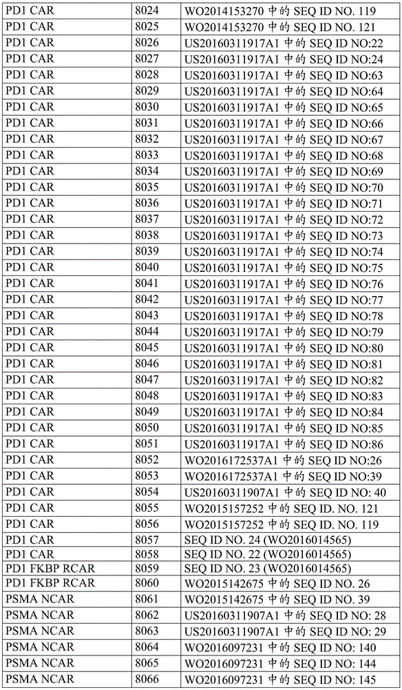


In one embodiment of the invention, the payloads of the invention are CD19 specific CARs targeting different B cell malignancies and Her2 specific CARs targeting sarcomas, glioblastomas and advanced Her2 positive lung malignancies.
In some embodiments, the CAR is a CD19 CAR. The amino acid sequences of the CD19CAR components and the CD19CAR construct are presented in table 17A and table 17B. Table 17B also provides alternative aliases for a given construct identifier. These aliases are identified by the prefix OT.
TABLE 17A CD19CAR construct Components


Table 17B: CD19CAR constructs


Tandem CAR (TanCAR)
In some embodiments, the CAR of the invention can be a tandem chimeric antigen receptor (TanCAR) capable of targeting two, three, four, or more tumor-specific antigens. In some aspects, the CAR is a bispecific TanCAR comprising two targeting domains that recognize two different TSAs on a tumor cell. A bispecific CAR can be further defined as comprising an extracellular region comprising a targeting domain (e.g., an antigen recognition domain) specific for a first tumor antigen and a targeting domain (e.g., an antigen recognition domain) specific for a second tumor antigen. In other aspects, the CAR is a multispecific TanCAR comprising three or more targeting domains configured in a tandem arrangement. The length of the interval between targeting domains in the TanCAR can be between about 5 amino acids and about 30 amino acids, e.g., 6,7, 8,9, 10, 11, 12, 13, 14, 15,16, 17, 18, 19, 20, 21, 22, 23, 24, 25, 26, 27, 28, 29, and 30 amino acids.
Split CAR
In some embodiments, the components of the invention, including the targeting moiety, transmembrane domain, and intracellular signaling domain, may be divided into two or more portions such that it relies on multiple inputs that facilitate assembly of a fully functional receptor. In one embodiment, a split synthetic CAR system can be constructed in which the assembly of the activated CAR receptor is dependent on the binding of the ligand to the SRE (e.g., small molecule) and the binding of a particular antigen to the targeting moiety. As a non-limiting example, a split CAR consists of two parts assembled in a small molecule-dependent manner; one portion of the receptor is characterized by having an extracellular antigen-binding domain (e.g., scFv), and the other portion has an intracellular signaling domain such as a CD3 ζ intracellular domain.
In one example, the second portion of the cytoplasmic fragment can be anchored to the plasma membrane by incorporating a transmembrane domain (e.g., the CD8 α transmembrane domain) into the construct.
In some aspects, the two parts of the split CAR system contain heterodimerization domains that conditionally interact upon binding of heterodimerization small molecules. Thus, receptor components assemble in the presence of small molecules to form an intact system that can then be activated by antigen conjugation. Any known heterodimerization component can be incorporated into the split CAR system. Other small molecule-dependent heterodimerization domains may also be used, including but not limited to the gibberellin-induced dimerization system (GID1-GAI), trimethoprim-SLF-induced dimerization of ecDHFR and FKBP (Czlapinski et al, J Am Chem Soc.,2008,130(40):13186-13187), and ABA (abscisic acid) -induced dimerization of PP2C and PYL domains (Cutler et al, Annu Revplant biol.2010,61: 651-679). Dual regulation achieved using inducible assembly (e.g., ligand-dependent dimerization) and degradation (e.g., destabilization domain-induced degradation of the CAR) of the split CAR system may provide greater flexibility to control the activity of CAR-modified T cells.
Switchable CAR
In some embodiments, the CAR of the invention can be a switchable CAR. Juilerat et al (Juilerat et al, sci. rep.,2016,6: 18950; the contents of which are incorporated herein by reference in their entirety) have recently reported controllable CARs that can be turned on transiently in response to a stimulus, such as a small molecule. In this CAR design, the system is integrated directly in the hinge domain separating the scFv domain from the cell membrane domain in the CAR. The system makes it possible to split or combine different key functions of the CAR, such as activation and co-stimulation, within different chains of the receptor complex, thereby mimicking the complexity of TCR native architecture. This integrated system allows switching of the scFv and antigen interaction between on/off states controlled by the absence/presence of a stimulus.
Reversible CAR
In other embodiments, the CAR of the invention can be a reversible CAR system. In this CAR configuration, the LID domain (ligand-induced degradation) is incorporated into the CAR system. CARs can be temporarily down-regulated by addition of ligands for the LID domain. The combination of LID-mediated and DD-mediated regulation would provide tunable control of persistently activated CAR T cells, thereby reducing CAR-mediated tissue toxicity.
Inhibitory CAR (iCAR)
In some embodiments, the payload of the invention can be an inhibitory CAR. Inhibitory CAR (icar) refers to a bispecific CAR design in which negative signals are used to enhance tumor specificity and limit normal tissue toxicity. This design incorporates a second CAR with a surface antigen recognition domain in combination with an inhibitory signal domain to limit T cell responsiveness, even in the case of concurrent engagement with an activating receptor. This antigen recognition domain is directed against a normal tissue specific antigen such that T cells can be activated in the presence of the first target protein, but if a second protein that binds to iCAR is present, T cell activation is inhibited.
As a non-limiting example, iCAR against prostate specific membrane antigen (PMSA) based on CTLA4 and PD1 inhibitory domains was shown to be able to selectively limit cytokine secretion, cytotoxicity and proliferation induced by T cell activation (Fedorov V.D et al, 2013, Sci Transl Med, 11; 5(215):215ra 172; the contents of which are incorporated herein in their entirety).
Chimeric transducible receptors
In some embodiments, the payloads of the present invention can be chimeric switch receptors that can switch from a negative signal to a positive signal. As used herein, the term "chimeric switch receptor" refers to a fusion protein comprising a first extracellular domain and a second transmembrane and intracellular domain, wherein the first domain comprises a negative signaling region and the second domain comprises a positive intracellular signaling region. In some aspects, the fusion protein is a chimeric switch receptor comprising an extracellular domain of an inhibitory receptor on a T cell fused to transmembrane and cytoplasmic domains of a costimulatory receptor. This chimeric switch receptor converts a T cell inhibitory signal into a T cell stimulatory signal.
As a non-limiting example, a chimeric switch receptor may comprise the extracellular domain of PD-1 fused to the transmembrane and cytoplasmic domains of CD28, as taught by Liu et al (Liu et al, Cancer Res.,2016,76(6): 1578-1590; the contents of which are incorporated herein by reference in their entirety). In some aspects, the extracellular domains of other inhibitory receptors such as CTLA-4, LAG-3, TIM-3, KIR and BTLA may also be fused to transmembrane and cytoplasmic domains derived from co-stimulatory receptors such as CD28, 4-1BB, CD27, OX40, CD40, GTIR and ICOS. In the context of the present invention, the SRE domain (e.g., DD) may be inserted at the N-terminus or C-terminus of the chimeric switch receptor.
In some embodiments, chimeric switch receptors of the invention may comprise a recombinant receptor comprising an extracellular cytokine binding domain of an inhibitory cytokine receptor (e.g., IL13 receptor α (IL13R α 1), IL10R, and IL4R α) fused to an intracellular signaling domain of a stimulatory cytokine receptor such as IL2R (IL2R α, IL2R β, and IL2R γ) and IL7R α one example of such a chimeric cytokine receptor is a recombinant receptor comprising a cytokine-binding extracellular domain of IL4R α linked to an intracellular signaling domain of IL7R α (see U.S. patent publication No. 2014/0050709; the contents of which are incorporated herein by reference in their entirety).
In one embodiment, a chimeric transforming receptor of the present invention may be a chimeric TGF β receptor, a chimeric TGF β receptor may comprise an extracellular domain derived from a TGF β receptor, such as TGF β receptor 1, TGF β receptor 2, TGF β receptor 3, or any other TGF β receptor or variant thereof, and a non-TGF β receptor intracellular domain, a non-TGF β receptor intracellular domain may be an intracellular domain or fragment thereof derived from TLR1, TLR2, TLR3, TLR4, TLR5, TLR6, TLR7, TLR8, TLR9, TLR10, CD28, 4-1BB (CD137), OX40(CD134), CD3 zeta, CD40, CD27, or a combination thereof an example of the chimeric TGF β receptor is discussed in U.S. patent publication No. US2016/0075755, the contents of which are incorporated herein by reference in their entirety.
In some embodiments, the payload of the present invention may be a fusion receptor composed of two parts. In one aspect, a two-part fusion receptor can comprise an antigen binding domain that binds a tumor antigen and an activation domain that binds one or more cell surface molecules. In some aspects, the antigen binding domain of the two-part receptor is an scFv antibody. In other aspects, the activation domain binds a T cell surface stimulatory molecule that can be selected from CD3, CD27, CD28, CD40, OX40(CD134), and 4-1BB (CD137), or an NK cell surface stimulatory molecule that is selected from CD16, NKG2D, and NKp 30. The activation domain may also be an scFv specific for stimulatory molecules on the surface of immune cells (e.g., T cells and NK cells). Immune cells can be genetically modified to express a two-part molecule comprising at least one antigen binding domain and an activation domain. The antigen binding domain binds to one or more molecules present on a target cell, such as a cancer cell. Immune cells expressing the molecule recognized by the activation domain will be activated and attack the recognized cancer cells. As a non-limiting example, a two-part molecule can be, for example, U.S. patent publication nos.: a linker molecule comprising an antigen recognizing scFv specific for CD19 or EphA2 as described in US 2016/0015749; the contents of said patent are incorporated herein by reference in their entirety.
Activation conditioned CAR
In some embodiments, the payload of the invention can be an activated conditional chimeric antigen receptor that is expressed only in activated immune cells. Expression of the CAR can be linked to an activation-conditional control region, which refers to one or more nucleic acid sequences that induce transcription and/or expression of a sequence under its control, e.g., a CAR. The activation conditional control region can be a promoter of a gene that is upregulated during activation of an immune effector cell, such as the IL2 promoter or the NFAT binding site. In some embodiments, activation of immune cells can be achieved by constitutively expressing a CAR (International publication No.: WO 2016126608; the contents of which are incorporated herein by reference in their entirety).
CAR targeting tumor cells with specific proteoglycan markers
In some embodiments, the payload of the invention can be a CAR that targets a particular type of cancer cell. Human cancer cells and metastases may express unique and otherwise abnormal proteoglycans, such as polysaccharide chains (e.g., Chondroitin Sulfate (CS), dermatan sulfate (DS or CSB), Heparan Sulfate (HS), and heparin). Thus, the CAR can be fused to a binding moiety that recognizes cancer-associated proteoglycans. In one example, the CAR can be fused to a VAR2CSA polypeptide that binds with high affinity to a specific type of Chondroitin Sulfate A (CSA) attached to proteoglycans (VAR 2-CAR). The extracellular ScFv portion of the CAR can be replaced with a VAR2CSA variant comprising at least a minimal CSA binding domain, thereby producing a CAR specific for Chondroitin Sulfate A (CSA) modification. Alternatively, the CAR may be fused to a split-protein binding system to produce a Spy-CAR (Spy-CAR), wherein the scFv portion of the CAR is replaced with a portion of the split-protein binding system, such as a Spy tag (SpyTag) and Spy-catcher, and a cancer recognition molecule (e.g., scFv and/or VAR2-CSA) is linked to the CAR by the split-protein binding system (see, e.g., PCT publication No.: WO 2016/135291; the contents of which are incorporated herein by reference in their entirety).
SUPRA CAR
In some implementations, the payload of the present invention can be a Split Universal Programmable (SUPRA) CAR. SUPRACAR can be a two-component receptor system comprising a universal receptor expressed on T cells (zip CAR) and a tumor-targeting scFv adaptor. The zip CAR universal receptor may be generated by fusion of an intracellular signaling domain and a leucine zipper as the extracellular domain. Tumor-targeting scFv adaptors or zipFv can be generated by fusion of homologous leucine zippers and scFv. The scFv of zipFv can bind to a tumor antigen, and the leucine zipper can bind to and activate the zip CAR on T cells. Unlike conventional fixed CAR designs, the modular design of the SUPRA CAR allows multiple antigens to be targeted without further genetic manipulation of immune cells. Any of the CAR designs disclosed by Cho et al, 2018, Cell 173, 1-13 may be suitable for use in the present invention (the contents of which are incorporated herein by reference in their entirety).
6. Cytokines, chemokines and other soluble factors
According to the invention, the payloads of the invention can be cytokines, chemokines, growth factors and soluble proteins produced by immune cells, cancer cells and other cell types that act as chemical communication between cells and tissues within the body. These proteins mediate a wide range of physiological functions, ranging from effects on cell growth, differentiation, migration and survival to many effector activities. For example, activated T cells produce multiple cytokines for achieving cytotoxic functions to eliminate tumor cells.
In some embodiments, the payloads of the present invention can be cytokines and fragments, variants, analogs and derivatives thereof, including but not limited to Tumor Necrosis Factor (TNF), Interferon (IFN), TGF β and chemokines.
In some embodiments, the payloads of the present invention may be cytokine receptors, recombinant receptors, variants, analogs and derivatives thereof; or a signal component of a cytokine.
In some embodiments, the cytokines of the invention may be used in immune cells such as CD8+ T for use in immunotherapyEMExpansion, survival, persistence and efficacy improvement of natural killer cells and Tumor Infiltrating Lymphocytes (TILs). In other embodiments, T cells engineered with two or more DD-regulated cytokines are used to provide dynamic control of T cell activation and tumor microenvironment remodeling. In one aspect, the present invention provides biological circuits and compositions to minimize toxicity associated with cytokine therapy. Although systemic cytokine therapy has been successful in reducing tumor burden, it is not without its successIt often results in the manifestation of severe dose-limiting side effects. Two factors contribute to the toxicity observed: (a) gene pleiotropic, in which cytokines affect different cell types and, as the case may be, exert opposing effects on the same cells, (b) cytokines have a short serum half-life and therefore need to be administered at high doses to achieve a therapeutic effect, which exacerbates gene pleiotropic effects. In one aspect, the cytokines of the invention can be used to modulate cytokine expression in the event of an adverse effect. In some embodiments, the cytokines of the present invention may be designed to have an extended lifespan or enhanced specificity to minimize toxicity.
In some embodiments, the payload of the invention can be an Interleukin (IL) cytokine, an Interleukin (IL) is a class of glycoproteins produced by leukocytes for modulating an immune response as used herein, the term "Interleukin (IL)" refers to an interleukin polypeptide from any species or source and includes full-length proteins as well as fragments or portions of such proteins, hi some aspects, the interleukin payload is selected from IL1, IL1 α (also known as erythropoietin-1), IL1 β (catabolin)), IL1 δ, IL1 epsilon, IL1 η, IL1 ζ, interleukin-1 family members 1 to 11(IL1F1 to IL1F11), interleukin-1 homolog 1 to 4(IL1H 11 to IL1H 11), IL11 related proteins 1 to 3(IL 111 to IL1RP 72, IL11, CD11, CD11, 3614, CD11, CD11, 3614, 11, CD11, CD11, and 3614, 11, CD11, and 3614, 11, and 3614, 11, and 11, 3614, and 11, and 3614 IL11, and 11 are IL11, 3614, 11, and other IL 3614, 11, and 11, and 3614, 11-3614, 11, and 11-like IL 3614, 11, and.
In certain embodiments, the cytokine may be a type I Interferon (IFN), including IFN α subtypes (IFN α, IFN α b, IFN α c), IFN α, IFN δ subtypes (IFN δ 1, IFN δ 2, IFN δ 8), IFN γ, IFN κ, and IFN ε, IFN λ, IFN ω, IFN τ, and IFN ζ in certain embodiments, the cytokine may be a member of the Tumor Necrosis Factor (TNF) superfamily, including TNF- α, TNF- β (also known as lymphotoxin- α (LT- α)), lymphotoxin- β (LT- β), CD40L (CD154), CD27L (CD70), CD30L (CD153), FASL (CD178), 4-1BBL (CD137L), OX40L, TRAIL (TNF-related apoptosis-inducing ligand), APRIL (proliferation inducing ligand), TRATALEAK, TNFRNCE 631, TRNL-68692, and GHT superfamily (TNFRL-20).
In one embodiment, the payload of the invention may comprise IL2(SEQ ID No.8299 encoded by SEQ ID nos. 8300 and 8301). In one aspect, the effector moiety of the invention may be a DD-IL2 fusion polypeptide.
In some aspects of the invention, an IL2 mutein may be used as the payload as used herein, the term "mutein" is a construct, molecule or sequence with mutations, changes or alterations in the protein, thus also referred to as a mutant, e.g., a protein mutant, i.e., a mutein. thus, an "IL 2 mutein" is an IL2 mutant. in some embodiments, an IL2 mutein is a variant of a wild-type IL2 protein, wherein the wild-type IL2 consists of the amino acid sequence SEQ ID No. 8299. in some aspects, it refers to an IL2 variant in which only IL2R αβ γ is bound and activated, while not significantly binding or activating only IL2R β γ expressing cells IL2 mutein in some examples, an IL 6326 mutein may be an IL2 protein in which the residues of IL2 responsible for binding IL2R β or IL2R γ are substituted, thus eliminating their interactionSA) In some embodiments, the IL2 mutein may be one that preferentially binds lower affinity IL2R β γ, but not to NK cells, to the high affinity IL2 receptor (i.e., IL2R αβ γ) necessary for selective activation of T cells over NK cellsIL2 protein with reduced affinity of CD 25.
In some embodiments, the IL2 mutein can be used to preferentially expand or stimulate Treg cells. As used herein, "preferentially expand or stimulate Treg cells" means that the IL2 mutein promotes the proliferation, survival, activation, and/or function of T regulatory cells.
Exemplary IL muteins may include, but are not limited to, the N88 substitution (Shanafelt et al, Nature Biotech.,2000,18: 1197;. 1202), the IL with the V91 substitution (e.g., U.S. patent publication No. US20140286898), the V91 substitution, the C125 substitution, the IL with three mutations: V69, N71, Q74, the IL mutein with high affinity for IL2 (N29, Y31, K35, T37, K48, V69, N71, Q74), the IL mutein with high affinity and reduced signaling activity for IL2 (N29, Y31, K35, T37, K48, V69, N71, Q74, N74), and the D20, N88, and Q126 substitutions as described in PCT application No. and the contents of each are incorporated herein by reference in their entirety, in other aspects, the IL muteins may include U.A. Pat. publication No. US 20188, N88, N126, EP 869, EP 020989, EP 869, EP 0203, EP 989, EP 869, EP 863, EP 989, and EP 0209, each incorporated herein incorporated by reference in their entirety.
In some aspects, the IL2 mutein can be fused to a polypeptide that extends the serum half-life of the IL2 mutein, such as an IgG Fc fragment. Preferably, the Fc region is derived from human IgG, including IgG1, IgG2, IgG3, and IgG 4. In other aspects, the payload of the invention can be an IL2 fusion protein comprising a second functional polypeptide. In a non-limiting example, an IL2 fusion protein can comprise an IL2 or IL2 mutein polypeptide fused to a pro-apoptotic Bcl-2 family polypeptide (such as Bad, Bik/Nbk, Bid, Bim/Bod, Hrk, Bak, or Bax); the fusion protein may be capable of inhibiting cell survival of a target cell expressing the IL2 receptor, inhibiting cell proliferation of the target cell, orEnhancing cell death or apoptosis of the target cell. Alternatively, IL2 or IL2 mutein polypeptides may be combined with anti-apoptotic Bcl-2 family polypeptides (such as Bcl-x)LBcl-w or Bcl-2). The fusion protein may be capable of enhancing cell survival of a target cell expressing a receptor for IL2, enhancing cell proliferation of the target cell, or inhibiting cell death or apoptosis of the target cell. See, e.g., U.S. patent publication nos.: US 2016/0229901.
In addition, the IL2 fusion protein can be an IL2-GMCSF fusion protein, which can promote cell-cell interactions; thus, an anti-cancer immune response is enhanced (Wen et al, J.Translational Med.,2016,14: 41).
In one embodiment, the payload of the present invention may include IL 12. IL12 is a heterodimeric protein with two subunits (p35, p40) that is secreted by antigen presenting cells such as macrophages and dendritic cells. IL12 acts on Natural Killer (NK) cells, macrophages, CD8 via the STAT4 pathway+Cytotoxic T cells and CD4+T helper cells to induce IFN γ production in these effector immune cells type 1 cytokines (reviewed by Trinchieri G, Nat RevImmunol.2003; 3(2): 133-146). IL12 promotes NK cells and CD8+Cytotoxic activity of T cells and thus have anti-tumor function. Intravenous injection of recombinant IL12 demonstrated modest clinical efficacy in a few patients with advanced melanoma and renal cell carcinoma (Gollob et al, Clin. cancer Res.2000; 6(5): 1678-. IL12 has been used as an adjuvant to enhance cytotoxic immunity in the case of use of melanoma antigen vaccines or use of peptide-pulsed peripheral blood mononuclear cells; and promoting NK cell activity in the case of breast cancer treatment with trastuzumab. In several tumor models, local delivery of IL12 into the tumor microenvironment promoted tumor regression. These studies all indicate that locally increased IL12 levels may promote anti-tumor immunity. One major obstacle to systemic or local administration of recombinant IL12 protein or administration by oncolytic viral vectors is the severe side effects when IL12 is present at high levels. Development of a system that tightly controls IL12 levels may provide for safe use of IL12 in cancer therapy.
In one aspect, the effector moiety of the invention may be a DD-IL12 fusion polypeptide. This regulatable DD-IL12 fusion polypeptide can be used directly as an immunotherapeutic or transduced into immune effector cells (T cells and TIL cells) to generate modified T cells with greater capacity for in vivo expansion and survival for adoptive cell transfer. The need for harsh preconditioning regimens in current adoptive cell therapies can be minimized using regulated IL 12; DD-IL12 can be used to modulate the tumor microenvironment and increase persistence in solid tumors that are currently refractory to tumor antigen-targeted therapies. In some embodiments, CAR-expressing T cells can be armored with DD-regulated IL12 to mitigate immunosuppression without systemic toxicity.
In some embodiments, IL12 may be Flexi IL12, where both the p35 subunit and the p40 subunit are encoded by a single cDNA that produces a single chain polypeptide. In one embodiment, IL12 may comprise a p40 subunit comprising amino acids 23 to 328 of wild-type IL12B and comprising the amino acid sequence SEQ ID NO.8302 (encoded by SEQ ID NO.8303 to 8312), and a p35 subunit comprising amino acids 57 to 253 of wild-type IL12A and comprising the amino acid sequence SEQ ID NO.8313 (encoded by SEQ ID NO.8314 and 8323). Any portion of IL12 that retains one or more functions of full-length or mature IL12 may be suitable for use in the present invention.
In some embodiments, the DD regulated IL12 compositions of the present invention can be used to minimize cytotoxicity associated with systemic IL12 administration treatment with IL12 has been associated with systemic influenza-like symptoms (fever, chills, fatigue, joint myalgia, headache), toxic effects on bone marrow and liver.
The form of the IL12 construct used as the payload in the present invention can be optimized. In one embodiment, the payload of the invention may be a bicistronic IL12 comprising a P40 subunit and a P35 subunit separated by an internal ribosome entry site or cleavage site such as P2A or furin cleavage site to allow independent expression of the two subunits from a single vector. This results in a secreted IL12 configuration more similar to naturally occurring IL12 than the flexi IL12 construct, and the payload of the present invention may be the p40 subunit of IL 12. DD-regulated p40 can be co-expressed with a constitutive p35 construct to produce "regulatable IL 12" expression. Alternatively, DD-regulated p40 can heterodimerize with endogenous p 35. P40 has been shown to stabilize p35 expression and stimulate the export of p35 (Jalah R et al (2013) Journal of biol. chem.288,6763-6776, the contents of which are incorporated herein by reference in their entirety).
In some embodiments, a modified form of IL12 may be used as a payload. These modified forms of IL12 may be engineered to have a reduced half-life in vivo compared to the unmodified forms, particularly when used in combination with the tunable systems described herein.
Human Flexi IL12 has a reported half-life of 5-19 hours, which when Administered as a therapeutic compound can lead to systemic cytotoxicity (Car et al (1999) The clinical of Interleukin-12: A Review "clinical Path.27A, 58-63; Robertson et al (1999)" Immunological Effects of Interleukin 12Administered by respiratory intention to Patients with cancer "cancer Res.5: 9-16; Atkins et al (1997)" Phase evolution of intraviral combinant Human Interleukin 12in Patients with Advantage Maglignnaceae "cancer Res.3:409 Res 417). Ligand-induced control of IL12 may modulate production in a dose-dependent manner, and the time from cessation of ligand administration until cessation of protein synthesis and clearance of IL12 may not be sufficient to prevent toxic accumulation of IL12 in plasma.
In one embodiment, the modified form of IL12 for use as a payload may be Topo-sc IL12 having the following configuration from N-terminus to C-terminus: (i) a first IL12 p40 domain (p40N), (ii) optionally a first peptide linker, (iii) an IL12 p35 domain, (iv) optionally a second peptide linker, and (v) a second IL12 p40 domain (p 40C). In one embodiment, the modified topo sc IL12 polypeptide exhibits an increased susceptibility to proteolysis. Topo-sc IL12 is described in International patent publication No. WO 2016048903; the contents of said patent are incorporated herein by reference in their entirety.
IL12 polypeptides may also be modified (e.g., genetically engineered, synthetically engineered, or recombinantly engineered) to increase susceptibility to a protease to reduce the biologically active half-life of the IL12 complex compared to the corresponding IL12 lacking protease susceptibility. Protease susceptible forms of IL12 are described in international patent publication No. WO 2017062953; the contents of said patent are incorporated herein by reference in their entirety.
Systemic toxicity of IL12 may also be limited or tightly controlled by mechanisms involving tethering IL12 to the cell surface to limit its therapeutic efficacy to the tumor site. The membrane tethered IL12 form achieved using a Glycosylphosphatidylinositol (GPI) signal peptide or using a CD80 transmembrane domain has been previously described (Nagarajan S et al (2011) J Biomed Mater Res A.99(3): 410-7; Bozeman EN et al (2013) vaccine.7; 31(20): 2449-56; Wen-Yu Pan et al (2012), mol. Ther.20:5, 927-937; the contents of each of which are herein incorporated by reference in their entirety) in some embodiments, the transmembrane domain may be selected from any of those transmembrane domains described in tables 13, 14 and 15.
In some embodiments, the level of IL12 secreted by the immune cells of the invention may be approximately similar to the level of IL12 secreted by human myeloid dendritic cells (mdcs 1) when activated with a TLR agonist. In one embodiment, the TLR agonist can be a combination of lipopolysaccharide administered with R848.
In some embodiments, IFN γ secreted due to IL 12-induced immune cell activation is at least 5-fold greater in the presence of the ligand as compared to levels in the absence of the ligand.
In some embodiments, regulation of IL12 will provide the necessary safety switch. In some embodiments, IL-12 secretion in the tumor microenvironment recruits and/or activates effector cells. In some embodiments, IL12 modulation will provide benefits on CAR T function without causing toxicity.
In one embodiment, the payload of the present invention may include IL 15. Interleukin 15 is a potent immunostimulatory cytokine and a survival factor essential for T cells and natural killer cells. Preclinical studies comparing IL2 and IL15 have shown that IL15 is associated with less toxicity compared to IL 2. In some embodiments, an effector moiety of the invention may be a DD-IL15 fusion polypeptide. The IL15 polypeptide may also be modified to increase its binding affinity for the IL15 receptor. For example, at position 72 of IL15, asparagine can be replaced with aspartic acid (SEQ ID NO.2 of U.S. patent publication No. 20140134128A 1; the contents of which are incorporated herein by reference in their entirety). In some aspects, IL15 comprises the amino acid sequence SEQ ID No.8234 (encoded by SEQ ID No. 8236) including amino acids 49-162 of wild-type IL 15. In some embodiments, the IL15 sequence may include a stop codon and may be encoded by the nucleotide sequences SEQ ID NO.8235 and 8237-8238.
One unique feature of IL 15-mediated activation is a trans-presentation mechanism, in which IL15 is presented as a complex with the α subunit of the IL15 receptor (IL15Ra), which binds to and activates membrane-bound IL15 β/gamma receptor on the same or different cells IL15/IL15Ra complex activates IL15 signaling more efficiently than IL15 alone, thus, in some embodiments, the effector module of the present invention may include a DD-IL15/IL15Ra fusion polypeptide.
Regulated IL15/IL15Ra can be used to promote CD8TEMExpansion, survival and potency of the cell population without affecting regulatory T cells, NK cells and TIL cells. In one embodiment, DD-IL15/IL15Ra may be used to enhance CD19 directed T cell therapy in the context of B cell leukemias and lymphomas. In one aspect, IL15/IL15Ra can be used as a payload of the invention to reduce the need for a preconditioning regimen in the current CAR-T therapeutic paradigm.
The effector modules comprising the DD-IL15, DD-IL15/IL15Ra and/or DD-IL15/IL15Ra sushi domains can be designed to be secreted (using, for example, IL2 signaling) or membrane bound (using, for example, the IgE or CD8a signal sequences).
In some embodiments, IFN γ secreted due to IL 15-induced immune cell activation is at least 10-fold greater in the presence of the ligand as compared to levels in the absence of the ligand.
In some embodiments, modulation of an IL15-IL15Ra fusion protein provides a safety switch compared to a constitutively expressed IL15-IL15 Ra. In some embodiments, IL15-IL15Ra results in better expansion and/or persistence of CAR T cells.
In some aspects, DD-IL115/IL15Ra comprises an amino acid sequence provided in table 18. In some embodiments, the linker used in table 18 may be an SG linker. In table 18, asterisks indicate translation of stop codons. In some embodiments, the DD described in table 18 can contain an additional stop codon. As used herein, IL15 of Wild Type (WT) refers to Uniprot ID: p40933, and IL15Ra of Wild Type (WT) refers to UniProt ID: q13261. Table 18 also provides alternative aliases for a given construct identifier. These aliases are identified by the prefix OT.
Table 18: DD-IL15-IL15Ra construct sequences


In some embodiments, the sequences described in table 18 may contain additional stop codons. For example, the construct "hPDE 5 (amino acids 535-860, R732L of WT); the termination sequence "can be encoded by the nucleotide sequence SEQ ID NO.8362 and the construct" hPDE5 (amino acids 535-860, R732L, F736A of WT); the termination sequence "can be encoded by the nucleotide sequence SEQ ID NO. 8363.
In one embodiment, the payload of the invention may include IL 18. IL18 is a pro-inflammatory and immunoregulatory cytokine that promotes IFN γ production by T cells and NK cells IL18 belongs to the IL1 family secreted IL18 binds to the heterodimeric receptor complex consisting of the IL18R α and β chains and initiates signal transduction IL18 acts in concert with other cytokines to modulate immune system function, including induction of IFN γ production in response to pathogen products, Th1 responses, and NK cell activation IL18 at several sitesIn the case of tumors, they exhibit an anticancer effect. Administration of recombinant IL18 protein or IL18 transgene would be by rendering CD4+T and/or NK cell mediated response activation to induce melanoma or sarcoma regression (reviewed by Srivastava et al, curr. Med. chem.,2010,17: 3353-one 3357). The combination of IL18 with other cytokines such as IL12 or co-stimulatory molecules (e.g. CD80) increases the anti-tumor effect of IL 18. For example, IL18 and IL12A/B or CD80 genes have been successfully integrated into the genome of oncolytic viruses with the aim of synergistically triggering T-cell mediated anti-tumor immune responses (Choi et al, Gene ther.,2011,18: 898-909). The IL2/IL18 fusion protein also showed enhanced antitumor properties relative to either cytokine alone in preclinical models as well as low toxicity (Acres et al, Cancer Res.,2005,65: 9536-9546).
IL18 activates NK cells either alone or in combination with IL12 and IL 15. Preclinical studies have demonstrated that adoptively transferred NK cells preactivated with IL12, IL15 and IL18 show enhanced effector function in vivo against established tumors (Ni et al, J Exp med.2012,209: 2351-. Activation of human NK cells by IL12/IL15/IL18 also showed memory-like characteristics and secreted more IFN γ in response to cytokines (e.g. low concentration of IL 2). In one embodiment, the effector moiety of the present invention may be a DD-IL18 fusion polypeptide.
In one embodiment, the payload of the present invention may include IL 21. IL21 is another gene pleiotropic type I cytokine, produced primarily by T cells and Natural Killer T (NKT) cells. IL21 has diverse effects on a variety of cell types including, but not limited to, CD4+And CD8+T cells, B cells, macrophages, monocytes and Dendritic Cells (DCs). The functional receptor for IL21 consists of the IL21 receptor (IL21R) and the common cytokine receptor gamma chain, which is also a subunit of the receptors for IL2, IL4, IL7, IL9 and IL 15. The study provides strong evidence that IL21 is a promising immunotherapeutic for cancer immunotherapy. IL21 promotes maturation, enhances cytotoxicity, and induces production of IFN γ and perforin (perforin) by NK cells. These effector functions inhibit B16 melanomaGrowth (Kasaian et al, Immunity.2002,16(4): 559-. IL21 together with IL15 makes antigen-specific CD8+T cell numbers and their effector functions are expanded, leading to tumor regression (Zeng et al, J Exp Med.2005,201(1): 139-. IL21 may also be used to rejuvenate a variety of immune effector cells in the tumor microenvironment. IL21 may also directly induce apoptosis in certain types of lymphomas such as diffuse large B-cell lymphoma, mantle cell lymphoma, and chronic lymphocytic leukemia cells by activating the STAT3 or STAT1 signaling pathway. IL21 activates NK cell-dependent cytotoxicity either alone or in combination with anti-CD 20 mAb (rituximab). Interestingly, the finding of immunosuppressive effects on IL21 suggests that this cytokine is a "twolip sword" -stimulation with IL21 can lead to induction or suppression of immune responses. Both stimulatory and inhibitory effects of IL21 must be considered when using IL 21-related immunotherapeutics. The level of IL21 needs to be tightly controlled by regulatory elements. In one aspect, the effector moiety of the invention may be a DD-IL21 fusion polypeptide.
In some embodiments, the payloads of the present invention may comprise type I interferons (IFN-I) are soluble proteins important to combat human viral infections, IFN-I comprises IFN α subtypes (IFN α, IFN α B, IFN α c), IFN β, IFN δ subtypes (IFN δ 1, IFN δ 2, IFN δ 8), IFN γ, IFN κ and IFN ε, IFN λ, IFN ω, IFN τ and IFN ζ. IFN α and IFN β are the predominant IFN I subtypes in the immune response all subtypes of IFN I signal transduction via the unique heterodimeric receptor interferon α receptor (IFNAR) consisting of 2 subunits, IFNAR1 and IFNAR2 IFNR activation may modulate viral infections and in the case of adaptive immunity host responses several signaling cascades may be activated by IFNR, including the Janus activated kinase-signal transductants and the transcriptional activation of the transcriptional activation pathway of the protein (IFNAR) and transcriptional pathway of the kinase (IFNAR) may be determined as a direct phosphokinase inhibition of the avian kinase pathway (MAPl) and oncogenic pathway (MAPl) 3, the receptor kinase pathway of the IFN-activating the IFN 364 receptor kinase (IFNk) and the IFNk pathwayProliferation of cells and virus infected cells and increased MHC class I expression, resulting in enhanced antigen recognition. IFN-I has also been shown to be involved in immune system regulation. IFNs can modulate the immune system directly through interferon receptors (IFNR) or indirectly through the induction of chemokines and cytokines. Type I IFNs potentiate NK cell function and promote NK cell survival. Type I IFNs also affect monocytes, supporting differentiation of monocytes into DCs with high antigen presenting capacity; and stimulate macrophage function and differentiation. Several studies have also demonstrated that IFN-I promotes CD8+T cell survival and function. In some cases, it may be desirable to use the biological circuits of the invention to tune the expression of type I IFNs to avoid immunosuppression caused by chronic treatment with IFNs.
Although type I IFNs are powerful tools to directly and indirectly regulate the function of the immune system, the side effects of systemic long-term therapy and lack of sufficiently high efficacy have suppressed concerns for IFN α for oncology clinical use it is believed that type I IFNs may be more effective if IFN levels at malignant tissues are tightly regulated.
The invention also relates to a method of inducing apoptosis in a cell, comprising administering to the cell a composition comprising a TNF- α, TNF- β (also known as lymphotoxin- α (LT- α)), lymphotoxin- β (LT- β), CD40L (CD154), CD27L (CD70), CD30L (CD153), FASL (CD178), 4-1BBL (CD137L), OX40L, TRAIL (TNF-related apoptosis inducing ligand), APRIL (proliferation inducing ligand), TWEAK, TRANCE, TALL-1, GITRL, LIGHT, and TNFSF1 through TNFSF20(TNF ligand superfamily members 1 through 20), and in one embodiment, the invention's payload may be TNF- α, TNF- α may cause cell lysis of tumor cells, and in one embodiment, the invention's TNF- α fusion polypeptide may also induce cell proliferation.
In one embodiment, the payload of the invention can be a cytokine fused to the ectodomain of TNF α, which is produced in the form of a membrane-associated cytokine fused to the ectodomain of TNF, hi one embodiment, the cytokine can be shed from the cell surface by the action of a membrane-associated protease and/or a protease in the extracellular space, such as MMP 9.
In some embodiments, the payloads of the present invention can include inhibitory molecules that block inhibitory cytokines. The inhibitor may be a blocking antibody specific for an inhibitory cytokine, an antagonist against an inhibitory cytokine, or the like.
In some aspects, the payloads of the present invention can include inhibitors of the secondary cytokine IL35 IL35 belongs to the interleukin-12 (IL12) cytokine family and is a heterodimer consisting of IL27 β chain Ebi3 and IL12 α chain p35 biologically active IL35 has been described only in forkhead box protein 3(Foxp3)+Regulated T cells (tregs) (resting and activated tregs). Unlike other members of the family, IL35 appears to act only in an anti-inflammatory manner by inhibiting effector T cell proliferation and perhaps other parameters (Collison et al, Nature,2007,450(7169): 566-.
In some embodiments, the payloads of the present invention may include inhibitors that block transforming growth factor β (TGF- β) subtypes (TGF- β, TGF- β, and TGF- β). TGF- β is secreted by many cell types including macrophages, and is often complexed with both proteins LTBP and LAP.
In some embodiments, the payload fused to the DD of the invention may be an immunosuppressive molecule such as an inhibitor of TGF- β and IDO.
In some embodiments, the payloads of the present invention can include chemokines and chemokine receptors. Chemokines are a family of secreted small cytokines or signaling proteins that induce directional chemotaxis of neighboring responsive cells. The chemokine may be an SCY (small cytokine) selected from the group consisting of: SCYA1-28(CCL1-28), SCYB1-16(CXCL1-16), SCYC1-2(XCL1-2), SCYD-1 and SCYE-1; or a C chemokine selected from: XCL1 and XCL 2; or a CC chemokine selected from: CCL1, CCL2, CCL3, CCL4, CCL5, CCL6, CCL7, CCL8, CCL9, CCL10, CCL11, CCL12, CCL13, CCL14, CCL15, CCL16, CCL17, CCL18, CCL19, CCL20, CCL21, CCL22, CCL23, CCL24, CCL25, CCL26, CCL27, and CCL 28; or a CXC chemokine selected from: CXCL1, CXCL2, CXCL3, CXCL4, CXCL5, CXCL6, CXCL7, CXCL8, CXCL9, CXCL10, CXCL11, CXCL12, CXCL13, CXCL14, CXCL15, CXCL16 and CXCL 17; or CX3C chemokine CX3CL 1. In some aspects, the chemokine receptor can be a receptor for C chemokine, including XCR 1; or receptors for CC chemokines, including CCR1, CCR2, CCR3, CCR4, CCR5, CCR6, CCR7, CCR8, CCR9 and CCR 10; or receptors for CXC chemokines, including CXCR1, CXCR2, CXCR3, CXCR4 and CXCR 5; or the CX3C chemokine receptor CX3CR 1.
In some embodiments, the payloads of the present invention may include other immunomodulators that play a critical role in immunotherapy, such as GM-CSF (granulocyte-macrophage colony stimulating factor), Erythropoietin (EPO), MIP3a, Monocyte Chemotactic Protein (MCP) -1, intracellular adhesion molecule (ICAM), macrophage colony stimulating factor (M-CSF), interleukin-1 receptor activating kinase (iRAK-1), lactotransferrin (lactotransferrin), and granulocyte colony stimulating factor (G-CSF).
In some embodiments, the payloads of the present invention may comprise amphiregulin. Amphiregulin (AREG) is an EGF-like growth factor that binds to the EGFR receptor and potentiates CD4+ regulatory T cell (Treg) function. AREG promotes immunosuppression in the tumor environment. Thus, in a certain embodiment, the payload of the invention may include amphiregulin to suppress the immune response during immunotherapy.
In some embodiments, the payloads of the present invention can include fusion proteins in which cytokines, chemokines, and/or other soluble factors can be fused to other biomolecules such as antibodies and/or ligands for receptors. The fusion molecule can increase the half-life of the cytokine, reduce systemic toxicity, and increase the local concentration of the cytokine at the tumor site. Fusion proteins containing two or more cytokines, chemokines, and/or other soluble factors can be used to obtain synergistic therapeutic benefits. In one embodiment, the payload may be a GM-CSF/IL2 fusion protein.
In some embodiments, any of the hinge and transmembrane domains described herein can be used as a scaffold for soluble cytokine presentation. Cytokines may be operably linked to the CD8 hinge and transmembrane domains through a protease cleavage site. Lysis at the lysis site will release cytokines from the cell surface membrane. In some aspects, the cytokine may be in a precursor form. The production of the active form of the cytokine from the precursor form occurs by lysis at the lysis site. Any of the cytokines described herein can be engineered using any of the hinge and transmembrane domains described herein as a scaffold.
7. Immune modulating agent
In some embodiments, the payloads of the present invention may comprise inhibitors (antagonists) of co-inhibitory molecules (e.g., immune checkpoints) including, without limitation, PD-1, PD-L1, PD-L2, CTLA-4, TIM-3, VISTA, BTLA, TIGIT, LAIRA, CD160, 2B4, and TGFR. In some aspects, the inhibitor may be a blocking/antagonistic antibody or fragment thereof, as previously discussed, or a ligand for a co-inhibitory receptor.
In some embodiments, the payloads of the present invention may comprise agonists of co-stimulatory molecules including, but not limited to, CD27, CD28, CD30, CD40, OX40(CD134), 4-1BB (CD137), CDS, ICAM-1, LFA-1(CD11a/CD18), ICOS (CD278), GITR, BAFFR, HVEM, CD7, LIGHT, NKG2C, SLAMF7, NKp80, CD160, B7-H3, and CD 83. As a non-limiting example, an agonist of the costimulatory molecule ICOS (CD278) can be the international patent publication no: ICOS binding proteins comprising the amino acid sequences SEQ ID NO 1,2, 3, 4,5 and 6 as disclosed in WO 2016120789; the contents of said patent are incorporated herein by reference in their entirety.
In some aspects, the agonist of the co-stimulatory molecule can be an agonistic antibody or fragment thereof as previously discussed; or a ligand for a co-stimulatory receptor; or any binding molecule that enhances the biological activity of its target. For example, an agonist ligand of OX40 may be OX40L (CD 252). As used herein, OX40 ligands include intact OX40 ligand, soluble OX40 ligand, fusion proteins comprising a functionally active portion of OX40 ligand covalently linked to a second moiety, e.g., a protein domain, and variants that may vary in amino acid sequence from naturally occurring OX4L, but retain the ability to specifically bind OX40 receptor, or even enhance the biological activity of OX 40L.
In general, an agonist of a costimulatory molecule substantially enhances the biological activity of its target molecule, such as T cell activation. Desirably, the biological activity is enhanced by 10, 20, 30, 50, 70, 80, 90, 95, or even 100%.
In some embodiments, the payloads of the present invention may include immunomodulators, including stress proteins and Heat Shock Proteins (HSPs), that can integrate both innate and adaptive immune responses. They may also be other chaperones and adaptors that stimulate an immune response. As a non-limiting example, the payload of the present invention can be a fusion protein comprising the NF-KB activating domain of Flagellin (Flagellin) fused to glucose regulatory protein 170(Grp170) with the ATP-binding domain truncated (see U.S. patent publication No.: US 2015/0315255; the contents of which are incorporated herein by reference in their entirety). The fusion construct forms a secretable Grp 170-flagellin hybrid chaperone protein (Flagrp170) that can be used to stimulate an anti-cancer immune response.
In other embodiments, the payloads of the present invention may include as an adaptor molecule in the cytoplasm a STING (stimulator of interferon genes) protein that as a component of the host cytoplasmic surveillance pathway activates the TANK binding kinase (TBK1) -IRF3 signaling axis, resulting in induction of IFN β and other IRF-3 dependent gene products that strongly activate innate immunity, thus resulting in production of antigen-specific CD4+And CD8+T cells and pathogen-specific antibodies.
Demaria et al reported that potentiation of STING protein activation by intratumoral injection of the cyclic dinucleotide GMP-AMP (cGAMP), an agonist of STING, resulted in antitumor CD8+The T cell response was enhanced, leading to growth control in both injected and contralateral tumors in mouse melanoma and colon cancer models. STING-dependent anti-tumor immunity depends on type I IFN produced by endothelial cells in the tumor microenvironment (Demaria et al, proc. natl. acad. sci. usa,2015,112(5): 15408-15413). These studies demonstrate that STING promotes anti-tumor immune responses by enhancing type I IFN signaling in the tumor microenvironment. The biological circuits, effector modules comprising STING may be applied to the tumor microenvironment alone or in combination with other immunotherapeutic agents of the invention to enhance the anti-tumor response.
In addition to STING proteins, the payloads of the present invention may also include PRRs (pattern recognition receptors) involved in sensing cell infection by viruses and microorganisms to activate innate immune inflammatory responses. The PRRs include Toll-like receptors (TLRs), RIG-I-like receptors (RLRs), NOD-like receptors (NLRs), and C-type lectin receptors (CLRs). The RLR family is an RNA sensing system comprising a retinoid-inducible Gene-I (RIG-1), a melanoma markerThe methylation-associated gene 5(MDA5) and the genetic and physiological laboratory protein 2(LGP 2). RIG-1 recognizes relatively short dsRNA (up to 1kb), whereas MDA5 detects long dsRNA (over 2kb) to activate the synthesis of type I IFN including IFN α and IFN β (Wilkins et al, Curr Opin Immunol, 2010,22: 41-47). RLR activates downstream signaling proteins that stimulate type I IFN production+And CD8+A cascade of intracellular signaling pathways of T cell activation. Among the ten TLRs identified in humans, TLR-1, TLR-2, TLR-4, TLR-5 and TLR-6 are expressed in the cell surface, while TLR-3, TLR-7/TLR-8 and TLR-9 are expressed within the ER compartment. In some embodiments, a payload of the invention can be a PRR or an agonist of a PRR.
8. Metabolic factor/metabolic checkpoint
In some embodiments, immune cells such as T cells used in immunotherapy can be metabolically reprogrammed to enhance anti-tumor T cell responses. Upon activation by T cell receptors or CARs and costimulatory signals, metabolic activity is necessary to support immune cell (particularly T cell) growth, expansion, differentiation and effector function. Metabolic competition between cancer cells and infiltrating immune effector cells leads to T cell anergy and dysfunction.
In some embodiments, the payload of the invention can be a modulator of glycolysis. The Warburg effect (Warburg effect) in cancer cells results in the production of large amounts of lactic acid that can inhibit T cell cytotoxicity and effector function. As a non-limiting example, immune cells used for adoptive transfer can be modified to overexpress phosphoenolpyruvate carboxykinase 1(PCK1) which increases PEP production in T cells. Increased production of the glycolytic metabolite phosphoenolpyruvate (PEP) can repress sarco/ER Ca (2+) -ATPase (SERCA) activity, thus maintaining T Cell receptor-mediated Ca (2+) -NFAT signaling and effector functions (Ho et al, Cell,2015,162(6): 1217-.
In some embodiments, the payload of the present invention may comprise a protein involved in the OXPHOS pathway. For example, LEM (lymphocyte expansion molecule) is a CD8 that promotes cytotoxicity in response to lymphocytic Choriomeningitis (CMV) infection+T cell proliferation and effector function and memory of T cell produced proteins. LEM is part of a complex of CRIF1(CR6 interacting factor 1) that mediates translation and insertion of OXPHOS (oxidative phosphorylation) proteins into the inner mitochondrial membrane, thereby modulating OXPHOS activity. Thus, LEM is a positive regulator of T cell metabolism (mitochondrial respiration levels) and expansion (Okoye et al, Science,2015,348(6238): 995-.
In some embodiments, the payload of the present invention may be an inhibitor of a metabolic enzyme involved in amino acid regulation. Metabolic competition between immune cells and tumor cells can lead to the establishment and maintenance of an immunosuppressive tumor microenvironment due to T cell anergy. Non-limiting examples include nitric oxide synthase and inhibitors of arginase I which degrades extracellular arginine or indoleamine 2, 3-dioxygenase (IDO) which degrades tryptophan. Elimination of these enzymes secreted by tumor cells and immunosuppressive cells can promote anti-tumor immunity.
In other embodiments, the payloads of the present invention may include proteins critical for the biosynthesis of de novo fatty acids and cholesterol. This may include proteins such as SREBP1 (also known as SREBF1), SREBP2(SREBF2), HMGCR, HMGCS, FASN, ACACA and SQLE, and transport pathways such as LDLR. Modulating cytotoxicity CD8+Cholesterol metabolism of T cells can enhance their anti-tumor effector function and proliferation. Cholesterol is a key component of membrane lipids, and has been shown to be CD8+An increase in plasma membrane cholesterol levels in T cells enhances T cell receptor clustering and signaling and more efficient formation of immune synapses (Moln. sup. r et al, J Biol chem.2012,287: 42664-42674). T cells for adoptive transfer (e.g., anti-tumor CAR T cells) can be further engineered to express proteins that enhance cholesterol biosynthesis and/or trafficking.
9. Safety switch
In some embodiments, the payloads of the present invention can include a safety switch for SRE regulation that can eliminate adoptively transferred cells in cases of severe toxicity, thereby mitigating the adverse effects of T cell therapy. Adoptive transfer of T cells in immunotherapy can attack normal cells in response to normal tissue expression of TAAs. Even the mesotumor target activity of adoptively transferred T cells can lead to toxicity, such as tumor lysis syndrome, cytokine release syndrome and associated macrophage activation syndrome.
In one embodiment, the payloads of the present invention can eliminate improperly activated cells by inducing apoptosis or by immunological surveillance that has been developed in the art.
In some embodiments, the payloads of the present invention can include an inducible killing/suicide gene that acts as a safety switch. The killer/suicide genes control their alloreactivity when introduced into adoptively transferred immune cells. The killing/suicide gene may be an apoptotic gene (e.g., a caspase) that allows for conditional apoptosis of the transduced cell by administration of a non-therapeutic ligand of an SRE (e.g., DD).
In some embodiments, the payload of the invention may comprise caspase 9. In some cases, caspase 9 may be modified to have low basal expression and lack the caspase recruitment domain (CARD) (SEQ ID NO: 26 and SEQ ID NO: 28 of U.S. Pat. No. US9434935B 2; the contents of which are incorporated herein by reference in their entirety).
In one embodiment, the payload of the invention is a suicide gene system, i.e. iCasp 9/Chemically Induced Dimerization (CID) system, consisting of a polypeptide derived from the caspase 9 gene fused to a drug binding domain derived from the human FK506 protein. Administration of the biologically inert small molecule AP1903 (rimoumid) induces cross-linking of the drug binding domain and dimerization of the fusion protein, and in turn induces dimerization of caspase 9. This leads to activation of the downstream effector caspase 3 and subsequent induction of apoptosis (Straathof et al, Blood,2005,105: 4247-4254; incorporated herein by reference in its entirety). Preclinical trials using CART comprising the iCasp9 gene have shown effective elimination of CAR T cells in vivo in a mouse model and demonstrate the potential efficacy of this approach. (Budde et al, PlosOne,2013,8: e82742.10.1371; Hoyos et al, Leukemia, 2010; 24(6): 1160-1170). In one embodiment, the payload of the invention may comprise caspase 9. In one aspect, the effector moiety of the invention may be a DD-caspase 9 fusion polypeptide. In some embodiments, the payload of the present invention may be full length caspase 9(SEQ ID No.8364, encoded by SEQ ID nos. 8365, 8366) or caspase 9 δ CD (SEQ ID No.8367, encoded by SEQ ID No. 8368). The caspase 9 sequences described herein may optionally include a stop codon at the C-terminus of the sequence.
In some cases, it has been shown that the iCasp9/CID system has a basal dimerization rate even in the absence of brix, resulting in unintended cell death. Modulation of the expression level of iCasp9/CID is critical to maximizing the efficacy of the iCasp9/CID system. The biological circuits of the invention and/or any of their components can be used to modulate or tune the iCasp9/CID system to optimize its utility. Other examples of proteins useful in the dimerization-induced apoptosis paradigm may include, but are not limited to, the Fas receptor, the death effector domain of Fas associated proteins, FADD, caspase 1, caspase 3, caspase 7, and caspase 8. (Belshaw P.J. et al, Chem biol.,996,3: 731-.
In some embodiments, the safety switch of the present invention may include metabolic enzymes such as herpes simplex virus thymidine kinase (HSV-TK) and Cytosine Deaminase (CD). HSV-TK phosphorylates nucleoside analogs including acyclovir (acyclovir) and ganciclovir (ganciclovir, GCV) to produce nucleoside triphosphate forms. When incorporated into DNA, it leads to chain termination and cell death. Unlike mammalian thymidine kinase, HSV-TK is characterized by a 1000-fold higher affinity for nucleoside analogs such as GCV, making it suitable as a suicide gene in mammalian cells. Cytosine Deaminase (CD) converts 5-fluorocytosine (5-FC) into cytotoxic 5-fluorouracil (5-FU) (Tiraby et al, FEMS Lett.,1998,167: 41-49).
In some embodiments, the safety switch of the present invention can include a CYP4B1 mutant (as a suicide gene), which can be co-expressed in CAR-engineered T cells (Roellecker et al, Gen ther.,2016, 5/19/2016, doi: 10.1038/gt.2016.38.).
In some embodiments, the payloads of the present invention may comprise fusion constructs that induce cell death, such as polypeptides having the formula St-R1-S1-Q-S2-R2, wherein St is a stem sequence, R1/2 and Q are different epitopes; and S1/2 is an optional spacer sequence (see international patent publication No.: WO 2013/153391; the contents of which are incorporated herein by reference in their entirety).
In some embodiments, the safety switch may be mediated by a therapeutic antibody that specifically binds to an antigen expressed in the plasma membrane of the adoptively transferred cell. Antigen-antibody interactions allow cell removal to be achieved after administration of specific monoclonal antibodies to the antigen. As non-limiting examples, the payloads of the present invention may include antigen and antibody pairs for mediating safety switching such as CD20 and anti-CD 20 antibodies (griffien et al, haematologic, 2009,94: 1316-; truncated human EGFR polypeptides and anti-EGFR monoclonal antibodies (Wang et al, Blood,2011,118: 1255-1263); and have a structure as disclosed in U.S. patent application publication nos.: 2015/0093401; the contents of each of the documents and patents are incorporated by reference herein in their entirety.
10. Adjustable switch
The utility of Adoptive Cell Therapy (ACT) has been limited by the high incidence of Graft Versus Host Disease (GVHD). GVHD occurs when adoptively transferred T cells elicit an immune response, leading to damage to host tissues. Recognition of host antigens by graft cells triggers a pro-inflammatory cytokine storm cascade suggesting acute GVHD. GVHD is characterized by an imbalance between the effector and regulatory branches of the immune system. In some embodiments, the payloads of the present invention can be used as a regulatory switch. As used herein, a "regulatory switch" refers to a protein that, when expressed in a target cell, increases tolerance to a transplant by enhancing the regulatory branches of the immune system.
In one embodiment, the regulatory switch may include a payload that preferentially facilitates expansion of regulatory T cells (Treg cells). Tregs are a unique population of cells in the thymus that are positively selected for high affinity ligands and play an important role in tolerance to self-antigens. Furthermore, tregs have also been shown to play a role in peripheral tolerance to foreign antigens. Because tregs promote immune tolerance, expansion of tregs with the compositions of the invention may be desirable to limit GVHD.
In some embodiments, regulatory switches may include, but are not limited to, Treg activating factors such as NF κ B, FOXO, nuclear receptor Nr4a, retinoic acid receptor α, NFAT, AP-1, and smad, which may lead to expression of the forkhead box P3(FOXP3) in T cells, leading to activation of regulatory T cell programs and expansion of T cells.
In one embodiment, the regulatory switch may be the transcriptional regulator FOXP3 in T cells. FOXP3 functions by inhibiting the function of NFAT, which results in the inhibition of the expression of a number of genes including IL2 and effector T cell cytokines. FOXP3 also acts as a transcriptional activator for genes such as CD2S, cytotoxic T lymphocyte antigen 4(CTLA4), glucocorticoid-induced TNF receptor family Genes (GITR), and folate receptor 4. FOXP3 also inhibits differentiation of IL 17-producing helper T cells (Th17) by antagonizing RORC (RAR-associated orphan receptor C). The exon 2-deficient subtype of FOXP 3(FOXP3 Δ 2) or exon 7-deficient subtype (FOXP3 Δ 7) may also be used as a regulatory switch. In one aspect, the effector moiety of the invention may be a DD-FOXP3 fusion polypeptide. FOXP3 can be full length FOXP3(SEQ ID No.8369, encoded by SEQ ID No. 8370); or FOXP3 (amino acids 2-431 of WT) (SEQ ID No.8371, encoded by SEQ ID No. 8372), Δ 2FOXP3(SEQ ID No.8373, encoded by SEQ ID No. 8374); or FOXP3 Δ (amino acids 2-396 of WT) (SEQ ID No.8375, encoded by SEQ ID No. 8376).
11. Homing receptor
In some embodiments, the payloads of the present invention may include homing receptors that direct immunotherapeutic cells to different anatomical compartments such as designated tumor sites for example, T cells expressing chimeric antigen receptors may be further modified to express homing receptors not normally expressed by T cells As used herein, the term "homing receptor" is a receptor that directs cells expressing receptors to designated organs, specific tissues or specific types of cells, the migration receptor facilitates the accumulation of T cells in certain target organs+Expression of T cells modified CD8+T cells home to CCL2 (a CCR2 ligand) to increase CCR2 at the site of prostate cancer where they are located (Garetto et al, Oncotarget,2016, 5/10. doi: 10.18632/oncotarget.9280); and CD103 as a gut homing receptor.
12. Immune signal transduction
Treatment with immunotherapeutics can induce immune cell signaling, leading to activation of cell-type specific immune activity, ultimately leading to an immune response. In some embodiments, the payload of the present invention may be an immune signaling biomolecule for achieving exogenous control of a signaling pathway. Exemplary immune signaling biomolecules include transcription factors such as nuclear factor of activated T cells (NFAT) (e.g., NFAT2, NFAT3, and NFAT 4), nuclear factor KB (nfkb), Signal Transducers and Activators of Transcription (STAT), activator protein-1 (AP-1), Rel, Fos, and Jun; kinases such as Janus kinase (JAK), extracellular signal-regulated kinase (ERK), mitogen-activated protein kinase (MAPK), mammalian target of rapamycin (mTOR), phosphoinositide-dependent kinase (PDK), protein kinase b (pkb), IkB kinase (IKK), calcium/calmodulin-dependent kinase (CaMK); and other signaling molecules such as Ras, Cbl, calmodulin (CaM), Calpain (Calpain), and IkkB kinase.
In one embodiment, the payloads of the present invention may be administered in combination with an inhibitor of SHP-1 and/or SHP-2. The tyrosine protein phosphatases SHP1 (also known as PTPN6) and SHP2 (also known as PTPN11) are involved in the inhibitory signaling pathway of programmed cell death (PD 1). The intracellular domain of PD1 contains an immunoreceptor tyrosine-based inhibitory motif (ITIM) and an immunoreceptor tyrosine-based switching motif (ITSM). ITSM has been shown to recruit SHP-1 and SHP-2. This results in negative co-stimulatory micro-clusters that induce dephosphorylation of proximal TCR signaling molecules, thereby resulting in inhibition of T cell activation, which can lead to T cell depletion. In one embodiment, inhibitors of SHP-1 and SHP-2 can include dominant negative forms of protein expression in T cells, TILs, or other cell types to mitigate depletion. The mutants can bind endogenous catalytically active proteins and inhibit their function. In one embodiment, the dominant negative mutant of SHP-1 and/or SHP-2 lacks the phosphatase domain required for catalytic activity. In some embodiments, Bergeron S et al (2011). 152(12) 4581-8; dustin JB et al (1999) J Immunol.3 month 1; 162(5) 2717-24; berchtol S (1998) Mol Endocrinol.4; 556-67 and Schram et al (2012) Am J Physiol Heart Physiol.1; 302(1) H231-43 any dominant negative SHP-1 mutant taught may be suitable for use in the present invention (the contents of each of which are incorporated herein by reference in their entirety).
13. Oncolytic virus
In some embodiments, the payload of the invention may comprise an oncolytic virus or any component of an oncolytic virus. In some embodiments, the payload can be an oncolytic virus or a component of an oncolytic virus that has been genetically modified for use in oncolytic viral therapy. As used herein, the term "viral therapy" refers to the therapeutic use of oncolytic viruses (replication competent viruses) to attack and destroy cancer cells. Oncolytic viruses are those that are capable of eliminating malignant tumors by directly targeting and killing cancer cells within the tumor without causing damage to normal tissues. Exemplary oncolytic viruses and genetically engineered oncolytic viruses having a cancer-specific tropism may include parvovirus, adenovirus, Coxsackievirus (Coxsackievirus), Herpes Simplex Virus (HSV), measles Virus, mumps Virus, moloney leukemia Virus (Moloneyleukemia Virus), myxovirus, Newcastle Disease Virus (Newcastle Disease Virus), reovirus, rhabdovirus, vesicular stomatitis Virus, and Vaccinia Virus (VV). It may also be a chimeric virus with increased oncolytic potential, such as U.S. patent No.: 9,441,246. Oncolytic viruses can be modified to be less susceptible to immunosuppression while more specifically targeting a particular class of cancer cells, or modified to insert and express cancer-suppressing transgenes. Oncolytic viruses can also be modified to improve the replication potential of the virus, to increase viral titer, and/or to increase the range of cancer cells that can be infected by the virus. Examples of modified oncolytic viruses that can be used as payloads include U.S. patent nos.: US8282917B2, international patent publication No.: those of WO2011070440, WO2004078206a1, WO2016144564, WO2016119052, WO 2009111892; the contents of each of these patents are incorporated herein by reference in their entirety.
In some embodiments, the payload of the invention may be one or more viral coat proteins, inserted transgenes, other factors that can increase viral replication in tumors, and combinations.
In certain instances, two or more oncolytic viruses may also be used as payloads within the same SRE or in two or more SREs to achieve synergistic killing of target cancer cells, such as international patent publication nos: as described in WO2010020056 (the contents of which are incorporated herein by reference in their entirety).
14. Genome editing system
In some embodiments, the payload of the invention can be a component of a gene editing system, including CRISPR (clustered regularly interspaced short palindromic repeats), CRISPR enzyme (Cas9), CRISPR-Cas9, or a CRISPR system and CRISPR-Cas9 complex. It can also be other genome editing systems, such as zinc finger nucleases, TALENs (transcription activator-like effector-based nucleases) and megabase meganucleases.
Additional features
The effector modules of the invention may further comprise a signal sequence that modulates the distribution of the payload, a cleavage and/or processing feature that facilitates cleavage of the payload from the effector module construct, a targeting and/or penetration signal that modulates cellular localization of the effector module, and/or one or more linker sequences that link the different components of the effector module (e.g., DD and payload). In some embodiments, the effector moiety may further comprise one or more additional features, such as linker sequences (of a particular sequence and length), cleavage sites, regulatory elements (that regulate expression of a target protein, such as a microrna targeting site), signal sequences that direct the effector moiety to a particular cellular or subcellular location, penetration sequences, or tags and biomarkers for tracking the effector moiety.
1. Signal sequence
In addition to SREs (e.g., DDs) and payload regions, the effector module of the present invention may also further comprise one or more signal sequences. Signal sequences (sometimes referred to as signal peptides, targeting signals, target peptides, localization sequences, transit peptides, leader sequences, or leader peptides) direct proteins (e.g., effector moieties of the invention) to their designated cellular and/or extracellular locations. Protein signal sequences play a major role in the targeting and translocation of almost all secreted proteins as well as many integral membrane proteins.
The signal sequence is a short (5-30 amino acids in length) peptide present at the N-terminus of most newly synthesized proteins designated to be oriented to a specific position. The signal sequence is recognized by a Signal Recognition Particle (SRP) and cleaved using type I and type II signal peptide peptidases. Signal sequences derived from human proteins can be incorporated as regulatory modules for the effector modules to direct the effector modules to specific cells and/or extracellular locations. These signal sequences were experimentally verified and can be cleaved (Zhang et al, Protein Sci.2004,13: 2819-2824).
In some embodiments, the signal sequence may be located at the N-terminus or C-terminus of the effector moiety, although not necessarily, and may be cleaved off from the desired effector moiety to produce a "mature" payload, i.e., an immunotherapeutic agent as discussed herein, although not necessarily.
In some examples, the signal sequence may be a secretory signal sequence derived from a native secretory protein, as well as variants thereof. In some cases, the secretory signal sequence may be a cytokine signal sequence, such as, but not limited to, the IL2 signal sequence comprising amino acid SEQ ID NO.261 (encoded by nucleotide sequence SEQ ID NO. 262-265) and/or the p40 signal sequence comprising amino acid sequence SEQ ID NO.266 (encoded by nucleotide sequence SEQ ID NO. 267-275).
In some cases, a signal sequence that directs the payload to the surface membrane of the target cell may be used. Expressing the payload on the surface of the target cell may be useful in limiting the diffusion of the payload into the non-target in vivo environment, thereby potentially improving the safety profile of the payload. In addition, the membrane presentation of the payload may allow physiological and qualitative signaling to be achieved, as well as stabilization and recirculation of the payload for a longer half-life. The membrane sequence may be an endogenous signal sequence of the N-terminal component of the payload. Optionally, it may be desirable to exchange this sequence for a different signal sequence. The signal sequences may be selected based on their compatibility with the secretory pathway of the target cell type such that the payload is present on the surface of the T cell. In some embodiments, the signal sequence may be the IgE signal sequence comprising amino acid SEQ ID NO.276 (encoded by nucleotide sequence SEQ ID NO. 277) or the CD8a signal sequence comprising amino acid SEQ ID NO.278 (encoded by nucleotide sequences SEQ ID NO. 279-283).
Other examples of signal sequences include those that can be U.S. patent nos. 8,148,494, 8,258,102, 9,133,265, 9,279,007 and U.S. patent application publication No. 2007/0141666; and variants of the modified signal sequence discussed in international patent application publication No. WO 1993/018181; the contents of each of these patents are incorporated herein by reference in their entirety. In other examples, the signal sequence may be a heterologous signal sequence from other organisms such as viruses, yeasts and bacteria that can direct the effector module to a particular cellular location such as the nucleus (e.g. EP 1209450). Other examples may include aspartic protease (NSP24) signal sequences from Trichoderma (Trichoderma) that may increase secretion of fused proteins such as enzymes (e.g., U.S. patent No.8,093,016 to Cervin and Kim), bacterial lipoprotein signal sequences (e.g., PCT application publication No. 1991/09952 to Lau and Rioux), escherichia coli (e.coli) enterotoxin II signal peptides (e.g., U.S. patent No. 6,605,697 to Kwon et al), escherichia coli secretion signal sequences (e.g., U.S. patent publication No. 2016/090404 to Malley et al), lipase signal sequences from methylotrophic yeasts (e.g., U.S. patent No.8,975,041), and signal peptides of dnase derived from corynebacterium (corynebacterium bacteria) (e.g., U.S. patent No. 4,965,197); the contents of each of these patents are incorporated herein by reference in their entirety.
Signal sequences may also include Nuclear Localization Signals (NLS), Nuclear Export Signals (NES), polarized cell microvalveolar structure localization signals (see, e.g., U.S. Pat. No.: 8,993,742; Cour et al, Nucleic Acids Res.2003,31(1): 393. sup. 396; the contents of each of which are incorporated herein by reference in their entirety), extracellular localization signals, signals used to achieve access to subcellular locations (e.g., lysosomes, endoplasmic reticulum, Golgi, mitochondria, plasma membranes, peroxisomes, etc.) (see, e.g., U.S. Pat. No.7,396,811; and Negi et al, Database,2015, 1-7; the contents of each of which are incorporated herein by reference in their entirety).
2. Cleavage site
In some embodiments, the effector module comprises a cleavage and/or processing feature. The effector moiety of the present invention may comprise at least one protein cleavage signal/site. The protein cleavage signal/site can be located at the N-terminus, the C-terminus, any gap between the N-terminus and the C-terminus, such as but not limited to halfway between the N-terminus and the C-terminus, halfway between the N-terminus and halfway point, halfway point and C-terminus, and combinations thereof.
The effector moiety may comprise one or more cleavage signals/sites of any protease. The protease can be a serine protease, a cysteine protease, an endopeptidase, a dipeptidase, a metalloprotease, a glutamine protease, a threonine protease, and an aspartic protease. In some aspects, the cleavage site can be a signal sequence of: furin, Actinidinase (actinoidan), calpain-1, carboxypeptidase A, carboxypeptidase P, carboxypeptidase Y, caspase-1, caspase-2, caspase-3, caspase-4, caspase-5, caspase-6, caspase-7, caspase-8, caspase-9, caspase-10, cathepsin B, cathepsin C, cathepsin G, cathepsin H, cathepsin K, cathepsin L, cathepsin S, cathepsin V, clostripain (clostripain), chymase, chymotrypsin, elastase, intracellular protease, enterokinase, factor Xa, formic acid, granzyme B, matrix metallopeptidase-2, matrix metallopeptidase P-5, and the like, Matrix metallopeptidase-3, pepsin, protease K, SUMO protease, subtilisin, TEV protease, thermolysin, thrombin, trypsin, and a taginase (TAGZyme).
In one embodiment, the cleavage site is a furin cleavage site comprising the amino acid sequence SARNRQKRS (SEQ ID NO.284 encoded by the nucleotide sequence SEQ ID NO. 285), or a modified furin cleavage site comprising the amino acid sequence ARNRQKRS (SEQ ID NO.286 encoded by the nucleotide sequence SEQ ID NO. 287), or a modified furin cleavage site comprising the amino acid sequence ESRRVRRNKRSK (SEQ ID NO.288 encoded by the nucleotide sequence SEQ ID NO. 289-291).
3. Protein tag
In some embodiments, the effector moiety of the present invention may comprise a protein tag. Protein tags can be used to detect and monitor the process of effector modules. The effector moiety may include one or more tags such as an epitope tag (e.g., FLAG or Hemagglutinin (HA) tag). A number of protein tags may be used for the effector moieties of the present invention. They include, but are not limited to, self-labeling polypeptide tags (e.g., haloalkane dehalogenase (halotag2 or halotag7), ACP tags, clip tags, MCP tags, snap tags), epitope tags (e.g., FLAG, HA, His, and Myc), fluorescent tags (e.g., Green Fluorescent Protein (GFP), Red Fluorescent Protein (RFP), Yellow Fluorescent Protein (YFP) and variants thereof), bioluminescent tags (e.g., luciferase and variants thereof), affinity tags (e.g., Maltose Binding Protein (MBP) tags, glutathione-S-transferase (GST) tags), immunogenic affinity tags (e.g., protein A/G, IRS, AU1, AU5, glu-glu, KT3, S-tag, HSV, VSV-G, Xpress, and V5), and other tags (e.g., biotin (small molecule), Streptag (StrepidI), SBP, Biotin Carboxyl Carrier Protein (BCCP) eXact, CBP, CYD, HPC, CBD intein-chitin binding domain, Trx, NorpA, and NusA).
In other embodiments, the label may also be selected from U.S. patent nos.: 8,999,897, respectively; 8,357,511, respectively; 7,094,568, respectively; 5,011,912, respectively; 4,851,341; and 4,703,004; U.S. patent application publication No.: 2013/115635 and 2013/012687; and international application publication No.: those disclosed in WO 2013/091661; the contents of each of these patents are incorporated herein by reference in their entirety.
In some aspects, multiple protein tags can be used, either as the same tag or different tags; each tag may be located at the same N-terminus or C-terminus, while in other cases, the tags may be located at each terminus.
4. Targeting peptides
In some embodiments, the effector moiety of the present invention may further comprise a targeting peptide and/or a penetrating peptide. Small targeting and/or penetrating peptides that selectively recognize cell surface markers (e.g., receptors, transmembrane proteins, and extracellular matrix molecules) can be used to target the effector module to a desired organ, tissue, or cell. Short peptides (5-50 amino acid residues) and naturally occurring peptides or analogs, variants, derivatives thereof synthesized in vitro can be incorporated into the effector moiety to home the effector moiety to a desired organ, tissue, and cell, and/or to subcellular locations within the cell.
In some embodiments, targeting sequences and/or penetrating peptides can be included in the effector moiety to drive the effector moiety to a target organ or tissue or cell (e.g., a cancer cell). In other embodiments, targeting and/or penetrating peptides can direct the effector moiety to a specific subcellular location inside the cell.
The targeting peptide has any number of amino acids from about 6 to about 30 inclusive. The peptide may have 6,7, 8,9, 10, 11, 12, 13, 14, 15,16, 17, 18, 19, 20, 21, 22, 23, 24, 25, 26, 27, 28, 29, or 30 amino acids. Typically, the targeting peptide may have 25 or fewer amino acids, for example 20 or fewer, for example 15 or fewer.
Exemplary targeting peptides can include, but are not limited to, those disclosed in the art, such as U.S. patent nos. 9,206,231, 9,110,059, 8,706,219; and 8,772,449 and U.S. application publication numbers 2016/089447.2016/060296.2016/060314.2016/060312.2016/060311.2016/009772.2016/002613.2015/314011 and 2015/166621, and those disclosed in international application publication numbers WO2015/179691 and WO 2015/183044; the contents of each of these patents are incorporated herein by reference in their entirety.
5. Joint
In some embodiments, the effector moiety of the present invention may further comprise a linker sequence. The linker region serves primarily as a spacer between two or more polypeptides within the effector module. As used herein, a "linker" or "spacer" refers to a molecule or a group of molecules that connects two molecules or two portions of a molecule, such as two domains of a recombinant protein.
In some embodiments, a "linker" (L) or "linker domain" or "linker region" or "linker module" or "peptide linker" as used herein refers to an oligopeptide or polypeptide region of about 1 to 100 amino acids in length that links together any domains/regions of the effector modules (also referred to as peptide linkers). The peptide linker may be 1-40 amino acids, or 2-30 amino acids, or 20-80 amino acids, or 50-100 amino acids in length. The length of the linker may also be optimized depending on the type of payload used and based on the crystal structure of the payload. In some cases, it may be preferable to select a shorter linker length. In some aspects, the peptide linker consists of amino acids, preferably 1 to 20 amino acids, linked together by peptide bonds, wherein the amino acids are selected from the 20 naturally occurring amino acids: glycine (G), alanine (a), valine (V), leucine (L), isoleucine (I), serine (S), cysteine (C), threonine (T), methionine (M), proline (P), phenylalanine (F), tyrosine (Y), tryptophan (W), histidine (H), lysine (K), arginine (R), aspartic acid (D), glutamic acid (E), asparagine (N) and glutamine (Q). One or more of these amino acids may be glycosylated, as is understood by those skilled in the art. In some aspects, the amino acid of the peptide linker may be selected from alanine (a), glycine (G), proline (P), asparagine (R), serine (S), glutamine (Q), and lysine (K).
In one example, an artificially designed peptide linker may preferably be composed of a polymer of flexible residues such as glycine (G) and serine (S) to allow free movement of adjacent protein domains relative to each other. Longer linkers can be used when it may be desirable to ensure that two adjacent domains do not interfere with each other. The selection of a particular linker sequence may relate to whether it affects the biological activity, stability, folding, targeting, and/or pharmacokinetic characteristics of the fusion construct. Examples of peptide linkers include, but are not limited to: SG, MH, GGSG (SEQ ID NO.292), encoded by the nucleotide sequence SEQ ID NO. 293; GGSGG (SEQ ID NO.294) encoded by the nucleotide sequence SEQ ID NO. 295-299; GGSGGG (SEQ ID NO.78) encoded by the nucleotide sequences SEQ ID NO.93 and 300; SGGGS (SEQ ID NO.301) encoded by the nucleotide sequence SEQ ID NO. 302-303; GGSGGGSGG (SEQ ID NO.77), encoded by the nucleotide sequence SEQ ID NO. 92; GGGGG (SEQ ID No.304), GGGGS (SEQ ID No.305) or (GGGGS) n (n ═ 2(SEQ ID No.306), 3(SEQ ID No. 307; encoded by SEQ ID nos. 308 to 313), 4(SEQ ID No.314), 5(SEQ ID No.315) or 6(SEQ ID No.316)), SSSSG (SEQ ID No.317) or (SSSSG) n (n ═ 2(SEQ ID No.318), 3(SEQ ID No.319), 4(SEQ ID No.320), 5(SEQ ID No.321) or 6(SEQ ID No.322)), SGGGSGGGGSGGGGSGGGGSGGGSLQ (SEQ ID No.323) encoded by the nucleotide sequence SEQ ID No. 324; EFSTEF (SEQ ID NO.325) encoded by the nucleotide sequence SEQ ID NO. 326-327; GKSSGSGSESKS (SEQ ID NO.328), GGSTSGSGKSSEGKG (SEQ ID NO.329), GSTSGSGKSSSEGSGSTKG (SEQ ID NO.330), GSTSGSGKPGSGEGSTKG (SEQ ID NO.331), VDYPYDVPDYALD (SEQ ID NO.332), encoded by the nucleotide sequence SEQ ID NO. 333; or EGKSSGSGSESKEF (SEQ ID NO. 334); or gsgsgsgs (SEQ ID No.8330) encoded by the nucleotide sequence SEQ ID No. 8347; or gsgsgsgsgs (SEQ ID No.8331) encoded by the nucleotide sequence SEQ ID No. 8348; or GS GSGGGSGS (SEQ ID NO.8332), encoded by the nucleotide sequence SEQ ID NO. 8349; SGSGSGS linker (SEQ ID NO:8382), or SG linker, encoded by AGT GGT; or an LD linker comprising lysine aspartate, encoded by CTAGAT. The linker may also be a DNA restriction enzyme recognition site or modified form thereof, such as a GS-or G/S-rich flexible linker; BamH1 site encoded by GGATCC; a G/S-rich flexible linker or BamH1 site; SR/Xba I site encoded by TCTAGA; or a GSG linker (BamH1-Gly) encoded by GGAT CCGGA.
In other examples, the peptide linker may be composed of a majority of sterically unhindered amino acids such as glycine (G) and alanine (a). An exemplary linker is polyglycine (such as (G)4(SEQ ID NO:8378)、(G)5(SEQ ID NO:8379)、(G)8(SEQ ID NO:8380)), poly (GA), and poly (alanine). The linkers described herein are exemplary, and the invention encompasses linkers that are much longer and include other residues.
The linker sequence may be a native linker derived from a multidomain protein. A natural linker is a short peptide sequence that separates two distinct domains or motifs within a protein.
In some aspects, the joint may be flexible or rigid. In other aspects, the linker can be cleavable or non-cleavable. As used herein, the terms "cleavable linker domain or region" or "cleavable peptide linker" are used interchangeably. In some embodiments, the linker sequence may be cleaved enzymatically and/or chemically. Examples of enzymes suitable for cleaving the peptide linker (e.g., proteases/peptidases) include, but are not limited to, Arg-C protease, Asp-N endopeptidase, chymotrypsin, clostripain, enterokinase, factor Xa, glutamyl endopeptidase, granzyme B, leucotrichin I, pepsin, proline endopeptidase, proteinase K, staphylococcal peptidase I, thermolysin, thrombin, trypsin, and members of the caspase family of proteolytic enzymes (e.g., caspases 1-10). Chemosensitive cleavage sites may also be included in the linker sequence. Examples of chemical cleavage agents include, but are not limited to, cyanogen bromide, which cleaves methionine residues; n-chlorobutyldiimide, iodobenzoic acid, or BNPS-skatole (BNPS-skatole) [2- (2-nitrophenylsulfinyl) -3-methylindole ], which cleaves a tryptophan residue; a dilute acid that cleaves at an aspartyl-prolyl bond; and an aspartic acid-proline cleavable recognition site (i.e., a cleavable peptide linker comprising one or more D-P dipeptide moieties). The fusion module may include a plurality of regions encoding the peptide of interest separated by one or more cleavable peptide linkers.
In other embodiments, the cleavable linker may be a "self-cleaving" linker peptide, such as a 2A linker (e.g., T2A), a 2A-like linker, or functional equivalents thereof, and combinations thereof. In some embodiments, the linker comprises a picornavirus 2A-like linker, a CHYSEL sequence of porcine teschovirus (teschovirus) (P2A), a CHYSEL sequence of medusa virus (Thosea asignavirus) (T2A), or a combination, variant, and functional equivalent thereof. In some embodiments, a biological circuit of the invention can include a 2A peptide. The 2A peptide is a sequence of about 20 amino acid residues from a virus that is recognized by a protease (2A peptidase) endogenous to the cell. 2A peptides were identified in picornaviruses, a typical example of which is podovirus (Robertson BH et al, J Virol 1985,54: 651-. 2A-like sequences have also been found in picornaviruses such as the equine rhinitis A virus, and unrelated viruses such as porcine teschovirus-1 and the insect Globius gipernici virus (TaV). In the virus, multiple proteins are derived from a large polyprotein encoded by one open reading frame. The 2A peptide mediates cotranslational cleavage of this polyprotein at a single site that forms the junction between the viral capsid and the domain of the replicating polyprotein. The 2A sequence contains the consensus motif D-V/I-E-X-N-P-G-P (SEQ ID NO: 8381). These sequences are thought to act in a co-translational manner, preventing the normal peptide bond formation between glycine and the last proline, leading to ribosome skipping to the next codon (Donnelly ML et al (2001) J Gen Virol,82: 1013-. After cleavage, the short peptide remains fused to the C-terminus of the protein upstream of the cleavage site, while proline is added to the N-terminus of the protein downstream of the cleavage site. Among the 2A peptides identified, four 2A peptides have been widely used, namely FMDV 2A (abbreviated herein as F2A); equine Rhinitis A Virus (ERAV)2A (E2A); porcine teschovirus-12A (P2A) and Spodoptera litura virus 2A (T2A). In some embodiments, the 2A peptide sequence suitable for use in the present invention is selected from SEQ ID nos. 8-11 of international patent publication WO2010042490, the contents of which are incorporated herein by reference in their entirety. In some embodiments, the cleavage site may be a P2A cleavable peptide (SEQ ID No.8239), encoded by the nucleotide sequence SEQ ID No. 8269.
Other linkers will be apparent to those skilled in the art and may be used in connection with alternative embodiments of the invention.
The linker of the invention may also be a non-peptide linker. For example, alkyl linkers such as-NH- (CH) may be used2) a-c (o) -, wherein a ═ 2-20. These alkyl linkers may further be substituted with any non-sterically hindered group such as lower alkyl (e.g., C)1-C6) Lower acyl, halogen (e.g. Cl, Br), CN, NH2Phenyl, and the like.
In some aspects, the linker can be a manufactured linker from U.S. patent nos. 4,946,778, 5,525,491, 5,856,456, and international patent publication No. WO 2012/083424; the contents of each of these patents are incorporated herein by reference in their entirety.
6. Embedded stimuli, signals, and other regulatory features
Micrornas (or mirnas) are non-coding RNAs of 19-25 nucleotides in length that bind to the 3' UTR of a nucleic acid molecule and down-regulate gene expression by decreasing the stability of the nucleic acid molecule or by inhibiting translation. The polynucleotides of the invention may comprise one or more microrna target sequences, microrna sequences or microrna seeds. The sequence may correspond to any known microRNA, such as those taught in U.S. publication No. US2005/0261218 and U.S. publication No. US2005/0059005, the contents of which are incorporated herein by reference in their entirety. As a non-limiting embodiment, known micrornas in the human genome, their sequences, and their binding site sequences are listed in commonly owned U.S. provisional patent application nos. 62/320,864 (filed 2016,4, 11), 62/466,596 (filed 2017, 3), and table 14 of international publication WO2017/180587 (the contents of each of which are incorporated herein by reference in their entirety).
The microRNA sequence comprises a "seed" region, i.e., a sequence in the region of positions 2-8 of the mature microRNA, which has full Watson-Crick complementarity with the miRNA target sequence. The microRNA seed may comprise positions 2-8 or 2-7 of the mature microRNA. In some embodiments, a microrna seed can comprise 7 nucleotides (e.g., nucleotides 2-8 of a mature microrna), with the seed complementarity site in the corresponding miRNA target flanked by adenine (a) opposite position 1 of the microrna. In some embodiments, a microrna seed can comprise 6 nucleotides (e.g., nucleotides 2-7 of a mature microrna), with the seed complementarity site in the corresponding miRNA target flanked by adenine (a) opposite position 1 of the microrna. See, e.g., Grimson A, Farh KK, Johnston WK, Garrett-Engel P, Lim LP, Bartel DP; mol cell.2007, 7 months and 6 days; 27(1):91-105. The bases of the microRNA seeds have complete complementarity with the target sequence. By engineering microrna target sequences into polynucleotides encoding a biological circuit component, effector module, SRE, or payload of the invention, molecules can be targeted for degradation or reduced translation, provided that the microrna in question is available. This process will reduce the risk of off-target effects after delivery of the nucleic acid molecule.
Identification of microRNAs, microRNA target regions, and their patterns of expression and role in biology has been reported (Bonauer et al, Curr Drug Targets 201011: 943-.
For example, if a polynucleotide is not intended to be delivered into the liver but ends there, microrna miR-122 abundant in the liver can inhibit expression of the polynucleotide, provided that one or more target sites of miR-122 are engineered into the polynucleotide. The introduction of one or more binding sites with different micrornas can be engineered to further reduce the longevity, stability and protein translation of the polynucleotide, thus providing an additional layer of rationality beyond stimulus selection, SRE design and payload variation.
As used herein, the term "microrna site" refers to any nucleotide sequence to which a microrna target site or microrna recognition site or microrna binds or associates. It will be appreciated that "binding" may follow traditional Watson-Crick hybridization rules, or may reflect any stable association of microRNAs with target sequences at or adjacent to microRNA sites.
In contrast, microRNA binding sites may be engineered (i.e., removed from) the sequence in which they naturally occur for the purposes of the polynucleotides of the present invention, such that protein expression is increased in a particular tissue. For example, the miR-122 binding site can be removed to improve protein expression in the liver.
Regulation of expression in multiple tissues can be achieved by introduction or removal of one or several microrna binding sites.
Specifically, micrornas are known to be differentially expressed in immune cells (also referred to as hematopoietic cells), such as Antigen Presenting Cells (APCs) (e.g., dendritic cells and macrophages), macrophages, monocytes, B lymphocytes, T lymphocytes, granulocytes, natural killer cells, and the like. Immune cell specific micrornas are implicated in immunogenicity, autoimmunity, immune response to infection, inflammation, and undesirable immune response following gene therapy and tissue/organ transplantation. Immune cell-specific micrornas also regulate many aspects of development, proliferation, differentiation and apoptosis of hematopoietic cells (immune cells). For example, miR-142 and miR-146 are expressed only in immune cells, and are abundant in myeloid dendritic cells in particular. Introduction of a miR-142 binding site into the 3' -UTR of a polypeptide of the invention can selectively inhibit gene expression in antigen presenting cells through miR-142 mediated mRNA degradation, thereby limiting antigen presentation in professional APCs (e.g., dendritic cells) and thereby preventing antigen-mediated immune responses following gene delivery (see Annoni a et al, blood,2009,114,5152 and 5161, the contents of which are incorporated herein by reference in their entirety).
In one embodiment, a microrna binding site known to be expressed in immune cells, particularly antigen presenting cells, can be engineered into a polynucleotide to suppress expression of the polynucleotide in APCs through microrna-mediated RNA degradation, thereby suppressing antigen-mediated immune responses while maintaining expression of the polynucleotide in non-immune cells in which the immune cell-specific microrna is not expressed.
A number of microrna expression studies have been conducted and described in the art to dissect the differential expression of micrornas in various cancer cells/tissues and other diseases. Some micrornas are abnormally over-expressed in some cancer cells, while others are under-expressed. For example, micrornas in cancer cells (WO2008/154098, US2013/0059015, US2013/0042333, WO 2011/157294); cancer stem cells (US 2012/0053224); pancreatic cancer and diseases (US2009/0131348, US2011/0171646, US2010/0286232, US 8389210); asthma and inflammation (US 8415096); prostate cancer (US 2013/0053264); hepatocellular carcinoma (WO2012/151212, US2012/0329672, WO2008/054828, US 8252538); lung cancer cells (WO2011/076143, WO2013/033640, WO2009/070653, US 2010/0323357); cutaneous T cell lymphoma (WO 2013/011378); colorectal cancer cells (WO2011/0281756, WO 2011/076142); cancer positive lymph nodes (WO2009/100430, US 2009/0263803); nasopharyngeal carcinoma (EP 2112235); chronic obstructive pulmonary disease (US2012/0264626, US 2013/0053263); thyroid cancer (WO 2013/066678); ovarian cancer cells (US2012/0309645, WO 2011/095623); breast cancer cells (WO2008/154098, WO2007/081740, US2012/0214699), leukemias and lymphomas (WO2008/073915, US2009/0092974, US2012/0316081, US2012/0283310, WO2010/018563, the contents of each of which are herein incorporated by reference in their entirety).
In one embodiment, micrornas can be used as described herein to support the creation of tunable biological circuits.
In some embodiments, the effector moiety may be designed to encode (in DNA or RNA or mRNA form) one or more payloads, SREs, and/or regulatory sequences such as micrornas or microrna binding sites. In some embodiments, any encoded payload or SRE can be stabilized or destabilized by mutation, followed by combination with one or more regulatory sequences to produce a dual or multi-tuned effector module or biological circuit system.
The various aspects or tuned modes can bring features to the effector module or biological circuit that are tuned in a differential manner. For example, an SRE may represent a destabilizing domain, while a mutation in the protein payload may alter its cleavage site or dimerization properties or half-life, and the inclusion of one or more micrornas or microrna binding sites may confer cell-retargeting or migration characteristics. Thus, the present invention includes biological circuits that are multifactorial in their rationality.
In some embodiments, the compositions of the invention may include an optional proteasome adaptor. As used herein, the term "proteasome adaptor" refers to any nucleotide/amino acid sequence that targets an attached payload for degradation. In some aspects, the adaptor targets the payload directly for degradation, thereby circumventing the need for ubiquitination reactions. Proteasome adaptors can be used in conjunction with destabilizing domains to reduce basal expression of the payload. Exemplary proteasome adaptors include the UbL domain of Rad23 or hHR23 b; HPV E7 that binds with high affinity both the target protein Rb and the S4 subunit of the proteasome, allowing direct proteasome targeting, bypassing the ubiquitination machinery; a protein gankyrin that binds Rb and the proteasome subunit S6.
The biological circuit can be engineered to contain one, two, three, four, or more tuned features.
Polynucleotide
The present invention provides polynucleotides encoding the novel hPDE5 DD of the invention, effector modules comprising a payload and associated DD, biological circuit systems comprising DD and effector modules, and other components.
The present invention provides isolated biological circuit polypeptides, effector modules, stimulus-responsive elements (SREs), and payloads, and polynucleotides encoding any of the foregoing; a vector comprising a polynucleotide of the invention; and cells expressing the polypeptides, polynucleotides and vectors of the invention. The polypeptides, polynucleotides, viral vectors and cells are suitable for inducing an anti-tumor immune response in a subject.
Exemplary nucleic acids or polynucleotides of the invention include, but are not limited to, ribonucleic acid (RNA), deoxyribonucleic acid (DNA), Threose Nucleic Acid (TNA), Glycol Nucleic Acid (GNA), Peptide Nucleic Acid (PNA), locked nucleic acid (LNA, including LNA having β -D-ribose configuration, α -LNA having α -L-ribose configuration (diastereomer of LNA), 2 '-amino-LNA having 2' -amino functionality, and 2 '-amino- α -LNA having 2' -amino functionality), or hybrids thereof.
In some embodiments, the polynucleotides of the invention may be messenger rna (mrna) or any nucleic acid molecule, and may or may not be chemically modified. In one aspect, the nucleic acid molecule is an mRNA. As used herein, the term "messenger rna (mrna)" refers to any polynucleotide that encodes a polypeptide of interest and that is capable of being translated in vitro, in vivo, in situ, or ex vivo to produce the encoded polypeptide of interest.
Traditionally, the basic components of an mRNA molecule include at least the coding region, the 5' UTR, the 3' UTR, the 5' cap, and the multiple a tail. Based on this wild-type modular structure, the present invention extends the functional range of traditional mRNA molecules by providing a payload construct that maintains modular organization but contains one or more structural and/or chemical modifications or alterations that confer useful properties, such as functional rationality, on the polynucleotide. As used herein, a "structural" feature or modification is one in which insertion, deletion, duplication, inversion or randomization of two or more linked nucleosides is performed in a polynucleotide, without significant chemical modification of the nucleosides themselves. The structural modification is chemical in nature and thus chemical because the chemical bond will necessarily be broken and reformed to achieve the structural modification. However, structural modifications will result in different nucleotide sequences. For example, the polynucleotide "ATCG" may be chemically modified to "AT-5 meC-G". The above polynucleotides may be structurally modified from "ATCCG" to obtain "ATCCCG". Here, a dinucleotide "CC" has been inserted, resulting in structural modifications to the polynucleotide.
In some embodiments, a polynucleotide of the invention may have a 5' UTR sequence that plays a role in translation initiation. The 5' UTR sequence may include features such as a Kozak sequence, which is commonly known to be involved in the process employed by ribosomes to initiate translation of a gene, having the consensus XCCR (a/G) CCAUG, where R is a purine (adenine or guanine) three bases upstream of the start codon (AUG), and X is any nucleotide. In one embodiment, the Kozak sequence is ACCGCC. By engineering features commonly found in a large number of expressed genes in target cells or tissues, the stability and protein production of the polynucleotides of the invention can be enhanced.
Further provided are polynucleotides that can contain an Internal Ribosome Entry Site (IRES) that plays an important role in initiating protein synthesis in the absence of a 5' cap structure in the polynucleotide. The IRES may serve as the sole ribosome binding site, or may serve as one of multiple binding sites. Polynucleotides of the invention comprising more than one functional ribosome binding site may encode several peptides or polypeptides that are independently translated by the ribosome, thereby producing a bicistronic and/or polycistronic nucleic acid molecule.
In some embodiments, the polynucleotide encoding the biological circuit, effector moiety, DD, and payload may comprise about 30 to about 100,000 nucleotides (e.g., 30 to 50, 30 to 100, 30 to 250, 30 to 500, 30 to 1,000, 30 to 1,500, 30 to 3,000, 30 to 5,000, 30 to 7,000, 30 to 10,000, 30 to 25,000, 30 to 50,000, 30 to 70,000, 100 to 250, 100 to 500, 100 to 1,000, 100 to 1,500, 100 to 3,000, 100 to 5,000, 100 to 7,000, 100 to 10,000, 100 to 25,000, 100 to 50,000, 100 to 70,000, 100 to 100,000, 500 to 1,000, 500 to 1,500, 500 to 2,000, 500 to 3,000, 500 to 5,000, 500 to 7,000, 500 to 10,000, 500 to 25,000, 500 to 1,000, 1 to 1,000, 500 to 1,000, 500 to 5,000, 1,000, 500,000, 1,000, 500 to 5,000, 1,000, 500,000, 1,000, 500,000, 1,000, 1,500 to 100,000, 2,000 to 3,000, 2,000 to 5,000, 2,000 to 7,000, 2,000 to 10,000, 2,000 to 25,000, 2,000 to 50,000, 2,000 to 70,000, and 2,000 to 100,000 nucleotides). In some aspects, a polynucleotide of the invention can comprise more than 10,000 nucleotides.
Regions of polynucleotides encoding certain features such as cleavage sites, linkers, migration signals, tags, or other features can independently range from 10 to 1,000 nucleotides in length (e.g., greater than 20, 30, 40, 45, 50, 55, 60, 70, 80, 90, 100, 120, 140, 160, 180, 200, 250, 300, 350, 400, 450, 500, 600, 700, 800, and 900 nucleotides, or at least 10, 15, 20, 25, 30, 35, 40, 45, 50, 55, 60, 70, 80, 90, 100, 120, 140, 160, 180, 200, 250, 300, 350, 400, 450, 500, 600, 700, 800, 900, and 1,000 nucleotides).
In some embodiments, the polynucleotides of the invention may further comprise a microrna binding site embedded within a regulatory portion, such as within the 3' UTR of a nucleic acid molecule, which, when binding to a microrna molecule, down-regulates gene expression by decreasing the stability of the nucleic acid molecule or by inhibiting translation. In contrast, microRNA binding sites may be engineered (i.e., removed from) the sequence in which they naturally occur for the purposes of the polynucleotides of the present invention, such that protein expression is increased in a particular tissue. For example, miR-142 and miR-146 binding sites can be removed to improve protein expression in immune cells. In some embodiments, any encoded payload can be regulated by SRE, then combined with one or more regulatory sequences to produce a dual or multi-tuned effector module or biological circuit system.
In some embodiments, the polynucleotides of the invention may encode fragments, variants, derivatives of the polypeptides of the invention. In some aspects, the variant sequences may retain the same or similar activity. Alternatively, a variant may have an altered activity (e.g., increased or decreased) relative to the starting sequence. Typically, a variant of a particular polynucleotide or polypeptide of the invention will have at least about 40%, 45%, 50%, 55%, 60%, 65%, 70%, 75%, 80%, 85%, 90%, 91%, 92%, 93%, 94%, 95%, 96%, 97%, 98%, 99% but less than 100% sequence identity to that particular reference polynucleotide or polypeptide as determined by sequence alignment programs and parameters described herein and known to those of skill in the art. Such alignment tools include those of the BLAST suite (Stephen et al, Gapped BLAST and PSI-BLAST: a new generation of protein database search programs, Nucleic Acids Res.,1997,25: 3389-.
In some embodiments, the polynucleotides of the invention may be modified. As used herein, the term "modified" or "modification" where appropriate refers to chemical modifications with respect to A, G, U (a T in DNA) or a C nucleotide. Modifications may be on the nucleobase and/or sugar portion of the nucleosides that comprise the polynucleotide. In some embodiments, a plurality of modifications are included in the modified nucleic acid or in one or more individual nucleosides or nucleotides. For example, modifications to a nucleoside can include one or more modifications to a nucleobase and a sugar. Modifications to the polynucleotides of the invention may include, for example, any of those taught in international publication No. WO2013/052523, the contents of which are incorporated herein by reference in their entirety.
As used herein, a "nucleoside" is defined as a compound containing a sugar molecule (e.g., a pentose or ribose) or derivative thereof in combination with an organic base (e.g., a purine or pyrimidine) or derivative thereof (also referred to herein as a "nucleobase"). As used herein, "nucleotide" is defined as a nucleoside that includes a phosphate group.
In some embodiments, the modification may be on an internucleoside linkage (e.g., a phosphate backbone). Herein, in the context of a polynucleotide backbone, the phrases "phosphate ester" and "phosphodiester" are used interchangeably. The backbone phosphate group can be modified by replacing one or more oxygen atoms with different substituents. In addition, modified nucleosides and nucleotides can include the replacement of an unmodified phosphate moiety with another internucleoside linkage global disk. Examples of modified phosphate groups include, but are not limited to, phosphorothioates, phosphoroselenoates, boranophosphates (boranophosphates), boranophosphates (boranophosphate esters), hydrogen phosphonates, phosphoramidates, phosphorodiamidates, alkyl or aryl phosphonates, and phosphotriesters. In the case of phosphorodithioates, both non-linking oxygens are replaced by sulfur. The phosphate linker may also be modified by replacing the linking oxygen with nitrogen (bridging phosphoramidate), sulfur (bridging phosphorothioate) and carbon (bridging methylene-phosphonate). Other modifications that can be used are for example in international application No.: the contents of said application are taught in WO2013/052523, incorporated herein by reference in their entirety.
Chemical modifications and/or substitutions of nucleotides or nucleobases of the polynucleotides of the invention suitable for use in the invention include any modified substitutions known in the art, such as (±)1- (2-hydroxypropyl) pseudouridine TP, (2R) -1- (2-hydroxypropyl) pseudouridine TP, 1- (4-methoxy-phenyl) pseudoutp, 2 '-O-dimethyladenosine, 1, 2' -O-dimethylguanosine, 1,2 '-O-dimethylinosine, 1-hexyl-pseudoutp, 1-homoallylpseudouridine TP, 1-hydroxymethylpseudouridine TP, 1-isopropyl-pseudoutp, 1-Me-2-thio-pseudoutp, 1-Me-4-thio-pseudoutp, 1-Me- α -thio-pseudouridine p, 1-Me-GTP, 2' -amino-2 '-deoxy-ATP, 2' -amino-2 '-deoxy-CTP, 2' -deoxy-amino-2 '-deoxy-guanosine, 2' -amino-2 '-lar-p, 2' -azido-uridine, 2 '-deoxy-cytidine, 2' -deoxy-uridine, 3-hydroxy-uridine, 3-acetyluridine, 3-hydroxy-butyluridine, 3-butyluridine, and/or 5-butyluridine.
The polynucleotides of the invention may comprise one or more of the modifications taught herein. Different sugar modifications, base modifications, nucleotide modifications, and/or internucleoside linkages (e.g., backbone structures) may be present at various positions in the polynucleotides of the invention. One of ordinary skill in the art will appreciate that nucleotide analogs or other modifications can be located at any position of a polynucleotide such that the function of the polynucleotide is not substantially reduced. The modification may also be a5 'or 3' terminal modification. A polynucleotide may contain from about 1% to about 100% modified nucleotides (relative to the total nucleotide content, or relative to any one or more of one or more types of nucleotides, i.e., A, G, U or C), or any intervening percentage (e.g., 1% to 20%, 1% to 25%, 1% to 50%, 1% to 60%, 1% to 70%, 1% to 80%, 1% to 90%, 1% to 95%, 10% to 20%, 10% to 25%, 10% to 50%, 10% to 60%, 10% to 70%, 10% to 80%, 10% to 90%, 10% to 95%, 10% to 100%, 20% to 25%, 20% to 50%, 20% to 60%, 20% to 70%, 20% to 80%, 20% to 95%, 20% to 100%, 50% to 60%, 50% to 70%, 50% to 80%, 50% to 90%, 50% to 95%, "or, 50% to 100%, 70% to 80%, 70% to 90%, 70% to 95%, 70% to 100%, 80% to 90%, 80% to 95%, 80% to 100%, 90% to 95%, 90% to 100%, and 95% to 100%).
In some embodiments, one or more codons of a polynucleotide of the invention can be replaced with other codons encoding the native amino acid sequence to tune expression of an SRE by a process referred to as codon usage. Because mRNA codons and tRNA anticodon pools tend to vary between organisms, cell types, subcellular locations, and over time, the codon usage described herein is Spatiotemporal (ST) codon usage.
In some embodiments of the invention, certain polynucleotide features may be codon optimized. Codon optimization refers to the process of modifying a nucleic acid sequence to achieve enhanced expression in a host cell by: at least 1,2, 3, 4,5, 10, 15, 20, 25, 50 or more codons of the native sequence are replaced with codons most commonly used in the gene of that host cell while maintaining the native amino acid sequence. Codon usage can be balanced using a Codon Adaptation Index (CAI) that measures the deviation of an encoding polynucleotide sequence from a reference gene set. Codon usage tables are available in the codon usage database (www.kazusa.or.jp/codon /), and CAI can be calculated by EMBOSS CAI program (embos. Codon optimization methods are known in the art and may be adapted in an attempt to achieve one or more of several goals. These goals include matching codon frequencies in the target and host organisms to ensure proper folding, biasing nucleotide content to alter stability or reduce secondary structure, minimizing tandem repeat codon or base extension that can impair gene construction or expression, tailoring transcriptional and translational control regions, inserting or removing protein signaling sequences, removing/adding post-translational modification sites (e.g., glycosylation sites) in the encoded protein, adding, removing or shuffling protein domains, inserting or deleting restriction sites, modifying ribosome binding sites and degradation sites, adjusting translation rates to allow proper folding of various domains of a protein, or reducing or eliminating problematic secondary structures within a polynucleotide. Codon optimization tools, algorithms and services are known in the art, and non-limiting examples include services from geneart (life technologies), DNA2.0(Menlo Park CA), OptimumGene (GenScript, Piscataway, NJ), such as, but not limited to, algorithms and/or proprietary methods of DNAWorks v3.2.3. In one embodiment, the polynucleotide sequence or portion thereof is codon optimized using an optimization algorithm. Codon usage for each amino acid is well known in the art, as are various species tables used to optimize expression in that particular species.
In some embodiments of the invention, certain polynucleotide features may be codon optimized. For example, preferred regions for codon optimization may be upstream (5 ') or downstream (3') of the region encoding the polypeptide. These regions can be incorporated into the polynucleotide before and/or after codon optimization of the payload coding region or Open Reading Frame (ORF).
After optimization (if necessary), the polynucleotide components are reconstituted and transformed into vectors such as, but not limited to, plasmids, viruses, cosmids, and artificial chromosomes.
Spatiotemporal codon usage may affect the expression of a polynucleotide of the invention, as codon composition determines the rate of translation of an mRNA species as well as its stability. For example, the tRNA anticodon for the optimized codon is abundant and thus translation can be enhanced. In contrast, fewer tRNA anticodons are less common for less common codons, and thus translation can proceed at a slower rate. Presnyak et al have shown that the stability of mRNA material depends on codon content, and that higher stability and thus higher protein expression can be achieved by utilizing optimized codons (Presnyak et al (2015) Cell 160, 1111-1124; the contents of which are incorporated herein by reference in their entirety). Thus, in some embodiments, ST codon usage can include selection of optimized codons to enhance expression of the SREs, effector modules, and biological circuits of the invention. In other embodiments, spatio-temporal codon usage may involve selecting codons that are less commonly used in genes of the host cell such that expression of the compositions of the invention is reduced. The ratio of optimized codons to codons used less commonly in the gene of the host cell can also be varied to tune expression.
In some embodiments, certain regions of a polynucleotide may be modified using codon usage methods. For example, preferred regions for codon usage may be upstream (5 ') or downstream (3') of the region encoding the polypeptide. These regions can be incorporated into the polynucleotide before and/or after codon usage of the payload coding region or Open Reading Frame (ORF).
The stop codon of the polynucleotides of the invention may be modified to include sequences and motifs to alter the expression level of the SRE, payload and effector modules of the invention. The sequence may be incorporated to induce read-through of a stop codon, where the stop codon may specify an amino acid such as selenocysteine or pyrrolysine. In other cases, the stop codon can be skipped entirely to continue translation through the alternative open reading frame. Stop codon readthrough can be used to tune the expression of components of the effector moiety at a particular ratio (e.g., as determined by the stop codon situation). Examples of preferred stop codon motifs include UGAN, UAAN and UAGN, wherein N is C or U.
Polynucleotide modification and manipulation can be accomplished by methods known in the art such as, but not limited to, site-directed mutagenesis and recombinant techniques. The resulting modified molecules can then be tested for activity using in vitro or in vivo assays, such as those described herein or any other suitable screening assay known in the art.
In some embodiments, a polynucleotide of the invention may comprise two or more effector module sequences or two or more payload sequences, in a pattern such as ABABAB or AABBAABBAABB or abccabbc or variations thereof, repeated one, two, or more than three times. In these versions, each letter A, B or C represents a different effector module component.
In another embodiment, a polynucleotide of the invention may comprise two or more effector module component sequences, wherein each component has one or more SRE sequences (DD sequences), or two or more payload sequences. As a non-limiting example, the sequence may be in a pattern such as ABABAB or AABBAABBAABB or abccabc or variations thereof, repeated once, twice or more than three times in each region. As another non-limiting example, the sequence may be repeated once, twice, or more than three times across the entire polynucleotide in a pattern such as ABABAB or AABBAABBAABB or abccabbc or variations thereof. In these patterns, each letter A, B or C represents a different sequence or component.
According to the present invention, polynucleotides encoding unique biological circuits, effector modules, SREs, and payload constructs can be linked together by the 3 'end using modified nucleotides at the 3' end. Chemical conjugation can be used to control the stoichiometry delivered into cells. Polynucleotides can be designed to conjugate to other polynucleotides, dyes, intercalating agents (e.g., acridine), crosslinking agents (e.g., psoralen (psoralene), mitomycin C (mitomycin C)), porphyrins (TPPC4, texaphyrin), Sapphyrin, polycyclic aromatic hydrocarbons (e.g., phenazine, dihydrophenazine), artificial endonucleases (e.g., EDTA), alkylating agents, phosphate esters, amino groups, sulfhydryl groups, PEG (e.g., PEG-40K), MPEG, [ MPEG ] or]2A polyamino group, an alkyl group, a substituted alkyl group, a radiolabelled label, an enzyme, a hapten (e.g. biotin), a transport/absorption enhancer (e.g. aspirin (aspirin), vitamin E, folic acid), a synthetic ribonuclease, a protein, e.g. a glycoprotein, or a peptide, e.g. a molecule with a specific affinity for a co-ligand, or an antibody, hormone and hormone receptor, non-peptide substance such as lipids, lectins, carbohydrates, vitamins, cofactors or drugs that bind to a given cell type such as cancer cells, endothelial cells or bone cells. As a non-limiting example, they may be conjugates with other immunoconjugates.
In some embodiments, compositions of polynucleotides of the invention can be produced by combining various components of the effector moiety using a Gibson assembly method. The Gibson assembly reaction consists of three isothermal reactions, each dependent on different enzymatic activities, including a 5' exonuclease to produce long overhangs, a polymerase to fill the gaps of the annealed single stranded regions, and a DNA ligase to seal the annealed and gap-filled nicks. Polymerase chain reactions useful for generating PCR products with overlapping sequences were performed prior to Gibson assembly. These methods can be repeated sequentially to assemble larger and larger molecules. For example, a method may comprise repeating the method above to join a second set of two or more target DNA molecules to each other, then repeating the method again to join the first set of target DNA molecules and the second set of target DNA molecules, and so on. At any stage during these multiple rounds of assembly, the assembled DNA can be amplified by transforming it into a suitable microorganism, or it can be amplified in vitro (e.g., using PCR).
In some embodiments, a polynucleotide of the invention may encode an effector moiety comprising a Destabilizing Domain (DD) and at least one payload as taught herein. The DD domain may be an hPDE5 mutant comprising one, two, three, four, five or more mutations.
In some embodiments, the effector moiety may be a polypeptide consisting of SEQ ID nos. 95-106; 205-222; 234, 236; 256-260; 378-379; 469-503; and 526-533 encoded PDE5-GFP fusion. In some embodiments, the effector moiety may be an hPD 5-CAR construct encoded by SEQ ID NO.8285-8298 or an hPD 5-IL15-IL15Ra construct encoded by SEQ ID NO. 8352-8361.
Cells
According to the invention, there is provided a cell genetically modified to express at least one of the biological circuits, SREs (e.g., DDs), effector moieties, and immunotherapeutic agents of the invention. Cells of the invention may include, but are not limited to, immune cells, stem cells, and tumor cells. In some embodiments, the immune cell is an immune effector cell, including but not limited to a T cell such as CD8+T cells and CD4+T cell (e.g. Th1, Th2, Th17, Foxp3+Cells), memory T cells such as T memory stem cells, central T memory cells and effector memory T cells, terminally differentiated effector T cells, Natural Killer (NK) cells, NK T cells, Tumor Infiltrating Lymphocytes (TILs), Cytotoxic T Lymphocytes (CTLs), regulatory T cells (tregs) and Dendritic Cells (DCs), other immune cells that can elicit effector function, or mixtures thereof.
In some embodiments, the cells of the invention may be autologous, allogeneic, syngeneic, or xenogeneic with respect to a particular individual subject.
In some embodiments, the cells of the invention may be mammalian cells, particularly human cells. The cells of the invention may be primary cells or immortalized cell lines.
In some embodiments, the cells of the invention may include an expansion factor as a payload to trigger the proliferation and expansion of the cells. Exemplary payloads include RAS such as KRAS, NRAS, RRAS2, MRAS, ERAS, and HRAS, DIRAS such as DIRAS1, DIRAS2, and DIRAS3, NKIRAS such as NKIRAS1 and NKIRAS2, RAL such as RALA and RALB, RAP such as RAP1A, RAP1B, RAP2A, RAP2B, and RAP2C, RASD such as RASD1 and RASD2, RASL such as RASL10A, RASL10B, RASL11A, RASL11B, and RASL12, REM such as REM1 and REM2, GEM, RERG, RERGL, and RRAD.
Engineered immune cells can be achieved by: a cellular composition is transduced with a polypeptide or a polynucleotide encoding the polypeptide of a biological circuit, effector moiety, SRE, and/or payload of interest (i.e., an immunotherapeutic agent) or a vector comprising the polynucleotide. The vector may be a viral vector such as a lentiviral vector, a gamma retroviral vector, a recombinant AAV, an adenoviral vector and an oncolytic viral vector. In other aspects, non-viral vectors, such as nanoparticles and liposomes, can also be used. In some embodiments, the immune cells of the invention are genetically modified to express at least one immunotherapeutic agent of the invention that can be tuned using a stimulant. In some examples, two, three, or more immunotherapeutic agents constructed in the same biological circuit and effector moiety are introduced into a cell. In other examples, two, three, or more biological circuits, effector modules, each of which comprises an immunotherapeutic agent, can be introduced into a cell.
In some embodiments, the immune cells of the invention can be T cells modified to express an antigen-specific T Cell Receptor (TCR) or T cells modified to express an antigen-specific Chimeric Antigen Receptor (CAR) as taught herein (referred to as CAR T cells). Thus, at least one polynucleotide encoding a CAR system (or TCR) described herein, or a vector comprising the polynucleotide, is introduced into a T cell. T cells expressing the CAR or TCR bind to a particular antigen through the extracellular targeting portion of the CAR or TCR, whereby a signal is transmitted into the T cell through the intracellular signaling domain, and thus, the T cell is activated. Activated CAR T cells change its behavior, including release of cytotoxic cytokines (e.g., tumor necrosis factor and lymphotoxin, etc.), improve cell proliferation rates, change cell surface molecules, and the like. The change results in the destruction of target cells expressing the antigen recognized by the CAR or TCR. In addition, cytokine release or changes in cell surface molecules can stimulate other immune cells, such as B cells, dendritic cells, NK cells, and macrophages.
The CAR introduced into the T cell may be a first generation CAR comprising only the intracellular signaling domain from TCR CD3 ζ, or a second generation CAR comprising the intracellular signaling domain from TCR CD3 ζ and a costimulatory signaling domain, or a third generation CAR comprising the intracellular signaling domain from TCR CD3 ζ and two or more costimulatory signaling domains, or a split CAR system, or an on/off switch CAR system. In one example, expression of the CAR or TCR is controlled by a Destabilizing Domain (DD) such as an hDHFR mutant in the effector moiety of the invention. The presence or absence of a hDHFR binding ligand such as TMP is used to tune CAR or TCR expression in transduced T cells or NK cells.
In some embodiments, the CAR T cells of the invention can be further modified to express one, two, three, or more additional immunotherapeutic agents. The immunotherapeutic agent may be an additional CAR or TCR specific for a different target molecule; cytokines such as IL2, IL12, IL15 and IL18, or cytokine receptors such as IL15 Ra; a chimeric switch receptor that converts an inhibitory signal to a stimulatory signal; targeting adoptively transferred cells to a homing receptor at a target site such as tumor tissue; an agent that optimizes metabolism of immune cells; or a safety switch gene (e.g., suicide gene) that kills activated T cells when severe events are observed after adoptive cell transfer or when transferred immune cells are no longer needed. These molecules may be included in the same effector moiety or in separate effector moieties.
In one embodiment, the CAR T cells (including TCR T cells) of the invention can be "armed" CAR T cells transformed with an effector moiety comprising a CAR and an effector moiety comprising a cytokine. Inducible or constitutive secretion of active cytokines will further armor CAR T cells to improve efficacy and persistence. In this context, the CART cells are also referred to as "armored CAR T cells". The "armor" molecule may be selected based on the tumor microenvironment as well as other elements of the innate and adaptive immune systems. In some embodiments, the molecule may be a stimulatory factor that has been shown to further enhance the efficacy and persistence of CAR T cells in the hostile tumor microenvironment through different mechanisms, such as IL2, IL12, IL15, IL18, type I IFN, CD40L, and 4-1BBL (Yeku et al, Biochem Soc Trans,2016,44 (2): 412-.
In some aspects, the armed CAR T cells of the invention are modified to express CD19CAR and IL 12. Following CAR-mediated activation in tumors, the T cells release inducible IL12, which potentiates T cell activation and attracts and activates innate immune cells to eliminate CD19 negative cancer cells.
In one embodiment, the T cell of the invention can be modified to express an effector moiety comprising a CAR and an effector moiety comprising a suicide gene.
In one embodiment, CAR T cells (including TCR T cells) of the invention can be transformed with an effector module comprising a cytokine and a safety switch gene (e.g., a suicide gene). The suicide gene may be an inducible caspase, such as caspase 9, which induces apoptosis when activated by an extracellular stimulator of the biological circuit system. The induced apoptosis eliminates the transferred cells, as required to reduce the risk of direct toxicity and uncontrolled cell proliferation.
In some embodiments, the immune cells of the invention can be NK cells modified to express an antigen-specific T Cell Receptor (TCR) or an antigen-specific Chimeric Antigen Receptor (CAR) as taught herein.
Natural Killer (NK) cells are members of the innate lymphoid cell family and are characterized in humans by the expression of the phenotypic marker CD56 (a neuronal cell adhesion molecule) in the absence of CD3 (a T cell co-receptor). NK cells are potent effector cells of the innate immune system that mediate cytotoxic attack without the need for prior antigen priming, thereby forming the first line of defense against diseases including cancer malignancies and viral infections.
Several preclinical and clinical trials have demonstrated that adoptive transfer of NK cells is a promising treatment for cancer such as acute myelogenous leukemia (Ruggeri et al, Science; 2002,295: 2097-. Adoptive transfer of NK cells expressing a CAR such as an activating CAR based on DAP12 revealed improved eradication of tumor cells (Topfer et al, J Immunol.2015; 194: 3201-3212). NK cells engineered to express CS-1 specific CAR also show enhanced cell lysis and interferon- γ (IFN γ) production in the case of multiple myeloma (Chu et al, Leukemia,2014,28(4): 917-.
NK cell activation is characterized by a series of receptors with activating and inhibitory functions.important activating receptors on NK cells include CD94/NKG2C and NKG2D (C-type lectin-like receptors) and Natural Cytotoxic Receptors (NCR) NKp30, NKp44 and NKp46, which recognize ligands on tumor cells or virally infected cells NK cell inhibition is mediated essentially by the interaction of polymorphic inhibitory killer immunoglobulin-like receptors (KIRs) with their cognate Human Leukocyte Antigen (HLA) ligands through the α -1 helix of HLA molecules.
NK cells can be isolated from Peripheral Blood Mononuclear Cells (PBMC), or derived from human Embryonic Stem (ES) cells and Induced Pluripotent Stem Cells (iPSC). Primary NK cells isolated from PBMCs can be further expanded for adoptive immunotherapy. Strategies and protocols suitable for expanding NK cells may include interleukin 2(IL2) stimulation and the use of autologous feeder cells or the use of genetically modified allogeneic feeder cells. In some aspects, NK cells can be selectively expanded with a combination of stimulatory ligands including IL15, IL21, IL2, 41BBL, IL12, IL18, MICA, 2B4, LFA-1, and BCM1/SLAMF2 (e.g., U.S. patent publication No.: US 20150190471).
Immune cells expressing effector moieties comprising CARs and/or other immunotherapeutic agents are useful as cancer immunotherapy. The immunotherapy comprises cells expressing CAR and/or other immunotherapeutic agents as active ingredients, and may further comprise suitable excipients. Examples of excipients may include pharmaceutically acceptable excipients including various cell culture media and isotonic sodium chloride.
In some embodiments, the cells of the invention can be dendritic cells that have been genetically modified to express the compositions of the invention. The cells are useful as cancer vaccines.
Pharmaceutical compositions and formulations
The invention further provides pharmaceutical compositions comprising one or more of the biological circuits, effector modules, SREs (e.g., DDs), stimulators, and target payloads (i.e., immunotherapeutic agents), carriers, cells, and other components of the invention, and optionally at least one pharmaceutically acceptable excipient or inert ingredient.
As used herein, the term "pharmaceutical composition" refers to a formulation of the biological circuit, SRE, stimulant and target payload (i.e., immunotherapeutic agent), other components, carriers, cells, or pharmaceutically acceptable salts thereof described herein, optionally along with other chemical components such as physiologically suitable carriers and excipients. The pharmaceutical compositions of the present invention comprise an effective amount of one or more of the active compositions of the present invention. The preparation of Pharmaceutical compositions containing at least one of the compositions of the present invention and/or additional active ingredients will be known to those skilled in the art in view of this disclosure, as exemplified by Remington's Pharmaceutical Sciences, 18 th edition Mack Printing Company,1990, which is incorporated herein by reference.
The term "excipient" or "inert ingredient" refers to an inactive substance added to pharmaceutical compositions and formulations to further aid in the administration of the active ingredient. For the purposes of this disclosure, the phrase "active ingredient" generally refers to any one or more of the biological circuits, effector modules, SREs, stimulators, and target payloads (i.e., immunotherapeutic agents), other components, carriers, and cells to be delivered as described herein. The phrase "pharmaceutically acceptable" means that the molecular entities and compositions do not produce an adverse, allergic, or other untoward reaction when administered to an animal, such as, for example, a human when appropriate.
In some embodiments, pharmaceutical compositions and formulations are administered to humans, human patients, or subjects. Although the description of the pharmaceutical compositions provided herein primarily refers to pharmaceutical compositions suitable for administration to humans, the skilled artisan will appreciate that the compositions are generally suitable for administration to any other animal, e.g., to a non-human animal such as a non-human mammal. Subjects to which the pharmaceutical compositions are intended to be administered include, but are not limited to, non-human mammals, including agricultural animals such as cows, horses, chickens and pigs, domestic animals such as cats, dogs, or research animals such as mice, rats, rabbits, dogs and non-human primates. It will be appreciated that for human administration, the formulations should meet sterility, pyrogenicity, overall safety and purity Standards as required by the FDA Office of Biological Standards.
The pharmaceutical compositions and formulations of the present invention may be prepared, packaged and/or sold in bulk in a single unit dosage form and/or in multiple single unit dosage forms. As used herein, a "unit dose" is an individual amount of a pharmaceutical composition that contains a predetermined amount of active ingredient. The amount of active ingredient is typically equal to the dose of active ingredient to be administered to the subject, and/or a suitable fraction of such dose, such as, for example, one-half or one-third of such dose.
The compositions of the present invention may be formulated in any manner suitable for delivery. The agent can be, but is not limited to, nanoparticles, poly (lactic-co-glycolic acid) (PLGA), microspheres, lipidoids, lipid complexes, liposomes, polymers, carbohydrates (including simple sugars), cationic lipids, and combinations thereof.
In one embodiment, the formulation is a nanoparticle that may comprise at least one lipid. The lipid can be selected from, but is not limited to, DLin-DMA, DLin-K-DMA, 98N12-5, C12-200, DLin-MC3-DMA, DLin-KC2-DMA, DODMA, PLGA, PEG-DMG, and PEGylated lipids. In another aspect, the lipid can be a cationic lipid, such as, but not limited to, DLin-DMA, DLin-D-DMA, DLin-MC3-DMA, DLin-KC2-DMA, and DODMA.
For the polynucleotides of the invention, the formulation may be selected from any of those taught, for example, in international application PCT/US2012/069610, the contents of which are incorporated herein by reference in their entirety.
The relative amounts of the active ingredient, pharmaceutically acceptable excipients or inert ingredients, and/or any additional ingredients in the pharmaceutical compositions of the invention will vary depending upon the identity, size and/or condition of the subject being treated, and further depending upon the route by which the composition will be administered. For example, the composition may comprise between 0.1% and 100%, such as between 0.5% and 50%, between 1% -30%, between 5% -80%, at least 80% (w/w) of the active ingredient.
The efficacy of treating or ameliorating a disease can be assessed, for example, by: measuring disease progression, disease remission, symptom severity, pain reduction, quality of life, dosage of drug required to maintain therapeutic effect, level of disease marker or any other measurable parameter appropriate for a given disease being treated or targeted for prevention. It is well within the ability of the person skilled in the art to monitor the efficacy of a treatment or prevention by measuring any one of the parameters or any combination of the parameters. With respect to administering the compositions of the present invention, administration in a clinically appropriate manner that is "effective against" e.g., an indication of cancer will produce a beneficial effect for at least a statistically significant fraction of patients, such as an improvement in symptoms, a cure, a reduction in disease burden, a reduction in tumor mass or cell number, an increase in life, an improvement in quality of life, or other effects that are generally recognized as positive by physicians familiar with treating particular types of cancer.
When there is a statistically significant improvement in one or more parameters of the disease condition, or by no worsening or no manifestation of symptoms in which they would otherwise be expected, a therapeutic or prophylactic effect is evident. As an example, a favorable change in a measurable parameter of a disease of at least 10%, and preferably at least 20%, 30%, 40%, 50% or greater, may indicate effective treatment. The efficacy of a given composition or formulation of the invention can also be assessed using experimental animal models for a given disease as known in the art. When using experimental animal models, treatment efficacy was confirmed when statistically significant changes were observed.
Delivery modalities and/or vectors
Carrier
The invention also provides vectors packaging polynucleotides of the invention encoding biological circuits, effector modules, sre (dd), and payload constructs, and combinations thereof. In some embodiments, polynucleotides encoding destabilizing domains, effector modules, and biological circuit systems are provided. Vectors comprising the polynucleotides of the invention are provided. In some aspects, the vector may be a non-viral vector or a viral vector. In some embodiments, the vector of the invention is a viral vector. Viral vectors may include, but are not limited to, retroviral vectors, adenoviral vectors, adeno-associated viral vectors, or lentiviral vectors. In some embodiments, the vectors of the invention may be non-viral vectors such as nanoparticles and liposomes.
The vectors of the invention can also be used to deliver the packaged polynucleotide to a cell, a local tissue site, or a subject. These vectors may be of any kind, including DNA vectors, RNA vectors, plasmids, viral vectors and particles. Viral vector technology is well known and described in Sambrook et al (2001, Molecular Cloning: A Laboratory Manual, Cold Spring Harbor Laboratory, New York). Viruses suitable for use as vectors include, but are not limited to, lentiviral vectors, adenoviral vectors, adeno-associated virus (AAV) vectors, herpes simplex virus vectors, retroviral vectors, oncolytic viruses, and the like.
Generally, vectors contain an origin of replication, a promoter sequence and a suitable restriction endonuclease, which are functional in at least one organism, as well as one or more selectable markers, such as a drug resistance gene.
As used herein, a promoter is defined as a DNA sequence recognized by the cell's transcription machinery that is required for specific transcription initiation of a polynucleotide sequence of the present invention.A vector may comprise a native or non-native promoter operably linked to a polynucleotide of the present invention.A promoter selected may be a strong, weak, constitutive, inducible, tissue-specific, developmental stage-specific, and/or organism-specific.an example of a suitable promoter is the immediate early Cytomegalovirus (CMV) promoter sequence.this promoter sequence is a strong constitutive promoter sequence capable of driving high levels of expression of the polynucleotide sequence operably linked thereto.Another example of a preferred promoter is the elongation growth factor-1. α (EF-1.α). other constitutive promoters may also be used, including but not limited to simian virus 40(SV40), Mouse Mammary Tumor Virus (MMTV), Human Immunodeficiency Virus (HIV) Long Terminal Repeat (LTR) promoter, avian virus promoter, Einstein-Barr promoter, sarcoma virus promoter, and promoters including, human sarcoma virus promoter, including, human cytomegalovirus promoter, Human Immunodeficiency Virus (HIV) Long Terminal Repeat (LTR) promoter, Myxoplasma kinase, PGC promoter, and optionally including but not limited to the promoters including the promoters found in the case where the promoters include the promoters found in the ubiquitin protein SEQ ID, CMV promoter, CMV, VEGF-1, VEGF-11, the promoter, VEGF-inducible promoters, the promoter, the following, the promoter, the promoters may include but are included in the sequences found in the case where the sequences found in the case.
In some embodiments, an optimal promoter can be selected based on its ability to achieve minimal expression of the SRE and payload of the invention in the absence of a ligand, and to achieve detectable expression in the presence of a ligand.
Additional promoter elements such as enhancers may be used to regulate the frequency of transcription initiation. The region may be located 10-100 base pairs upstream or downstream of the initiation site. In some cases, two or more promoter elements may be used to activate transcription in a cooperative manner or in an independent manner.
In some embodiments, the recombinant expression vector may comprise regulatory sequences specific to the type of host cell into which the vector is introduced, such as transcription and translation initiation and termination codons.
1. Lentiviral vectors
In some embodiments, lentiviral vectors/particles can be used as a vehicle and delivery modality. Lentiviruses are a subset of the family of retroviridae, which are so-called because of the need for reverse transcription of the viral RNA genome into DNA prior to integration into the host genome. Thus, the most important feature of lentiviral vectors/particles is their integration of the genetic material into the genome of the target/host cell. Some examples of lentiviruses include human immunodeficiency virus: HIV-1 and HIV-2, Simian Immunodeficiency Virus (SIV), Feline Immunodeficiency Virus (FIV), Bovine Immunodeficiency Virus (BIV), Jembranana Virus (JDV), Equine Infectious Anemia Virus (EIAV), equine infectious anemia Virus, Weissen-Meidi Virus (visna-maedi), and Caprine Arthritis Encephalitis Virus (CAEV).
Typically, the lentiviral particles that make up the gene delivery vehicle are individually replication-defective (also referred to as "self-inactivating"). Lentiviruses can infect both dividing and non-dividing cells by means of an entry mechanism achieved through an intact host nuclear envelope (Naldini L et al, curr. Opin. Biotechnol,1998,9: 457-. Recombinant lentiviral vectors/particles have been produced by attenuating HIV virulence genes in a multiplex fashion, such as deleting the genes Env, Vif, Vpr, Vpu, Nef and Tat, making the vector biologically safe. Accordingly, lentiviral vectors derived from, for example, HIV-1/HIV-2 can mediate efficient delivery, integration and long-term expression of transgenes into non-dividing cells. As used herein, the term "recombinant" refers to a vector or other nucleic acid that contains both lentiviral sequences and non-lentiviral retroviral sequences.
Lentivirus particles can be produced by co-expressing the viral packaging element and the vector genome itself in a producer cell, such as a human HEK293T cell. These elements are usually provided in three (in the case of second generation lentiviral systems) or four separate plasmids (in the case of third generation lentiviral systems). The producer cells are co-transfected with a plasmid encoding the lentiviral components (referred to as the packaging system) including the core (i.e., structural proteins) and enzymatic components of the virus and the envelope proteins, and a plasmid encoding the genome, i.e., the transporter itself, including the foreign transgene to be transferred into the target cell (also referred to as the transfer vector). Generally, the plasmid or vector is included in a producer cell line. The plasmid/vector is introduced into the producer cell line by transfection, transduction or infection. Methods for transfection, transduction, or infection are well known to those skilled in the art. By way of non-limiting example, the packaging construct and transfer construct may be introduced into the producer cell line by: calcium phosphate transfection, lipofection or electroporation, usually together with a dominant selectable marker such as neo, DHFR, Gln synthetase or ADA, is followed by selection and isolation of clones in the presence of appropriate drugs.
The producer cell produces a recombinant viral particle containing a foreign gene, such as the effector moiety of the invention. Recombinant viral particles are recovered from the culture medium and titrated by standard methods used by those skilled in the art. Recombinant lentiviral vectors can be used to infect target cells.
Cells useful for producing high titer lentiviral particlesMay include, but is not limited to, HEK293T cells, 293G cells, STAR cells (Relander et al, mol. Ther.,2005,11:452-TM293 expression System (ThermoFisher, Waltham, MA) and other HEK 293T-based production cell lines (e.g., Stewart et al, HumGene Ther.2011,22(3): 357-369; Lee et al, Biotechnol Bioeng,2012,10996) 1551-1560; throm et al, blood.2009,113(21): 5104-5110; the contents of each of which are incorporated herein by reference in their entirety).
In some aspects, the envelope protein may be a heterologous envelope protein from other viruses, such as the G protein of vesicular stomatitis virus (VSV G) or the baculovirus gp64 envelope protein. The VSV-G glycoprotein may be chosen, inter alia, among the species classified in the vesiculovirus (vesiculovirus): carragus virus (CJSV), Kidipura virus (CHPV), Cocarla virus (COCV), Isfah virus (Isfahanvirus, ISFV), Maraba virus (MARAV), Piry virus (PIRYV), Alagos Vesicular stomatitis virus (VSAV), Indiana Vesicular stomatitis virus (VSIV) and New Zephyr Vesicular stomatitis virus (Vesicular Vesicular New ersjey virus, VSNNNje) and/or viruses classified temporarily in the genus Vesicular such as Grass carp virus (CJV), Canada virus (CQkura virus strain 157575), Canada virus (CQkura virus, Vekura virus (CQkura virus), Canava virus strain of Margarita virus (Vesicular virus, Vesico virus), Vesicular Vesicular stomatitis virus (VSAV), Vesicular Vesicular stomatitis virus III virus (VSN-virus), Vesicular Vesicular virus strain 35virus (CQkura virus, Vekura virus strain 157575), jury), Kraft virus (KLAV), Kwatta virus (KWAV), Lajojoba virus (La Joyavirus, LJV), Marpais Spring virus (Malpais Spring virus, MSPV), Elongshan bat virus (MeBV), Pelvet virus (PERV), Pike fry rhabdovirus (Pike fry rhabdovirus, RV), Borden virus (Porton virus, PORV), Laodi virus (Radi virus, IV), Spring viremia virus (Spring viremia of carp virus, SVCV), Tree virus (Tupavirus, UDPV), ulcer disease rhabdovirus (Ulva virus, Boyday virus, Yb virus). gp64 or other baculovirus envelope proteins may be derived from Autographa californica nuclear polyhedrosis virus (AcMNPV), Heliothis apiacea nuclear polyhedrosis virus (Anagra falcifera nuclear polyhedrosis virus), Bombyx mori (Bombyxmori) nuclear polyhedrosis virus, Spanish glorybowera (Choristoneura fumigaana) nuclear polyhedrosis virus, Flammulina yezoensis (Orgypsis pseudotsuga) monocoque virus, Ephyxas pallida (Ephypias pomfret a) nuclear polyhedrosis virus, Spanish webworm (Hydria cunea) nuclear polyhedrosis virus, Heliothis domestica (Galaminella) nuclear polyhedrosis virus, Hypericum pseudoperonospora virus (Hydrana) nuclear polyhedrosis virus, Spanish webworm (Hydria cunea) nuclear polyhedrosis virus, Marsdenia sylvestris (Duck virus), Hypericum pseudoperonospora pernici virus (Dtovora), or Periplaneta nuclear polyhedrosis virus (Toothrix).
Additional elements provided in the lentiviral particle may include a retroviral LTR (long terminal repeat) at the 5 'end or the 3' end; a retroviral export element, optionally including a lentiviral Reverse Response Element (RRE); a promoter or an active portion thereof and a Locus Control Region (LCR) or an active portion thereof. Other elements include the central polypurine tract (cPPT) sequence to improve transduction efficiency in non-dividing cells, the post-transcriptional regulatory element of the hepatitis virus of Woodchuck (WHP) (WPRE) to enhance expression of the transgene, and to increase potency. The effector module is connected to the carrier.
Methods for producing recombinant lentiviral particles are discussed in the art, for example, in U.S. patent nos.: 8,846,385, respectively; 7,745,179, respectively; 7,629,153, respectively; 7,575,924, respectively; 7,179,903, respectively; and 6,808,905; the contents of each of these patents are incorporated herein by reference in their entirety.
The lentiviral vector used may be selected from, but is not limited to, pLVX, pLenti6, pLJM1, FUGW, pWPXL, pWPI, pLenti CMV puro DEST, pLJM1-EGFP, pULTRA, pInducer20, pHIV-EGFP, pCW57.1, pTRPE, pELPS, pRRL and pLionII.
Lentiviral vehicles known in the art may also be used (see U.S. Pat. Nos. 9,260,725; 9,068,199; 9,023,646; 8,900,858; 8,748,169; 8,709,799; 8,420,104; 8,329,462; 8,076,106; 6,013,516; and 5,994,136; International patent publication No. WO 2012079000; the contents of each of which are incorporated herein by reference in their entirety).
2. Retroviral vector (gamma-retroviral vector)
In some embodiments, retroviral vectors can be used to package and deliver a biological circuit, biological circuit component, effector module, SRE, or payload construct of the invention. Retroviral Vectors (RVs) allow for permanent integration of a transgene into a target cell. In addition to complex HIV-1/2-based lentiviral vectors, simple gamma-retrovirus-based retroviral vectors have also been widely used to deliver therapeutic genes, and have clinically proven to be one of the most efficient and powerful gene delivery systems capable of transducing a wide range of cell types. Example species of gamma retroviruses include Murine Leukemia Virus (MLV) and feline leukemia virus (FeLV).
In some embodiments, the gamma retroviral vector derived from a mammalian gamma retrovirus, such as Murine Leukemia Virus (MLV), is recombinant. The MLV family of gamma retroviruses includes the ecotropic, amphotropic, heterophilic and polyhomotropic subfamilies. The ecotropic virus can only infect murine cells using the mCAT-1 receptor. Examples of ecotropic viruses are moloney MLV and AKV. Amphotropic viruses infect mice, humans and other species through the Pit-2 receptor. An example of an amphotropic virus is the 4070A virus. Heterophilic and polytropic viruses utilize the same (Xpr1) receptor, but differ in their species tropism. Heterophilic viruses such as NZB-9-1 infect humans and other species rather than murine species, whereas polytropic viruses such as focus-forming virus (MCF) infect mice, humans and other species.
Gamma retroviral vectors can be produced in packaging cells by co-transfecting cells with several plasmids including a plasmid encoding the retroviral structural and enzyme (gag-pol) polyprotein, a plasmid encoding the envelope (env) protein, and a plasmid encoding the vector mRNA comprising the polynucleotide encoding the composition of the present invention to be packaged in newly formed viral particles.
In some aspects, the recombinant gamma retroviral vector is pseudotyped with envelope proteins from other viruses. The incorporation of envelope glycoproteins into the outer lipid layer of the virion can increase/alter cell tropism. Exemplary envelope proteins include gibbon leukemia virus envelope protein (GALV) or vesicular stomatitis virus G protein (VSV-G), or simian endogenous retrovirus envelope protein, or measles virus H and F proteins, or human immunodeficiency virus gp120 envelope protein, or kokar vesicular virus envelope protein (see, e.g., U.S. application publication No.: 2012/164118; the contents of which are incorporated herein by reference in their entirety). In other aspects, the envelope glycoprotein may be genetically modified to incorporate into the gamma retroviral vector a targeting/binding ligand, including but not limited to peptide ligands, single chain antibodies, and growth factors (Waehler et al, nat. Rev. Genet.2007,8(8): 573-587; the contents of which are incorporated herein by reference in their entirety). These engineered glycoproteins can retarget (target) vectors to cells expressing their respective target portions. In other aspects, a "molecular bridge" can be introduced to direct the vector to a particular cell. The molecular bridge has dual specificity: one end can recognize viral glycoproteins and the other end can bind to molecular determinants on the target cell. Such molecular bridges as ligand-receptors, avidin-biotin, and chemical conjugates, monoclonal antibodies and engineered fusion-inducing proteins can direct the attachment of viral vectors to target cells for transduction (Yang et al, Biotechnol. Bioeng.,2008,101(2): 357-368; and Maetzig et al, Virus, 2011,3, 677-713; the contents of each of which are incorporated herein by reference in their entirety).
In some embodiments, the recombinant gamma retroviral vector is a self-inactivating (SIN) gamma retroviral vector. The vector is not replication competent. The SIN vector may have a deletion in the 3' U3 region that initially contains enhancer/promoter activity. In addition, the 5' U3 region may be replaced with a potent promoter derived from cytomegalovirus or RSV (as required in packaging cell lines), or selected internal promoters, and/or enhancer elements. The internal promoter may be selected according to the particular gene expression requirements required to achieve a particular object of the invention.
In some embodiments, the polynucleotide encoding the biological circuit, biological circuit components, effector modules, SRE is inserted within a recombinant viral genome. Other components of the viral mRNA of a recombinant gamma retroviral vector may be modified by insertion or removal of naturally occurring sequences (e.g., insertion of an IRES, insertion of a heterologous polynucleotide encoding a polypeptide of interest or inhibitory nucleic acid, shuffling with more efficient promoters from different retroviruses or viruses instead of the wild-type promoter, etc.). In some examples, the recombinant gamma retroviral vector may comprise a modified packaging signal, and/or a Primer Binding Site (PBS), and/or a5 'enhancer/promoter element in the U3 region of the 5' Long Terminal Repeat (LTR), and/or a modified 3'-SIN element in the U3 region of the 3' -LTR. These modifications may increase the titer and capacity of the infection.
Gamma retroviral vectors suitable for delivery of a biological circuit component, effector module, SRE or payload construct of the present invention may be selected from U.S. patent nos.: 8,828,718, respectively; 7,585,676, respectively; 7,351,585, respectively; U.S. application publication No.: 2007/048285, respectively; PCT application publication No.: WO 2010/113037; WO 2014/121005; WO 2015/056014; and european patent number: EP 1757702; those disclosed in EP1757703 (the contents of each of which are herein incorporated by reference in their entirety).
3. Adeno-associated virus vectors (AAV)
In some embodiments, a polynucleotide of the invention can be packaged into a recombinant adeno-associated virus (rAAV) vector. The vector or virion can be designed to utilize any known serotype capsid or combination of serotype capsids. The serotype capsid may include capsids from any identified AAV serotype and variants thereof, such as AAV1, AAV2, AAV2G9, AAV3, AAV4, AAV4-4, AAV5, AAV6, AAV7, AAV8, AAV9, AAV10, AAV11, AAV12, and AAVrh 10.
In one embodiment, the AAV serotype may be or have a sequence as described in U.S. publication No. US20030138772, such as, but not limited to, AAV1 (SEQ ID NOs: 6 and 64 of US 20030138772), AAV 2(SEQ ID NOs: 7 and 70 of US 20030138772), AAV 3(SEQ ID NOs: 8 and 71 of US 20030138772), AAV 4(SEQ ID NO:63 of US 20030138772), AAV 5(SEQ ID NO:114 of US 20030138772), AAV 5(SEQ ID NO:65 of US 20038772), AAV7 (SEQ ID NOs: 1-3 of US 20030138772), AAV8 (SEQ ID NOs: 4 and 95 of US 20030138772), AAV 9(SEQ ID NOs: 5 and 100 of US 20030138772), AAV10 (SEQ ID NO:117 of US 20030138772), AAV 3014 (SEQ ID NO: 20030138772), vr30138772), AAV 20030138772, SEQ ID variants thereof (SEQ ID NOs: 3638772) or AAV 3638772 variants thereof, such as described in US20030138772, which are incorporated herein by reference in its entirety. Non-limiting examples of variants include SEQ ID NOs 9, 27-45, 47-62, 66-69, 73-81, 84-94, 96, 97, 99, 101-113 of US20030138772, the contents of which are incorporated herein by reference in their entirety.
In one embodiment, the AAV serotype can have a sequence as described by Pulichela et al (Molecular Therapy,2011,19(6): 1070-: 6,156,303, respectively; 7,198,951, respectively; U.S. patent publication nos.: US2015/0159173 and US 2014/0359799; and international patent publication nos.: the sequences described in WO1998/011244, WO2005/033321 and WO 2014/14422; the contents of each of the documents and patents are incorporated by reference herein in their entirety.
AAV vectors include not only single-stranded vectors, but also self-complementary AAV vectors (scAAV). scAAV vectors contain DNA that anneal together to form a double stranded vector genome. scAAV allows rapid expression in cells by skipping second strand synthesis.
rAAV vectors can be made by standard methods in the art, such as by triple transfection in sf9 insect cells or in suspension cell cultures of human cells such as HEK293 cells.
The biological circuit, biological circuit component, effector module, SRE, or payload construct can be encoded in one or more viral genomes for packaging in an AAV capsid as taught herein.
In addition to at least one or two ITRs (inverted terminal repeats), the vector or viral genome may also include certain regulatory elements necessary for expression from the vector or viral genome. Such regulatory elements are well known in the art and include, for example, promoters, introns, spacers, stuffer sequences and the like.
In some embodiments, more than one effector module or SRE (e.g., DD) may be encoded in the viral genome.
4. Oncolytic viral vectors
In some embodiments, the polynucleotides of the invention can be packaged into oncolytic viruses, such as vaccine viruses. Oncolytic vaccine viruses may include virus particles of Thymidine Kinase (TK) deficient, Granulocyte Macrophage (GM) Colony Stimulating Factor (CSF) expressible, replication competent vaccinia virus vectors sufficient to induce oncolytic effects on cells in tumors (e.g., U.S. Pat. No.: 9,226,977).
In some embodiments, a viral vector of the invention may comprise two or more immunotherapeutic agents taught herein, wherein the two or more immunotherapeutic agents may be included in one effector moiety, regulated by the same DD. In this case, two or more immunotherapeutic agents are simultaneously tuned by the same stimulant. In other embodiments, the viral vectors of the invention may comprise two or more effector modules, wherein each effector module comprises a different immunotherapeutic agent. In this case, the two or more effector moieties and the immunotherapeutic agent are tuned by different stimuli, thereby providing independent modulation of the two or more components separately.
5. Messenger RNA (mRNA)
In some embodiments, the effector moiety of the present invention may be designed as messenger rna (mrna). As used herein, the term "messenger RNA" (mRNA) refers to any polynucleotide that encodes a polypeptide of interest and that is capable of being translated in vitro, in vivo, in situ, or ex vivo to produce the encoded polypeptide of interest. The mRNA molecules may have structural components or characteristics of any of those mRNA molecules taught in international application No. PCT/US2013/030062, the contents of which are incorporated herein by reference in their entirety.
Polynucleotides of the invention may also be designed as taught, for example, in: uk patent application serial No. 0316089.2 (now abandoned) filed on 9.7.2003 by Ribostem limited, PCT application number PCT/GB2004/002981 published on WO2005005622 filed on 9.7.2004, US patent application national phase accession No. 10/563,897 (now abandoned) published on 8.6.2006, and european patent application national phase accession No. EP 20060247195 (now withdrawn) published on 9.7.2004 by Ribostem limited, in serial No. EP 2004743322; PCT application number PCT/US2007/88060 published as WO2008140615, filed by Novozymes inc 12/19 of 2007, US patent application national phase published as US20100028943, filed 7/2 of 2009, and european patent application national phase published as EP2104739, filed 7/7 of 2009, are entered into serial number 12/520,072 and EP 2007874376; the University of Rochester (University of Rochester) in PCT application No. PCT/US2006/46120 published as WO2007064952 filed on 4.12.2006 and US patent application serial No. 11/606,995 published as US20070141030 filed on 1.12.2006; european patent application serial number EP2007024312 (now abandoned), published by BioNTech gmbh, filed on 12/14/2007, PCT application number PCT/EP2008/01059 published on 12/2008, published on WO2009077134, published on 6/2/2010, published on EP2240572, published as national phase of european patent application serial number EP2008861423, published as US20110065103, published on 24/11/2010, serial number 12/,735,060, published as german patent application serial number DE 102005046490, published as WO2007036366, published as PCT application PCT/EP2006/0448 published on 28/2006, published as european patent EP1934345 published on 3/21/2012, and published as national phase US patent application serial number 11/992,638, published as 20100129877, published on 14/2009 and 8/14/28, filed by BioNTech; U.S. patent application serial No. 13/088,009 published as US20120046346, filed on day 4/15 2011 and PCT application PCT/US2011/32679 published on day 4/15 20110130624, filed on day 4/2011; U.S. patent application serial No. 12/957,340, published as US20110244026, filed on 20.11.2010 by shine Human Genetic Therapeutics; PCT application PCT/US1998/019492, published as WO1999014346, filed by Sequitur, Inc. at 18.9.1998; the PCT application number PCT/US2010/00567 published as WO2010098861 filed 24/2010 by ScrippsResearch Institute and the national phase of U.S. patent application published as US20120053333 filed 3/11/2011 are entered in serial No. 13/203,229; in PCT application No. PCT/EP2010/004681 published as WO2011012316, filed on 30/7/2010, at the university of Ludwig-maxmilans (Ludwig-maximilians university); cellscript gmbh is filed on 30.6.2008 and granted US patent No. 8,039,214 on 18.10.2011, US patent application serial No. 12/962,498 (filed on 7.12.2010, published in US 20110143436), 12/962,468 (filed on 7.12.2010, published in US 20110143397), 13/237,451 (filed on 20.9.2011, published in US 20120009649), and PCT applications PCT/US2010/59305 (filed on 7.2010, published in WO 2011071931) and PCT/US2010/59317 (filed on 7.12.2010, published in WO 2011071936); the board of The University of pennsylvania (The treasures of The University of pennsylvania) entered serial No. 11/990,646 at PCT application number PCT/US2006/32372 published as WO2007024708, filed on 21.8.2006, and at US patent application national phase published as US20090286852, filed on 27.3.2009; curevac Limited liability companies are described in German patent application Ser. Nos. DE 102001027283.9 (filed 5/6/19/2001), DE 102001062480.8 (filed 12/19/2001) and DE 202006051516 (filed 31/10/2006) (all of which have been abandoned), European patent Nos. EP1392341 (filed 30/3/2005) and EP1458410 (filed 2/1/2008), PCT application Nos. PCT/EP2002/06180 (filed 5/6/2002, published as WO 2002098443), PCT/EP2002/14577 (filed 19/12/30519/2002, published as WO 2002001401), PCT/EP2007/09469 (filed 31/12/2007, published as WO 52770), PCT/EP2008/03033 (filed 16/2008, published as WO 2009127230/200802006/004784 (filed 19/2005, filed as WO 20061828, published as WO 94612281, published as WO 2007/EP 2008/0009/2007 (filed 2007, published as WO 20091839/2008, published as 20080 10/729,830 9, published as US patent No. 20080 10/729,830 83), published as US 20050032730), 10/870,110 (filed 6/18/2004, published as US 20050059624), 11/914,945 (filed 7/2008, published as US 20080267873), 12/446,912 (filed 27/10/2009, published as US 2010047261) (now abandoned), 12/522,214 (filed 4/1/2010, published as US 20100189729), 12/787,566 (filed 26/5/2010, published as US 20110077287), 12/787,755 (filed 26/2010, published as US 20100239608), 13/185,119 (filed 18/7/2011, published as US 02620119950) and 13/106,548 (filed 12/2011, published as US 0311472), which are all incorporated herein by reference in their entirety.
In some embodiments, the effector moiety may be designed to self-amplify RNA. As used herein, "self-amplifying RNA" refers to an RNA molecule that can replicate in a host, resulting in an increase in the amount of RNA and proteins encoded by the RNA. The self-amplifying RNA can have structural features or components of any of those taught in international patent application publication No. WO2011005799 (the contents of which are incorporated herein by reference in their entirety).
V. administration, delivery and administration
The compositions of the invention may be delivered to a cell or subject by one or more routes and modalities. Viral vectors containing one or more of the effector moieties, SREs, immunotherapeutics, and other components described herein can be used to deliver them into cells and/or subjects. Other modalities, such as mRNA, plasmids, and use in recombinant protein form may also be used.
1. Delivery into cells
In another aspect of the invention, polynucleotides encoding the biological circuits, effector modules, SREs (e.g., DDs), payloads of interest (immunotherapeutics), and compositions of the invention, and vectors comprising the polynucleotides, can be introduced into cells, such as immune effector cells.
In one aspect of the invention, polynucleotides encoding the biological circuits, effector modules, SREs (e.g., DDs), payloads of interest (immunotherapeutics), and compositions of the invention can be packaged into viral vectors or integrated into the viral genome, thereby allowing for transient or stable expression of the polynucleotides. Preferably, the viral vector is a retroviral vector, including a lentiviral vector. To construct retroviral vectors, polynucleotide molecules encoding biological circuits, effector modules, DDs, or target payloads (i.e., immunotherapeutic agents) are inserted into the viral genome in place of certain viral sequences to produce replication-defective viruses. The recombinant viral vector is then introduced into a packaging cell line containing the gag, pol and env genes, but not the LTR and packaging components. The recombinant retroviral particles are secreted into the culture medium, then collected, optionally concentrated, and used for gene transfer. Lentiviral vectors are particularly preferred because they are capable of infecting both dividing and non-dividing cells.
The vector may also be prepared by non-viral methods, by physical methods such as needles, electroporation, sonoporation, hydrodynamic perforation; chemical carriers such as inorganic particles (e.g., calcium phosphate, silica, gold) and/or chemical methods to transfer into cells. In some embodiments, synthetic or natural biodegradable agents, such as cationic lipids, lipid nanoemulsions, nanoparticles, peptide-based carriers, or polymer-based carriers, may be used for delivery.
In some embodiments, the polypeptides of the invention can be delivered directly into a cell. In one embodiment, the polypeptides of the invention may be delivered using a synthetic peptide comprising an Endosomal Leakage Domain (ELD) fused to a cell penetrating domain (CLD). The polypeptide of the present invention is co-introduced into cells together with an ELD-CLD synthetic peptide. ELDs facilitate the escape of proteins trapped in endosomes into the cytosol. The domains are derived from proteins of microbial and viral origin and have been described in the art. CPD allows protein transport across the plasma membrane and has also been described in the art. The ELD-CLD fusion proteins increase transduction efficiency in a synergistic manner when compared to co-transduction with either of the separate domains. In some embodiments, as another method of allowing the escape of the cargo from the endosome into the cytosol, a histidine-rich domain may optionally be added to the shuttle construct. The shuttle may also include a cysteine residue at the N-terminus or C-terminus to produce multimers of fusion peptides. ELD-CLD fusion peptide multimers generated by adding cysteine residues into the termini of the peptides showed even greater transduction efficiency when compared to single fusion peptide constructs. The polypeptides of the invention may also be attached to appropriate localization signals to direct the cargo to appropriate subcellular locations such as the nucleus. In some embodiments, any ELD, CLD or fusion ELD-CLD synthetic peptide taught in international patent publications WO2016161516 and WO2017175072 may be suitable for use in the present invention (the contents of each of which are incorporated herein by reference in their entirety).
2. Administration of drugs
The present invention provides methods comprising administering to a subject in need thereof any one or more compositions for immunotherapy. These compositions can be administered to a subject using any amount and any route of administration effective to prevent or treat clinical conditions such as cancer, infectious diseases, and other immunodeficiency disorders.
The compositions of the present invention are generally formulated in dosage unit form to facilitate administration and uniformity of dosage. It will be appreciated, however, that the total daily amount of the composition of the invention will be determined by the attending physician within the scope of sound medical judgment. The specific therapeutically or prophylactically effective dose level for any particular patient will depend upon a variety of factors including the disorder being treated and the severity of the disorder; the activity of the particular compound used; the specific composition used; the age, weight, general health, sex, and diet of the patient; the time of administration, route of administration, prior or concurrent therapeutic intervention, and rate of excretion of the particular compound employed; the duration of the treatment; drugs used in combination or concomitantly with the specific compound employed; and similar factors well known in the medical arts.
For example, a low dose of a composition of the invention can be used to initially treat a patient with a high tumor burden, while a patient with a low tumor burden can be treated with a higher and repeated dose of a composition of the invention to ensure that a minimum tumor antigen burden is recognized.
In one embodiment, a polypeptide and/or polynucleotide expressing a composition described herein, e.g., an effector moiety, is administered to a subject in need thereof to treat cancer. In one embodiment, a population of cells comprising a biological circuit, effector moiety, and/or SRE described herein is administered to a subject in need thereof to treat cancer.
In one embodiment, cells expressing a composition described herein are administered at a dosage and/or time course of administration described herein.
In one embodiment, the composition is introduced into an immune cell (e.g., T cell, NK cell). The compositions can be introduced into immune cells by methods including, but not limited to, viral transduction, transfection, and/or in vitro transcription. In some embodiments, the immune cells are transduced with a retrovirus. In one aspect, the retrovirus may be a lentivirus. In some aspects, lentivirus titers can be used to tune the expression of a payload. In some embodiments, the titer of the lentivirus can have a multiplicity of infection (MOI) in the range of 0.001-0.01, 0.01-0.1, 0.1-1, or 1-10. In some embodiments, the MOI may be 1, 2,3, 4,5,6, 7, 8,9, or 10.
In some embodiments, a subject (e.g., a human) receives an initial administration with immune cells comprising a composition described herein, e.g., an SRE, and one or more subsequent cellular administrations. In some embodiments, a therapeutically effective amount of a composition and/or cell described herein can be administered to a subject. In some embodiments, a therapeutically effective amount may be indicated as the precise amount required to inhibit a tumor, reduce tumor flow and/or tumor burden. A therapeutically effective amount may be determined taking into account the age, weight, tumor size, sex, extent of infection or metastasis of the subject. In some embodiments, expression is described hereinThe cells of the composition of (a) may be present at 10 per kg body weight of the subject4To 109At the dose of individual cells. The cells can be administered by using infusion techniques commonly known in immunotherapy (see, e.g., Rosenberg et al, New Eng.J.of Med.319:1676,1988; the contents of which are incorporated herein by reference in their entirety).
In some aspects, a subject may be subjected to leukapheresis, wherein leukocytes are collected, enriched, or depleted ex vivo to select and/or isolate cells of interest, such as T cells. These T cell isolates can be expanded and treated by methods known in the art so that the compositions of the invention can be introduced into cells. A subject in need thereof may then undergo standard treatment with high dose chemotherapy followed by peripheral blood stem cell transplantation. In certain aspects, after or concurrent with transplantation, the subject receives an infusion of the immune cells of the invention. In another aspect, the expanded cells are administered before or after surgery.
The dosage to be administered to a patient for the above treatment will vary with the exact nature of the condition being treated and the recipient of the treatment. The dose for administration to a human may be scaled according to art-accepted specifications.
In one embodiment, the composition is introduced into an immune cell (e.g., T cell, NK cell), e.g., using in vitro transcription, and the subject (e.g., human) receives an initial administration with an immune cell of the invention (e.g., T cell, NK cell), and one or more subsequent administrations with an immune cell of the invention (e.g., T cell, NK cell), wherein the one or more subsequent administrations are administered less than 15 days, e.g., 14, 13, 12, 11, 10, 9,8, 7, 6,5, 4, 3, or 2 days after the previous administration. In one embodiment, a subject (e.g., a human) is administered more than one administration with an immune cell of the invention (e.g., T cell, NK cell) per week, e.g., 2,3, or 4 administrations with an immune cell of the invention (e.g., T cell, NK cell) per week. In one embodiment, a subject (e.g., a human subject) receives more than one administration (e.g., 2,3, or 4 administrations per week) with immune cells (e.g., T cells, NK cells) per week (herein)Also referred to as circulation), followed by one week without administration of immune cells (e.g., T cells, NK cells), followed by one or more additional administrations with immune cells (e.g., T cells, NK cells) to the subject (e.g., more than one administration with immune cells (e.g., T cells, NK cells) per week). In another embodiment, the subject (e.g., human subject) receives more than one cycle of immune cells (e.g., T cells, NK cells), and the time between cycles is less than 10, 9,8, 7, 6,5, 4, or 3 days. In one embodiment, immune cells (e.g., T cells, NK cells) are administered every other day to achieve 3 administrations per week. In one embodiment, the immune cells (e.g., T cells, NK cells) of the invention are administered for at least two, three, four, five, six, seven, eight or more weeks. In some embodiments, the dose of immune cells expressing the compositions of the invention described herein comprises about 1x10⁶、1.1x10⁶、2x10⁶、3.6x10⁶、5x10⁶、1x10⁷、1.8x10⁷、2x10⁷、5x10⁷、1x10⁸、2x10⁸、3x10⁸Or 5x10⁸Individual cells/kg. In some embodiments, the dose of immune cells comprises at least about 1x10⁶、1.1x10⁶、2x10⁶、3.6x10⁶、5x10⁶、1x10⁷、1.8x10⁷、2x10⁷、5x10⁷、1x10⁸、2x10⁸、3x10⁸Or 5x10⁸Individual cells/kg. In some embodiments, the dose of immune cells comprises up to about 1x10⁶、1.1x10⁶、2x10⁶、3.6x10⁶、5x10⁶、1x10⁷、1.8x10⁷、2x10⁷、5x10⁷、1x10⁸、2x10⁸、3x10⁸Or 5x10⁸Individual cells/kg. In some embodiments, the dose of immune cells comprises about 1.1x10⁶-1.8x10⁷Individual cells/kg. In some embodiments, the dose of immune cells comprises about 1x10⁷、2x10⁷、5x10⁷、1x10⁸、2x10⁸、3x10⁸、5x10⁸、1x10⁹、2x10⁹Or 5x10⁹And (4) cells. In some embodiments, the dose of immune cells comprises at least about 1x10⁷、2x10⁷、5x10⁷、1x10⁸、2x10⁸、3x10⁸、5x10⁸、1x10⁹、2x10⁹Or 5x10⁹And (4) cells. In some embodiments, the dose of immune cells comprises up to about 1x10⁷、2x10⁷、5x10⁷、1x10⁸、2x10⁸、3x10⁸、5x10⁸、1x10⁹、2x10⁹Or 5x10⁹And (4) cells. In some embodiments, the dose of immune cells comprises up to about 1x10⁷、1.5x10⁷、2x10⁷、2.5x10⁷、3x10⁷、3.5x10⁷、4x10⁷、5x10⁷、1x10⁸、1.5x10⁸、2x10⁸、2.5x10⁸、3x10⁸、3.5x10⁸、4x10⁸、5x10⁸、1x10⁹、2x10⁹Or 5x10⁹And (4) cells. In some embodiments, the dose of immune cells comprises up to about 1-3x10⁷To 1-3x10⁸And (4) cells.
In one embodiment, a cell expressing a composition described herein is administered as a first line therapy for a disease, e.g., a cancer described herein. In another embodiment, cells expressing a composition described herein are administered as a second, third, fourth line therapy against a disease, e.g., a cancer described herein. In some embodiments, the subject may be subject to preconditioning prior to administration of the cells.
Also provided herein are methods of administering a ligand of the invention to a subject in need thereof. The ligand can be administered to the subject or to the cell using any amount and any route of administration effective to tune the biological circuit of the invention. The precise amount required will vary from subject to subject depending on the species, age and general condition of the subject, the severity of the disease, the particular composition, its mode of administration, its mode of activity, and the like. The subject may be a human, a mammal, or an animal. The compositions of the present invention are generally formulated in unit dosage form to facilitate administration and uniformity of dosage. It will be appreciated, however, that the total daily amount of the composition of the invention will be determined by the attending physician within the scope of sound medical judgment. In certain embodiments, the ligands of the invention may be administered at a dosage level sufficient to deliver from about 0.0001mg to about 100mg, from about 0.001mg to about 0.05mg, from about 0.005mg to about 0.05mg, from about 0.001mg to about 0.005mg, from about 0.05mg to about 0.5mg, from about 0.01mg to about 50mg, from about 0.1mg to about 40mg, from about 0.5mg to about 30mg, from about 0.01mg to about 10mg, from about 0.1 mg/to about 10mg, or from about 1mg to about 25mg, from about 10mg to about 100mg, from about 50mg to about 500mg, from about 100mg to about 1000mg per kg of body weight of the subject per day, once or more times a day, to achieve the desired effect. In some embodiments, a dosage level may be 1mg, 5mg, 10mg, 20mg, 30mg, 40mg, 50mg, 60mg, 70mg, 80mg, 90mg, 100mg, 110mg, 120mg, 130mg, 140mg, 150mg, 160mg, 170mg, 180mg, 190mg or mg per kg body weight of a subject per day divided one or more times a day to achieve the desired effect.
The present disclosure provides methods for delivering any of the ligands described herein to a cell or tissue, the methods comprising contacting the cell or tissue with the ligand, and can be effected in vitro, ex vivo, or in vivo. In certain embodiments, a ligand of the invention can be administered to a cell at a dosage level sufficient to deliver about 1nM to about 10nM, about 5nM to about 50nM, about 10nM to about 100nM, about 50nM to about 500nM, about 100nM to about 1000nM, about 1 μ M to about 10 μ M, about 5 μ M to about 50 μ M, about 10 μ M to about 100 μ M, about 25 μ M to about 250 μ M, about 50 μ M to about 500 μ M. In some embodiments, the ligand can be selected from but not limited to 0.0064M, 0.0032M, 0.016M, 0.08M, 0.4M, 1M, 2M, 5M, 10M, 25M, 50M, 75M, 100M, 150M, 200M, 250M dose to cell administration.
The desired dose of the ligand of the invention may be delivered only once, three times a day, twice a day, once a day, every other day, every third day, every week, every two weeks, every three weeks, or every four weeks. In certain embodiments, multiple administrations (e.g., two, three, four, five, six, seven, eight, nine, ten, eleven, twelve, thirteen, fourteen or more administrations) can be used to deliver the desired dose. When multiple administrations are employed, a split dosing regimen, such as those described herein, can be used. As used herein, a "divided dose" is a division of a "single unit dose" or total daily dose into two or more doses, e.g., administration of a "single unit dose" in two or more divided doses. As used herein, a "single unit dose" is a dose of any therapeutic agent administered in one dose/one route/one point of contact, i.e., in a single administration event. The required dose of the ligands of the invention may be administered in the form of a "pulsed dose" or in the form of a "continuous flow". As used herein, a "pulsed dose" is a series of single unit doses of any therapeutic agent administered at a fixed frequency over a period of time. As used herein, "continuous flow" is a dose of a therapeutic agent that is administered continuously in a single route/single point of contact for a period of time, i.e., in a continuous administration event. The total daily dose, i.e. the amount given or specified over a 24 hour period, may be administered by any of these methods, or in a combination of these methods, or by any other method suitable for drug administration.
3. Administration of
In some embodiments, the composition for immunotherapy may be administered to cells ex vivo, and subsequently administered to a subject. Immune cells can be isolated and expanded ex vivo using a variety of methods known in the art. For example, methods of isolating cytotoxic T cells are described in U.S. patent nos. 6,805,861 and 6,531,451; the contents of each of these patents are incorporated herein by reference in their entirety. The isolation of NK cells is described in U.S. patent nos.: 7,435,596, respectively; the contents of said patent are incorporated herein by reference in their entirety.
In some embodiments, depending on the nature of the cells, the cells can be introduced into the host organism, e.g., mammal, in a wide variety of ways, including by injection, infusion, topical instillation, or implantation. In some aspects, the cells of the invention can be introduced at a tumor site. The number of cells employed will depend on a number of things, i.e., the purpose of introduction, the lifespan of the cells, the protocol to be used, e.g., number of administrations, the ability of the cells to multiply, etc. The cells may be in a physiologically acceptable medium.
In some embodiments, the cells of the invention can be administered to a subject having a disease or disorder in multiple doses. Administration typically achieves an improvement in one or more symptoms of the cancer or clinical condition, and/or treatment or prevention of the cancer or clinical condition or symptoms thereof.
In some embodiments, the composition for immunotherapy may be administered in vivo. In some embodiments, a polypeptide of the invention comprising a biological circuit of the invention, an effector molecule, an SRE, a payload of interest (immunotherapeutic agent), and a composition can be delivered to a subject in vivo. The in vivo delivery of immunotherapeutics is well described in the art. For example, methods of cytokine delivery are described in european patent nos.: the contents of said patent are incorporated herein by reference in EP0930892a 1.
Route of delivery
The pharmaceutical compositions, biological circuits, biological circuit components, effector moieties including their SREs (e.g., DDs), payloads (i.e., immunotherapeutic agents), carriers, and cells of the invention can be administered by any route to achieve a therapeutically effective result.
These routes include, but are not limited to, enteral (into the intestine), gastrointestinal, epidural (into the dura), oral (per os), transdermal, epidural, intracerebral (into the brain), intracerebroventricular (into the ventricles), epithelial (applied to the skin), intradermal (into the skin itself), subcutaneous (under the skin), nasal (via the nose), intravenous (into the veins), intravenous bolus injection, intravenous drip, intraarterial (into the arteries), intramuscular (into the muscles), intracranial (into the heart), intraosseous infusion (into the bone marrow), intrathecal (into the spinal canal), intraperitoneal (infusion or injection into the peritoneum), intraspinal, intravitreal (via the eyes), intravenous injection (into the pathological cavity), intracavitary (into the base of the penis), intravaginal administration, intrauterine, extraamniotic, transdermal (diffusion through intact skin to achieve systemic distribution), and the like, Transmucosal (diffusion through the mucosa), transvaginal, insufflation (snuff), sublingual, sublabial, enema, eye drop (over the conjunctiva), ear drop, otic (in or through the ear), buccal (towards the cheek), conjunctival, transdermal, dental (to one or more teeth), electroosmosis, endocervical, sinus, intratracheal, extracorporeal, hemodialysis, infiltration, interstitial, intra-abdominal, intra-amniotic, intra-articular, intra-biliary, intrabronchial, intracapsular, intracartilaginous (in cartilage), intracolic (in the cauda equina), intracisternal (in the cisterna magna), intracorneal (in the cornea), intracoronary (in the coronary artery), intracavernosal (in the expandable space of the corpus cavernosum of the penis), intradiscal (in the intervertebral disc), intraductal (in the duct of the gland), intraduodenal (in the duodenum), intraintestinal (in the duodenum), intratracheal (in the mouth), oral cavity (in the mouth of the eye, Within the dura (within or below the dura mater), within the epidermis (toward the epidermis), within the esophagus (toward the esophagus), within the stomach (within the stomach), within the gingiva (within the gingiva), within the ileum (within a distal portion of the small intestine), within a lesion (within a localized lesion or directly introduced into a localized lesion), intraluminal (within a lumen), intralymphatic (within a lymph), intramedullary (within the medullary cavity of the bone), intracerebroventricular (within the cerebrospinal fluid), intramyocardial (within the myocardium), intraocular (within the eye), intraovarian (within the ovary), pericardium (within the pericardium), intrapleural (within the pleura), intraprostatic (within the prostate), intrapulmonary (within the lung or its bronchi), intraspinal (within the nasal or periorbital sinus), intraspinal (within the spinal column), intrasynovial (within the joint), intratendon (within the tendon), intratesticular (within the testis), Intrathecal (within cerebrospinal fluid at any level of the cerebrospinal axis), intrathoracic (within the chest), intratubular (within the tubules of the organ), intratumoral (within the tumor), intratympanic (within the middle ear), intravascular (within one or more vessels), intraventricular (within the chamber), iontophoretic (by means of an electric current, wherein soluble salt ions migrate into the tissues of the body), irrigation (soaking or irrigating an open wound or body cavity), translaryngeal (directly on the larynx), nasogastric (through the nose and into the stomach), occlusive dressing techniques (topical application followed by covering with a dressing that occludes the area), transocular (outward eye), oropharyngeal (directly to the mouth and pharynx), parenteral, transdermal, periarticular, epidural, perineural, periodontal, rectal, respiratory (within the respiratory tract, by oral or nasal inhalation to achieve a local or systemic effect), Retrobulbar (behind the pons or behind the eyeball), intramyocardial (into the myocardium), soft tissue, subarachnoid, subconjunctival, submucosal, trans-superficial, trans-placental (across or across the placenta), trans-tracheal (across the tracheal wall), trans-tympanic (across or across the tympanic cavity), trans-ureteral (toward the ureter), trans-urethral (toward the urethra), trans-vaginal, sacral block, diagnosis, nerve block, biliary perfusion, cardiac perfusion, photopheresis, or transspinal.
Medicine box
Also provided is a kit comprising any of the polynucleotides or expression vectors described herein.
The invention encompasses various kits for suitably and/or effectively carrying out the methods of the invention. Typically, the kit will include a sufficient number and/or number of components to allow the user to perform one or more treatments and/or perform one or more experiments on the subject.
In one embodiment, the invention provides a kit for inhibiting a gene in vitro or in vivo, comprising a biological circuit of the invention or a combination of biological circuits of the invention, optionally in combination with any other suitable active agent.
The kit may further include packaging and instructions and/or a delivery agent to form a formulated composition. Delivery agents may include, for example, saline, buffered solutions.
In additional embodiments, an assay screening kit is provided. The kit includes a container for the screening assay. Instructions for use of the assay and information about the screening method will be included in the kit.
In some embodiments, the DDs, effector modules and biological circuit systems and compositions of the invention can be used as research tools for exploring protein activity in biological systems, such as cells and subjects. In other embodiments, the DDs, effector modules and biological circuit systems and compositions of the invention can be used to treat diseases such as cancer and genetic disorders.
Use of
In one aspect of the invention, a method for reducing tumor volume or burden is provided. The method comprises administering to a subject having a tumor a pharmaceutically effective amount of a pharmaceutical composition comprising at least one biological circuit system, effector moiety, DD and/or target payload (i.e., immunotherapeutic agent), at least one carrier, or cell. The biological circuit system and effector module with any of the immunotherapeutic agents as described herein can be in the form of a polypeptide, or a polynucleotide such as mRNA, or a viral vector comprising the polynucleotide, or a cell modified to express the biological circuit, effector module, DD, and target payload (i.e., immunotherapeutic agent).
In another aspect of the invention, methods are provided for inducing an anti-tumor immune response in a subject. The method comprises administering to a subject having a tumor a pharmaceutically effective amount of a pharmaceutical composition comprising at least one biological circuit system, effector moiety, DD and/or target payload (i.e., immunotherapeutic agent), at least one carrier, or cell. The biological circuit and effector moiety having any of the immunotherapeutic agents described herein may be in the form of a polypeptide, or a polynucleotide such as mRNA, or a viral vector comprising the polynucleotide, or a cell modified to express the biological circuit, effector moiety, DD, and payload of interest (i.e., immunotherapeutic agent).
The methods of the invention may be Adoptive Cell Transfer (ACT) using genetically engineered cells such as immune effector cells of the invention, cancer vaccines comprising a biological circuit system of the invention, an effector moiety, a DD, a target payload (i.e., an immunotherapeutic agent), or a composition that manipulates a tumor immunosuppressive microenvironment, or a combination thereof. These treatments may further be employed with other cancer treatments such as chemotherapy and radiation therapy.
1. Adoptive cell transfer (adoptive immunotherapy)
Recent advances in the field of cancer immunology have allowed the development of several approaches to help the immune system keep cancer in a desperate state. The immunotherapeutic methods include targeting cancer antigens by monoclonal antibodies or by adoptive transfer of ex vivo engineered T cells (e.g., which contain chimeric antigen receptors or engineered T cell receptors). The invention also provides methods for inducing an immune response in a subject using the compositions of the invention. Also provided are methods for using the compositions of the invention to reduce tumor burden in a subject. The invention also provides immune cells engineered to include one or more of the polypeptides, polynucleotides, or vectors of the invention. The cells may be immune effector cells, including T cells such as cytotoxic T cells, helper T cells, memory T cells, regulatory T cells, Natural Killer (NK) cells, NK T cells, cytokine-induced killer (CIK) cells, Cytotoxic T Lymphocytes (CTLs), and Tumor Infiltrating Lymphocytes (TILs). The engineered cells can be used for adoptive cell transfer to treat diseases (e.g., cancer).
In some embodiments, the pharmaceutical compositions, biological circuits, biological circuit components, effector moieties or payloads of SREs comprising them of the present invention can be used to modulate or alter or utilize the immune system to target one or more cancers. This method is also contemplated with other described biological methods, for example immune response modulating therapies such as administration of interferons, interleukins, colony stimulating factors, other monoclonal antibodies, vaccines, gene therapies and non-specific immunomodulators are also contemplated as anti-cancer therapies to be combined with the pharmaceutical compositions, biological circuits, biological circuit components, effector moieties or payloads of SREs comprising them of the present invention.
Cancer immunotherapy refers to a set of different treatment strategies designed to induce the patient's autoimmune system against cancer. In some embodiments, the pharmaceutical compositions, biological circuits, biological circuit components, effector moieties or payloads of SREs comprising them of the present invention are designed as immunooncology therapeutic agents.
In some embodiments, cells genetically modified to express at least one biological circuit system, effector moiety, DD, and/or target payload (immunotherapeutic agent) may be used for Adoptive Cell Therapy (ACT). As used herein, adoptive cell transfer refers to the administration of immune cells (from autologous, allogeneic, or genetically modified hosts) with targeted anti-cancer activity. ACT has shown promise in clinical applications for malignant and infectious diseases. For example, T cells genetically engineered to recognize CD19 have been used to treat follicular B cell lymphomas (Kochenderfer et al, Blood,2010,116: 4099-.
Immune cells for adoptive cell therapy may be selected from, but are not limited to, CD8+ T cells, CD4+ T cells, helper T cells, Natural Killer (NK) cells, NKT cells, Cytotoxic T Lymphocytes (CTLs), Tumor Infiltrating Lymphocytes (TILs), memory T cells, regulatory T (treg) cells, cytokine-induced killer (CIK) cells, dendritic cells, human embryonic stem cells, mesenchymal stem cells, hematopoietic stem cells, or mixtures thereof.
There are several types of cellular immunotherapy, including Tumor Infiltrating Lymphocyte (TIL) therapy, genetically engineered T cells carrying Chimeric Antigen Receptors (CARs), and recombinant TCR technology. In some embodiments, the compositions of the invention may be used to alter a TIL (tumor infiltrating lymphocyte) population in a subject. In one embodiment, any of the payloads described herein can be used to alter the ratio of CD 4-positive cells to CD 8-positive populations. In some embodiments, the TIL may be sorted ex vivo and engineered to express any of the cytokines described herein. The payloads of the present invention may be used to expand the CD4 and/or CD8 population of TILs to enhance TIL-mediated immune responses.
In one embodiment, the Chimeric Antigen Receptor (CAR) of the invention can be a conditionally active CAR. Wild-type proteins or domains thereof such as those described herein can be used to produce conditionally active biological proteins that are reversibly or irreversibly inactivated under wild-type normal physiological conditions, and the conditionally active biological proteins and domains and uses of the conditionally active biological proteins and domains are provided. The methods and conditionally active proteins are taught, for example, in international publication No. WO2016033331, the contents of which are incorporated herein by reference in their entirety. As a non-limiting example, a CAR comprises at least one antigen-specific targeting region that has evolved from a wild-type protein or domain thereof, and comprises one or more of: reduced activity in an assay under normal physiological conditions compared to the antigen-specific targeting region of the wild-type protein or a domain thereof; and an increased activity in the assay under aberrant conditions compared to the antigen-specific targeting region of the wild-type protein or a domain thereof.
According to the present invention, the biological circuits and systems may be used to develop and administer cell therapies, such as adoptive cell therapies. Certain effector moieties suitable for use in cell therapy are given in figures 7-12. The biological circuits, their components, effector modules and their SREs and payloads can be used in cell therapy to achieve TCR removal-TCR gene disruption, TCR engineering, for modulating epitope-tagged receptors, for use in APC platforms for stimulating T cells, as a tool to enhance ex vivo APC stimulation, for method improvement of T cell expansion, for ex vivo stimulation with antigens, in TCR/CAR combinations, for manipulation or regulation of TIL, in allogeneic cell therapy, in combination T cell therapy with other therapeutic modalities (e.g. radiation, cytokines), for encoding engineered or modified TCRs, or for enhancing T cells in aspects other than TCR (e.g. by introducing cytokine genes; genes for PD1, CTLA4 checkpoint inhibitors).
In some embodiments, improved response rates are obtained to support cell therapy.
Expansion and persistence of cell populations can be achieved by modulating or fine-tuning the payload, such as receptor or pathway components in T cells, NK cells, or other immune-related cells. In some embodiments, the biological circuits, their components, SREs, or effector modules are designed to spatially and/or temporally control the expression of proteins that enhance T cell or NK cell responses. In some embodiments, the biological circuits, their components, SREs, or effector modules are designed to spatially and/or temporally control the expression of proteins that inhibit T cell or NK cell responses.
Provided herein are methods for use in adoptive cell therapy. The methods involve preconditioning a subject in need thereof, conditioning immune cells with the SREs, biological circuits, and compositions of the invention, administering engineered immune cells expressing the compositions of the invention to the subject, and successfully engrafting the engineered cells within the subject.
In some embodiments, the SREs, biological circuits, and compositions of the invention can be used to minimize preconditioning regimens associated with adoptive cell therapy. As used herein, "preconditioning" refers to any treatment regimen administered to a subject to improve the outcome of an adoptive cell therapy. Preconditioning strategies include, but are not limited to, whole body irradiation and/or lymphocyte-depleting chemotherapy. Clinical trials of adoptive therapy without preconditioning have failed to show any clinical benefit, indicating its importance in ACT. However, preconditioning is associated with significant toxicity and limits the group of subjects eligible for ACT. In some cases, immune cells for ACT may be engineered to express cytokines such as IL12 and IL15 as payloads in the case of using the SRE of the present invention, to reduce the need for preconditioning (Pengaram et al (2012) Blood 119(18): 4133-41; the contents of which are incorporated herein by reference in their entirety).
In some embodiments, neurotoxicity can be associated with CAR or TIL therapy, which neurotoxicity can be associated with CD19-CAR toxicity can be attributed to excessive infiltration of T cells into the brain.
In some embodiments, the effector module may encode one or more cytokines. In some embodiments, the cytokine is IL 15. The effector module encoding IL15 can be designed to induce the proliferation of cytotoxic populations and avoid the stimulation of tregs. In other cases, effector modules that induce proliferation of cytotoxic populations may also stimulate NK and NKT cells.
In some embodiments, the effector module can encode or be tuned or induced to produce one or more cytokines for achieving cell expansion in the context of the biological circuit of the invention. In such cases, the actual expansion of the cells can be tested. Amplification may be at least 5%, 10%, 20%, 30%, 40%, 50%, 60%, 70%, 80%, 90% or greater.
In some embodiments, the tumor microenvironment can be remodeled using a biological circuit containing an effector moiety encoding IL 17.
In some embodiments, the biological circuits, their components, SREs, or effector modules are designed to modulate tregs to attenuate autoimmune disorders. In this case, IL2 may be regulated using a module that is singly tuned or a module with features that are multiply tuned as described herein.
In some embodiments, the immune cell used in ACT can be a dendritic cell, a T cell such as CD8+T cells and CD4+T cells, Natural Killer (NK) cells, NK T cells, Cytotoxic T Lymphocytes (CTLs), Tumor Infiltrating Lymphocytes (TILs), lymphokine-activated killer (LAK) cells, memory T cells, regulatory T cells (tregs), helper T cells, cytokine-induced killer (CIK) cells, and any combination thereof. In other embodiments, the immunostimulatory cells for ACT can be generated from Embryonic Stem Cells (ESC) and Induced Pluripotent Stem Cells (iPSC). In some embodiments, autologous or allogeneic immune cells are used in ACT.
In some embodiments, the cells for ACT can be T cells engineered to express a T Cell Receptor (TCR) with a novel specificity or a CAR comprising an antigen binding domain specific for an antigen on a target tumor cell. In other embodiments, the cell for ACT can be an NK cell engineered to express a CAR comprising an antigen binding domain specific for an antigen on a tumor cell of interest. In addition to adoptive transfer of genetically modified T cells (e.g., CAR T cells) for immunotherapy, alternative types of CAR-expressing leukocytes, alone or in combination with CAR T cells, can also be used for adoptive immunotherapy. In one example, a mixture of T cells and NK cells can be used for ACT. According to the invention, the expression level of CAR in T cells and NK cells is tuned and controlled by a small molecule that binds to a DD operably linked to a CAR in an effector moiety.
NK cell activation, which also induces cytokine secretion such as IFN γ, TNF- α, and GM-csf, enhances phagocytic function of macrophages and their antimicrobial activity, and enhances adaptive immune responses by up-regulating antigen presentation by antigen presenting cells such as Dendritic Cells (DCs) (reviewed by viier et al, nat. immunol., 503, 9(5): 5).
Other examples of genetic modifications may include the introduction of Chimeric Antigen Receptors (CARs) and the down-regulation of inhibitory NK cell receptors such as NKG 2A. Examples of CARs include, but are not limited to, CD19 and CD20 specific CARs for B cell malignancies, CARs targeting CD33 on leukemia cells, CS1 CARs and CD138 CARs for myeloma cells, GD2 CARs for neuroblastoma cells, Her2/Neu and erbB2 CARs for breast cancer cells, carcinoembryonic antigen (CEA) CARs for colon cancer, EpCAM CARs for epithelial tumors, GPA7 CARs for melanoma, NKG2D ligand CARs for leukemia and solid tumors, and TRAIL R1 CARs for various tumor targets. CAR can be the POI of an effector moiety and is regulated by the binding of DD to its corresponding ligand.
In another example, NK cells used for ACT may be modified to express an effector moiety comprising a high affinity CD16-158V polymorphism (HA-CD16) that potentiates NK cell-mediated antibody-dependent cellular cytotoxicity (ADCC) against the tumor. Infusion of NK cells genetically modified to express HA-CD16 can be used as a strategy to improve the efficacy of antibody-based therapies for cancer patients.
NK cells can also be genetically reprogrammed to circumvent NK cell inhibitory signals following interaction with tumor cells. For example, genetic modification of NK cells using CRISPR, ZFN, or TALENs to silence their inhibitory receptors may enhance the anti-tumor ability of NK cells.
Immune cells can be isolated and expanded ex vivo using a variety of methods known in the art. For example, methods of isolating and expanding cytotoxic T cells are described in U.S. patent nos. 6,805,861 and 6,531,451; U.S. patent publication nos.: US20160348072a1 and international patent publication nos.: in WO2016168595A 1; the contents of each of these patents are incorporated herein by reference in their entirety. Isolation and expansion of NK cells is described in U.S. patent publication nos.: US20150152387a1, U.S. patent No.: 7,435,596, respectively; and eye, J.L. (2016.) cytotherapy.18(5): 653-63; the contents of each of the patents and documents are incorporated by reference herein in their entirety. Specifically, human primary NK cells can be expanded in the presence of feeder cells such as myeloid cell lines that have been genetically modified to express membrane-bound IL-15, IL-21, IL-12, and 4-1 BBL.
In some cases, a sub-population of immune cells may be enriched for ACT. Methods for immune cell enrichment are described in international patent publication nos.: as taught in WO2015039100a 1. In another example, B and T lymphocyte attenuator markers BTLA positive T cells can be used to enrich for anti-cancer reactive T cells, as described in U.S. patent nos.: 9,512,401 (the contents of each of which are incorporated herein by reference in their entirety).
In some embodiments, a selected subset of the immune cells available for ACT are depleted to enhance T cell expansion. For example, U.S. patent publication nos.: the method taught in US20160298081a1 to deplete Foxp3+ T lymphocytes in immune cells that minimize anti-tumor immune responses; the contents of said patent are incorporated herein by reference in their entirety.
In some embodiments, activation and expansion of T cells for ACT is achieved by antigen stimulation of a Chimeric Antigen Receptor (CAR) transiently expressed on the cell surface. The activation method is described in international patent no: WO2017015427, the contents of which are incorporated herein by reference in their entirety.
In some embodiments, the immune cell can be activated by an antigen associated with an Antigen Presenting Cell (APC). In some embodiments, the APC can be an antigen-specific or non-specific dendritic cell, macrophage, or B cell. The APCs may be autologous or homologous in their origin. In some embodiments, the APC can be an artificial antigen presenting cell (aAPC), such as a cell-based aAPC or a cell-free aAPC. The cell-based aapcs can be selected from genetically modified allogeneic cells such as human erythroleukemia cells or xenogeneic cells such as murine fibroblasts and Drosophila (Drosophila) cells. Alternatively, the APC may be a cell-free APC, wherein the antigen or co-stimulatory domain is presented on a synthetic surface such as a latex bead, a polystyrene bead, a lipid vesicle, or an exosome.
In some embodiments, the adoptive cell therapy is performed by autologous transfer, wherein the cells are derived from the subject in need of treatment, and the cells are administered to the same subject after isolation and processing. In other cases, ACT may involve allogeneic transfer, wherein cells are isolated and/or prepared from a donor subject other than the recipient subject who ultimately received the cell therapy. The donor subject and recipient subject may be genetically identical or similar, or may express the same HLA class or subtype.
In some embodiments, multiple immunotherapeutic agents introduced into immune cells (e.g., T cells and NK cells) for ACT may be controlled by the same biological circuit system. In one example, a cytokine such as IL12 and a CAR construct such as CD19 CAR are linked to the same hDHFR destabilizing domain. Expression of IL12 and CD19 CAR were simultaneously tuned using TMP. In other embodiments, the multiple immunotherapeutic agents introduced into immune cells (e.g., T cells and NK cells) for ACT may be controlled by different biological circuit systems. In one example, cytokines such as IL12 and CAR constructs such as CD19 CAR are linked to different DDs in two separate effector modules, thus can be individually tuned using different stimuli. In another example, the suicide gene and CAR construct can be linked to two separate effector moieties.
Following genetic modulation using the SREs, biological circuits, and compositions of the invention, the cells are administered to a subject in need thereof. Methods for administering cells for adoptive cell therapy are known and can be used in connection with the provided methods and compositions. For example, adoptive T cell therapy methods are described, for example, in U.S. patent application publication nos. 2003/0170238 to Gruenberg et al; U.S. Pat. No. 4,690,915 to Rosenberg; rosenberg (2011) Nat Rev Clin Oncol.8(10): 577-85. See, e.g., Themeli et al (2013) Nat Biotechnol.31(10): 928-933; tsukahara et al (2013) Biochem Biophys Res Commun 438(1) 84-9; davila et al (2013) PLoS ONE 8(4) e 61338; the contents of each are incorporated herein by reference in their entirety.
In some embodiments, the immune cells used in ACT can be modified to express one or more immunotherapeutic agents that contribute to immune cell activation, infiltration, expansion, survival, and anti-tumor function. The immunotherapeutic agent may be a second CAR or TCR specific for a different target molecule; a cytokine or cytokine receptor; a chimeric switch receptor that converts an inhibitory signal to a stimulatory signal; targeting adoptively transferred cells to a homing receptor at a target site such as tumor tissue; an agent that optimizes metabolism of immune cells; or a safety switch gene (e.g., suicide gene) that kills activated T cells when severe events are observed after adoptive cell transfer or when transferred immune cells are no longer needed.
In some embodiments, immune cells used for adoptive cell transfer can be genetically manipulated to improve their persistence in the body, cytotoxicity, tumor targeting ability, and ability to home to the site of disease, with the overall goal being to further improve their ability to kill tumors in cancer patients. One example is the introduction of effector modules of the invention comprising cytokines such as gamma-cytokines (IL2 and IL15) into immune cells to promote immune cell proliferation and survival. Transduction of cytokine genes (e.g., gamma-cytokines IL2 and IL15) into cells would enable immune cells to multiply without the addition of exogenous cytokines, and cytokine-expressing NK cells have enhanced tumor cytotoxicity.
Another example includes the use of genetic modifications to direct infused NK cells to the appropriate tumor tissue. NK cells may be genetically engineered with effector modules encoding CCR7 receptors to improve their migration towards its one ligand CCL 19. Other strategies may involve the use of chemokine receptors such as CXCR3 to improve migration of NK cells to inflamed tissues such as those infiltrated by metastatic tumors.
For example, NK cells may be genetically modified to express a dominant negative mutant form of TGF- β type II receptor (DNT β RII) on their surface that renders the NK cell resistant to inhibition by TGF- β.
In some embodiments, the biological circuits, their components, SREs, or effector modules are designed to be significantly less immunogenic than other biological circuits or switches in the art.
As used herein, "substantially less immunogenic" refers to a detectable decrease in immunogenicity. In another embodiment, the term refers to a fold reduction in immunogenicity. In another embodiment, the term refers to a method such that an effective amount of the biological circuit, components thereof, SRE, or effector moiety can be administered without triggering a decrease in a detectable immune response. In another embodiment, the term refers to such that the biological circuits, their components, SREs, or effector modules can be repeatedly administered without eliciting a decrease in an immune response. In another embodiment, the reduction is such that the biological circuits, their components, SREs, or effector modules can be repeatedly administered without eliciting an immune response.
In another embodiment, the immunogenicity of the biological circuits, their components, SREs or effector modules is reduced to 1/2 compared to its unmodified counterpart or reference compound. In another embodiment, the immunogenicity is reduced to 1/3. In another embodiment, the immunogenicity is reduced to 1/5. In another embodiment, the immunogenicity is reduced to 1/7. In another embodiment, the immunogenicity is reduced to 1/10. In another embodiment, the immunogenicity is reduced to 1/15. In another embodiment, the immunogenicity is reduced to 1/. In another embodiment, the immunogenicity is reduced to 1/50. In another embodiment, the immunogenicity is reduced to 1/100. In another embodiment, the immunogenicity is reduced to 1/200. In another embodiment, the immunogenicity is reduced to 1/500. In another embodiment, the immunogenicity is reduced to 1/1000. In another embodiment, the immunogenicity is reduced to 1/2000. In another embodiment, the immunogenicity is reduced by another fold difference.
Methods of determining immunogenicity are well known in the art and include, for example, measuring secretion of cytokines (e.g., IL12, IFN α, TNF- α, RANTES, MIP-1 α or MIP-1 β, IL6, IFN β, or IL8), measuring expression of DC activation markers (e.g., CD83, HLA-DR, CD80, and CD86), or measuring the ability to act as an adjuvant to an adaptive immune response.
The present invention provides methods of inducing an immune response in a cell. As used herein, the term "immune response" refers to the activity of cells of the immune system in response to a stimulus or antigen. In some embodiments, the antigen may be a cancer antigen. In some aspects, a method of inducing an immune response can involve administering to a cell a therapeutically effective amount of any of the compositions described herein. In one aspect, the methods may involve administering a pharmaceutical composition described herein. In one aspect, the methods may involve administering a polynucleotide, vector. In some embodiments, induction of an immune response occurs as a result of expression and/or function of an immunotherapeutic agent described herein. The method of inducing an immune response may further involve administering to the cell an effective amount of a stimulus to tune expression of the immunotherapeutic agent. In some embodiments, the immunotherapeutic agent is capable of inducing an immune response in response to a stimulus. Induction of an immune response may occur in cells within the subject, i.e., in vivo, ex vivo, or in vitro. Induction of an immune response can be assessed by measuring the release of cytokines such as IL2 and IFN γ from cells. In some embodiments, induction of an immune response may be measured by assessing cell surface markers such as, but not limited to, CD3, CD4, CD8, CD14, CD20, CD11b, CD16, CD45, and HLA-DR, CD 69, CD28, CD44, IFN γ. Examples of cell surface markers for antigen presenting cells include, but are not limited to, MHC class I, MHC class II, CD40, CD45, B7-1, B7-2, IFN γ receptor and IL2 receptor, ICAM-1 and/or Fc γ receptor. Examples of cell surface markers for dendritic cells include, but are not limited to, MHC class I, MHC class II, B7-2, CD18, CD29, CD31, CD43, CD44, CD45, CD54, CD58, CD83, CD86, CMRF-44, CMRF-56, DCIR, and/or lectin-1 (Dectin-1), and the like; and in some cases also CD2, CD3, CD4, CD8, CD14, CD15, CD16, CD19, CD20, CD56, and/or CD 57. Examples of cell surface markers for NK cells include, but are not limited to, CCL3, CCL4, CCL5, Granulysin (Granulysin), granzyme B, granzyme K, IL10, IL22, IFNg, LAP, perforin, and TNFa.
2. Cancer vaccine
In some embodiments, the biological circuits, effector modules, target payloads (immunotherapeutics), vectors, cells, and compositions of the invention are useful in cancer vaccines. In one aspect, dendritic cells are modified to express the compositions of the invention and are useful as cancer vaccines.
In some embodiments, the cancer vaccine may comprise peptides and/or proteins derived from a Tumor Associated Antigen (TAA). The strategies may be used to provoke an immune response in a subject, which in some cases may be a Cytotoxic T Lymphocyte (CTL) response. Peptides for cancer vaccines can also be modified to match the mutation profile of a subject. For example, EGFR-derived peptides having mutations that match those found in subjects in need of therapy have been successfully used in patients with lung cancer (Li F et al (2016) Oncoimmunology.10 months 7 days; 5(12): e 1238539; the contents of which are incorporated herein by reference in their entirety).
In one embodiment, the cancer vaccine of the present invention may be a super agonist Altered Peptide Ligand (APL) derived from TAA. These are mutant peptide ligands that have one or more amino acid deviations from the native peptide sequence, which more efficiently activate specific CTL clones than the native epitope. These changes may allow the peptide to better bind to a defined class I MHC molecule or to interact more favorably with the TCR of a given tumor-specific CTL subset. APL may be used with U.S. patent publication nos.: the method taught in US20160317633a1, the contents of which are incorporated herein by reference in their entirety.
3. Tumor Microenvironment (TME)
In some embodiments, the biological circuits, their components, SREs, or effector modules are designed to remodel the tumor microenvironment such that the utility of the biological circuits or pharmaceutical compositions is extended beyond directed cell killing.
In some embodiments, the biological circuits, their components, SREs, or effector modules are designed to reduce, mitigate, or eliminate CAR cytokine storm. In some embodiments, the reduction, alleviation and/or elimination occurs in a solid tumor or tumor microenvironment.
In some embodiments, the biological circuits, effector modules, target payloads (immunotherapeutic agents), vectors, cells, and pharmaceutical compositions of the invention can be used to switch immunosuppressive microenvironments to increase immune responses.
In some embodiments, the invention provides methods for using the inventive compositions to convert inhibitory immunomodulatory signals from immunosuppressive cytokines secreted by cancer cells or surrounding tumor stroma into stimulatory signals immunosuppressive cytokines including, without limitation, IL13, IL4, TGF- β, IL6, IL8, and il10 in one aspect, genetically modified tumor-specific T cells (e.g., CAR T cells) or T cells with native tumor specificity can be further engineered to express effector modules comprising chimeric transduction receptors that bind inhibitory/suppressor cytokines and convert their intracellular consequences into immunostimulatory/activating signals, thereby improving the efficacy of tumor-specific T cells, the chimeric transduction receptors are composed of inhibitory cytokine receptors (e.g., IL13R, IL4R, IL10R, TGF β R1/ALK 6866, β R2, and 3929R 84/42-glycans) fused with extracellular domains of tumor-specific cytokine receptors such as TGF receptor CD 462, CD 467, CD 465, CD 4624, and CD 4624, which share a common signaling domain to tumor-specific receptors such as IL13, CD 462, CD 463, CD 465, and CD 465.
In some embodiments, the present invention provides methods for abrogating the immunosuppressive effects produced by Myeloid Derived Suppressor Cells (MDSCs). Tumor cells secrete indoleamine 2, 3-dioxygenase (IDO), which promotes immunosuppression by recruiting MDSCs. The immunosuppressive environment is further promoted by MDSCs through Nitric Oxide Synthase (NOS) and arginase 1(ARG1) which degrades extracellular arginine. Amino acid deprivation within the tumor microenvironment inhibits T cell anti-tumor activity. In some embodiments, the payloads of the present invention may include inhibitors of NOS, ARG1, and tryptophan metabolic pathways such as IDO. In one embodiment, the payload may include an inhibitor of colony stimulating factor receptor 1(CSFR1) that is required for the proliferation and function of MDSCs.
In some embodiments, dominant negative mutants of inhibitory co-receptors such as PD-1, CTLA-4, LAG-3, TIM-3, KIR, or BTLA may be used as payloads of the present invention to overcome inhibitory signaling in the tumor microenvironment.
4. Combination therapy
In some embodiments, it may be desirable to combine the compositions, vectors, and cells of the invention for administration to a subject. Compositions of the invention comprising different immunotherapeutic agents may be used in combination to enhance immunotherapy.
Immunotherapeutic agent
In some embodiments, it may be desirable to combine the compositions of the present invention with adjuvants that can enhance the efficacy and longevity of antigen-specific immune responses. Adjuvants used as immunostimulants in combination therapy include biomolecules or delivery vehicles that deliver antigens. As non-limiting examples, the compositions of the present invention may be combined with biological adjuvants such as cytokines, Toll-like receptors, bacterial toxins and/or saponins (saponins). In other embodiments, the compositions of the present invention may be combined with a delivery vehicle. Exemplary delivery vehicles include polymeric microspheres, immunostimulatory complexes, emulsions (oil-in-water or water-in-oil), aluminum salts, liposomes, or virosomes.
In some embodiments, immune effector cells modified to express the biological circuits, effector modules, DDs, and payloads of the invention can be combined with a biological adjuvant described herein. Dual regulation of the CAR and cytokines and ligands is performed to separate dynamic control of target-mediated activation from intrinsic cell T cell expansion. The dual regulation also minimizes the need to employ a preconditioning regimen in the patient. As a non-limiting example, a DD-regulated CAR such as a CD19 CAR can be combined with a cytokine such as IL12 to enhance the anti-Tumor efficacy of the CAR (Pegram H.J. et al, moving-targeted cells modified to cells IL12 operating system molecules with out connected for prior conditioning. blood.2012; 119: 4133-41; the contents of each of which are incorporated herein by reference in their entirety). As another non-limiting example, Merchant et al combine dendritic cell-based vaccination with recombinant human IL7 to improve outcomes in patients with High Risk Pediatric sarcoma (Merchant, M.S. et al, Adjuvant immunology to Improject in High-Risk Pediatric Sarcomas. Clin Cancer Res.2016.22(13): 3182-91; the contents of each are incorporated herein by reference in their entirety).
In some embodiments, immune effector cells modified to express one or more antigen-specific TCRs or CARs may be combined with the compositions of the invention comprising an immunotherapeutic agent that switches the immunosuppressive tumor microenvironment.
In one aspect, effector immune cells modified to express CARs specific for different target molecules on the same cell can be combined. In another aspect, different immune cells, such as NK cells and T cells, modified to express the same CAR construct may be used in combination for tumor therapy, e.g., a T cell modified to express a CD19 CAR may be combined with a NK cell modified to express the same CD19 CAR to treat a B cell malignancy.
In other embodiments, the immune cell modified to express the CAR can be combined with a checkpoint blockade agent.
In some embodiments, immune effector cells modified to express the biological circuits, effector modules, DDs, and payloads of the invention can be combined with a cancer vaccine of the invention.
In some embodiments, an effector moiety comprising a CAR can be used in combination with an effector moiety comprising a cytokine, or an effector moiety comprising a safety switch, or an effector moiety comprising a metabolic factor, or an effector moiety comprising a homing receptor.
In one embodiment, an effector module comprising a CD19 CAR may use international patent application no: the method taught in WO2016164580, the contents of which are incorporated herein by reference in their entirety, is used in combination with an aminopyrimidine derivative such as a Burkit's tyrosine receptor kinase (BTK) inhibitor.
Cancer treatment
In some embodiments, the methods of the invention may comprise the combination of the compositions of the invention with other agents effective in the treatment of cancer, infectious diseases, and other immunodeficiency disorders, such as anti-cancer agents. As used herein, the term "anti-cancer agent" refers to any agent that is capable of negatively affecting cancer in a subject, for example, by killing cancer cells, inducing apoptosis of cancer cells, reducing the growth rate of cancer cells, reducing the occurrence or number of metastases, reducing tumor size, inhibiting tumor growth, reducing blood supply to tumors or cancer cells, promoting an immune response against cancer cells or tumors, preventing or inhibiting the progression of cancer, or increasing the lifespan of a subject with cancer.
In some embodiments, the anti-cancer agent or anti-cancer therapy may be a chemotherapeutic agent, or radiation therapy, an immunotherapeutic agent, surgery, or any other therapeutic agent that, when combined with the present invention, results in an improvement in the therapeutic efficacy of the treatment.
In some embodiments, the compositions of the invention may be used in combination with immunotherapeutic agents other than the inventive therapies described herein, such as antibodies specific for some target molecules on the surface of tumor cells.
Examples include but are not limited to acervocine (Acivicin), doxorubicin (Aclarubicin), oxycodone hydrochloride (Acodazole hydrochloride), Crohn (Acronenine), Aldocosan (Adozelesin), adriamycin (such as a prodrug), a prodrug (such as a prodrug, prodrug), a prodrug (such as a prodrug), a prodrug, a compound, a prodrug, a compound, a prodrug, a compound, a prodrug, a compound, a prodrug, a compound, a prodrug, a compound, a prodrug, a compound, a prodrug, a compound, a prodrug, a compound, a prodrug, a compound, a prodrug, a compound, a prodrug, a compound, a prodrug, a compound, a prodrug, a compound, a prodrug, a compound, a prodrug, a compound, a prodrug, a compound, a prodrug, a compound, a prodrug, a compound, a prodrug, a compound, a prodrug, a compound.
In one embodiment, the present invention further relates to the use of the pharmaceutical compositions, biological circuits, biological circuit components, effector modules or payloads of SREs comprising the same of the present invention for the treatment of one or more forms of cancer in combination with other drugs and/or other therapeutic methods, e.g., with known drugs and/or known therapeutic methods such as those currently used to treat these conditions, for example, the pharmaceutical compositions, biological circuits, biological circuit components, effector modules or payloads of SREs comprising the same may also be administered in combination with one or more additional anti-cancer treatments such as biologicals, chemotherapeutics and radiation therapies, thus, treatments may include, for example, imatinib (gleevec), all-trans retinoic acid, monoclonal antibody therapeutics (ozomicin), chemotherapeutics (e.g., chlorambucil, prednisone, prednisolone, vincristine, cytarabine, loratadine, nipagine, nigernesin (e), inhibitors of eotaxifolin), or antineoplastic drugs such as the antineoplastic drugs cited in co-pending applications for doxorubicin, cisplatin, doxorubicin, an agonist, an antagonist or a prodrug, an antibiotic, an anti-doxorubicin, an antibiotic, such as an antibiotic, such as described in a prodrug, an antibiotic, an antimycolide, an antibiotic, an antimycolide, an antimutaline, an antimalutaline, an antibiotic.
Radiotherapeutic agents and radiotherapeutic factors include radiation and waves that induce DNA damage, such as gamma irradiation, X-rays, UV irradiation, microwaves, electron emission, radioisotopes, and the like. Therapy may be achieved by irradiating a localised tumour site with radiation of the form described above. Most likely all of these factors achieve a wide range of damage to DNA, precursors of DNA, replication and repair of DNA, and assembly and maintenance of chromosomes. The dose range of X-rays is in the range of 50 to 200 roentgens per daily dose for an extended period (3 to 4 weeks) up to a single dose of 2000 to 6000 roentgens. The dosage of radioisotopes varies widely, and depends on the half-life of the isotope, the intensity and type of radiation emitted, and the uptake achieved by neoplastic cells.
Radiotherapy (also known as radiation therapy, X-ray therapy, or irradiation) is the use of ionizing radiation to kill cancer cells and shrink tumors. Radiation therapy may be administered externally by External Beam Radiotherapy (EBRT), or internally by brachytherapy. The effect of radiation therapy is localized and limited to the treated area. Radiotherapy can be used to treat almost every type of solid tumor, including brain, breast, cervical, laryngeal, lung, pancreatic, prostate, skin, stomach, uterus or soft tissue sarcoma. Radiation is also used to treat leukemia and lymphoma.
In some embodiments, the chemotherapeutic agent may be an immunomodulatory agent such as Lenalidomide (LEN). Recent studies have demonstrated that lenalidomide can enhance the anti-tumor function of CAR-modified T cells (Otahal et al, Oncoimmunology,2015,5(4): e 1115940). Some examples of anti-tumor antibodies include tositumumab, cetuximab.
Chemotherapy is the treatment of cancer with drugs that destroy cancer cells. In current use, the term "chemotherapy" generally refers to cytotoxic drugs that generally affect rapidly dividing cells, as opposed to targeted therapies. Chemotherapeutic drugs interfere with cell division in various possible ways, for example interfering with DNA replication or segregation of newly formed chromosomes. Most forms of chemotherapy target all rapidly dividing cells and are not specific to cancer cells, but a certain degree of specificity may result from many cancer cells failing to repair DNA damage, while normal cells can usually repair DNA damage.
Exemplary chemotherapeutic agents include, but are not limited to, 5-FU enhancers, 9-AC, AG2037, AG3340, protease inhibitors, aminoglutethimide, amsacrine (m-AMSA), asparaginase, azacitidine, palmostat (Batimastat, BB94), BAY 12-9566, BCH-4556, dinaphthalimide, busulfan, capecitabine, carboplatin, carmustine + polifeprosan (Polifepr), cdk4/cdk2 inhibitors, chlorambucil, CI-994, cisplatin, cladribine, CS-682, Cytarabine hydrochloride (Cytarabine HCl), D2163, dactinomycin, daunorubicin hydrochloride, diphoteracil (DEPOCytocyt), doxetazine (VLD), doxycycline), doxetazine (VLR), doxetazine, doxylamine (Dolat), doxylamine (Dolatrine (Dolastine), doxycycline hydrochloride (DOTMR), doxycycline hydrochloride (TM-D), doxycycline hydrochloride (VLD), doxycycline hydrochloride (MDT), doxycycline (I), doxycycline hydrochloride (TM) (MDT-D), doxycycline (1-NO (TM) (MDT), doxycycline-NO (TM) (MDT), doxycycline-NO (1/doxycycline), doxycycline-NO (TM) (MDT), doxycycline-NO (TM) (MDT-Na (MDT), doxycycline-NO (TM) (MDT), doxycycline-NO (TM) (MDT), doxycycline-1-Na (MDT), doxycycline-NO (TM) (MDT), doxycycline-Na (TM) (MDT), doxycycline-D) inhibitor, doxycycline-Na-DL-Na-D), doxycycline-Na (TM) (MDT), doxycycline-Na (MDT), doxycycline-Na (TM) (MDT), doxycycline-1, doxycycline-Na (MDT-1-Na (MDT-DL-Na (MDT-DL-NO (MDT), doxycycline-DL-Na (MDT), doxycycline-Na (MDT-D) inhibitor, doxycycline-Na-D), doxycycline-Na (MDT-D) inhibitor, doxycycline-1, doxycycline-D) inhibitor, doxycycline-DL-Na (MDT-DL-1-DL-Na (MDT-DL-D) inhibitor, doxycycline-DL-Na (MDT-DL-Na-DL-1, doxycycline-DL-Na-DL-D), doxycycline-D (MDT-DL-Na-DL-D (MDT.
Other agents that may be used in combination with the compositions of the present invention may also include, but are not limited to, agents that affect the upregulation and gap junctions of cell surface receptors and their ligands such as Fas/Fas ligand, DR4 or DR5/TRAIL, cytostatics and differentiation agents, inhibitors of cell adhesion such as Focal Adhesion Kinase (FAK) inhibitors and Lovastatin (Lovastatin), or agents that increase the sensitivity of hyperproliferative cells to apoptosis-inducing agents such as antibody C225.
Combinations may include the simultaneous or separate administration of the composition of the invention and the other agent. Alternatively, the immunotherapy of the invention may be preceded or followed by other agents/therapies with intervals ranging from minutes, days, weeks to months.
5. Therapeutic uses
Provided herein is a method of reducing tumor volume or burden in a subject in need thereof, the method comprising introducing into the subject a composition of the invention.
The invention also provides a method for treating cancer in a subject, the method comprising administering to the subject an effective amount of an immune effector cell genetically modified to express at least one effector moiety of the invention.
Cancer treatment
Various cancers can be treated with the pharmaceutical compositions, biological circuits, biological circuit components, effector moieties or payloads of SREs comprising them of the present invention. As used herein, the term "cancer" refers to any of a variety of malignant neoplasms characterized by the proliferation of anaplastic cells with a propensity to invade surrounding tissues and metastasize to new body sites, and refers to pathological conditions characterized by said malignant neoplastic growth. The cancer may be a tumor or hematologic malignancy, and includes, but is not limited to, all types of lymphomas/leukemias, carcinomas, and sarcomas, such as those found in: anus, bladder, bile duct, bone, brain, breast, cervix, colon/rectum, endometrium, esophagus, eye, gallbladder, head and neck, liver, kidney, larynx, lung, mediastinum (thorax), mouth, ovary, pancreas, penis, prostate, skin, small intestine, stomach, spinal cord, coccyx, testis, thyroid, and uterus.
Types of carcinomas that may be treated with the compositions of the present invention include, but are not limited to, papilloma/carcinoma, choriocarcinoma, embryonal sinus tumor, teratoma, adenoma/adenocarcinoma, melanoma, fibroma, lipoma, leiomyoma, rhabdomyoma, mesothelioma, hemangioma, osteoma, chondroma, glioma, lymphoma/leukemia, squamous cell carcinoma, small cell carcinoma, large cell undifferentiated carcinoma, basal cell carcinoma, and sinus undifferentiated carcinoma.
Types of carcinomas that can be treated with the compositions of the present invention include, but are not limited to, soft tissue sarcomas such as alveolar soft tissue sarcoma, angiosarcoma, dermatofibrosarcoma, hard fibrous tumors, desmoplastic small round cell tumors, extraskeletal chondrosarcoma, extraskeletal osteosarcoma, fibrosarcoma, angioepithelioma, angiosarcoma, Kaposi's sarcoma (Kaposi's ssarcoma), leiomyosarcoma, liposarcoma, lymphangiosarcoma, lymphosarcoma, malignant fibrous histiocytoma, neurofibrosarcoma, rhabdomyosarcoma, synovial sarcoma, and astemius tumors (Askin's tumor), Ewing's sarcoma (Ewing's ssarcoma) (primitive neuroectodermal tumor), malignant vascular endothelioma, malignant schwanoma, osteosarcoma, and chondrosarcoma.
As a non-limiting example, the cancer that can be treated can be acute myelogenous leukemia, acute lymphocytic leukemia, acute myelogenous leukemia, adenocarcinoma, adenosarcoma, adrenal cancer, adrenocortical cancer, anal cancer, anaplastic astrocytoma, angiosarcoma, appendiceal cancer, astrocytoma, basal cell carcinoma, B-cell lymphoma, bile duct cancer, bladder cancer, bone cancer, intestinal cancer, brain stem glioma, brain tumor, breast cancer, carcinoid tumor, cervical cancer, bile duct cancer, chondrosarcoma, chronic lymphocytic leukemia, chronic myelogenous leukemia, colon cancer, colorectal cancer, craniopharyngeal carcinoma, skin lymphoma, skin melanoma, diffuse astrocytoma, ductal carcinoma in situ, endometrial cancer, ependymoma, epithelioid sarcoma, esophageal cancer, Ewing's sarcoma (Ewing sarcoma), extrahepatic bile duct cancer, ependymoma, neuroblastoma, melanoma, and combinations thereof, Eye cancer, fallopian tube cancer, fibrosarcoma, gallbladder cancer, stomach cancer, gastrointestinal carcinoid, gastrointestinal stromal tumor, general cancer, germ cell tumor, glioblastoma multiforme, glioma, hairy cell leukemia, head and neck cancer, angioendothelioma, hodgkin lymphoma, hodgkin's disease, hodgkin's lymphoma, hypopharynx cancer, invasive ductal cancer, invasive lobular cancer, inflammatory breast cancer, intestinal cancer, intrahepatic bile duct cancer, invasive/invasive breast cancer, islet cell carcinoma, jaw cancer, kaposi's sarcoma, kidney cancer, larynx cancer, leiomyosarcoma, leptomeningeal metastasis, leukemia, lip cancer, liposarcoma, liver cancer, lobular carcinoma in situ, lower astrocytoma, lung cancer, lymph node cancer, lymphoma, male breast cancer, medullary cancer, medulloblastoma, melanoma, meningioma, Merkel (Merkel) cell carcinoma, Merkel cell carcinoma, colon cancer, mesenchymal chondrosarcoma, interstitial cancer, mesothelioma, metastatic breast cancer, metastatic melanoma, metastatic squamous neck cancer, mixed glioblastomas, oral cancer, mucous cancer, mucosal melanoma, multiple myeloma, nasal cavity cancer, nasopharyngeal cancer, neck cancer, neuroblastoma, neuroendocrine tumor, non-hodgkin's lymphoma, non-small cell lung cancer, oat cell cancer, eye cancer, ocular melanoma, oligodendroglioma, oral cancer, oropharyngeal cancer, osteogenic sarcoma, osteosarcoma, ovarian cancer, epithelial ovarian cancer, ovarian germ cell tumor, primary peritoneal carcinoma of the ovary, ovarian stromal tumor, Paget's disease, pancreatic cancer, papillary carcinoma, sinus carcinoma, parathyroid carcinoma, pelvic carcinoma, penile carcinoma, peripheral nerve cancer, peritoneal carcinoma, pharyngeal carcinoma, pheochromocytoma, squamous cell carcinoma, carcinoma of the head carcinoma of the mouth, squamous cell carcinoma, squamous cell, Hairy cell astrocytoma (Pilocytic astrocytoma), pineal region tumor, pineal blastoma, pituitary adenocarcinoma, primary central nervous system lymphoma, prostate cancer, rectal cancer, renal cell carcinoma, renal pelvis carcinoma, rhabdomyosarcoma, salivary gland carcinoma, sarcoma, osteosarcoma, soft tissue sarcoma, uterine cancer, sinus cancer, skin cancer, small cell lung cancer, soft tissue sarcoma, spinal cancer, spinal cord cancer, spinal tumors, squamous cell carcinoma, gastric cancer, synovial sarcoma, T-cell lymphoma, testicular cancer, laryngeal cancer, thymoma/thymus cancer, thyroid cancer, tongue cancer, tonsil cancer, transitional cell cancer, triple negative breast cancer, fallopian tube cancer, tubular cancer, ureter cancer, urethral cancer, uterine adenocarcinoma, uterine sarcoma, vaginal cancer, and vulvar cancer.
In some embodiments, the CAR of the invention can be a CAR suitable for treating multiple myeloma, such as a CS1CAR, a CD38 CAR, a CD138 CAR, and a BCMA CAR. In some embodiments, the CAR of the invention may be a CAR suitable for treating acute myeloid leukemia, such as a CD33 CAR, a CD123 CAR, and a CLL1 CAR. In some embodiments, the CAR of the invention may be a CAR suitable for use in the treatment of T cell leukemia, such as a CD5 CAR and a CD7 CAR. In some embodiments, the CAR of the invention can be a CAR suitable for treating a solid tumor, such as a mesothelin CAR, a GD 2CAR, a GPC3 CAR, a Her 2CAR, an EGFR CAR, a Muc 1CAR, an EpCAM CAR, a PD-L1 CAR, a CEA CAR, a Muc16 CAR, a CD133 CAR, a CD171 CAR, a CD70 CAR, a CLD18 CAR, a cMET CAR, an EphA 2CAR, a FAP CAR, a folate receptor CAR, an IL13Ra 2CAR, an MG7CAR, a PSMA CAR, a ROR 1CAR, and a2 CAR.
The invention also provides methods of reducing tumor burden in a subject. In some embodiments, as used herein, "tumor burden" refers to the number of cancer cells or the number of cancers in a subject. In some aspects, tumor burden is also referred to as tumor burden. In some embodiments, the tumor may be disseminated throughout the body of the subject. In one aspect, the tumor can be a liquid tumor such as a leukemia or lymphoma. Methods of reducing tumor burden can involve administering a therapeutically effective amount of immune cells to a subject. Immune cells can be engineered to express the compositions described herein. In some embodiments, immune cells expressing a composition of the invention can be administered to a subject by any of the delivery routes described herein. Dosing regimens for administration of immune cells are also provided herein. In some embodiments, a therapeutically effective amount of a stimulant may also be administered to the subject to tune expression of the immunotherapeutic agent. In some aspects, the immunotherapeutic agent may be capable of reducing tumor burden. Protocols for ligand/stimulant administration are also provided. The reduction in tumor burden can be measured by any method known in the art, including tumor imaging and measurement of marker proteins. In some aspects, bioluminescent imaging can be used to measure tumor burden. Bioluminescent imaging utilizes the natural light emission from a bioluminescent protein such as luciferase. By adding a suitable substrate, the bioluminescent protein can participate in a chemical reaction that releases photons. The release of photons can be captured and quantified by sensitive detection methods. Tumor cells can be engineered to express luciferase, and the efficacy of the compositions described herein to reduce tumor burden can be quantified by imaging. In some aspects, tumor burden can be measured by photon flux (photons per second). In some embodiments, the photon flux is positively correlated with tumor burden.
Disease and toxin
Various infectious diseases can be treated with the pharmaceutical compositions, biological circuits, biological circuit components, effector moieties or payloads of SREs comprising them of the present invention. As used herein, the term "infectious disease" refers to any condition caused by an organism such as a bacterium, virus, fungus, or parasite. As a non-limiting example, the infectious disease can be acute bacterial sinusitis, 14-day measles, acne, chronic atrophic limb dermatitis (ACA) - (late skin manifestation of Lyme disease), acute hemorrhagic conjunctivitis, acute hemorrhagic cystitis, acute sinusitis, adult T-cell leukemia-lymphoma (ATLL), African narcolepsy (African Sleeping Sickness), AIDS (acquired immunodeficiency syndrome), Alveolar hydatid disease (alveolarhydatid), Amebiasis (Amebias), amebic meningitis, borderline disease, anthrax, arbovirus disease or infectious-like disease, ascariasis- (ascaris infection), aseptic meningitis, tinea pedis (tinea pedis), Australian tick-borne typhus (Australian tick typhus), avian influenza, babesia, bacillary angiomatosis, bacterial meningitis, bacterial vaginosis, acute hemorrhagic conjunctivitis, Amebiasis, amebic, and bacterial vaginosis, Balanitis, balantidiasis, brucellosis (Bang's Disease), babesia Forest virus Infection (Barmah Forest Infection), bartonella Disease (Bartonellosis) (verrugoperiana), crimean's Disease (Carrion's Disease), Oroya fever (Oroya febrile), Bat litura virus Infection (Bat lysavirus inffection), Bay sore (bayssore) (chilo's ulcer), bailey roundworm Infection (baysasaris Infection) (raccoon Infection), river fever (Beaver), beef tapeworm, venereal Disease syphilis (endemic syphilis), diphasic meningitis, black accident, black nodulation, uroschesis, baby's fever, paronychia, botuli malacia, botuli, bursitis, bursiti, Bronchiolitis, bronchitis, brucellosis (brucellosis), bubonese, bullous impetigo, Burkholderia rhinocercosa (Melioidosis), Burkholderia pseudomelissi (Burkholderia mali) (Melioidosis), Burkholderia mellea (also mycobacterial burbulius), buss shift's disease (buss), buss-bessek's disease (buss-bushel disease) (cryptococcosis), California subgroup encephalitis (California gropencephalitis), campylobacter disease (campylobacter), candida (Candidiasis), acacialis (canine leptospiritis; 7 hea; weibull spirillosis; leptospermaria viridis; leptospermaria heat), leptospermaria subclinica (leptospermaria), leptospermaria (leptospermaria), leptospermonaceae heat (leptospermaria), leptospermaria, leptospiritopathy (leptospiritopathy), leptospiriasis (leptospiriasis, leptospiritopathy, leptospiriasis (leptospiriasis), leptospiriasis, leptosp, Carbuncles, Cardioblast's disease, Cat scrapie, cave diseases, Medium Asian hemorrhagic fever, Medium Europe tick disease, cervical cancer, Chagas disease (Chagas disease), chancroid (chancroid), Chicago disease (Chicago disease), fowl pox (chickenpox), glue ulcers, Chevrong tick fever (Chikungunya fe), chlamydia infection, cholera, pigmented blastomycosis (Chromoblastomycosis), botulis (Ciguerrea), gonorrhea, Sinomenoraria Wallicheni (Fasciola hepatica infection), Clostridium Difficile (Clostridium Difficile) infection, Clostridium Perfringens disease (Clostridia) pyogenes (Epsilosis), coccidioidomycosis mycosis fungal infection (Valley fever), coccidioidomycosis coccosis (desermatosis), Tortosis vaginosis, Coladiella virus disease, condyloma latus (Cochloan), condyloma acuminata (Critium), Crohn fever (Critium acuminatum), Crohn fever (Critium contagiosum), Crohn fever, Crohn's, Laryngitis, cryptococcosis, cryptosporidiosis (Crypto), cutaneous larval immigration, Cyclosporidiosis (Cyclosporiasis), Cystic echinococcosis (Cystic hydatid), cysticercosis, Czechloroslavonosis (Czechloroslavovak tick), D68(EV-D68), dacryocystitis, dengue, Darling's Disease, Megastillus fever, dengue (1, 2,3 and 4), coccidioidomycosis, chest wall pain (Devil's grip), dual phase lactasis, diphtheria, disseminated intravascular coagulation, dog taeniasis, Donovalis (Donovanosis), Dunosomycosis (inguinalis), Duchenopodium granulomatosis (Dusonantiasis), filariasis, Duke's Disease (Duke's Disease), Darunadedis (Discodex), Falitas-Disease-young-Disease-Nippon Disease (Dvpora), Duvinosa-short-Disease-brown Disease-Disease (Dvchrosis-s-Disease-brown Disease), Coli infection (escherichia coli), eastern equine encephalitis, ebola hemorrhagic fever (ebola virus disease EVD), trichophytosis, Ehrlichiosis (Ehrlichiosis) (senrett threo (Sennetsu feber)), encephalitis, endemic fever, endemic syphilis, endophthalmitis, trichophytosis, enterobiasis (enterobiasis), enterotoxin-B poisoning (staphylococcal food poisoning), enterovirus infection, epidemic keratoconjunctivitis, epidemic relapsing fever, epidemic typhus fever, epiglottitis, erysipelas-like (erysipelosis), chronic migratory erythema, infectious erythema, marginal erythema, erythema multiforme erythema, erythema nodosum leprosum, erythrasma, nasopharyngeal leishmaniasis (espondia), mycosis foot mycosis, european mycosis, infantile acute eruption (sixth disease), ophthalmia, eastern otitis, eastern tick-induced fever (ebola virus disease), trichophytosis, enterobiasis, enterotoxoplasia, enterotoxemia, entero, Fascioliasis, southern European maculopathy (tick borne typhus), fifth Disease (erythema infectiosum), Phragopyron-Dukes (Filatow-Dukes ' Disease) (scalded skin syndrome; Retter's Disease), Taenia, Fittz-Houtt syndrome-perihepatitis, Frands Island maculopathy (Flinders Island Spotted Fever), influenza (influenza), folliculitis, tetragonal (Four Cornerse), tetragonal (human Lung syndrome (HPS)), Yatosia (Frambesia), Francisis (Francissense), furunculosis, aeronecrosis, gastroenteritis, genital herpes, genital warts, German measles, Gerstmann-Straussler-Gehriske-Strauser-Straussler-Gilch Disease, Gilcers ' Disease (Gilcers Disease), Gingivitis, melittis, adenitis, adenopyrexia (infectious mononucleosis), jaw nematode disease, gonococcal infection (gonorrhea), gonorrhea, inguinal granuloma (donova), filariasis, haemophilus influenzae disease, Hamburger disease (Hamburger disease), Hansen's disease-leprosy, Hantaan disease, Hantaan-Korea hemorrhagic fever, Hantaan virus pulmonary syndrome (HPS), chancroid, scleromeasles, Haverfevers (Haverhifeverlasting) -murine bite heat, head and body pediculosis, abdominal fever, helicobacter disease, Hemolytic Uremic Syndrome (HUS), hepatitis A, hepatitis B, hepatitis C, hepatitis D, hepatitis E, herpangina, genitals, labial, neonatal, sudorsalis, herpes histoplasmosis (plasmosis), plasmosis (cytoplasma), Hiss-Warner disease (His-Werner disease), HIV infection, hookworm infection, hordeolum, HTLV disease, HTLV-associated myelopathy (HAM), human granulocytic Ehrlichia disease, human monocytic Ehrlichia disease, Human Papillomavirus (HPV) disease, human lung syndrome, hydatid cyst, rabies, impetigo including congenital (German measles), inclusion body conjunctivitis-swimming pool conjunctivitis, pannus, infantile diarrhea, infectious mononucleosis, infectious myocarditis, infectious pericarditis, influenza, isosporosis, Israeli rash of the marshall (Israeli spottedfeber), Japanese encephalitis, tinea cruris, Georgia labo-disease (Joger Lobodisc disease) -Bolboothyria disease, Junlin yellow fever, Huntingen sanguinin hemorrhagic fever (Jungang fever), black kalagac black fever (Kalazar), Gray fever, Gray yellow fever, Huntingen, Kaposi's sarcoma, keloid blastomycosis, keratoconjunctivitis, Kuru (Kuru), Kasanou forest disease (Kyasanur forest disease), Laplace encephritis (Lacrose encephalitis), Lassa hemorrhagic fever (Lassa hemorrhagic farm), Legionella disease (Legionnaire disease), Legionella pneumonia, Lemierre Syndrome (Lemierre's Syndrome) (septicemia after angina), somnadenia fever, leprosy, leptospirosis (seven day fever; Welchard disease), Leersia hexandra disease (Listeria), Fasciola hepatica infection, Labao's mycosis, tetanus, Royafilopsis, Tapustulosis (Louping Ill), Lodvidedown LG's angina (Ludwig's angina), Lung lung fluke infection, paragonia paragonimiasis (paragonia), Lepidogrisy disease (Marmorbid), lymphomatosis granulosa (Boehringer), lymphomatosis (Maclea lymphogranulomatosis (Boeh) infection (Maclea fever), Ludwig's lymphogranulomatosis (Ludwigia), Lepidium gorgiae disease (Ludwi) and Ludwi, Mycosis pedis, malarial disease (Mall del pinto), malaria, pustule, Maltesis fever, Marburg hemorrhagic fever, Masterse disease, maternal septicemia, measles, mediterranean macula fever, melioidosis (Whitmore's disease), meningitis, meningococcal disease, MERS, Milker's nodule (Miller's), contagious molluscum, candidiasis, monkeypox, mononucleosis syndrome, Montezuma's diarrhea, measles, MRSA (methicillin-resistant Staphylococcus aureus), mucormycosis-conjugal disease, multiple organ dysfunction syndrome or MODS, Multiple System Atrophy (MSA), mumps, murine typhus, inflammatory bowel disease, neuromeningitis, multiple sclerosis, Mycobacteria, and Mycobacteria, Mycotic vulvovaginitis, myositis, heat of seven days, necrotizing fasciitis type 1, necrotizing fasciitis type 2, radiculopathy (Negishi), New world spotted fever (New world spotted fever), nocardiosis, non-gonococcal urethritis, non-poliomyelitis (non-polio enterovirus), Norovirus (Norovirus) infection, North American blastomycosis, northern tick-borne typhus, Norwalk virus (Norwalk virus) infection, Norwegian itch (Norwegian itch), Ohara disease (O' Hardiease), Omskhemirhagic fever (Omskhemorrhhagic), Konoceropathy (Onchoceriosis), onycis, posttesticular trematosis, neonatal ophthalmia (Oprather), oral leukoplakia, Orientia, eastern boil, parodontia (Orthodonovatus), parodontosis, paragonitis, paronychiasis, parodontosis, Paragonimiasis, paralytic shellfish (paralytic shellfish), parotitis (crown inflammation), parotitis, PCP pneumonia, pediculosis, hepatic purpura, pelvic inflammatory disease, pertussis (also known as wheezing cough), Phaeohyphomycosis (Phaeohyphomycosis), pharyngoconjunctival fever, trichosporosis (trichosporidiosis albus), trichosporosis (trichosporidiosis nigra), necrotizing enteronitis (Pigbel), conjunctivitis, Pinta, pinworm infection, fossa cuticle separation, discoloring psoriasis (tinea versicolor), plague; inguinal gland inflammation, pleurodynia, pneumococcal disease, pneumocystis, pneumonia, pneumonitis (plague), poliomyelitis or poliomyelitis, polycystic echinococcosis, Pontiac fever (Pontiac feber), taeniasis of pork, Posada-Wernike disease (Posada-Wernicke disease), sepsis after angina, Powassan disease (Powassan), progressive multifocal leukoencephalopathy, progressive rubella panencephalitis, prostatitis, pseudomembranous colitis, psittacosis, puerperal fever, pustular rash (smallpox), pyelonephritis, portal phlebitis, Q fever, peritonsillar abscess, pentasolar fever (5 days fever), rabbit fever, rabies, raccoon ascaris infection, rat bite fever, Reiter Syndrome (Reiter Syndrome), recurrent fever, respiratory tract fusion virus (RSV) infection, rheumatic fever, Rhodotorula, leporidosis, rickettisis, rickettsialosis, poliomyelism (poliomyelism), poliomyelitis, poliosis, poliomyelitis, poliosis, taenia, Rickettsial Disease, Rift Valley Fever (Rift Valley River), ringworm, retter's Disease (Ritter's Disease), heorectary Disease, Rocky Mountain spotted Fever (Rocky Mountain spotted Fever), Ross Handler's Disease (sporotrichosis), infant roseola, Ross River Fever (Ross River farm), rotavirus infection, ascaris infection, rubella, measles, russian spring type Disease (russeling spring), salmonellosis gastroenteritis, saint warfare Valley Fever (San joquin Valley River), saint Paulo Encephalitis (Sao Paulo enceis), saint Paulo Fever, SARS, scabies (scabies), scabies (scabies), schistosomiasis-like skin syndrome, Scarlet Fever (Scarlet/Scarlet), scory Fever (Scarlet), scolymus respiratory Disease, scotch Fever (scotland Fever), scolec Fever, scolec Disease (scolecid Fever), scolec Fever, scolecid Fever, scolecular Fever, scolec, Severe Acute Respiratory Syndrome (SARS), Shigella toxigenic escherichia coli (STEC/VTEC) disease, shigellasis gastroenteritis (Shigella), shin-Fever, shingles, shipping Fever, siberian tick-borne typhus, sinusitis, sixth disease, hamacal disease (Slapped chek disease), narcolepsy, smallpox (pox), schistosomiasis (Snail river), chancroid, southern tick-associated rash disease (southern assiaced rash disease), schizocystitis (sparganisis), cavernous explorer disease (Spelunker's disease), sporadic typhus, sporotrichosis, macula Fever, spring season, st Acute respiratory syndrome, rash outbreak, otic swimmer, itchy swimmer, swimming pool conjunctivitis, yellow forest fever, syphilis, Systemic Inflammatory Response Syndrome (SIRS), tabes spinosus, taeniasis, taenia, taigeerencephalitis, Tanner's disease, taeniasis, temporal lobe encephalitis, tetanus (denticleisis), tetanus infection, pinworm infection, thrush, tick's disease, tick-borne typhus, syphilis, tinea corporis, tinea cruris, tinea manuum, tinea nigra, tinea unguium, tinea versicolor, coccidioidomycosis, cryptococcosis, toxic shock syndrome, toxoplasmosis, transmissible spongiform disease (CJD), Traveler's cramp, mojazz 5, mojavasiasis, trichomoniasis, flagellosis, and acridomycosis (TSP), lower extremity paraplegionellosis (TSP), dermatosis, tajoseptember's disease, taenia purpurea, trichoderma, syphilis, ichthyosis, taenia horia, tajohns, Trypanosomiasis, Tuberculosis (TB), tuberculosis, protection from the sun, typhoid, typhus, chancroid (Ulcu molle), wavy fever (Undulent feber), urban yellow fever (Urbanyellow feber), urethritis, vaginitis, vaginosis, vancomycin intermediate resistance Staphylococcus aureus disease (VISA), vancomycin resistance Staphylococcus aureus disease (VRSA), varicella, Venezuelan equine encephalitis, Peruvian warts, Vibrio cholerae, Vibrio disease (Vibrio), Wensert's disease or trench stomatitis, viral conjunctivitis, viral meningitis, viral meningoencephalitis, viral rash, visceral larval migration, yellow fever, vulvovaginitis, wart, Waterhouse (Waterhouse), Welcher's disease, West Nile fever, Western equine encephalitis, Whipple's disease, trichuris infection, white trichome, Whitchenoderma coronaria, Whitleyish disease, Vibrio koreanti, Vibrio's disease, Vibrio's fever, Vi, Winter diarrhea, trench fever (wolfanic roof), Wool disease (wolfono ' disease), Yaws disease (Yaws), yellow fever, Yersinia (Yersinia), zajohnsen's disease (Yersinia), zaholski disease (Zahorsky's disease), zika Virus disease, shingles, zygomycosis, johnkangnian Virus (John Cunningham Virus, JCV) disease, human immunodeficiency Virus (human immunodeficiency Virus) disease, influenza Virus disease, hepatitis b, hepatitis c, hepatitis d, Respiratory Syncytial Virus (RSV), herpes simplex Virus 1 and 2, human cytomegalovirus disease, eberman-balvirus disease, varicella zoster disease, coronavirus disease, vaccinia Virus disease, enterovirus 71 disease, rubella Virus disease, papillomavirus disease, pneumoconiosis (Streptococcus viridis), Streptococcus pneumoniae (Streptococcus viridis, Streptococcus pneumoniae) disease, Streptococcus pneumoniae (Streptococcus viridis Virus), Streptococcus pneumoniae Virus (Streptococcus pneumoniae Virus), Streptococcus pneumoniae Virus (Streptococcus viridis Virus), Streptococcus pneumoniae, Streptococcus, Staphylococcus aureus (Staphyloccus aureus/S.aureus) disease, methicillin-resistant Staphylococcus aureus (MRSA) disease, vancomycin intermediate-resistant Staphylococcus aureus (VISA) disease, vancomycin-resistant Staphylococcus aureus (VRSA) disease, Staphylococcus epidermidis (Staphyloccocus epidermidis/S.epidermidis) disease, Clostridium Tetani (Clostridium Tetani) disease, Bordetella pertussis (Bordetella pertussis) disease, bordetella parapertussis (Bordetella paratatus) disease, mycobacterial (Mycobacterium) disease, Francisella tularensis (Francisella tularensis) disease, Toxoplasma gondii (Toxoplasma gondii) disease, Candida (Candida) (Candida albicans, Candida glabrata (c. glabrata), Candida parapsilosis (c. parapsilosis), Candida tropicalis (c. tropicalis), Candida krusei (c.krusei), and Candida vitis (c.lusitaniae)) disease, and/or any other infectious disease, disorder or syndrome.
Various toxins may be treated with the pharmaceutical compositions, biological circuits, biological circuit components, effector modules or payloads of SREs comprising them of the present invention. Non-limiting examples of toxins include ricin, bacillus anthracis toxin, shiga toxin and shiga-like toxin, botulinum toxin.
Various tropical diseases can be treated with the pharmaceutical compositions, biological circuits, biological circuit components, effector modules or payloads of SREs comprising them of the present invention. Non-limiting examples of tropical diseases include chikungunya fever, dengue fever, chagas disease, rabies, malaria, ebola virus disease, maberg virus disease, west nile virus disease, yellow fever, japanese encephalitis virus disease, st.
Various food-borne diseases and gastroenteritis may be treated with the pharmaceutical compositions, biological circuits, components of biological circuits, effector modules or payloads of SREs comprising them of the present invention. Non-limiting examples of food-borne diseases and gastroenteritis include rotavirus disease, norovirus (norovirus) disease, Campylobacter jejuni (Campylobacter jejuni) disease, clostridium difficile disease, entamoeba histolytica disease, Helicobacter pylori (Helicobacter pylori) disease, enterotoxin B disease of staphylococcus aureus, Hepatitis A Virus (HAV) disease, hepatitis e, listeriosis monocytogenes, salmonellosis, clostridium perfringens disease and salmonellosis.
Non-limiting examples of infectious agents include adenovirus, phagocytophilic anaplasmosis (Anaplama phagocophilis), human Ascaris (Ascaris Lumbricides), Bacillus anthracis (Bacillus anthrasis), Bacillus cereus (Bacillus cereus), Bacteroides bacteroides (Bacteroides), Bacteroides bacteroides (Bacteroides), Bacteroides parainfluenza (Corynebacterium), Escherichia coli (Escherichia coli), Escherichia coli (Corynebacterium parvus), Escherichia coli (Lactobacillus), Escherichia coli (Corynebacterium parvus, Escherichia coli), Escherichia coli (Lactobacillus typhus, Escherichia coli), Escherichia coli (Corynebacterium parvus, Escherichia coli), Escherichia coli (Corynebacterium parvus, Escherichia coli), Escherichia coli (Corynebacterium parvus), Escherichia coli (Corynebacterium typhus, Escherichia coli), Escherichia coli (Corynebacterium typhus, Escherichia coli), Escherichia coli (Corynebacterium typhus, Escherichia coli), Escherichia coli, and Escherichia coli (Escherichia coli) are useful viruses.
Various rare diseases can be treated with the pharmaceutical compositions, biological circuits, biological circuit components, effector modules or payloads of SREs comprising them of the present invention. As used herein, the term "rare disease" refers to any disease that affects a small percentage of the members of a population. As a non-limiting example, the rare Disease may be Alpeller's Syndrome (Acrochelosyrdatylia), limb dermatitis, Addison Disease (Addison Disease), Addison Syndrome (Adie Synrome), Alagile Syndrome (Alagile Synrome), amyloidosis, amyotrophic lateral sclerosis, Angelman Syndrome (Angelman Synrome), angiolymphoid hyperplasia with Eosinophilia (angiolyphidia with eosinophyllia), Arnold-Giardia Malformation (Arnold-Chiari Malformation), arthritis, juvenile rheumatoid Disease, Asperger Synrome, Barder-Biedl Synrome (Barrett-Barber Synnine), Barrett-Barber Syndrome (Barrett-Beedman Synrome), Barker-Beardson Syndrome (Beardson-Willd Synrome), Barker Syndrome (Beardson's Syndrome), Barker's Syndrome (Beardson-Barker Synrome), Barker's Syndrome (Beardson-Barker's Syndrome), brachyurman's Syndrome (Beardson-Barker's Syndrome), Brown-Squar Syndrome (Brown-Sequard Syndrome), Pad-Giardia Syndrome (bud-Chiari Syndrome), Burkitt lymphoma, carcinoma 256, Walker's Disease (Walker), Carolinia Disease (Carolidisease), Chack-Marie-Dus Disease (Charcot-Marie-Tooth Disease), Chelida-east Syndrome (Chediak-Higashi Syndrome), Kiriley-Fromel Syndrome (Chiari-Frommel Syndrome), Chordostasis (Chordyspora Punctata), pseudoobstruction of colon, neoplasms of colon, Hereditary non-polyposis (Herediary Noolyplosis), skull bone dysplasia (Craniofacial Dysostosis), Creutzfeldt-Jaquard Syndrome (Waldkey-Jaundi Syndrome), Wald-Jaundi Syndrome (Wald-Jaundi-Crohn Syndrome), Wald-Giardia Syndrome (Walldley Syndrome), Wald-Giardia Syndrome (Walldriley-Girdiza-Tokuse Disease), Creutzfeld-Jaundi Syndrome (Wald-Jaundi Syndrome), Crohn Syndrome (Wald-Jaundi Syndrome), Crohn Syndrome, Crohn-Jaundi Syndrome, Crohn, Delange Syndrome (De Lange Syndrome), vascular dementia, dermatitis herpetiformis, DiGeorge Syndrome (DiGeorge Syndrome), Diffuse brain Sclerosis of Hildebrand (Diffuse Central Sclerosis of Schilder), Duaner's retrogression Syndrome (Duane Reconnection Syndrome), Depiderkran's Contracture (Dupuytren Contracture), Ebbstein malformation (Ebstein Anomaly), Eisengeger Syndrome (Eisenenmenger Syndrome), Ellis-Vankerverie Syndrome (Ellis-Van Cren veld Syndrome), encephalitis, chondrodysplasia, epidermal necrolysis, poisoning, unilateral atrophy, factor XII deficiency, fanconi anemia (Fanconiniemia), Filtiy's Syndrome (Felty's Syndrome), hypertrophic fibrodysplasia, Fabry-Syndrome, Fabry-De-mediated Syndrome (Disjogrex-Disease), Guinea-Syndrome (Folder-des Syndrome), Gray's Syndrome (fog-richardson-Syndrome), Graves's Syndrome (Fusarium disorder), Graves's Disease (Fusarium Disease), Graves's Syndrome (Fusarium Disease, Graves's Syndrome (Fabricius), Graves's Syndrome, Graves's Disease, Graves's Syndrome, Graves's, Gerstmann-Straussler-Scheinder Syndrome (Gerstmann-Syndrome), giant lymph node hyperplasia, glycogen storage Disease type I, glycogen storage Disease type II, glycogen storage Disease type IV, glycogen storage Disease type V, glycogen storage Disease type VII, Goldngham Syndrome (Goldnhar-Syndrome), Guillain-Barre Syndrome (Guillain-Barre Syndrome), Harlerman's Syndrome (Hallermann's Syndrome), multiple hamartoma Syndrome, Hardnop Disease (Hartnop Disease), Hepatolenticular Degeneration (Hepodencular Degeneration), Hepatolenticular Degeneration, hereditary sensory and motor neuropathy, Hirschrung Disease (Hirschrung Disease), histiocytic lymphadenitis, histiocytosis, Langerhans Cell (Langerhans-Cell) Disease, Hodgkin Disease, Huntington Syndrome (Huntington Syndrome), bone hypotrophy Syndrome, diffuse hypertrophic ketosis, diffuse ketosis, and hypertrophic hyperkeratosis, ADH Syndrome, intestinal polyps, Isaacs Syndrome (Isaacs Syndrome), Katager's Syndrome (Kartager Syndrome), Karens-Sell Syndrome (Kearns-SayresSyndrome), Kleperel-Ferl Syndrome (Klippel-Feil Syndrome), Kleperel-Trarore-Weber Syndrome (Klippel-Trenay-Weber Syndrome), Kluer-Bucy Syndrome (Kluper-Bucy Syndrome), Korsakoff Syndrome (Korsakoff Syndrome), Laford's Disease (Lafora Disase), Lanbert-Eaton Syndrome (Lambert-Eastenon Syndrome), Landar-Klauf Syndrome, Langere-Gillendow Syndrome (Langere-Gilles-Gillee Syndrome), Langere-Gillee Syndrome (Langere-Leigh-like encephon Syndrome), Langere's-Leigh-Hawth Syndrome (Langere-Leigh-like Syndrome), Langerber Syndrome (Langerber-Leigh Syndrome, Langerber Syndrome (Langerber-Leigh, Li-Fraumeni Syndrome (Li-Fraumeni Syndrome), Long QT Syndrome, Machado-Joseph Disease (Machado-Joseph Disease), Malolly-Wess Syndrome (Mallorey-Weiss Syndrome), Marek Disease (Markek Disease), Marfan Syndrome (Marfan Syndrome), Meckel diverticulum (MeckelDivertiumum), Meiger Syndrome (Meige Syndrome), Melkenson-Rosemata Syndrome (Melkerson-Rosenthal Syndrome), Meniere Disease (Meniere Disease), Mikuriley's Disease (Mikuriley's Disease), Miller snow Syndrome (Mullerian snow Syndrome), Mubius Syndrome, Smoke Disease (Moyamo Disease), Muyamori mucopolysaccharid I, Murray polysaccharid, Murray I, Mucorase III, Murray-forming Homox Syndrome, Murray-type I, Murray-Rosemary Syndrome, Murray-Rosemary-type I Syndrome, Spinal muscular Atrophy, sudden sleep disorder, neurite dystrophy (Neuroaxonal dysuropathies), neuromyelitis optica, Neuronal Ceroid lipofuscinosis (neuronic-Lipofuscinoses), Niemann-Pick Disease (Niemann-Pick Disease), Noonan Syndrome (Noonan Syndrome), Hereditary Optic Atrophy (Optic Atrophy, Hereditary), Osteitis Deformans (osteoreformation), osteochondritis chondritis, osteochondritis dyschondrogenesis, primary Osteolysis (oseltasis, Essential), Paget Disease externality of Mammary gland (Paget Disease exsomamy), Paget's Disease of Mammary gland (Paget's Disease, Mammary), tuberculous non-suppurative panulitis (nonproductive nosporal Disease, dermato-parotid Disease), peyrosis, peyronieri-peyronieri (peyronieri-peyronieri), peyronieri-peyronieri Disease of Mammary gland, peyronieri-peyronie-spongitis, peyronieri-peyronieri, peyronie-peyronie Disease, peyronie's-peyronie's Disease, peyronie's, peyronie, Pertz-Jacobs Syndrome (Peutz-Jeghers Syndrome), cerebral Pick Disease (Pick Disease of the brain), Bilobine Syndrome (Pierre Robin Syndrome), pigmentation disorders, pityriasis licheniformis, polycystic ovary Syndrome, autoimmune polyendocrinopathy, Prader-Willi Syndrome (Prader-Willi Syndrome), Pupil disorders (Pupil Disorder), Reit Syndrome (ReSyndrome), Reye Syndrome (Reye Syndrome), Binstan-Taybi Syndrome (Rustein-Taybi Syndrome), Sandhoff Disease (Sandhoff Syndrome), Ewing's sarcoma, Schnitz Syndrome (Schnitz Syndrome), Sjogren's Syndrome (Sjogren's Syndrome), Sjogren-Larson Syndrome (Sjogren-Marz Syndrome), Sjogren-Leitz Syndrome (Sjogren-Leimz Syndrome), Sjogren-Larson-Leimz Syndrome (Sjogren-Leimz Syndrome), Leimz Syndrome (Leimz Syndrome), Leimz Syndrome, Leimt Syndrome, Leimsis, Le, Steckey-Weber Syndrome (Sturge-Weber Syndrome), gustatory sweating, Takayasu Arteritis (Takayasu Arteritis), Dangers Disease (Tangierdisease), Tay-Sass Disease (Tay-Sachs Disease), thromboangiitis obliterans, autoimmune thyroiditis, Tayzis Syndrome (Tietz's Syndrome), Togaviridae Infection, Toloxa-Hunt Syndrome (Toloxa-Hunt Syndrome), Tourette Syndrome (Tourette Syndrome), uveal meningitis Syndrome, Waldenstrom Syndrome (Waardenburg's Syndrome), Wegener granulomatosis (Wegener Graulomatosis), Welch's Disease (Wernerse), Warner Syndrome Syndrome (Welcher Syndrome), Willims Syndrome (Willimes Syndrome), Williams Syndrome (Williams Synwilson-Wallstre-Wallstrom Syndrome), Wolframwater Syndrome (Wolson Syndrome), Wolson Syndrome (Wolframve-Wallstremul-Wallsm Syndrome (Wolson Syndrome), Wolson Syndrome (Willem-Wilson Syndrome), Wolson Syndrome (Wolson Syndrome), Wolson Syndrome, Wolson, Wolman's Disease (Wolman Disease), Zimunger Syndrome (Zellweger Syndrome), Zollinger Ellison Syndrome (Zollinger-Ellison Syndrome), and von Willebrand's Disease (von Willebrand Disease).
Various autoimmune diseases and autoimmune related diseases can be treated with the pharmaceutical compositions, biological circuits, biological circuit components, effector moieties or payloads of SREs comprising them of the present invention. As used herein, the term "autoimmune disease" refers to a disease in which the body produces antibodies that attack its own tissues. As a non-limiting example, the autoimmune disease can be Acute Disseminated Encephalomyelitis (ADEM), acute necrotizing hemorrhagic leukoencephalopathy, Addison's disease, agammaglobulinemia, alopecia areata, amyloidosis, ankylosing spondylitis, anti-GBM/anti-TBM nephritis, anti-phospholipid syndrome (APS), autoimmune vascular edema, autoimmune aplastic anemia, autoimmune autonomic abnormalities, autoimmune hepatitis, autoimmune hyperlipidaemia, autoimmune immunodeficiency, Autoimmune Inner Ear Disease (AIED), autoimmune myocarditis, autoimmune oophoritis, autoimmune pancreatitis, autoimmune retinopathy, Autoimmune Thrombocytopenic Purpura (ATP), autoimmune thyroid disease, autoimmune urticaria, axonal and neuronal neuropathies, baropathy (Balo disease), Behcet's disease, Bullous pemphigoid, cardiomyopathy, Castleman's disease, celiac disease, Chagas' disease, chronic fatigue syndrome, Chronic Inflammatory Demyelinating Polyneuropathy (CIDP), Chronic Relapsing Multifocal Osteomyelitis (CRMO), Schle-Strauss syndrome (Churg-Strauss syndrome), cicatricial pemphigoid/benign mucosal pemphigoid, Crohn's disease, Coganis syndrome, cold agglutinin disease, congenital heart block, Coxsackie myocarditis, CREST disease, primary mixed cryoglobulinemia, demyelinating neuropathy, dermatitis herpetiformis, dermatomyositis, Devic's disease (neuromyelitis optica), discoid lupus, Dresler's syndrome (Dressler's syndrome), endometriosis, eosinophilic granulocytic esophagitis, eosinophilic cytocytic vasculitis, Erythema nodosum, experimental allergic encephalomyelitis, Evans syndrome (Evans syndrome), fibromyalgia, fibrositis, giant cell arteritis (temporal arteritis), giant cell myocarditis, glomerulonephritis, Goodpasture's syndrome, granulomatosis with polyangiitis (GPA) (previously known as Wegener's granulomatosis), Graves ' disease, Guillain-Barre syndrome, Hashimoto's encephalitis, Hashimoto's thyroiditis, hemolytic anemia, Hunnke-Scheine purpura (Henoch-Schonein purpura), herpes gestationis, hypogammaglobulinemia, Idiopathic Thrombocytopenic Purpura (ITP), IgA nephropathy, IgG 4-related scleroderma, immune-regulated lipopathy, cystitis, interstitial arthritis, juvenile diabetes mellitus (type 1 diabetes mellitus), Juvenile myositis, kawasaki syndrome, lambert-eaton syndrome, leukocytoclastic vasculitis, Lichen planus, Lichen sclerosus (Lichen sclerosis), ligneous conjunctivitis, linear IgA disease (LAD), lupus (SLE), chronic lyme disease, meniere's disease, microscopic polyangiitis, Mixed Connective Tissue Disease (MCTD), moronen's ulcer, murphy-Habermann disease (micha-Habermann disease), multiple sclerosis, myasthenia gravis, myositis, sudden sleep disorder, neuromyelitis optica (debarker), neutropenia, ocular cicatricial pemphigoid, optic neuritis, recurrent rheumatism, PANDAS (pediatric autoimmune neuropsychiatric disorder associated with streptococcus), paraneoplastic cerebellar degeneration, nocturnal hemoglobinuria (paroxysma), pnberg syndrome (parryromy roman syndrome), etc, Parsonny-Turner syndrome, parsoney-Tryan's syndrome, pars plana ciliitis (peripheral uveitis), pemphigus, peripheral neuropathy, perivenous encephalomyelitis, pernicious anemia, POEMS syndrome, polyarteritis nodosa, I, II and type III autoimmune polyglandular syndrome, polymyalgia rheumatica, polymyositis, post-myocardial infarction syndrome, post-pericardiotomy syndrome, progesterone dermatitis, primary biliary sclerosis, primary sclerosing cholangitis, psoriasis, psoriatic arthritis, idiopathic pulmonary fibrosis, pyoderma gangrenosum, pure red blood cell regeneration disorder, Raynaud's phenomenon (Raynauds phenomenon), reactive arthritis, reflex sympathetic dystrophy, Laitel's syndrome, recurrent polychondritis, restless leg syndrome, retroperitoneal fibrosis, rheumatic fever, rheumatoid arthritis, chronic bronchitis, and chronic bronchitis, Sarcoidosis, Schmidt syndrome (Schmidt syndrome), scleritis, scleroderma, sjogren's syndrome, sperm and testis autoimmunity, stiff man syndrome, Subacute Bacterial Endocarditis (SBE), susacs syndrome, sympathetic ophthalmia, takayasu arteritis, temporal arteritis/giant cell arteritis, thrombocytopenic purpura (TTP), torosah-hunter syndrome, transverse myelitis, ulcerative colitis, Undifferentiated Connective Tissue Disease (UCTD), uveitis, vasculitis, bullous dermatosis, vitiligo and wegener's granulomatosis (now known as granulomatosis with polyangiitis (GPA)).
The renal diseases include acute glomerulonephritis, renal glomerular sclerosis, Nephrotic glomerular dysfunction (such as acute glomerulonephritis, glomerular Nephrotic dysfunction, Nephrotic glomerular sclerosis, Nephrotic glomerular dysfunction (such as acute glomerulonephritis, glomerular Nephrotic glomerular dysfunction, Nephrotic glomerular sclerosis, Nephrotic glomerular dysfunction (such as acute glomerulonephritis, Nephrotic glomerular Nephrotic dysfunction, Nephrotic glomerular Nephrotic dysfunction, Nephrotic glomerular dysfunction, Nephrotic dysfunction, Nephrotic glomerular sclerosis, Nephrotic glomerular Nephrotic glomerular dysfunction (such as a Nephrotic glomerular dysfunction, Nephrotic glomerular dysfunction, Nephrotic glomerular Nephrotic dysfunction, Nephrotic glomerular dysfunction, Nephrotic glomerular dysfunction, Nephrotic glomerular dysfunction, Nephrotic dysfunction, glomerular dysfunction, Nephrotic dysfunction, Nephrotic glomerular dysfunction in the Syndrome, Nephrotic glomerular dysfunction in the Kidney-vesicular complication, or renal Syndrome, or renal dysfunction in the Kidney-vesicular complication, neuro-vesicular complication, or renal Syndrome induced by the urinary dysfunction in the Kidney-vesicular complication, or renal Syndrome induced by the eye-vesicular complication, or renal Syndrome, or renal dysfunction in the eye (such as a, or renal Syndrome, or renal dysfunction in the eye-vesicular complication, or renal Syndrome, or renal dysfunction in the urinary Bladder (such as a.
Various cardiovascular diseases can be treated with the pharmaceutical compositions, biological circuits, biological circuit components, effector moieties or payloads of SREs comprising them of the present invention. As a non-limiting example, the cardiovascular disease can be ischemic heart disease, also known as coronary artery disease, cerebrovascular disease (stroke), peripheral vascular disease, heart failure, rheumatic heart disease, and congenital heart disease.
Various antibody deficiencies can be treated with the pharmaceutical compositions, biological circuits, biological circuit components, effector moieties or payloads of the SREs comprising them of the present invention. As a non-limiting example, antibody deficiency may be X chromosome-linked agammaglobulinemia (XLA), Autosomal Recessive Agammaglobulinemia (ARA), Common Variant Immunodeficiency (CVID), IgG (IgG1, IgG2, IgG3 and IgG4) subclass deficiency, selective IgA deficiency, Specific Antibody Deficiency (SAD), transient low gammagaglobulinemia during infancy, antibody deficiency with normal or elevated immunoglobulins, selective IgM deficiency, immunodeficiency with thymoma (Good's Syndrome), transcobalamin II deficiency, warts, low gammagaglobulinemia, infection, congenital myelogranulocytic deficiency (WHIM) Syndrome, drug-induced antibody deficiency, K chain deficiency, heavy chain deficiency, post-meiosis isolate (PMS2) disorder and unspecified low gammagaglobulinemia.
Various ocular diseases can be treated with the pharmaceutical compositions, biological circuits, biological circuit components, effector moieties or payloads of SREs comprising them of the present invention. As a non-limiting example, the ocular disease can be Thyroid Eye Disease (TED), Graves' disease (GD), and orbital disease, retinal degeneration, cataracts, optic atrophy, macular degeneration, Leber congenital amaurosis, retinal degeneration, cone-rod dystrophy, Usher syndrome, leopard syndrome, photophobia, and aversion to light.
Examples of such nerve diseases include drug compositions, biological circuits, biological circuit components, effector modules or payload-related disorders thereof, neuroleptic Disorder, neuroleptic neurodocentomopathy, neurodocentoneuropathy, neuroleptic Disorder, neuroleptic Disorder, neuroleptic neurodocoresinosis, neuroleptic Disorder, neuroleptic Disorder, neuroleptosis, neuroleptic Disorder, neurolept-dysnoese-dyscrasia, neuroleptosis, neuroleptinepheoresinomeniere-dyscrasia, neuroleptosis, neuroleptinephelen-dyscrasia, neuroleptosis, neuroleptic Disorder, or neuroleptosis, or neurolept-dysencephinemaclewy-dyscrassi-stroke-dyscrassi-or.
Various psychological disorders may be treated with the pharmaceutical compositions, biological circuits, biological circuit components, effector modules or payloads of the SREs comprising them of the present invention. As a non-limiting example, the psychological disorder can be mental loss, absence epilepsy, acute stress disorder, adjustment disorder, drug abuse (NOS), age-related cognitive decline, agoraphobia, alcohol Addiction, Alzheimer's Disease, amnesia (also known as amnestic disorder), Amphetamine Addiction (Amphetamine addition), anorexia nervosa, antegrade amnesia, antisocial personality disorder (also known as social morbidity), anxiety disorder (also known as generalized anxiety disorder), anxiolytic related disorder, Asiatic Burger's syndrome (which is currently part of autism spectrum disorder), attention deficit disorder (also known as ADD), attention deficit hyperactivity disorder (also known as ADHD), autism spectrum disorder (also known as autism), self-emaciation, avoidance disorder, barbiturate related disorder, Benzodiazepine-related disorders, loss of death, bookmania, binge eating disorders, bipolar disorders (also known as manic depression, including bipolar I and bipolar II), physical dysmorphic disorders, borderline intellectual function, borderline personality disorder, respiratory-related sleep disorders, transient psychotic disorders, nocturnal molars, bulimia nervosa, caffeine addiction, cannabis addiction, catatonic disorders, catatonic schizophrenia, childhood amnesia, childhood disintegration disorders (now part of autism spectrum disorder), childhood paroxysmal fluency disorders (previously known as stuttering), circadian rhythm disorders, claustrophobia, cocaine-related disorders, communication disorders, conduct disorders, switch over disorders, costal delusion (Cotard delusion), cyclothymic disorders (also known as cyclothymic disorders), delirium, delusional disorders, dementia, dependency personality disorders (also known as adynamic personality disorders), Personality disorder (currently referred to as personality disintegration/realistic disintegration disorder), depression (also referred to as major depressive disorder), depressive personality disorder, realistic disintegration disorder (currently referred to as personality disintegration/realistic disintegration disorder), obsessive-compulsive cutaneous scratching (Dermotillomania), dyssynchrony, developmental coordination disorder, Daozhenius Syndrome (Diogenes Syndrome), disorders of writing expression, affective distress, antisocial personality disorder, dissociative amnesia, dissociative wandering disorder, dissociative identity disorder (previously referred to as multiple personality disorder), Down Syndrome (Downdynme), difficulty in reading, pain in sexual intercourse, dysthymia (currently referred to as persistent depressive disorder), eating disorder (NOS), Ekkom's Syndrome (Ekbom's Syndrome) (delusional parasitic wander), mood unstable personality disorder, fecal incontinence, urine (bedwetting), urinary incontinence (bed), depression, or dysuria, Eromania, vulvar disorder, expressive language disorder, sexual dysfunction, female sexual dysfunction, amour disorder, bipolar disorder (Folie deux), fregli delusional (Fregoli delusional), friction disorder, psychomotor state, Ganser syndrome (Ganser syndrome), gambling addiction, sex dysphoria (previously referred to as sex recognition disorder), generalized anxiety disorder, systemic adaptation syndrome, self-delusions, hallucinogenic agent addiction, unstable personality disorder (halloysonality disorder), dramatic personality disorder, primary hypersomnia, huntington's disease, hypoactive sexual desire disorder, hypothesis, mild mania, hyperkinesia syndrome, hypersomnia, hysteria, impulse control disorder (NOS), inhalant addiction, insomnia, developmental disorder, intermittent explosive disorder, tubbert syndrome (jobert syndrome), neuro syndrome, Kleptomania, Korsakoff's Syndrome, interstitial amnesia, language disorders, learning disorders, major depression (also known as major depressive disorder), major depressive disorder, male sexual dysfunction, dress sickness, mathematic disorders, drug-related disorders, melancholia, mental retardation (now known as intellectual development disorder), phonophobia, morbid jealousy, multiple personality disorder (now known as segregative identity disorder), menjorsen Syndrome (munghasen Syndrome), menjorsen's disease (munghasen by Proxy), selfish personality disorder, sudden sleep disorder, neglect, neurocognitive disorder (previously known as dementia), neuroleptic-related disorder, nightmare disorder, non-rapid eye movement disorder, obsessive compulsive personality disorder (also known as obsessive compulsive personality disorder), sleep affecting paronychia, opiate addiction, oppositional-resistance disorder, dreaminal disorder, obsessive compulsive personality disorder (also known as obsessive compulsive personality disorder), sleep affecting sleep disorder, sleepiness disorder, addiction, Healthy food dementia disorder (ON), pain disorder, panic attack, panic disorder, delusional personality disorder, parkinson's disease, chaperone problem, passive aggressor personality disorder, morbid gambling, boy loving disorder, perfection, compulsive delusions, persistent depressive disorder (also known as dysthymia), personality change due to general medical condition, personality disorder, Pervasive Developmental Disorder (PDD), phencyclidine-related disorder, morbid phobia disorder, phonological disorder, physical abuse, pica, multiple substance-related disorder, postpartum depression, post-traumatic anger 25041disorder (PTED), post-traumatic stress disorder, premature ejaculation, premenstrual dysphoric disorder, psychogenic amnesia, psychological factors affecting medical condition, psychoneuropathic personality disorder, unspecified psychiatric disorder, pyromania, reactive dependency disorder, sexual love disorder Reading disorders, recurrent short-term depression, relationship disorders, REM sleep behavior disorders, restless leg syndrome, retrograde memory, Retts Disorder (currently part of autism spectrum Disorder), ruminating syndrome, abuse personality Disorder, schizoaffective Disorder, schizophreniform personality Disorder, schizophrenia, schizophreniform Disorder, schizotypal personality Disorder, seasonal affective Disorder, sedative addiction, hypnotic addiction or anxiolytic addiction, selective mutism, self-frustrating personality Disorder, separation anxiety Disorder, female sexual dysfunction, male sexual dysfunction, sexual addiction, sexual abuse Disorder, sexual arousal Disorder, shared psychotic Disorder, sleep disordered sleep, sleep paralysis, sleep terror sleep disorders (currently part of a Disorder of pernicious dream), social Disorder, somatoform Disorder, recurrent short-term depression, relationship disorders, REM sleep behavior disorders, restless leg syndrome, retrograde memory Disorder, schizoaffective Disorder, seasonal affective Disorder, sedation Disorder, selective mutism Disorder, sexual desire Disorder, sexual desire Disorder, Specific phobias, stringoda syndrome (stenhallsyn dreme), Stereotypic dyskinesia (sterotypic motor disorder), addiction to irritants, stuttering (now known as childhood onset dysfluency), substance-related disorders, tardive dyskinesia, tobacco addiction, tourette syndrome, transient spastic disorders, transient amnesia completely, dystrophia, trichotillomania, undifferentiated somatoform disorders, vaginism, and peeping disorders.
Various pulmonary diseases can be treated with the pharmaceutical compositions, biological circuits, biological circuit components, effector moieties or payloads of SREs comprising them of the present invention. As a non-limiting example, the pulmonary disease can be asbestosis, asthma, bronchiectasis, bronchitis, chronic cough, Chronic Obstructive Pulmonary Disease (COPD), laryngitis, cystic fibrosis, hantavirus disease, idiopathic pulmonary fibrosis, pertussis, pleuritis, pneumonia, pulmonary embolism, pulmonary hypertension, sarcoidosis, sleep apnea, spirometry, Sudden Infant Death Syndrome (SIDS), tuberculosis, alagilo's syndrome, autoimmune hepatitis, biliary atresia, cirrhosis, ERCP (endoscopic retrograde cholangiopancreatography), and hemochromatosis. Non-alcoholic steatohepatitis, purpura, primary biliary sclerosis, primary sclerosing cholangitis.
Various bone disorders can be treated with the pharmaceutical compositions, biological circuits, biological circuit components, effector modules or payloads of the SREs comprising them of the present invention. As a non-limiting example, the bone disease can be osteoporosis, neurofibromatosis, Osteogenesis Imperfecta (OI), rickets, osteosarcoma, achondroplasia, bone fracture, osteomyelitis, heumatosis, osteomalacia, hip dysplasia, paget's disease of bone, marble-like bone disease, osteochondrosis, bone cancer, bone disease, osteochondrosis, osteoma, fibrodysplasia, clavicular craniodynoplasia, osteoclastoma, bone cysts, metabolic bone disease, acrostriatal hypertrophy, callus, cafneysyndrome (cafneysyndrome), and mandibular hypoplasia.
Various hematological diseases can be treated with the pharmaceutical compositions, biological circuits, biological circuit components, effector moieties or payloads of SREs comprising them of the present invention. As a non-limiting example, the hematological disorder can be anemia and CKD (for healthcare professionals), aplastic anemia and myelodysplastic syndrome, deep vein thrombosis, hemochromatosis, hemophilia, Hennoke-Schonlein purpura (C.) (for example Purpurpurpus), idiopathic thrombocytopenic Purpura, iron deficiency anemia, pernicious anemia, pulmonary embolism, sickle cell anemia, sickle cell trait and other hemoglobinopathies, thalassemia, thrombotic thrombocytopenic Purpura, and von willebrand's disease(Von Willebrand Disease)。
In a certain embodiment, the biological circuit of the invention is useful for treating infectious diseases. The biological circuits of the invention may be introduced into cells suitable for adoptive cell transfer, such as macrophages, dendritic cells, natural killer cells and/or T cells. Infectious diseases to be treated by the biological circuit of the invention may be diseases caused by viruses, bacteria, fungi and/or parasites. The IL15-IL15Ra payloads of the present invention are useful for increasing immune cell proliferation and/or persistence of immune cells suitable for treatment of infectious diseases.
6. Microbial community
Changes in the composition of the microbiome may affect the effect of anti-cancer therapies. A diverse community of mutualistic symbiotic, metabisulpheach and pathogenic microorganisms is present in all environmentally exposed sites in the body and is referred to herein as a "microbiota". Environmentally exposed parts of the body that can be inhabited by the microflora include the skin, nasopharynx, oral cavity, respiratory tract, gastrointestinal tract and reproductive tract.
In some embodiments, microbiome engineered with the biological circuits of the present invention may be used to improve the efficacy of anti-cancer immunotherapy. Sivan et al found that in a subcutaneous melanoma tumor model, mice with bifidobacteria (Bifidobacterium) in their gut microbiome were more responsive to immune checkpoint blockade, such as anti-PD-L1 immunotherapy (Sivan A. et al, commercial Bifidobacterium antigens or immunity and lipids anti-PD-L1 efficacy. science 2015; 350: 1084-9; the contents of which are incorporated herein by reference in their entirety). In one embodiment, proteins, RNA and/or other biomolecules derived from the microbiome may be used as payloads to affect the efficacy of anti-cancer immunotherapy. In other embodiments, the microorganism can be delivered along with the immunotherapeutic composition of the invention to improve the efficacy of immunotherapy.
7. Means and reagents for the preparation of therapeutic agents
Tools and reagents useful for generating immunotherapeutic agents to reduce tumor volume or burden in a subject in need thereof are provided in the invention. A number of variables are involved in the generation of therapeutic agents, such as the structure of the payload, the type of cell, the method of gene transfer, the method and time of ex vivo expansion, preconditioning, and the number and type of tumor burden in the subject. The parameters may be optimized using the tools and reagents described herein.
Cell lines
The present disclosure provides a mammalian cell that has been genetically modified with a composition of the invention. Suitable mammalian cells include primary cells and immortalized cell lines. Suitable mammalian cell lines include, but are not limited to, the human embryonic kidney cell line 293, the fibroblast cell line NIH 3T3, the human colorectal cancer cell line HCT116, the ovarian cancer cell line SKOV-3, the immortalized T cell line Jurkat cells, the lymphoma cell line Raji cells, NALM-6 cells, K562 cells, HeLa cells, PC12 cells, HL-60 cells, NK cell lines (e.g., NKL, NK962 and YTS), and the like. In some cases, the cell is not an immortalized cell line, but a cell obtained from an individual, and is referred to herein as a primary cell. For example, the cell is a T lymphocyte obtained from an individual. Other examples include, but are not limited to, cytotoxic cells, stem cells, peripheral blood mononuclear cells, or progenitor cells obtained from an individual.
Tracking SRE, biological circuits and cell lines
In some embodiments, it may be desirable to track the present compositions or cells modified by the present compositions tracking may be achieved by using a payload such as a reporter moiety, as used herein, which refers to any protein capable of producing a detectable signal in response to an input examples include alkaline phosphatase, β -galactosidase, chloramphenicol acetyltransferase, β -glucuronidase, peroxidase, β -lactamase, catalytic antibodies, bioluminescent proteins such as luciferase, and fluorescent proteins such as Green Fluorescent Protein (GFP).
The reporter moiety can be used to monitor the response of the DD upon addition of a ligand corresponding to the DD. In other cases, the reporter moiety can be used to track cell survival, persistence, cell growth, and/or localization in vitro, in vivo, or ex vivo.
In some embodiments, it may be preferred that the reporter moiety may be a luciferase protein. In one embodiment, the reporter moiety is renilla luciferase or firefly luciferase.
Animal model
The utility and efficacy of the compositions of the invention can be tested in vivo in animal models, preferably mouse models. The mouse model used may be an isogenic mouse model in which mouse cells are modified with a composition of the invention and tested in mice with the same genetic background. Examples include pMEL-1 and 4T1 mouse models. Alternatively, xenograft models in which human cells such as tumor cells and immune cells are introduced into immunodeficient mice can also be utilized in the study. The immunodeficient mouse can be CByJ. Cg-Foxn1nu/J、B6;129S7-Rag1t m1Mom/J、B6.129S7-Rag1tm1Mom/J、B6.CB17-Prkdcscid/SzJ、NOD.129S7(B6)-Rag1tm1Mom/J、NOD.Cg-Rag1tm1MomPrf1tm1Sdz/Sz、NOD.CB17-Prkd cscid/SzJ、NOD.Cg-PrkdcscidB2mtm1Unc/J、NOD-scid IL2RgnullNude (nu) mice, SCID mice, NOD mice, RAG1/RAG2 mice, NOD-Scid mice, IL2rgnull mice, B2 mnell mice, NOD-SCID IL2r mnell mice, NOD-SCID-B2 mnell mice, and HLA transgenic mice.
Cell assay
In some embodiments, the effectiveness of the compositions of the invention as immunotherapeutics can be assessed using cellular assays. The expression level and/or identity of the compositions of the invention can be determined according to any method known in the art for identifying proteins and/or quantifying protein levels. In some embodiments, the methods may include western blotting, flow cytometry, and immunoprecipitation.
Provided herein are methods for functionally characterizing cells expressing SREs, biological circuits, and compositions of the invention. In some embodiments, functional characterization is performed in the context of primary immune cells or immortalized immune cell lines, and can be determined by expression of cell surface markers. Examples of cell surface markers for T cells include, but are not limited to, CD3, CD4, CD8, CD14, CD20, CD11b, CD16, CD45, and HLA-DR, CD 69, CD28, CD44, IFN γ. Examples of cell surface markers for antigen presenting cells include, but are not limited to, MHC class I, MHC class II, CD40, CD45, B7-1, B7-2, IFN γ receptor and IL2 receptor, ICAM-1 and/or Fc γ receptor. Examples of cell surface markers for dendritic cells include, but are not limited to, MHC class I, MHC class II, B7-2, CD18, CD29, CD31, CD43, CD44, CD45, CD54, CD58, CD83, CD86, CMRF-44, CMRF-56, DCIR, and/or lectin-1, and the like; and in some cases also CD2, CD3, CD4, CD8, CD14, CD15, CD16, CD19, CD20, CD56, and/or CD 57. Examples of cell surface markers for NK cells include, but are not limited to, CCL3, CCL4, CCL5, granulysin, granzyme B, granzyme K, IL10, IL22, IFNg, LAP, perforin, and TNFa.
8. Gene editing
The CRISPR-Cas9 system is a novel genome editing system that has been rapidly developed and implemented in numerous model organisms and cell types, and replaces other genome editing technologies such as TALENs and ZFNs. CRISPR is a sequence motif present in bacterial and archaeal genomes and consists of short (about 24-48 nucleotides) direct repeats separated by unique spacers of similar size (Grissa et al BMC Bioinformatics 8,172 (2007)). They are typically flanked by a set of CRISPR-associated (Cas) protein-encoding genes required for CRISPR maintenance and function (Barrangou et al, Science 315,1709(2007), Brouns et al, Science 321,960(2008), Haft et al ploscout Biol 1, e60 (2005)). The CRISPR-Cas system provides adaptive immunity against invasive genetic elements such as viruses, phages and plasmids (Horvath and Barrangou, Science,2010,327: 167-. Three different types of CRISPR-Cas systems in bacteria have been classified, and type II CRISPR-Cas systems have been most studied. In bacterial type II CRISPR-Cas systems, mini CRISPRRNA(crRNA) processed from a pre-repeat-spacer transcript (pre-crRNA) can form a duplex with the tracrRNA/Cas9 complex in the presence of trans-activating rna (tracrRNA)/Cas 9. The mature complex is recruited to the target double stranded DNA sequence complementary to the spacer sequence in the tracrRNA: crRNA duplex for cleavage of the target DNA by the Cas9 endonuclease (Garneau et al, Nature,2010,468: 67-71; Jinek et al, Science,2012,337: 816-821; Gasiunas et al, Proc. Natl Acad. Sci. USA.,109: E2579-2586; and Haurwitz et al, Science,2010,329: 1355-1358). In the case of a type II CRISPR-CAS system, target recognition and cleavage by the crRNA: tracrRNA/CAS9 complex requires not only a sequence in the tracrRNA: crRNA duplex that is complementary to the target sequence (also referred to as a "protospacer" sequence), but also a protospacer proximity motif (PAM) sequence located at the 3' end of the protospacer sequence of the target polynucleotide. The PAM motif may vary between different CRISPR-Cas systems.
The CRISPR-Cas9 system has been developed and improved for use in genetic editing and has proven to be a highly efficient and specific technique for editing nucleic acid sequences even in eukaryotic cells. Various researchers have disclosed various improvements to bacterial CRISPR-Cas systems, and have demonstrated that CRISPR-Cas systems can be used to manipulate nucleic acids in cells, such as mammalian cells, as well as in plant cells. Representative reference patents include U.S. patent nos.: 8,993,233, respectively; 8,999,641, respectively; 8,945,839, respectively; 8,932,814, respectively; 8,906,616, respectively; 8,895,308, respectively; 8,889,418, respectively; 8,889,356, respectively; 8,871,445, respectively; 8,865,406, respectively; 8,771,945, respectively; and 8,697,359; U.S. patent publication nos.: 20150031134, respectively; 20150203872, respectively; 20150218253, respectively; 20150176013, respectively; 20150191744, respectively; 20150071889, respectively; 20150067922, respectively; and 20150167000; each of which is incorporated herein by reference in its entirety.
However, controlling the role and activity of CRISPR-Cas systems (e.g., guide RNAs and nucleases) has been challenging and can often be problematic.
The biological circuits of the invention and/or any of their components can be used to modulate or tune the CRISPR/Cas9 system to optimize its utility.
In some embodiments, the payload of the effector moiety of the present invention may comprise an alternative subtype or ortholog of Cas9 enzyme.
The most commonly used Cas9 is derived from Streptococcus pyogenes (Streptococcus pyogenes), and the RuvC domain can be inactivated by a D10A mutation, and the HNH domain can be inactivated by an H840A mutation.
In addition to Cas9 derived from streptococcus pyogenes, other RNA-guided endonucleases (RGENs) can also be used for programmable genome editing. Cas9 sequence has been identified in more than 600 bacterial strains. While the Cas9 family shows a high diversity in amino acid sequence and protein size, all Cas9 proteins share a common architecture with a central HNH nuclease domain and a split RuvC/rnase H domain. Examples of Cas9 orthologs from other bacterial strains include, but are not limited to, Cas proteins identified in: acaryochloris marina MBIC 11017; acetobacter arabinosus (Acetohalobium arabaticum) DSM 5501; acidithiobacillus caldus (Acidithiobacillus caldus); acidithiobacillus ferrooxidans (Acidithiobacillus ferrooxidans) ATCC 23270; alicyclobacillus acidocaldarius (Alicyclobacillus acidocaldarius) LAA 1; alicyclobacillus acidocaldarius DSM 446(Alicyclobacillus acidocaldarius subsp. acidocaldarius DSM 446); wine color-like stain (allochromyces vinosus) DSM 180; ammoniella danesei (Ammoniflex degenesii) KC 4; anabaena variabilis (Anabaena variabilis) ATCC 29413; arthrospira maxima CS-328; arthrospira platensis (Arthrospira platensis) Paraca strain; arthrospira sp (Arthrospira sp.) PCC 8005; bacillus pseudomycoides (Bacillus pseudomycoides) DSM 12442; bacillus selenate (bacillus selenitier) MLS10 is reduced; burkholderia (Burkholderia bacterium)1_1_ 47; thermolytic cellulose bacteria (Caldcellosistor becscii) DSM 6725; candidatus disuforudisudaxviator MP 104C; hydrothermal oral pyrolytic cellulose bacteria _108 (Caldicellulosriptorhydrothermalis _ 108); bacillus pristinaespiralis phage (Clostridium phase) c-st; clostridium botulinum A3 mary strain (Clostridium botulinum A3 str. loch Maree); clostridium botulinum Ba 4657 strain; clostridium difficile (Clostridium difficile) QCD-63q 42; crocodile algae (crocosphera watsonii) WH 8501; certain species of the genus cyanobacterium (Cyanothece sp.) ATCC 51142; cyanobacteria certain CCY 0110; cyanobacteria certain PCC 7424; cyanobacteria certain PCC 7822; bacillus sibiricus (Exiguobacterium sibiricum) 255-15; large fengolds (Finegoldia magna) ATCC 29328; mycobacterium vaccae (Ktedobacter racemifer) DSM 44963; lactobacillus delbrueckii subspecies bulgaricus PB 2003/044-T3-4; lactobacillus salivarius (Lactobacillus salivarius) ATCC 11741; listeria innocua (Listeria innocula); sphingomonas sp (Lyngbya sp.) PCC 8106; marinobacter sp ELB 17; investigating Methanohalium evastiatum Z-7303; microcystis phage (Microcystis phase) Ma-LMM 01; microcysticerifera aeruginosa (Microcysticeriferyosa) NIES-843; marine microvibrillator (Microscilla marina) ATCC 23134; microcoleus chthonoplastus (Microcoleus chthonoplastes) PCC 7420; neisseria meningitidis (Neisseria meningitidis); nitrosococcus halophilus (Nitrosococcus halophilus) Nc 4; nocardiopsis daronensis, Dessonvillea daronensis, DSM 43111; nodularia cystokiniana (nodularia spumigena) CCY 9414; PCC 7120 of candida sp (Nostoc sp.); oscillatoria species (Oscilllaroriasp.) PCC 6506; hot propionate dark anaerobic saussurea (Pelotomaculum thermophilous) SI; shipao mobilis (Petroga mobilis) SJ 95; naphthalene degrading polar monas (Polaromonas naphthanenivorans) CJ 2; p.polaromonas sp.JS 666; pseudoalteromonas mobilis (pseudoalteromonas mobilis) TAC 125; streptomyces pristinaespiralis (Streptomyces pristinaespiralis) ATCC 25486; streptomyces pristinaespiralis ATCC 25486; streptococcus thermophilus (Streptococcus thermophilus); streptomyces viridochromogenes (Streptomyces viridochromogens) DSM 40736; alternaria rosea (Streptosporangiumroseum) DSM 43021; synechococcus sp (PCC 7335); and Pyrenophora africana (Thermosipho africanus) TCF52B (Chylinski et al, RNA biol., 2013; 10(5): 726-.
In addition to Cas9 orthologs, other Cas9 variants such as inactive dCas9 and fusion proteins with effector domains of different functions may serve as platforms for genetic regulation. Any of the foregoing enzymes may be suitable for use in the present invention.
9. Stem cell applications
The biological circuits of the invention and/or any of their components may be used in regulated cell reprogramming, stem cell engraftment, or other applications where controlled or tunable expression of the reprogramming factors is useful.
The biological circuits of the invention can be used to reprogram cells including stem cells or induced stem cells. Induction of Induced Pluripotent Stem Cells (iPSCs) was initially achieved by Takahashi and Yamanaka (Cell,2006.126(4): 663-76; incorporated herein by reference in its entirety) using viral vectors to express KLF4, c-MYC, OCT4 and SOX2 otherwise collectively referred to as KMOS.
Excisable lentiviral and transposon vectors; repeated use of transient plasmid vectors, episomal vectors, and adenovirus vectors have also been used in attempts to obtain iPSCs (Chang, C. -W. et al, Stem Cells,2009.27(5): 1042-.
DNA-free methods to generate human ipscs have also been obtained using the following: continuous protein transduction with recombinant proteins incorporating Cell penetrating peptide moieties (Kim, D. et al, Cell Stem Cell,2009.4(6): 472-; 476; Zhou, H. et al, Cell Stem Cell,2009.4(5): 381-4; each incorporated herein by reference in their entirety) and infectious transgene delivery using Sendai virus (Sendai virus) (Fusaki, N. et al, Proc Jpn Acad Ser B PhysBiol Sci,2009.85(8): pages 348-62; incorporated herein by reference in its entirety).
The effector moiety of the present invention may comprise a payload comprising any gene that supports reprogramming of a cell, including, but not limited to OCT such as OCT4, SOX such as SOX1, SOX2, SOX3, SOX15, and SOX18, NANOG, KLF such as KLF1, KLF2, KLF4, and KLF5, MYC such as c-MYC and n-MYC, REM2, TERT, and LIN28, and variants thereof. The sequence of the reprogramming factors is taught, for example, in International application PCT/US2013/074560, the contents of which are incorporated herein by reference in their entirety.
In some embodiments, the payload of the invention may be a cardiac lineage specification factor such as degermes protein (EOMES), a T-box transcription factor; WNT signaling pathway components such as WNT3 and WNT 3A. EOMES is extremely required for the development of the heart. Cardiomyocyte programming achieved by EOMES involves autocrine activation of the typical WNT signaling pathway, and vice versa. Under conditions conducive to promoting cardiac lineage, WNT signaling activates EOMES, and EOMES in turn promotes WNT signaling, creating self-sustaining loops that promote cardiac lineage. Activation loops that are too weak or too strong promote non-cardiac fates, such as endodermal fates and other mesodermal fates, respectively. The DDs of the invention can be used to tune EOMES and WNT payload levels to create activation loops that initiate and/or maintain cardiac specification during gastrulation.
VII. definition
Throughout this specification, the features or functions of the compositions of the present disclosure are disclosed in groups or ranges. It is expressly intended that the disclosure includes each and every individual subcombination of the members of the groups and ranges. The following is a non-limiting list of definitions for terms.
Activity: as used herein, the term "activity" refers to the condition under which an occurrence or performance is occurring. The compositions of the invention may have activity, and this activity may involve one or more biological events. In some embodiments, the biological event may comprise a cell signaling event. In some embodiments, a biological event can include an interaction of a cell signaling event-associated protein with one or more corresponding proteins, receptors, small molecules, or any of the biological circuit components described herein.
Adoptive Cell Therapy (ACT): the term "adoptive cell therapy" or "adoptive cell transfer" as used herein refers to a cell therapy that involves the transfer of cells into a patient, where the cells may have been derived from the patient or another individual and engineered (altered) and subsequently transferred back into the patient. The therapeutic cells may be derived from the immune system, such as immune effector cells: CD4+ T cells; CD8+ T cells, natural killer cells (NK cells); as well as B cells and Tumor Infiltrating Lymphocytes (TILs) derived from the resected tumor. The most commonly transferred cells are autologous anti-tumor T cells after ex vivo expansion or manipulation. For example, autologous peripheral blood lymphocytes can be genetically engineered to recognize specific tumor antigens by expressing a T Cell Receptor (TCR) or a Chimeric Antigen Receptor (CAR).
Medicament: as used herein, the term "agent" refers to a biological, pharmaceutical, or chemical compound. Non-limiting examples include simple or complex organic or inorganic molecules, peptides, proteins, oligonucleotides, antibodies, antibody derivatives, antibody fragments, receptors, and soluble factors.
Agonist(s): the term "agonist" as used herein refers to a compound that, in combination with a receptor, can produce a cellular response. An agonist may be a ligand that binds directly to a receptor. Alternatively, an agonist may be indirectly combined with a receptor, for example, by: (a) forming a complex with another molecule that directly binds to the receptor, or (b) additionally resulting in a modification of another compound such that the other compound directly binds to the receptor. An agonist may be referred to as an agonist of a particular receptor or family of receptors, e.g., an agonist of a co-stimulatory receptor.
Antagonists: the term "antagonist" as used herein refers to any agent that inhibits or reduces the biological activity of a target to which it binds.
Antigen: the term "antigen" as used herein is defined as a molecule that elicits an immune response when it is introduced into or produced by a subject, such as a tumor antigen that occurs as a result of the self-development of cancer. The immune response may involve antibody production, or the activation of specific immune competent cells such as cytotoxic T lymphocytes and T helper cells, or both. The antigen may be derived from an organism, a subunit of a protein/antigen, killed or inactivated whole cells or lysates. In the context of the present invention, the term "antigen of interest" or "desired antigen" refers to those proteins and/or other biomolecules provided herein that immunospecifically bind to or interact with the antibodies of the present invention and/or fragments, mutants, variants and/or altered forms thereof described herein. In some embodiments, the antigen of interest may comprise any of the polypeptides or payloads or proteins described herein or fragments or portions thereof.
Approximation: as used herein, the term "approximately" or "about" as applied to one or more target values means that the numerical value is similar to the reference value. In certain embodiments, unless stated otherwise or otherwise apparent from the context, the term "approximately" or "about" means that a range of values falls within 25%, 20%, 19%, 18%, 17%, 16%, 15%, 14%, 13%, 12%, 11%, 10%, 9%, 8%, 7%, 6%, 5%, 4%, 3%, 2%, 1%, or less of the referenced value in either direction (greater than or less than) (with the exception when the value will exceed 100% of the possible values).
Alkyl groups: the terms "alkyl", "alkoxy", "hydroxyalkyl", "alkoxyalkyl" and "alkoxycarbonyl" as used herein include both straight and branched chains containing from 1 to 12 carbon atoms, and/or which may be substituted or unsubstituted.
Alkenyl: the terms "alkenyl" and "alkynyl" as used herein alone or as part of a larger moiety are intended to include both straight and branched chains containing from 2 to 12 carbon atoms.
Aryl: the term "aryl" as used herein alone or as part of a larger moiety as in "aralkyl", "aralkoxy", or "aryloxyalkyl" refers to monocyclic, bicyclic, and tricyclic carbocyclic systems having a total of 5 to 14 ring members, wherein at least one ring is aromatic, and wherein each ring in the system contains 3 to 8 ring members. The term "aryl" is used interchangeably with the term "aryl ring".
Aromatic: the term "aromatic" as used herein refers to an unsaturated hydrocarbon ring structure having delocalized pi electrons. As used herein, "aromatic" may refer to monocyclic, bicyclic, or polycyclic aromatic compounds.
Aliphatic: the term "aliphatic" or "aliphatic group" as used herein refers to a straight or branched C1-C8 hydrocarbon chain or a monocyclic C3-C8 hydrocarbon or bicyclic C8-C12 hydrocarbon that is fully saturated or contains one or more units of unsaturation, but is not aromatic (also referred to herein as "carbocycle" or "cycloalkyl"), and has a single point of attachment to the rest of the molecule, wherein any individual ring in the bicyclic system has 3-7 members.
And … … association: as used herein, the terms "associated with … …," "conjugated," "linked," "connected," and "tethered," when used in reference to two or more moieties, mean that the moieties are physically associated or linked to each other, either directly or through one or more additional moieties that act as linkers, to form the following structure: the structure is sufficiently stable such that the moieties remain physically associated under the conditions, e.g., physiological conditions, under which the structure is used. The "association" need not be strictly achieved by direct covalent chemical bonding. It may also imply that it is stable enough for the "associated" entities to maintain ionic bonding or hydrogen bonding or hybridization-based connectivity associated in a solid manner.
Self-body: the term "autologous" as used herein means that any substance originates from the same individual into which it is to be reintroduced at a later time.
Bar code: the term "barcode" as used herein refers to a polynucleotide or amino acid sequence that distinguishes one polynucleotide or amino acid from another polynucleotide or amino acid.
Biological circuit system: as used herein, a "biological circuit" or "biological circuit system" is defined as a circuit within or suitable for use in a biological system comprising a stimulus and at least one effector moiety that is responsive to the stimulus, wherein the response to the stimulus is produced within the biological system, between biological systems, as an indication of the biological system, or at least one signal or result on the biological system. A biological system is generally understood to be any cell, tissue, organ system or organism, whether animal, vegetal, fungal, bacterial or viral. It is also understood that the biological circuit can be an artificial circuit that employs a stimulus or effector module taught by the present invention and that achieves a signal or result in a cell-free environment, such as employing a diagnostic reporter subsystem, device, assay, or kit. The artificial circuit may be associated with one or more electronic, magnetic or radioactive components or parts. In the context of the present invention, the biological circuit comprises a Destabilizing Domain (DD) biological circuit system.
Checkpoint factor: as used herein, a checkpoint factor is any moiety or molecule whose function is exerted at the boundary of a process. For example, checkpoint proteins, ligands or receptors may function to arrest or accelerate the cell cycle.
Co-stimulatory molecules: as used herein, by its meaning in immune T cell activation, is meant a set of immune cell surface receptors/ligands that engage between a T cell and an APC and produce a stimulatory signal in the T cell that is combined with a stimulatory signal in the T cell produced by the recognition of an antigen/MHC complex (pMHC) on the APC by the T Cell Receptor (TCR).
Cytokines: the term "cytokine" as used herein refers to a family of small soluble factors with pleiotropic functions, produced by many cell types that can affect and regulate the function of the immune system.
Delivering: the term "delivery" as used herein refers to the act or manner of delivering a compound, substance, entity, moiety, cargo, or payload. "delivery agent" refers to any agent that at least partially facilitates the in vivo delivery of one or more substances (including but not limited to the compounds and/or compositions of the invention) to a cell, subject, or other cell of a biological system.
Destabilization: as used herein, the term "destabilizing," "destabilizing region," or "destabilizing domain" means that the region or molecule is less stable than the starting, reference, wild-type, or native form of the same region or molecule.
Engineering: as used herein, embodiments of the invention are "engineered" when they are designed to have structural or chemical features or properties that vary from the starting point, wild-type or native molecule.
Expressing: as used herein, "expression" of a nucleic acid sequence refers to one or more of the following events: (1) generating an RNA template from the DNA sequence (e.g., by transcription); (2) processing RNA transcripts (e.g., by splicing, editing, 5 'cap formation, and/or 3' end processing); (3) translation of RNA into a polypeptide or protein; (4) folding of the polypeptide or protein; and (5) post-translational modifications to the polypeptide or protein.
Is characterized in that: as used herein, "feature" refers to a characteristic, property, or unique element.
Preparation: as used herein, "formulation" includes at least a compound and/or composition of the invention and a delivery agent.
Fragment (b): as used herein, "fragment" refers to a moiety. For example, a fragment of a protein may include a polypeptide obtained by digestion of a full-length protein. In some embodiments, a fragment of a protein comprises at least 3,4,5,6, 7, 8,9, 10,11, 12, 13, 14, 15, 16, 17, 18, 19, 20, 25, 30, 35, 40, 45, 50, 55, 60, 65, 70, 75, 80, 85, 90, 95, 100, 150, 200, 250 or more amino acids. In some embodiments, a fragment of an antibody comprises a portion of an antibody.
Functionality: as used herein, a "functional" biomolecule is a biological entity having a structure and in a form, in which case it exhibits a property and/or activity that it is characterized by.
Heterocyclic ring: the term "heterocycle", "heterocyclyl" or "heterocycle" as used herein refers to a monocyclic, bicyclic or tricyclic ring system having 3 to 14 ring members, wherein one or more ring members are heteroatoms, wherein each ring in the system contains 3 to 7 ring members, and is non-aromatic.
Hot spot: as used herein, "hot spot" or "mutational hot spot" refers to an amino acid position in a protein-encoding gene that is mutated (by substitution) more frequently than elsewhere within the same gene.
Hydrophilicity: as used herein, "hydrophilic" refers to molecules that interact with or have an affinity for water.
Hydrophobicity: as used herein, "hydrophobic" refers to molecules that do not interact with water or have an affinity for water.
Immune cells the term "immune cell" as used herein refers to any cell of the immune system that is derived from hematopoietic stem cells in the bone marrow that give rise to two major lineages, myeloid progenitor cells (which give rise to bone marrow cells such as monocytes, macrophages, dendritic cells, megakaryocytes, and granulocytes) and lymphoid progenitor cells (which give rise to lymphoid cells such as T cells, B cells, and Natural Killer (NK) cells) exemplary immune system cells include CD4+ T cells, CD8+ T cells, CD4-CD 8-double negative T cells, T γ δ cells, T αβ cells, regulatory T cells, natural killer cells, and dendritic cells macrophages and dendritic cells may be referred to as "antigen presenting cells" or "APCs," which are specialized cells that can activate T cells when a Major Histocompatibility Complex (MHC) receptor complexed with a peptide on the surface of an APC interacts with a TCR on the surface of a T cell.
And (3) immunotherapy: the term "immunotherapy" as used herein refers to a type of disease treatment that uses immunological tools such as monoclonal antibodies, receptor-immunoglobulin fusion proteins, vaccines and/or immune cells.
In vitro: as used herein, the term "in vitro" means that the event occurs in an artificial environment, e.g., in a test tube or reaction vessel, in cell culture, in a Petri dish (Petri dish), etc., rather than within an organism (e.g., an animal, plant, or microorganism).
In vivo: as used herein, the term "in vivo" refers to an event occurring within an organism (e.g., an animal, plant, or microorganism or a cell or tissue thereof).
And (3) jointing: as used herein, a linker refers to a moiety that connects two or more domains, moieties, or entities. In one embodiment, the linker may comprise 10 or more atoms. In another embodiment, the linker may comprise a set of atoms, for example 10 to 1,000 atoms, and may contain atoms or groups such as, but not limited to, carbon, amino, alkylamino, oxygen, sulfur, sulfoxide, sulfonyl, carbonyl, and imine. In some embodiments, a linker may comprise one or more nucleic acids comprising one or more nucleotides. In some embodiments, the linker may comprise an amino acid, a peptide, a polypeptide, or a protein. In some embodiments, the moiety bound by the linker may include, but is not limited to, an atom, a chemical group, a nucleoside, a nucleotide, a nucleobase, a sugar, a nucleic acid, an amino acid, a peptide, a polypeptide, a protein complex, a payload (e.g., a therapeutic agent), or a label (including, but not limited to, a chemical label, a fluorescent label, a radioactive label, or a bioluminescent label). The linker can be used for any suitable purpose, such as for forming multimers or conjugates, and for administering payloads, as described herein. Examples of chemical groups that may be incorporated into the linker include, but are not limited to, alkyl, alkenyl, alkynyl, amido, amino, ether, thioether, ester, alkylene, heteroalkylene, aryl, or heterocyclyl, each of which may be optionally substituted, as described herein. Examples of linkers include, but are not limited to, unsaturated alkanes, polyethylene glycols (e.g., a plurality of ethylene glycol or propylene glycol monomeric units, such as diethylene glycol, dipropylene glycol, triethylene glycol, tripropylene glycol, tetraethylene glycol, or tetraethylene glycol), and dextran polymers. Other examples include, but are not limited to, cleavable moieties within the linker that can be cleaved using a reducing agent or photolysis, such as, for example, a disulfide linkage (-S-) or an azo linkage (-N ═ N-). Non-limiting examples of selectively cleavable bonds include amide bonds, which can be cleaved, for example, by using tris (2-carboxyethyl) phosphine (TCEP) or other reducing agents and/or photolysis, and ester bonds, which can be cleaved, for example, by acidic or basic hydrolysis.
Lipophilicity: as used herein, the term "lipophilic" refers to having an affinity for a lipid or fat.
Metabolites: metabolites are intermediates of metabolic reactions catalyzed by enzymes naturally present in cells. This term is often used to describe small molecules, fragments of larger biomolecules, or processed products.
Modification: as used herein, the term "modified" means that the state or structure of a molecule or entity has been altered as compared to the parent or reference molecule or entity. Molecules can be modified in a number of ways, including chemically, structurally, and functionally. In some embodiments, the compounds and/or compositions of the present invention are modified by the introduction of unnatural amino acids.
Mutation: as used herein, the term "mutation" refers to a change and/or alteration. In some embodiments, mutations can be changes and/or alterations to proteins (including peptides and polypeptides) and/or nucleic acids (including polynucleic acids). In some embodiments, mutations include changes and/or alterations to the protein and/or nucleic acid sequence. The changes and/or alterations may include additions, substitutions and/or deletions of one or more amino acids (in the case of proteins and/or peptides) and/or nucleotides (in the case of nucleic acids and/or polynucleic acids). In some embodiments, wherein the mutation comprises an addition and/or substitution of amino acids and/or nucleotides, the addition and/or substitution may comprise 1 or more amino acid and/or nucleotide residues, and may comprise modified amino acids and/or nucleotides. The resulting construct, molecule or sequence having a mutation, alteration or change may be referred to herein as a mutant.
Novel antigens: the term "neoantigen" as used herein refers to a tumor antigen that is present in tumor cells, but not normal cells, and does not induce deletion (i.e., central tolerance) of their cognate antigen-specific T cells in the thymus. Like pathogens, these tumor neoantigens can provide "foreign" signals to induce an effective immune response needed for cancer immunotherapy. The neoantigen may be restricted to a particular tumor. The neoantigen is a peptide/protein with missense mutations (missense neoantigen), or a new peptide with long completely novel amino acid segments obtained from a novel open reading frame (neoORF). The neoORF may arise in some tumors as a result of an out-of-frame insertion or deletion (due to a defect in DNA mismatch repair leading to microsatellite instability), gene fusion, stop codon read-through mutation, or translation of improperly spliced RNA (e.g., Saeterdal et al, Proc Natl Acad Sci USA,2001,98: 13255-.
Off-target: as used herein, "off-target" refers to an unintended effect on any one or more targets, genes, cellular transcripts, cells, and/or tissues.
Operatively connected to: as used herein, the phrase "operably linked" refers to a functional linkage between two or more molecules, constructs, transcripts, entities, moieties, etc.
Phenyl group: as used herein, "phenyl" refers to a compound having the formula C6H5An atomic cyclic group of (1).
Payload or target Payload (POI): the terms "payload" and "target Payload (POI)" as used herein are used interchangeably. A payload of interest (POI) refers to any protein or compound whose function is to be altered. In the context of the present invention, a POI is a component of the immune system that includes both the innate immune system and the adaptive immune system. The payload of interest may be a protein, a fusion construct encoding a fusion protein, or a non-coding gene, or variants and fragments thereof. When based on amino acids, the target payload can be referred to as a target protein.
Pharmaceutically acceptable excipients: the term "pharmaceutically acceptable excipient" as used herein refers to any ingredient present in a pharmaceutical composition that is different from an active agent (e.g., an active agent as described herein), and that has properties that are substantially non-toxic and do not cause inflammation in a subject. In some embodiments, the pharmaceutically acceptable excipient is a vehicle capable of suspending and/or dissolving the active agent. Excipients may include, for example: antiadherents, antioxidants, binders, coatings, compression aids, disintegrants, dyes (colorants), emollients, emulsifiers, fillers (diluents), film formers or coatings, flavors, fragrances, glidants (flow enhancers), lubricants, preservatives, printing inks, adsorbents, suspending or dispersing agents, sweeteners, and water of hydration. Exemplary excipients include, but are not limited to: butylated Hydroxytoluene (BHT), calcium carbonate, calcium phosphate (dibasic), calcium stearate, cross-linked carboxymethylcellulose, cross-linked polyvinylpyrrolidone, citric acid, cross-linked povidone, cysteine, ethylcellulose, gelatin, hydroxypropyl cellulose, hydroxypropyl methylcellulose, lactose, magnesium stearate, maltitol, mannitol, methionine, methylcellulose, methyl paraben, microcrystalline cellulose, polyethylene glycol, polyvinylpyrrolidone, povidone, pregelatinized starch, propyl paraben, retinyl palmitate, shellac, silicon dioxide, sodium carboxymethyl cellulose, sodium citrate, sodium starch glycolate, sorbitol, starch (corn), stearic acid, sucrose, talc, titanium dioxide, vitamin a, vitamin E, vitamin C, and xylitol.
Pharmaceutically acceptable salts: pharmaceutically acceptable salts of the compounds described herein are those forms of the disclosed compounds in which the acid or base moiety is in its salt form (e.g., as produced by reacting a free base group with a suitable organic acid). Examples of pharmaceutically acceptable salts include, but are not limited to, inorganic or organic acid salts of basic residues such as amines; basic or organic salts of acidic residues such as carboxylic acids; and the like. Representative acid addition salts include acetate, adipate, alginate, ascorbate, aspartate, benzenesulfonate, benzoate, bisulfate, borate, butyrate, camphorate, camphorsulfonate, citrate, cyclopentanepropionate, digluconate, dodecylsulfate, ethanesulfonate, fumerate, glucoheptonate, glycerophosphate, hemisulfate, heptonate, hexanoate, hydrobromide, hydrochloride, hydroiodide, 2-hydroxy-ethanesulfonate, lactobionate, lactate, laurate, lauryl sulfate, malate, maleate, malonate, methanesulfonate, 2-naphthalenesulfonate, nicotinate, nitrate, oleate, oxalate, palmitate, pamoate, pectate, persulfate, 3-phenylpropionate, Phosphates, picrates, pivalates, propionates, stearates, succinates, sulfates, tartrates, thiocyanates, tosylates, undecanoates, valerates, and the like. Representative alkali or alkaline earth metal salts include sodium, lithium, potassium, calcium, magnesium, and the like, as well as non-toxic ammonium, quaternary ammonium, and amine cations including, but not limited to, ammonium, tetramethylammonium, tetraethylammonium, methylamine, dimethylamine, trimethylamine, triethylamine, ethylamine, and the like. Pharmaceutically acceptable salts include, for example, the conventional non-toxic salts obtained from non-toxic inorganic or organic acids. In some embodiments, pharmaceutically acceptable salts are prepared from the parent compound containing a basic or acidic moiety by conventional chemical methods. In general, the salts can be prepared by reacting the free acid or base forms of these compounds with a stoichiometric amount of the appropriate base or acid in water or in an organic solvent, or in a mixture of the two; generally, nonaqueous media such as diethyl ether, ethyl acetate, ethanol, isopropanol or acetonitrile are preferred. A list of suitable Salts is found in Remington's Pharmaceutical Sciences, 17 th edition, Mack Publishing Company, Easton, Pa.,1985, page 1418, Pharmaceutical Salts: Properties, Selection, and Use, P.H.Stahl and C.G.Wermuth (eds.), Wiley-VCH,2008, and Berge et al, Journal of Pharmaceutical science,66,1-19(1977), each of which is incorporated herein by reference in its entirety. Pharmaceutically acceptable solvates: the term "pharmaceutically acceptable solvate" as used herein refers to a crystalline form of a compound in which molecules of a suitable solvent are incorporated into the crystal lattice. For example, solvates may be prepared by crystallization, recrystallization or precipitation from solutions comprising organic solvents, water or mixtures thereof. Examples of suitable solvents are ethanol, water (e.g. monohydrate, dihydrate and trihydrate), N-methylpyrrolidone (NMP), Dimethylsulfoxide (DMSO), N '-Dimethylformamide (DMF), N' -Dimethylacetamide (DMAC), 1, 3-dimethyl-2-imidazolidinone (DMEU), 1, 3-dimethyl-3, 4,5, 6-tetrahydro-2- (1H) -pyrimidinone (DMPU), Acetonitrile (ACN), propylene glycol, ethyl acetate, benzyl alcohol, 2-pyrrolidone, benzyl benzoate and the like. When water is the solvent, the solvate is referred to as a "hydrate". In some embodiments, the solvent incorporated into the solvate is of a type or on a scale that is physiologically tolerable to the organism to which the solvate is administered (e.g., in a unit dosage form of a pharmaceutical composition).
Piperazine: as used herein, "piperazine" refers to a six-membered ring containing two nitrogen atoms at opposite positions in the ring.
Target protein: as used herein, the term "protein of interest" or "desired protein" includes those provided herein and fragments, mutants, variants and altered forms thereof.
Purine: as used herein, "purine" refers to an aromatic heterocyclic structure in which one heterocycle is an imidazole ring and one heterocycle is a pyrimidine ring.
Pyrimidine: as used herein, "pyrimidine" refers to an aromatic heterocyclic structure similar to benzene, but in which two carbon atoms are replaced by nitrogen atoms.
Pyridopyrimidine: as used herein, "pyridopyrimidine" refers to an aromatic heterocyclic structure in which one heterocyclic ring is a purine ring and one heterocyclic ring is a pyrimidine ring.
Quinazoline: as used herein, the term "quinazoline" refers to an aromatic heterocyclic structure in which one heterocyclic ring is a benzene ring and one heterocyclic ring is a pyrimidine ring.
And (3) stabilizing: as used herein, "stable" means that the compound or entity is sufficiently robust to survive isolation from the reaction mixture to obtain a useful purity, and preferably is capable of being formulated into an effective therapeutic agent.
And (3) stabilizing: as used herein, the terms "stabilize," "stabilization zone," and "stabilization zone" mean that the material is caused or becomes stable. In some embodiments, stability is measured relative to absolute values. In some embodiments, stability is measured relative to a secondary condition or state or a reference compound or entity.
Standard CAR: as used herein, the term "standard CAR" refers to the standard design of chimeric antigen receptors. The components of the CAR fusion protein, including the extracellular scFv fragment, transmembrane domain, and one or more intracellular domains, are constructed in a linear fashion into a single fusion protein.
Stimulus Responsive Element (SRE): the term "Stimulus Responsive Element (SRE)" as used herein is a component of an effector moiety that is joined, linked, connected, or associated with one or more payloads of the effector moiety and, in some cases, results in a responsive property of the effector moiety to one or more stimuli. As used herein, the "responsive" nature of an SRE to a stimulus can be characterized by covalent or non-covalent interaction, direct or indirect association, or structural or chemical reaction with the stimulus. Further, the response of any SRE to a stimulus may be a question as to degree or kind. The response may be a partial response. The response may be a reversible response. The response may ultimately result in a regulated signal or output. The output signal may have a property related to the stimulus, for example producing a modulation between 1% and 100%, or a fold increase or decrease, such as 2-fold, 3-fold, 4-fold, 5-fold, 10-fold or more. One non-limiting example of an SRE is a Destabilizing Domain (DD).
Subject: as used herein, the term "subject" or "patient" refers to any organism to which a composition of the present invention can be administered, e.g., for experimental, diagnostic, prophylactic and/or therapeutic purposes. Typical subjects include animals (e.g., mammals such as mice, rats, rabbits, non-human primates, and humans) and/or plants.
T cell: t cells are immune cells that produce T Cell Receptors (TCRs). T cells may be naive (no exposure to antigen; increased expression of CD62L, CCR7, CD28, CD3, CD127, and CD45RA, and decreased expression of CD45RO compared to TCM), memory T cells (TM) (primed and with long life), and effector cells (primed and cytotoxic). The TM can be further divided into subsets of central memory T cells (TCM, increased expression of CD62L, CCR7, CD28, CD127, CD45RO, and CD95, and decreased expression of CD54RA compared to naive T cells) and effector memory T cells (TEM, decreased expression of CD62L, CCR7, CD28, CD45RA, and increased expression of CD127 compared to naive T cells or TCM). Effector T cells (TEs) refer to antigen-experienced CD8+ cytotoxic T lymphocytes with reduced expression of CD62L, CCR7, CD28, and positive for granzyme and perforin compared to TCM. Other exemplary T cells include regulatory T cells such as CD4+ CD25+ (Foxp3+) regulatory T cells and Treg17 cells, as well as Tr1, Th3, CD8+ CD 28-and Qa-1-defined T cells.
T Cell Receptor (TCR) refers to a member of the immunoglobulin superfamily that is capable of specifically binding to an antigenic peptide that binds to an MHC receptor, having a variable antigen binding domain, a constant domain, a transmembrane domain and a short cytoplasmic tail.TCR is found on the surface of cells or in soluble form and typically comprises a heterodimer having a α chain and a β chain (also known as TCR β 0 and TCR β 2, respectively), or a γ chain and a δ chain (also known as TCR γ and TCR δ, respectively). the extracellular portion of a TCR chain (e.g. the β 1 chain, β 3 chain) comprises two immunoglobulin domains, i.e. a zeta domain at the N-terminus (e.g. the β 4 chain variable domain or V β 6, β 5 chain variable domain or V β 7) and one constant domain adjacent to the cell membrane (e.g. the β chain constant domain or C β and the β chain or C β chain constant domain or C β chain β) similar to the TCR 72. the TCR chain, a TCR 72 or β chain, a TCR γ chain, a TCR 72, a TCR chain comprising a complementary domain which is separated by a CD complex which is determined by the CD β, a CD β, or CD β, a CD β, or CD β, a CD β, or CD β, or CD.
Therapeutic agents: the term "therapeutic agent" refers to any agent that has a therapeutic, diagnostic, and/or prophylactic effect and/or elicits a desired biological and/or pharmacological effect when administered to a subject. Therapeutic agents of the present invention include any of the biological circuit components taught herein, alone or in combination with other therapeutic agents.
A therapeutically effective amount of: as used herein, the term "therapeutically effective amount" means an amount of an agent (e.g., a nucleic acid, a drug, a therapeutic agent, a diagnostic agent, a prophylactic agent, etc.) to be delivered that, when administered to a subject suffering from or susceptible to an infection, disease, disorder, and/or condition, is sufficient to treat the infection, disease, disorder, and/or condition, ameliorate the symptoms of the infection, disease, disorder, and/or condition, diagnose, prevent the infection, disease, disorder, and/or condition, and/or delay the onset of the infection, disease, disorder, and/or condition. In some embodiments, the therapeutically effective amount is provided in a single dose. In some embodiments, the therapeutically effective amount is administered in a dosage regimen comprising multiple doses. One skilled in the art will appreciate that in some embodiments, a unit dosage form can be considered to comprise a therapeutically effective amount of a particular agent or entity if it comprises an amount that is effective when administered as part of such a dosage regimen.
Triazine: as used herein, a "triazine" is a class of nitrogen-containing heterocycles that are structurally similar to benzene, but in which three carbon atoms are replaced by nitrogen atoms.
Treatment (Treatment/therapy): as used herein, the term "treatment" means a method for obtaining a beneficial or desired result, including and preferably a beneficial or desired clinical result. The beneficial or desired clinical results include, but are not limited to, one or more of the following: reducing the proliferation of cancerous cells or other diseased cells (or destroying cancerous cells or other diseased cells), reducing the metastasis of cancerous cells found in cancer, shrinking the size of tumors, alleviating symptoms caused by disease, increasing the quality of life of patients, reducing the dose of other drugs required to treat the disease, delaying the progression of the disease, and/or extending the survival of the individual.
Tuning: as used herein, the term "tune" means to adjust, balance, or adapt a thing in response to a stimulus or toward a particular result. In one non-limiting example, the SREs and/or DDs of the invention adjust, balance, or adapt the function or structure of the composition to which they are attached, connected, or associated in response to a particular stimulus and/or environment.
Variants: as used herein, the term "variant" refers to a first composition (e.g., a first DD or payload) associated with a second composition (e.g., a second DD or payload, also referred to as a "parent" molecule). The variant molecule may be derived from, isolated from, based on, or homologous to the parent molecule. The term variant may be used to describe a polynucleotide or polypeptide.
VIII. equivalents and ranges
Those skilled in the art will recognize, or be able to ascertain using no more than routine experimentation, many equivalents to the specific embodiments of the invention described herein. The scope of the invention is not intended to be limited by the above description but rather is as set forth in the following claims.
In the claims, articles such as "a" and "the" may mean one or more than one unless indicated to the contrary or otherwise evident from the context. Claims or descriptions that include an "or" between one or more members of a group are deemed to be eligible if one, more than one, or all of the group members are present in, used in, or otherwise relevant to a given product or process, unless indicated to the contrary or otherwise evident from the context. The invention includes embodiments in which exactly one member of the group is present in, used in, or otherwise relevant to a given product or process. The invention includes embodiments in which more than one or all of the group members are present in, used in, or otherwise associated with a given product or process.
It should also be noted that the term "comprising" is intended to be open-ended and allows, but does not require, the inclusion of additional elements or steps. When the term "comprising" is used herein, the term "consisting of … …" is therefore also encompassed and disclosed.
When ranges are given, endpoints are included. Moreover, it is to be understood that unless otherwise indicated or otherwise evident from the context and understanding of one of ordinary skill in the art, values that are expressed as ranges can in various embodiments of the invention be expressed as any specific value or sub-range within the stated range, up to one tenth of the unit of the lower limit of the range, unless the context clearly dictates otherwise.
Furthermore, it should be understood that any particular embodiment of the present invention that falls within the prior art may be explicitly excluded from any one or more claims. Because the embodiments are considered to be known to those of ordinary skill in the art, they may be excluded even if the exclusion is not explicitly set forth herein. Any particular embodiment of the compositions of the present invention (e.g., any antibiotic, therapeutic or active ingredient; any method of production; any method of use; etc.) may be excluded from any one or more claims for any reason whether or not related to the existence of prior art.
It is to be understood that the words which have been used are words of description rather than limitation, and that changes may be made within the purview of the appended claims without departing from the true scope and spirit of the invention in its broader aspects.
Although the present invention has been described in considerable detail and with a certain particularity with respect to several described embodiments, it is not intended that it should be limited to any such particulars or embodiments or any particular embodiment, but it should be construed with references to the appended claims so as to provide the broadest possible interpretation of such claims in view of the prior art and, therefore, to effectively encompass the intended scope of the invention. The invention is further illustrated by the following non-limiting examples.
Examples
Example 1 Generation of novel ligand-responsive DD by mutagenesis screening
Design of research
To engineer constructs that exhibit ligand-dependent stability, candidate Ligand Binding Domains (LBDs) were selected, and a cell-based screen using Yellow Fluorescent Protein (YFP) as a protein stability reporter was designed to identify mutants of the candidate LBDs with the following desired characteristics of the destabilizing domain: lower protein levels in the absence of LBD ligand (i.e., low basal stability), large dynamic range, robust and predictable dose-response behavior, and rapid degradation kinetics (Banaszynski et al, (2006) Cell; 126(5):995 and 1004). Candidate LBDs bind to the desired ligand and not the endogenous signaling molecule.
The candidate LBD sequences (as templates) were first mutated using a combination of nucleotide analogue mutagenesis and error-prone PCR to generate a library of mutants based on the template candidate domain sequences. The resulting library was cloned in-frame at the 5 'end or 3' end of the YFP gene and a retroviral expression system was used to stably transduce the library of YFP fusions into NIH3T3 fibroblasts.
Transduced NIH3T3 cells were subjected to three to four rounds of sorting using Fluorescence Activated Cell Sorting (FACS) to screen libraries of candidate DDs. Transduced NIH3T3 cells were cultured in the absence of high affinity ligands for the Ligand Binding Domain (LBD) and cells exhibiting low levels of YFP expression were selected by FACS.
Screening strategy I
The selected cell population is cultured in the presence of the high affinity ligand of the ligand binding domain for a period of time (e.g., 24 hours) at which time the cells are again sorted by FACS. Selecting cells exhibiting high levels of YFP expression by FACS and dividing the selected cell population into two groups and treating again with a high affinity ligand of the ligand binding domain at different concentrations; one group was treated with a lower concentration of ligand and the other group was treated with a higher concentration of ligand for a period of time (e.g., 24 hours) at which time the cells were again sorted by FACS. Cells expressing mutants that respond to lower concentrations of ligand are isolated.
Isolated cells that responded to lower concentrations of ligand were treated again with ligand and cells exhibiting low fluorescence levels were collected 4 hours after ligand removal from the medium. This fourth sort was designed to enrich for cells that exhibited rapid degradation kinetics (Iwamoto et al, Chem biol.2010, 9/24; 17(9): 981-.
Screening strategy II
In the absence of the high affinity ligand of LBD, the selected cell population is subjected to one or more additional sorting by FACS, and cells exhibiting low levels of YFP expression are selected for further analysis. Cells are treated with high affinity ligand of the ligand binding domain for a period of time (e.g., 24 hours) and sorted again by FACS. Cells expressing high levels of YFP were selected by FACS. Cells with high YFP expression were treated again with ligand and cells exhibiting low fluorescence levels were collected 4 hours after removal of ligand from the medium to enrich cells exhibiting rapid degradation kinetics. Any sorting step can be repeated to identify DDs with ligand-dependent stability.
Cells were recovered after sorting. The identified candidate cells are collected and genomic DNA is extracted. Candidate DD were amplified by PCR and isolated. The candidate DD is sequenced and compared to the LBD template to identify mutations in the candidate DD.
Example 2 novel DD obtained from human PDE5 by site-directed mutagenesis
To identify novel destabilizing domain mutations, mutagenic PCR was performed using non-natural nucleotides to the open reading frame of the catalytic domain of human PDE5(SEQ ID NO. 3). The mutant library was ligated in frame with AcGFP reporter at the C-terminus and cloned into pLVX-IRES-Puro vector. The lentivirus library was then used to infect HEK293T cells. Cells were selected with puromycin and the library was screened using the screening strategy described in example 1. DNA was extracted from the cell pool, cloned into a vector, and transformed into e. Each clone was sequenced and cloned C-terminal in frame with linker GGSGGGSGG (SEQ id No.77) and AcGFP into plvx. The catalytic domain of wild-type hPDE5 was also cloned into plvx. HEK293 cells were transduced with individual clones and selected with puromycin.
HEK293 cells expressing the OT-hPDE5N construct were incubated with 10 μ M sildenafil or vehicle control DMSO for 48 hours and the stability of the hPDE5 mutant was evaluated at the protein level. Cell lysates obtained from cells expressing the hPDE5N construct were immunoblotted with an AcGFP antibody (Clonetech, Mountain View, CA). Samples were also immunoblotted with GAPDH antibody to ensure uniform protein loading. Immunoblotting showed that OT-hPDE5N-002, OT-hPDE5N-003, OT-hPDE5N-006, OT-hPDE5N-008, OT-hPDE5N-009, OT-hPDE5N-010 showed increased levels of PDE5-GFP protein in the case of sildenafil treatment when compared to DMSO treatment, indicating sildenafil-independent stabilization of the constructs. Furthermore, the levels of hPD 5-GFP in the case of DMSO treatment were lower in OT-hPD 5N-002, OT-hPD 5N-003 and OT-hPD 5N-006 than in the wild-type hPD 5 construct OT-hPD 5N-001, indicating that these constructs were stabilized in the absence of ligand.
GFP expression in cells expressing the hPDE5 construct was measured by FACS. HEK293 cells expressing OT-hPDE5N-001 to OT-hPDE5N-010 were incubated with 10 μ M sildenafil or vehicle control DMSO for 48 hours and the Mean Fluorescence Intensity (MFI) was calculated. The stabilization ratio was calculated as the fold change in GFP intensity in sildenafil-treated samples compared to treatment with DMSO (i.e. in the absence of ligand) in the case of the same construct. The destabilizing mutation coefficient was calculated as the fold change in GFP intensity in the hPDE5 mutant construct (OT-hPDE5N-002 to OT-hPDE5N-010) in the absence of ligand compared to the hPDE5 wild-type construct (OT-hPDE 5N-001). Destabilizing mutation coefficients less than 1 and stabilizing ratios greater than 1 are desirable in DD. The results and ratios are presented in table 19.
Table 19: GFP expression in hPD 5-derived DD


As shown in table 19, all constructs showed a stabilization ratio of greater than 1, indicating that all constructs showed ligand (sildenafil) -dependent stabilization. The stabilization ratio of OT-hPDE5N-006, OT-hPDE5N-008 and OT-hPDE5N-009 was greater than 10, indicating a strongly ligand-dependent stabilization. The constructs OT-hPDE5N-002, OT-hPDE5N-003, OT-hPDE5N-005 and OT-hPDE5N-010 showed stabilization ratios in the range of 2 to 10, indicating moderate ligand-dependent stabilization. The destabilizing mutation coefficient observed with all constructs was less than 1, indicating that all hPDE5 mutants were destabilized compared to wild-type hPDE 5. Notably, the constructs OT-hPDE5N-002, OT-hPDE5N-003, OT-hPDE5N-004, 0T-hPDE5N-006, OT-hPDE5N-008, and OT-hPDE5N-009 showed destabilizing mutation coefficients less than 0.2, indicating strong destabilization in the absence of ligand. OT-hPDE5N-006, OT-hPDE5N-008 and OT-hPDE5N-009 were identified as mutants with the desirable characteristics of DD, i.e., low basal expression in the absence of ligand and high expression in the presence of ligand.
Example 3 sildenafil-dependent stabilization of hPDE5 mutants
HEK293 cells expressing the OT-hPDE5N-006, OT-hPDE5N-008 or OT-hPDE5N-009 constructs were incubated with sildenafil, or vehicle control (DMSO) at different concentrations in the range of 0.005 μ M to 10 μ M for 48 hours. The stability of the hPDE5 mutant was measured using FACS and the Mean Fluorescence Intensity (MFI) of GFP was calculated. The stabilization ratio was calculated as the fold change in GFP intensity in sildenafil-treated samples compared to treatment with DMSO (i.e. no ligand present) in the same construct. A stabilization ratio greater than 1 is desirable in DD. MFI and stabilization ratio are presented in table 20.
Table 20: sildenafil dose titration


As shown in table 20, all three constructs showed an increase in the stabilization ratio with increasing dose of sildenafil, indicating ligand dose-dependent stabilization of the hPDE5 mutant. Half maximal effective concentration or EC50Is about 1. mu.M.
Sildenafil-independent stabilization of the hPDE5 mutant was also measured over a period of time. HEK293 cells expressing OT-hPDE5N-006, OT-hPDE5N-008 and OT-hPDE5N-009 were treated with 10. mu.M sildenafil for 2, 4, 6, 16, 24 and 48 hours. The stability of the hPDE5 mutant was measured using FACS and the Mean Fluorescence Intensity (MFI) of GFP was calculated. The stabilization ratio was calculated as the fold change in GFP intensity in sildenafil-treated samples compared to treatment with DMSO (i.e. no ligand present) in the same construct. A stabilization ratio greater than 1 is desirable in DD. MFI and stabilization ratio are presented in table 21.
Table 21: time course of sildenafil treatment

As shown in table 21, all three constructs showed an increase in the rate of stabilization with increasing duration of treatment with sildenafil, indicating a time-dependent increase in stabilization of the hPDE5 mutant.
Example 4 hPDE5
Vardenafil-dependent stabilization of DD
Vardenafil has an IC of 0.7nM50And compared to having an IC of 3.9nM50Sildenafil (D) is a more potent inhibitor of hPD 5 (Doggrell S et al (2007) Int J Impot. Res 19(3): 281-95). To test whether vardenafil was able to stabilize hPD 5DD, HEK293 cells expressing OT-hPD 5-006, OT-hPD 5-008 and OT-hPD 5-009 were treated with vardenafil at a concentration in the range of 0.0005. mu.M to 10. mu.M for 48 hours. The stability of the hPDE5 mutant was measured using FACS and the Mean Fluorescence Intensity (MFI) of GFP was calculated. The stabilization ratio was calculated as the fold change in GFP intensity in vardenafil-treated samples compared to treatment with DMSO (i.e. no ligand present) in the same construct. A stabilization ratio greater than 1 is desirable in DD. MFI and stabilization ratio are presented in table 22.
Table 22: vardenafil dose titration

As shown in table 22, all three constructs showed an increase in the rate of stabilization with increasing dose of vardenafil, indicating dose-dependent stabilization of vardenafil by hPDE5 DD. Half maximal effective concentration or EC50Is about 0.1-0.3 μ M, which is less than the EC observed for sildenafil50(about 1 μ M), indicating that vardenafil may stabilize hPDE5DD more strongly than sildenafil.
The ability of vardenafil-stabilized hPDE5 derived DD was measured in cell lines stably transduced with hPDE5DD, such as HEK293 cells, HCT-116 cells and SKOV-3 cells. Cells were transduced with the OT-hPDE5N-006, 008 and 009 construct and incubated with 1. mu.M or 10. mu.M vardenafil or with DMSO for 48 hours. The stability of the hPDE5 mutant was measured using FACS and the Mean Fluorescence Intensity (MFI) of GFP was calculated. The stabilization ratio was calculated as the fold change in GFP intensity in vardenafil-treated samples compared to treatment with DMSO (i.e. no ligand present) in the same construct. A stabilization ratio greater than 1 is desirable in DD. MFI and stabilization ratio are presented in table 23.
Table 23: vardenafil dose titration

As shown in table 23, all three constructs showed an increase in the stabilization ratio with both doses of vardenafil, indicating ligand dose-dependent stabilization of hPDE5 DD. These data are consistent with the results observed with the dose response of vardenafil observed in table 22.
Example 5 hPDE5
C-terminal fusion proteins
The DD may be located upstream or downstream of the payload within the SRE. The hPDE5 mutant generated by site mutagenesis as discussed in example 2 was fused to the C-terminus of GFP to test whether the hPDE5 mutant when fused to the C-terminus of the protein of interest could destabilize the protein of interest. The linker was placed between GFP and hPDE5 and cloned into plvx. HEK293T cells stably expressing the GFP-hPDE5 (wild type and mutant) constructs were incubated with 10 μ M sildenafil or 10 μ M vardenafil or DMSO (control) for 48 hours. After incubation, FACS was used to measure Mean Fluorescence Intensity (MFI). When fused to the C-terminus, all hPDE5 (mutant) -GFP constructs were expected to stabilize GFP in the presence of ligand, while they were expected to destabilize GFP in the absence of ligand.
Example 6 novel DD obtained from human hPD by site-directed mutagenesis
Known mutations in phosphodiesterases that affect protein stability have been identified and used to identify novel hPDE derived DD. Previously identified mutations include, but are not limited to, hPD 5(I778T), or hPD 6C (H602L), hPD 6C (E790K), hPD 6C (R104W), hPD 6C (Y323N) and hPD 6C (P391L), or hPD 4D (S752A), hPD 4D (S754A), hPD 4D (S752A, S754A) and hPD 4D (E75 757A, E758A, D759A) (Zhu et al (2010) Mol Cell biol.4379-4390; Alexandre et al (2015). The human PDE mutants were fused to a linker and a reporter gene such as GFP. The reporter constructs were transfected into cells such as NIH3T3 cells and 293T cells. The transfected cells are incubated with appropriate ligands such as the ligands sildenafil and vardenafil for hPDE5 or the ligands aplet and roflumilast for hPDE 4. The fluorescence signal was measured by FACS, and the average fluorescence signal intensity was calculated.
Example 7 characterization of hPDE5 mutants Using a thermomigration assay
A thermomigration assay can be used to measure the heat denaturation temperature of a protein as an indicator of its stability in response to different conditions such as pH, ions, salts, additives, drugs, and/or mutations. In addition, a thermomigration assay can be used to understand the correlation between ligand binding, enzymatic activity, and stabilization efficacy. The human PDE5 mutant was mixed with a thermal assay dye, a thermal assay buffer and a ligand (or DMSO control). Samples are also treated with different concentrations of factors such as drugs, salts, ions or other parameters. The sample is loaded into an instrument, such as a real-time PCR instrument, and the temperature ramp rate is set in the range of about 0.1-10 degrees celsius per minute. Fluorescence under each condition was measured at regular intervals over a temperature range spanning 25-95 degrees celsius, the typical protein development temperature.
Example 8 characterization of hPDE5 truncation mutants
The data shown in the previous examples show that the catalytic domain of hPDE5 is a suitable template for the identification of destabilizing domains. Several truncation mutants of full-length hPDE5 were generated to identify regions within the catalytic domain other than the catalytic region that could serve as templates for the identification of DD. The tested truncation mutants comprise amino acids 535-860(E535-Q860) of hPDE5WT, amino acids 535-836 (E535-S836) of hPDE5WT, amino acids 590-860 (M590-Q860) of hPDE5WT, amino acids 590-836 (M590-S836) of hPDE5WT, where the amino acid positions are in relation to SEQ ID NO. 1. The DD mutation R732L was incorporated into all truncation mutants and its destabilization potential was compared to the E535-Q860 wild type by fusing the mutant to the SG linker and the reporter AcGFP. HCT-116 cells were stably transduced with the above construct and treated with 10. mu.M of the stabilizing ligand sildenafil or vardenafil or DMSO for 48 hours. As a control, parental untransduced cells and cells expressing OT-hPDE5N-001 or OT-hPDE5N-028 were also included in the assay. Mean Fluorescence Intensity (MFI) of GFP was analyzed by FACS. The stabilization ratio was calculated as the fold change in GFP intensity in sildenafil-treated samples compared to treatment with DMSO (i.e. in the absence of ligand) in the case of the same construct. The destabilizing mutation coefficient was calculated as the fold change in GFP intensity in the hPDE5 mutant construct compared to the hPDE5 wild-type construct (OT-hPDE5N-019) in the absence of ligand. A destabilizing mutation coefficient of less than 1 is desirable. A stabilization ratio greater than 1 is desirable in DD. The results and ratios are presented in table 24.
Table 24: expression of truncated mutant of hPDE5


As shown in table 24, the OT-hPDE5N-020 construct with the R732L mutation, consisting of the E535-Q860 amino acid of hPDE5, showed the most strong ligand-dependent stabilization at the highest stabilization ratio with both ligands. The destabilizing mutation coefficients were also calculated for construct 020-. This suggests that removal of residues from the catalytic domain does not appear to enhance the ligand-dependent stabilization potential of hPDE 5.
Protein expression analysis of the truncated mutants was performed in parallel by western blotting. HCT116 cells expressing OT-hPD 5N-019 to OT-hPD 5N-024 were treated with 10. mu.M vardenafil for 24 hours and immunoblotted against AcGFP using an anti-AcGFP antibody (Cat. No. 63277, Clonetech, Mountain View, California). GAPDH levels were also analyzed to ensure uniform protein loading. The OT-hPDE5N-020 construct with the R732L mutation, consisting of amino acids 535 to 860 of E535-Q860, i.e., hPDE5WT (SEQ ID NO.1), showed vardenafil-independent stabilization. This result is consistent with FACS analysis. No stabilization was observed with OT-hPDE5N-021, whereas constructs OT-hPDE5N-022, OT-hPDE5N-023, and OT-hPDE5N-024 showed AcGFP expression in neither the presence nor the absence of vardenafil, indicating that in these mutants, the expression of the constructs was below the level detected by the Western blot method.
Example 9 characterization of hPDE5 combinatorial mutants
The single destabilizing mutants identified were combined to test whether combining two or more mutations would produce domains with greater destabilizing potential in an additive or synergistic manner. Desirable qualities of DD include lower expression of SRE in the absence of ligand and ligand-dependent stabilization of SRE. DD generation constructs using SG linkers to link to GFP were used. HCT-116 cells were stably transduced with the above construct and treated with 10. mu.M of the stabilizing ligand sildenafil or vardenafil or DMSO for 48 hours. As a control, parental untransduced cells and cells expressing OT-hPDE5N-001 or OT-hPDE5N-028 were also included in the assay. Mean Fluorescence Intensity (MFI) of GFP was analyzed by FACS. The stabilization ratio was calculated as the fold change in GFP intensity in sildenafil-treated samples compared to treatment with DMSO (i.e. in the absence of ligand) in the case of the same construct. The destabilizing mutation coefficient was calculated as the fold change in GFP intensity in the hPDE5 mutant construct compared to the hPDE5 wild-type construct (OT-hPDE5N-019) in the absence of ligand. A destabilizing mutation coefficient of less than 1 is desirable. A stabilization ratio greater than 1 is desirable in DD. The results and ratios are presented in table 25.
Table 25: expression of hPD 5 combinatorial mutants

As shown in table 25, only the OT-hPDE5N-026 construct with the R732L and D764N mutations showed sildenafil-independent stabilization at a rate of 5.63. Similar results were obtained with OT-hPDE5N-026 after treatment with vardenafil. However, OT-hPDE5N-025 only showed vardenafil-independent stabilization at a rate of 4.26. Both constructs were destabilized in the absence of ligand. As expected, the hPDE5 wild-type construct did not show any significant ligand-dependent stabilization. These data indicate that strategies to combine mutations can be used to identify improved DDs as well as DDs stabilized by specific ligands.
Protein expression analysis of the combinatorial mutants was performed in parallel by western blotting. HCT116 cells expressing OT-hPDE5N-025 and 26 were treated with 10. mu.M vardenafil for 24 hours and immunoblotted against AcGFP using an anti-AcGFP antibody (Cat. No. 63277, Clonetech, Mountain View, California). GAPDH levels were also analyzed to ensure uniform protein loading. The OT-hPDE5N-025 construct with hPDE5(F736A, D764N) and the OT-hPDE5N-026 construct with hPDE5(R732L, D764N) showed vardenafil-independent stabilization of AcGFP protein levels as measured by Western blotting. This result is consistent with FACS analysis.
The hPDE5 combination mutants were tested for response to increasing doses of sildenafil. HCT116 cells transduced with the hPDE5 construct were treated with sildenafil at doses ranging from 0.04 μ M to 30 μ M for 24 hours. Mean Fluorescence Intensity (MFI) was measured by FACS. Parental HCT116 and cells expressing the OT-hPDE5N-019 wild-type construct were also included as controls. The response of the combination mutants to sildenafil was compared to the response of the single mutant construct OT-hPDE 5N-020. The stabilization ratio, indicated as SR in table 26, was calculated as the fold change in GFP intensity in sildenafil-treated samples compared to treatment with DMSO (i.e. in the absence of ligand) in the case of the same construct. The destabilizing mutation coefficient ratio was calculated as fold change in GFP intensity in the hPDE5 mutant construct compared to the hPDE5 wild-type construct (OT-hPDE5N-019) in the absence of ligand. It is desirable for the destabilizing mutation coefficient ratio to be less than 1. A stabilization ratio greater than 1 is desirable in DD. The results and stabilization ratios are presented in table 26.
Table 26: sildenafil dose response of combination mutants

As shown in table 26, the OT-hPDE5N-026 construct achieved higher stabilization ratios across multiple doses of sildenafil than the single mutant OT-hPDE5N-020 construct. For any given ligand dose, the combination mutant 025 achieved a much lower rate of stabilization than the 026 construct. The destabilizing mutation coefficient ratios obtained for OT-hPDE5N-020 (ratio 0.1), OT-hPDE5N-025 (ratio 0.17) and OT-hPDE5N-026 (ratio 0.1) indicate that all constructs destabilize in the absence of ligand. These data indicate that the combination mutant OT-hPDE5N-026 is a potent DD candidate when the stabilizing ligand is sildenafil.
The hPDE5 combination mutants were tested for response to increasing doses of vardenafil. HCT116 cells transduced with the hPD 5 construct were treated with vardenafil at a dose ranging from 0.04. mu.M to 30. mu.M for 24 hours. Mean Fluorescence Intensity (MFI) was measured by FACS. Parental HCT116 and cells expressing OT-hPDE5N-019 containing the wild-type construct hPDE5 were also included as controls. The response of the combination mutants to vardenafil was compared to the response of the single mutant construct OT-hPDE 5N-020. The Stabilization Ratio (SR) was calculated as the fold change in GFP intensity in vardenafil-treated samples in the case of the same construct compared to treatment with DMSO (i.e. in the absence of ligand). The destabilizing mutation coefficient ratio was calculated as fold change in GFP intensity in the hPDE5 mutant construct compared to the hPDE5 wild-type construct (OT-hPDE5N-019) in the absence of ligand. It is desirable for the destabilizing mutation coefficient ratio to be less than 1. A stabilization ratio greater than 1 is desirable in DD. The results and the stabilization ratio are presented in table 27.
Table 27: vardenafil dose response of combination mutants

As shown in Table 27, the OT-hPDE5N-026 construct achieved higher stabilization rates across multiple doses of vardenafil than the single mutant OT-hPDE5N-020 construct. For any given ligand dose, the combination mutant 025 achieved a much lower rate of stabilization than the 026 construct. The destabilizing mutation coefficients obtained for OT-hPDE5N-020 (ratio 0.13), OT-hPDE5N-025 (ratio 0.12) and OT-hPDE5N-026 (ratio 0.08) indicate that all constructs were destabilized in the absence of ligand. These data indicate that when the stabilizing ligand is vardenafil, the combination mutant OT-hPDE5N-026 is a potent DD candidate.
The hPDE5 combination mutants were tested for response to increasing doses of tadalafil. HCT116 cells transduced with the hPDE5 construct were treated with tadalafil at doses ranging from 0.14 to 100 μ M for 24 hours. Mean Fluorescence Intensity (MFI) was measured by FACS. Parental HCT116 and cells expressing OT-hPDE5N-019 containing the wild-type construct hPDE5 were also included as controls. The response of the combination mutants to tadalafil was compared to the response of the single mutant construct OT-hPDE 5N-020. The Stabilization Ratio (SR) was calculated as the fold change in GFP intensity in tadalafil treated samples compared to treatment with DMSO (i.e. in the absence of ligand) in the case of the same construct. The destabilizing mutation coefficient was calculated as the fold change in GFP intensity in the hPDE5 mutant construct compared to the hPDE5 wild-type construct (OT-hPDE5N-0019) in the absence of ligand. A destabilizing mutation coefficient of less than 1 is desirable. A stabilization ratio greater than 1 is desirable in DD. The results and stabilization ratios are presented in table 28.
Table 28: tadalafil dose response of combination mutants

As shown in table 28, the stabilization ratio across multiple doses of tadalafil obtained for the OT-hPDE5N-026 construct was higher than for the single mutant OT-hPDE5N-020 construct, except at a drug concentration of 100 μ M. The stabilization rates achieved by the combination mutant OT-hPDE5-025 were much lower for any given ligand dose than for the OT-hPDE5-026 construct. The destabilizing mutation coefficients obtained for OT-hPDE5N-020 (ratio 0.1), OT-hPDE5N-025 (ratio 0.17) and OT-hPDE5N-026 (ratio 0.1) indicate that all constructs were destabilized in the absence of ligand. These data indicate that the combination mutant OT-hPDE5N-026 is a potent DD candidate when the stabilizing ligand is tadalafil.
The stabilization ratios obtained for each construct at the highest concentrations of all three ligands sildenafil, vardenafil and tadalafil were compared and are shown in table 29.
Table 29: comparative analysis of hPDE5 ligand

As shown in Table 29, based on the stabilization ratio, it is evident that OT-hPDE5N-026 is effectively stabilized by all three ligands compared to the single mutant construct as well as another combinatorial mutant construct (OT-hPDE 5N-025). Both combination mutants were most effectively stabilized by vardenafil, the most potent inhibitor of hPDE 5. Tadalafil was more effective than sildenafil in stabilizing the construct OT-hPDE5N-025, although this was achieved with only a small margin-this observation is noteworthy because sildenafil is a much more potent inhibitor of hPDE5 than tadalafil.
The hPDE5 combination mutants were tested for response to increasing durations of sildenafil treatment. HCT116 cells transduced with the hPD 5 construct were treated with sildenafil at 0.5 or 5. mu.M for 0-72 hours. Mean Fluorescence Intensity (MFI) was measured by FACS. The response of the combination mutants to sildenafil was compared to the response of the single mutant construct OT-hPDE 5-020. The stabilization ratio was calculated as the fold change in GFP intensity in sildenafil-treated samples compared to treatment with DMSO (i.e. in the absence of ligand) in the case of the same construct. The destabilizing mutation coefficient was calculated as the fold change in GFP intensity in the hPDE5 mutant construct compared to the hPDE5 wild-type construct (OT-hPDE5N-019) in the absence of ligand. A destabilizing mutation coefficient of less than 1 is desirable. A stabilization ratio greater than 1 is desirable in DD. The results of the time course experiments with 5 or 0.5. mu.M and the stabilization ratio are presented in Table 30 and Table 31, respectively.
Table 30: response to sildenafil (5 μ M) over time

Table 31: response to sildenafil (0.5 μ M) over time

As shown in tables 30 and 31, the obtained stabilization ratio over time of the OT-hPDE5N-026 construct was higher than that of the single mutant OT-hPDE5N-020 construct at a 5 μ M dose of sildenafil. However, at lower doses of 0.5 μ M, this pattern was reversed, with the single mutation construct showing greater stabilization. For both ligand doses, the combined mutant OT-hPDE5-025 achieved a much lower stabilization rate at any given time compared to the OT-hPDE5-026 construct. These data indicate that the selection of a suitable DD can sometimes depend on the desired ligand treatment duration and the concentration of ligand available for use in the system.
Example 10 mutagenesis of DD hotspot
Analysis of mutants identified by site-directed mutagenesis identified an amino acid hotspot whose mutation conferred destabilizing and ligand-dependent stabilization properties to hPDE 5. To improve the DD characteristics of these constructs, the amino acid at the hotspot position is mutated to any known amino acid, including but not limited to lysine, aspartic acid, glutamic acid, glutamine, asparagine, histidine, serine, threonine, tyrosine, cysteine, methionine, tryptophan, alanine, isoleucine, leucine, phenylalanine, valine, proline, and glycine. Libraries of hot-spot mutations are generated by site-directed mutagenesis, and each mutant in the library is fused to a reporter protein such as AcGFP by a linker. The properties of DD in the presence and absence of ligand were analyzed by western blotting and FACS as described previously. Ligands evaluated include, but are not limited to, sildenafil, vardenafil and tadalafil.
Example 11 optimization of BioLoop behaviour
The biological circuit of the present invention comprises a plurality of modules that can be optimized. Libraries of components are generated to allow rapid generation of novel constructs with desired behavior. Ligand pharmacokinetics is a powerful tool for achieving payload-specific tuning in vivo, and depending on the regulatory factor, can be used to shift the ligand response curve of the effector moiety to the left or right. Several regulatory factors were tested, including but not limited to ligand dose, concentration, magnitude, duration, and route of administration. The destabilization domain may also be modified to allow for improved biological circuit behavior. The destabilization domain is a central determinant of the dynamic range of the biological circuit. Depending on the DD selected, the ligand response curve of the effector moiety can be shifted up or down. The nature, location, and number of DDs within the effector module are modified. DD selection was also varied depending on its desired degradation kinetics. Promoters that transcriptionally control the expression of the SRE are optimized. The choice of promoter will affect the basal off state and affect the dynamic range of stabilization. In addition, promoter selection will promote the extent of the resulting stabilized payload. Other optimizable elements of the biological circuit include vectors, translation elements, leader sequences, placement of components within the SRE, codon usage, protease sites, linkers and mRNA stability.
Example 12 Vardenafil dose response curves in hPDE5 mutants
Combinatorial mutants were generated as described in example 9. The response of hPDE5 combination mutants to increasing doses of vardenafil was tested in HCT116 cells transduced with the hPDE5 construct, which HCT116 cells were treated with vardenafil at doses ranging from 0.1 to 10 μ M for 48 hours, or vehicle control (DMSO). The Mean Fluorescence Intensity (MFI) of GFP was measured by FACS. The response of the combination mutants to vardenafil was compared to the response of the single mutant construct OT-hPDE 5N-020. The MFI fold change over DMSO is presented in table 32.
Table 32: vardenafil dose response
| Sildenafil (mu M) | OT-hPDE5-020 | OT-hPDE5-025 | OT-hPDE5-026 |
| 0.01 | 4026 | 4444 | 3815 |
| 0.04 | 6970 | 4934 | 3839 |
| 0.12 | 14832 | 5133 | 4870 |
| 0.37 | 26429 | 5007 | 8107 |
| 1.11 | 34932 | 5751 | 21857 |
| 3.33 | 49739 | 10516 | 49105 |
| 10.00 | 51306 | 24927 | 64734 |
As shown in table 32, the ligand-dependent stabilization obtained for the OT-hPDE5N-020 construct was observed at lower doses, i.e., 0.1 and 0.3 micromolar concentrations of vardenafil compared to the other two constructs, indicating that very low doses of ligand were sufficient to stabilize this hPDE5 mutant. At similar doses, the OT-hPDE5N-026 construct showed no ligand-dependent stabilization. However, at the highest dose of vardenafil, much higher GFP expression was obtained compared to OT-hPDE 5N-020. In contrast, at lower doses OT-hPDE5N-025 showed very little GFP expression, and at higher doses only modest GFP expression compared to the other two constructs. These experiments indicate that a combination of the single hPDE5DD mutations may produce a synergistic effect that is not predicted by the properties of the single mutants. Thus, based on the nature of the target payload and the desired expression level, a suitable hPDE5DD may be selected.
Example 13 DD modulated immunotherapeutic agent expression in vitro
Immunotherapeutic payloads of the invention such as cytokines (e.g., IL2, IL12, IL15, IL15-IL15Ra), chimeric antigen receptor (CD19 CAR), regulatory proteins (e.g., FOXP3), safety switches (e.g., caspase 9) were fused to any of the DDs described herein and cloned into an expression vector. Optional linkers and cleavage sites are added to optimize the conformation of the effector moiety.
To test for ligand-dependent immunotherapeutic payload production, cells were plated in growth medium, e.g., DMEM with 10% FBS, and incubated overnight at 37 ℃ under 5% CO 2. Cells were transiently transfected or transduced with the construct using Lipofectamine 2000 and incubated for 48 hours. After incubation, the growth medium is changed to a medium containing ligands (e.g., sildenafil, vardenafil and tadalafil). After 24 hours incubation with ligand, cells were lysed and immunoblotted with an antibody specific for the payload. For secreted payloads, such as cytokines, growth media is collected from cultures expressing the effector modules of the present invention. Similar to immunoblotting, payload expression in the culture medium was determined using methods known in the art such as ELISA and MSD assays. For cell surface-expressed payloads, cell surface expression analysis methods known in the art, such as flow cytometry, are also used to determine the expression of the payload. The expression of the payload obtained in the presence of the ligand is compared with the expression in the absence of the ligand. An increase in the level of payload with increasing ligand concentration and/or treatment duration is indicative of DD-mediated regulation of the payload. Expression was also compared to parental untransduced cells and cells expressing immunotherapeutic payloads (i.e. constitutively expressed) that were not attached to DD.
Example 14 in vitro function of DD modulated immunotherapeutics
To test whether DD-regulated IL12 expression could activate IL12 signaling, human T cells were isolated from PBMC and activated with phytohemagglutinin (PHA, 2 μ g/ml) for 3 days followed by treatment with 50IU/ml interleukin-2 (IL2) for 24 hours. Cells were washed, resuspended in fresh medium, and rested for 4 hours. T cells were transduced with the DD regulated IL12 construct and treated with ligands (e.g. sildenafil, vardenafil and tadalafil) or vehicle controls based on the DD used. Activation by IL12 results in phosphorylation of STAT 4. In addition, IL12 promotes differentiation of naive T cells into Th1 cells, which results in secretion of IFN γ from T cells. Cells were collected and STAT4 phosphorylation was measured using STAT4 antibody (cellsignaling technology, Danvers, MA). Cell supernatants and cell lysates were analyzed for IFN γ. Cells expressing DD-regulated IL12 would be expected to have increased STAT4 phosphorylation and increased IFN γ expression in the presence of ligand.
IL15 and IL15/IL15Ra fusion molecules confer a memory phenotype on T cells and increased proliferation of NK cells (Hurton L et al (2016), PNAS,113: E7788-7797). This dependence of NK cell proliferation on cytokines can be used to test the functionality of cytokines and cytokine fusion proteins that are regulated or constitutively expressed by DD. Activation of NK-92 cells in response to ligand treatment was assessed by FACS using a panel of markers whose increased expression correlates with NK activation. These include NKG2D, CD71, CD 69; chemokine receptors such as CCR5, CXCR4 and CXCR3, perforin, granzyme B and interferon gamma (IFNg). It is expected that expression of IL15 or IL15-IL15Ra fusion molecules under the regulation of DD will increase NK cell activation. To evaluate the effect of ligand-dependent stabilization of IL15 and IL15-IL15Ra on primary T cells, T cells were transduced with the DD-IL15Ra construct. T cell proliferation and memory phenotype markers (e.g., CD62L) in the presence or absence of ligand were measured by using flow cytometry.
To test the ability of DD-regulated CD19CAR cells to kill target cells in vitro, a population of primary T cells was transduced with a DD-regulated CD19CAR construct, co-cultured with CD 19-expressing K562 cells (target cells) in the presence or absence of a ligand specific for DD. Multiple combinations of T cells and target cells were established. These combinations include DD-regulated CAR-expressing T cells co-cultured with K562 cells (in the presence or absence of ligand), T cells co-cultured with K562 cells expressing CD19, and K562 cells expressing CD19 without T cell co-culture. Additional controls included target cells only; untransduced T cells; t cells transduced with the empty vector. Target cells were treated with mitomycin C to prevent their proliferation. K562 cells were fluorescently labeled with NucLight Red and co-cultured with T cells for 300 hours. Cell death was monitored by labeling cells with annexin v (annexin v), and cell death in the target K562 cells was monitored by using The living cell analysis system (Essen Biosciences, Ann Arbor, MI) evaluated cells positive for both annexin V and NucLight Red. Target cell killing is expected to be achieved with the DD regulated CAR construct only in the presence of the ligand and when using K562 target cells ectopically expressing CD 19. No cell killing is expected in the case of untreated controls with the same co-culture setup and when T cells are co-cultured with parental K562 cells that do not express CD19 in the presence or absence of ligand. Constitutive constructs are predicted to show cell killing in the presence as well as in the absence of ligand.
Example 15 in vivo function of DD modulated immunotherapeutics
The ligand-dependent in vivo function of the DD-regulated immunotherapeutic payload is characterized by: t cells expressing the DD regulated construct were evaluated for their ability to inhibit the growth of established tumors after treatment with the ligands of the invention. Tumor cells such as HCT116 cells are subcutaneously xenografted into mice. T cells stably transduced with the DD regulated constructs described herein were injected intravenously into mice. When tumors reach a size of about 300 cubic millimeters approximately two weeks after injection, mice are dosed with ligands (e.g., sildenafil, vardenafil, and tadalafil) or vehicle controls every two days at different concentrations. Tumor volume and body weight were monitored twice a week. Plasma and tumor samples were also collected after the last ligand administration, and payload levels as well as ligand levels were measured. It is expected that tumor volume is significantly smaller in mice treated with ligand compared to vehicle-treated animals. Tumor payload levels are expected to be positively correlated with tumor volume.
To determine whether the compositions of the invention promote immune cell persistence, T cells transduced with the DD-IL15/IL15Ra construct were injected into mice. Mice are treated with ligands (e.g., sildenafil, vardenafil and tadalafil) and monitored over a period of 40-50 days. T cells transduced with the constitutive OT-IL15-008 construct and untransduced parental T cells were injected into individual mice as controls. Blood was drawn from the mice in a conventional manner and tested for the presence of T cells. Mice treated with ligand are expected to retain T cells expressing hPDE5DD-IL15-IL15Ra, whereas T cells in vehicle-treated mice are not expected to persist.
Example 16 Co-expression of DD-regulated payloads
Toxicity associated with systemic administration of interleukins can be circumvented by using CAR-T cells to deliver the interleukins into the target tissue. This combination approach also has greater anti-tumor activity compared to interleukin and CAR therapy alone. Cells were co-transfected with CD19CAR (constitutive or under DD regulation) and DD-interleukins such as DD-IL2, DD-IL12, DD-IL15 and DD-IL15/IL15Ra constructs. Depending on the DD used, the transfected cells are treated with a stabilizing ligand. CD19CAR expression was assessed by immunoblotting for CD3 ζ. DD-IL2, DD-IL12, DD-IL15 and DD-IL15/IL15Ra expression in the medium were measured by ELISA.
DD-regulated caspase 9 has the potential to improve safety and minimize toxicity associated with CD19CAR therapy. Cells were co-transfected with CD19CAR (constitutive or under DD regulation) and DD-caspase 9 construct. Depending on the DD used, the transfected cells are treated with a stabilizing ligand. CD19CAR and caspase 9 expression was assessed by immunoblotting for CD3 ζ and caspase 9, respectively.
Example 17 sildenafil and tadalafil dose response curves in hPD 5 mutants
Combinatorial mutations were generated as previously described in example 9. HCT116 cells expressing hPDE5 combination mutants were tested for response to increasing doses of tadalafil and sildenafil (for 48 hours) or vehicle control (DMSO) in the range of 0.1 μ M to 100 μ M. Mean Fluorescence Intensity (MFI) was measured by FACS. The response of the combinatorial mutants to ligand treatment was compared to the single mutant construct OT-hPDE 5N-020. MFI in the case of ligand treatment is shown in tables 33 and 34.
Table 33: tadalafil dose response
| Tadalafil (mu M) | OT-hPDE5-020 | OT-hPDE5-025 | OT-hPDE5-026 |
| 0.14 | 3616 | 4328 | 3720 |
| 0.41 | 5811 | 5002 | 3804 |
| 1.23 | 11694 | 5503 | 4810 |
| 3.70 | 24185 | 5744 | 7893 |
| 11.11 | 40380 | 8299 | 24617 |
| 33.33 | 57233 | 15831 | 52927 |
| 100.00 | 72587 | 31020 | 84334 |
Table 34: sildenafil dose response
| Sildenafil (mu M) | OT-hPDE5-020 | OT-hPDE5-025 | OT-hPDE5-026 |
| 0.14 | 3771 | 4398 | 3401 |
| 0.41 | 5463 | 4832 | 3681 |
| 1.23 | 10479 | 5031 | 4477 |
| 3.70 | 22441 | 4974 | 7625 |
| 11.11 | 35561 | 5675 | 24104 |
| 33.33 | 52029 | 9677 | 51416 |
| 100.00 | 101226 | 37494 | 136010 |
As shown in table 33 and table 34, ligand dose-dependent stabilization was obtained with all three constructs. Dose-dependent stabilization at lower doses (e.g. up to 11 μ M of sildenafil and tadalafil) was more pronounced in the single mutant construct OT-hPDE5-020 compared to the other two constructs. At higher concentrations of ligand, both OT-hPDE5-025 and OT-hPDE5-026 showed ligand-dependent stabilization. Indeed, the OT-hPDE5-026 construct showed much higher MFI values at 33 and 100. mu.M compared to both OT-hPDE5-020 and OT-hPDE 5-025. These experiments indicate that the combination of the single hPDE5DD mutations results in a synergistic effect that is not predicted by the properties of the single mutants. Thus, a suitable hPDE5DD may be selected based on the nature of the target payload and the desired expression level.
Example 18 kinetics of hPDE5 mutants
The shutdown kinetics of hPDE5 mutants OT-hPDE5-020 and OT-hPDE5-026 were tested by treating HCT116 cells expressing these constructs or the wild-type control construct OT-hPDE 5-019. Cells were treated with ligand for 48 hours. The medium containing the ligand is removed and fresh medium without ligand is added. MFI was analyzed by FACS at 0 hours (i.e. before starting the experiment), 48 hours after ligand treatment, and several time points up to 96 hours after ligand elution. The stabilization ratio was calculated as the fold change in GFP intensity in the ligand-treated samples compared to the GFP intensity in the absence of ligand in the case of the same construct. The results are shown in table 35.
Table 35: closing dynamics

As shown in table 35, the stabilization ratio of the hPDE5 mutant decreased after removal of the ligand. The value of the stabilization ratio decreased more rapidly in the case of the combination mutant OT-hPDE5-026 compared to the OT-hPDE5-020 construct, indicating that destabilization of DD was achieved more rapidly in the OT-hPDE5-026 construct. The ligand-dependent stabilization as indicated by the stabilization ratio achieved with the OT-hPDE5-026 construct was lower compared to the OT-hPDE5-020 construct, which also promoted the superior shutdown kinetics observed with the OT-hPDE5-026 construct.
Example 19.hPDE5
Behavior of C-terminal mutants
The hPDE5 mutants were tested for their ability to tune the expression of the payload when attached to the C-terminus of the payload. OT-hPDE5C-030 was compared with OT-hPDE 5C-036. NIH3T3 cells were incubated with different doses of sildenafil or vardenafil for 48 hours and MFI was analyzed by FACS. The Stabilization Ratio (SR) was calculated as the fold change in GFP intensity in the ligand-treated sample compared to that in the absence of ligand in the case of the same construct. The results are shown in table 36.
Table 36: hPD 5
Response of C-terminal mutants to sildenafil and vardenafil


As shown in table 36, the OT-hPDE5C-030 construct showed a stabilization ratio of greater than 1, indicating that both vardenafil ligand and tadalafil ligand can stabilize GFP expression. As expected, the wild-type construct OT-hPDE5C-036 was much more destabilized than 1, indicating that no stabilization was actually achieved with the wild-type construct.
GFP expression at 24 and 48 hours was also measured to monitor the dependence of hPDE5-DD GFP constructs on ligand exposure duration. The results are shown in table 37 and table 38A, where SR indicates the stabilization ratio.
Table 37: hPD 5
Dose response of C-terminal mutants in case of sildenafil treatment

Table 38A: hPD 5
Dose response of C-terminal mutants in case of vardenafil treatment

Consistent with the response measured at 48 hours, the response measured at 24 hours showed that the mutant hPDE5 construct stabilized GFP expression as evidenced by a stabilization ratio of greater than 1, while the wild-type construct OT-hPDE5C-036 showed a stabilization ratio of about 1, indicating no ligand-dependent stabilization. The results were consistent between sildenafil treatment and vardenafil treatment. It was noted that the stabilization ratio obtained at 48 hours was much greater than the value obtained at 24 hours, indicating a time-dependent stabilization of GFP expression. Similar results were obtained by western blot analysis using GFP antibody. At 0.1 μ M sildenafil, a modest stabilization of GFP protein levels was observed by western blotting. When cells were treated with 1 μ M sildenafil, the stabilization of GFP increased. Both 0.1. mu.M and 1. mu.M doses of vardenafil showed stabilization of GFP levels. These trends were observed at both 24 hours and 48 hours. All comparisons were performed with respect to cells not treated with either ligand.
The hPDE5C terminal mutant was also tested for response to lower doses of vardenafil and is shown in table 38B.
Table 38B: hPD 5
Dose response of C-terminal mutants in case of vardenafil treatment
| Vardenafil (nM) | | Stabilization ratio | |
| 0 | 137 | - | |
| 1 | 163 | 1.19 | |
| 3 | 230 | 1.68 | |
| 10 | 477 | 3.48 | |
| 30 | 909 | 6.64 | |
| 100 | 1417 | 10.34 | |
| 1000 | 1789 | 13.06 |
As shown in table 38B, the hPDE5C terminal mutant showed dose-dependent stabilization even at lower doses of vardenafil, as indicated by the increased stabilization ratio. Thus, vardenafil at even low nanomolar doses is sufficient to induce ligand-dependent stabilization of hPDE5 DD.
Example 20 behaviour of hPDE5 luciferase constructs
PDE5 derived DD was attached to a reporter payload such as luciferase to produce constructs OT-hPDE5-031, OT-hPDE5-032, OT-hPDE5-033 and OT-hPDE 5-035. The constructs are suitable for studying the dynamics of destabilization and stabilization of DD in vivo. These constructs were transduced into HCT116 cells. Cells were seeded into 96-well plates at 2000 cells per well and incubated with 1 μ M or DMSO vardenafil for 48 hours. Luciferase activity was then measured using a plate reader assay. Parental untransduced cells were used as controls to measure background fluorescence levels. The results are shown in table 39. In table 39, the stabilization ratio was calculated as the fold change in luciferase signal in vardenafil-treated samples compared to treatment with DMSO (i.e. in the absence of ligand) in the case of the same construct. The destabilizing mutation coefficient was calculated as fold change in GFP intensity in the hPDE5 mutant construct compared to the hPDE5 wild-type construct (OT-hPDE5-031) in the absence of ligand. A destabilizing mutation coefficient of less than 1 is desirable. A stabilization ratio greater than 1 is desirable in DD.
Table 39: stability of luciferase constructs

The intensity of luciferase signal observed in parental cells not expressing the construct indicates that some of the signal may be a noise floor. Construct OT-hPDE5-033 showed the lowest destabilizing mutation coefficient, indicating that the construct is destabilized. An increase in the stabilization ratio was observed with all constructs including the wild-type hPDE5 construct OT-hPDE 5-031. Construct OT-hPDE5-035 showed the highest stabilization ratio, indicating the most strongly ligand-dependent stabilization. Of the tested constructs, OT-hPDE5-033 showed destabilization in the absence of ligand and stabilization in the presence of ligand.
Example 21 mutagenesis of amino acid 732 of hPDE5
Analysis of mutants identified by site-directed mutagenesis identified an amino acid hotspot whose mutation conferred destabilizing and ligand-dependent stabilization properties to hPDE 5. The amino acid at position 735 was selected for further analysis because the hPDE5(R732L) mutation has the properties described above. To improve the DD characteristics of this DD, the amino acid at position 732 is mutated to any known amino acid, including but not limited to aspartic acid, glutamic acid, glutamine, asparagine, histidine, serine, threonine, tyrosine, cysteine, methionine, tryptophan, alanine, isoleucine, leucine, phenylalanine, valine, proline and glycine. A library of mutations was generated by site-directed mutagenesis, and each mutant in the library was fused to a reporter protein such as AcGFP by a linker and transduced into HCT116 cells. The properties of the DD in the presence and absence of ligand were analyzed by FACS as previously described. Ligands evaluated include, but are not limited to, sildenafil and vardenafil. The results are shown in tables 40A and 40B.
Table 40A: ligand-responsive behavior of hotspot constructs

Table 40B: stabilization ratio and destabilization mutation coefficient of hotspot constructs


The destabilizing mutation coefficients and stabilization ratios obtained for each construct with the indicated ligands were analyzed to identify the construct with the lowest destabilizing mutation coefficient ratio, and then the data were second sorted to identify the construct with the highest stabilization ratio. This analysis allowed the identification of the construct with the lowest destabilizing mutation coefficient, and also with the highest stabilization ratio. Based on this analysis, it was recognized that virtually all tested mutants showed a destabilizing mutation coefficient of less than 1 in the case of the sildenafil-treated DMSO control, indicating that all constructs were destabilized in the absence of sildenafil. The OT-hPDE5-081 construct, with arginine-to-lysine substitutions, exhibits a high rate of sildenafil-independent stabilization in the presence of sildenafil. Similar analysis of the constructs in the case of vardenafil identified OT-hPDE5-081, OT-hPDE5-066, OT-hPDE5-067, and OT-hPDE5-065 as having a low destabilizing mutation coefficient and a high ligand-dependent stabilization ratio. Other constructs with low destabilizing mutation coefficients and high stabilization ratios include OT-hPDE5-067, OT-hPDE5-065, OT-hPDE5-069, OT-hPDE5-074, and OT-hPDE 5-075.
Selected mutants were also tested for response to increasing doses of sildenafil, vardenafil and tadalafil in HCT116 cells treated with ligand for 48 hours. The MFI values obtained with each ligand treatment are shown in tables 41A, 41B and 41C.
Table 41A: vardenafil dose response

Table 41B: sildenafil dose response

Table 41C: tadalafil dose response

At the highest dose, OT-hPDE5-074 with arginine to threonine substitutions showed the strongest increase in GFP expression with all three ligands tested. At doses of vardenafil below 1 μ M, all constructs showed ligand-dependent stabilization. Ligand-dependent stabilization was shown by OT-hPDE5-069, OT-hPDE5-074 and OT-hPDE5-075 at doses below 1. mu.M sildenafil. In the case of tadalafil doses below 1 μ M, OT-hPDE5-065, OT-hPDE5-069 and OT-hPDE5-074 showed ligand-dependent stabilization. By mutagenesis of the R732 locus DD with various properties was obtained. The selection of a particular DD may be based on the payload used and the desired degree of expression for the payload. EC50 values for each construct in the case of each ligand are shown in table 41D.
50Table 41D: EC against Hot Point mutants

These results are consistent with western blot analysis of GFP protein levels in HCT116 cells stably transduced with these mutants. The following constructs show strong stabilization of GFP levels in the presence of vardenafil and when compared to DMSO control: OT-hPD 5-009, OT-hPD 5-064, OT-hPD 5-065, OT-hPD 5-068, OT-hPD 5-070, OT-hPD 5-071, OT-hPD 5-072, OT-hPD 5-073, OT-hPD 5-074, OT-hPD 5-075, OT-hPD 5-076, OT-hPD 5-077, OT-hPD 5-078, OT-hPD 5-079 and OT-hPD 5-080. Moderate ligand-dependent stabilization of GFP levels was observed with OT-hPDE5-066, OT-hPDE5-067, OT-hPDE5-069, and OT-hPDE 5-081. All samples showed equal protein loading as measured by GAPDH protein levels.
Example 22: dynamic Range of hPDE5 Regulation
HCT116 cells were transduced with hPD 5-GFP constructs OT-hPD 5-083, OT-hPD 5-084, OT-hPD 5-085 and OT-hPD 5-094 and incubated for 48 hours with the indicated concentrations of ligand. GFP fluorescence was measured by FACS. Fold changes in GFP expression over the untreated DMSO control in the case of vardenafil, sildenafil and tadalafil are shown in tables 42A, 42B and 42C, respectively.
Table 42A: vardenafil dose response

Table 42B: sildenafil dose response

Table 42C: tadalafil dose response


As shown in Table 42A, Table 42B, and Table 42C, dynamic modulation of hPDE5DD was observed with OT-hPDE5-083, OT-hPDE5-084, and OT-hPDE 5-085. In the case of all three ligands, the hPDE5 wild-type construct showed little to no ligand-dependent stabilization. The stabilizing concentration 50 or (SC) for all four constructs in the case of each ligand was also calculated50) (which is the concentration of ligand required to achieve 50% stabilization), and the results are shown in table 42D.
50Table 42D: stabilized concentration 50(SC) of hPD 5DD
| Ligands | OT-hPDE5-83 | OT-hPDE5-84 | OT-hPDE5-85 | OT-hPDE5-94 |
| Vardenafil | 0.04 | 1.15 | >30 | 0.07 |
| Sildenafil | 0.38 | 6.82 | >30 | 0.67 |
| Tadalafil | 1.97 | 9.54 | >100 | 0.77 |
Taken together, these results indicate that structure-directed mutagenesis of the ligand binding site in PDE5 resulted in 23nM ligand-DD interactions. OT-hPDE5-083 with Y612F, R732L was able to react with SC of 23nM50Binds to vardenafil, indicating a potent DD-ligand interaction.
Example 23 DD regulated expression of IL15-IL15-Ra in vitro
Immunotherapeutic payloads of the invention such as IL15-IL15Ra fusion proteins were fused to any of the DDs described herein and cloned into expression vectors such as pLVX and pELNS vectors. To test ligand-dependent IL15-IL15Ra production, HEK293T cells were transiently transfected with the construct. The cells were then plated in DMEM with 10% FBS and incubated overnight at 37 ℃ under 5% CO 2. The growth medium is exchanged for a medium containing a ligand such as vardenafil at 10 μ M. After 48 hours of incubation with ligand, cells were lysed and immunoblotted with an antibody specific for IL15 Ra. In all three of the test constructs OT-IL15-043, OT-IL15-044 and OT-IL15-045, hPD 5-DD is capable of down-regulating IL15Ra levels in the presence of vardenafil. In contrast, in the case of the constitutive construct OT-IL15-008, the IL15Ra level remained unchanged in the presence and absence of ligand. It was noted that ligand-dependent stabilization was accompanied by an upward shift in the mobility of IL15Ra only in the presence of vardenafil, indicating that stabilization may be accompanied by post-translational protein modification. Uniform loading of the samples was demonstrated using actin as a loading control.
The effect of the vector backbone on the expression of the payload was examined by cloning the OT-IL15-031 construct into the pELPS vector backbone to produce the OT-IL15-079 construct. The constructs were transduced into HEK293T cells and IL15Ra expression was determined as described above. Only OT-IL15-031 showed vardenafil-independent stabilization of IL15Ra protein levels. Only modest ligand-dependent stabilization was observed with the OT-IL15-079 construct in the presence of vardenafil.
The level of IL15 was also investigated using the MSD assay. Growth media was collected from cells expressing IL15-IL15Ra and exposed to 10 μ M vardenafil or vehicle control for 48 hours. The results are shown in table 43, where the stabilization ratio is defined as the ratio of the expression, function or level of the target protein, i.e., IL15-IL15Ra, in response to the stimulus, i.e., vardenafil, to the expression of IL15-IL15Ra in the absence of the stimulus, i.e., in the case of DMSO control. The destabilization ratio was calculated as the ratio of the expression of IL15Ra in the absence of a stimulus specific for the effector moiety, i.e. vardenafil, to the expression, function or level of the target protein expressed in a constitutive manner (OT-IL15-008) and in the absence of a stimulus specific for SRE. A stabilization ratio greater than 1 and a destabilization ratio less than 1 is desirable.
Table 43: IL15 level (pg/ml)
| Construct | Control | Vardenafil | Destabilization ratio | Stabilization ratio |
| OT-IL15-008 | 710.73 | 799.39 | - | 1.12 |
| OT-IL15-043 | 63.37 | 93.63 | 0.09 | 1.48 |
| OT-IL15-044 | 193.09 | 203.21 | 0.27 | 1.05 |
| OT-IL15-045 | 255.05 | 320.39 | 0.36 | 1.26 |
As shown in table 43, all three hPDE 5-regulated constructs showed destabilization ratios less than 1, indicating destabilization in the absence of ligand. OT-IL15-043 appears to be the most destabilized construct. It is also the most stabilized construct of ligand, with the highest stabilization ratio.
Expression of the OT-IL15-031 construct in HCT-116 cells was also analyzed by Western blotting. Cells were exposed to 10 μ M vardenafil for 48 hours and IL15 and IL15Ra levels were analyzed by western blotting. Similar to HEK293T cells, HCT116 cells also showed vardenafil-independent stabilization with a concomitant upward shift in the mobility of IL15Ra protein, indicating protein modification.
Example 24 Effect of the adaptor on DD modulation of IL15-IL15Ra
The type of linker used and the length of the linker were tested for its effect on the regulation of payload expression. The IL15-IL15Ra fusion construct was linked to hPD 5(R732L) DD using GSGSGSGS (SEQ ID NO.8330) as a linker (as in OT-IL 15-111), or GSGSGSGS (SEQ ID NO.8331) as a linker (as in OT-IL 15-112), or GSGSGGGSGS (SEQ ID NO.8332) as a linker (as in OT-IL 15-113). The IL15 part of the construct was tagged with a Flag tag, whereas the IL15Ra part was tagged with an HA tag. These tags allow the two components of the construct to be tracked individually in the experiment. The construct was transiently transfected into HEK293T cells, followed by incubation with 1 μ M vardenafil for 24 hours. The properties of DD regulated IL15-IL15Ra payloads in the presence and absence of ligand were analyzed by FACS using HA antibodies as previously described. The results are shown in table 44, where the stabilization ratio is defined as the ratio of the expression, function or level of the target protein, i.e., IL15-IL15Ra, in response to the stimulus, i.e., vardenafil, to the expression of IL15-IL15Ra in the absence of the stimulus, i.e., DMSO control-treated samples.
Table 44: HA Positive cells in the case of different linkers%
| Construct name | Control | Vardenafil | Stabilization ratio |
| OT-IL15-111 | 37.5 | 63.2 | 1.69 |
| OT-IL15-112 | 41.6 | 74.6 | 1.79 |
| OT-IL15-113 | 42 | 69.1 | 1.65 |
As shown in table 44, all three constructs, each with a different linker, yielded somewhat similar stabilization ratios, indicating that linker length and identity did not affect regulation of payload by DD. All three constructs also had a stabilization ratio greater than 1, indicating that payload expression was stabilized in the presence of the ligand. Of the three constructs tested, OT-IL15-112 showed a slightly higher stabilization ratio than the other two constructs, indicating that the use of the gsgsgsgsgs linker may result in slightly improved ligand-dependent stabilization.
Example 25 DD regulated CD19
CAR expression and function
An immunotherapeutic payload of the invention, such as CD19CAR, was fused to any of the DDs described herein and cloned into the expression vectors pLVX and pELNS vectors. In this manner, OT-CD19-052(hPD 5(WT)) and OT-CD19-053(hPD 5 (R732L)). The HA tag was added to enable easy detection of the chimeric protein.
To test ligand-dependent CD19CAR production, NIH3T3 cells were transiently transfected with the construct. The cells are then plated in growth medium such as DMEM with 10% FBS and incubated overnight at 37 ℃ under 5% CO 2. The growth medium was changed to media containing ligands at various vardenafil concentrations in the range of 30nM to 10,000 nM. After 48 hours incubation with ligand, cells were analyzed by FACS using HA antibody and Mean Fluorescence Intensity (MFI) was calculated. The results are shown in table 45A, where the stabilization ratio is defined as the ratio of the expression, function, or level of the target protein, CD19CAR in response to the stimulus, vardenafil, to the expression of CD19CAR in the absence of the stimulus, i.e., in the case of DMSO control. A stabilization ratio greater than 1 is desirable.
Table 45A: CD19
CAR expression

As shown in table 45A, a stabilization ratio of greater than 1 was observed at all doses of vardenafil, and even at the lowest concentration of vardenafil (30nM), indicating that low doses of ligand may be sufficient to achieve ligand-dependent stabilization. It was also noted that the stabilization ratio obtained at the highest concentration of ligand, i.e., 10,000nM, was lower than that obtained with 1000nM ligand, indicating a bimodal mode of stabilization.
Dose-dependent regulation of OT-CD19-052 and OT-CD19-053 was measured in HEK293 cells transfected with 2ug DNA and stably selected using 2ug/ml puromycin. CAR surface expression was detected using protein L staining after 24 hours of ligand treatment at the indicated concentrations. Median protein L fluorescence is shown in table 45B.
Table 45B: ligand dose-dependent CAR expression

As shown in table 45B, only OT-CD19-053 responded to increasing doses of ligand with increased surface CAR expression, whereas the WT construct OT-CD19-052 did not. This was observed with all three ligands. However, the highest levels of CAR expression were observed with increasing doses of vardenafil. The EC50 for vardenafil, tadalafil and sildenafil were determined to be 25nM, 380nM and 1500nM, respectively.
Cells were also treated with ligand for different durations, with ligand concentrations shown in table 45C, and protein L expression was measured using FACS. The results are shown in tables 45C and 45D. The stabilization ratio is shown in table 45E.
Table 45C: time-dependent increase of CAR expression in case of vardenafil

Table 45D: time-dependent increase of CAR expression in the case of tadalafil and sildenafil

Table 45E: time-dependent increase in CAR expression

Analysis of the stabilization ratio indicated that OT-CD19-053 was stabilized by all three ligands as the duration of incubation with the ligands increased. As expected, the 10 μ M dose of ligand showed much greater CAR expression compared to the 1 μ M dose. Modulation was evident even at lower doses.
The constructs OT-CD19-130, OT-CD19-131, and OT-CD19-132 were cloned into pELNS vectors and transduced into T cells. Cells were tested for three different volumes of lentiviral supernatant, namely 1. mu.l, 10. mu.l and 20. mu.l. After transduction, cells were treated with 10 μ M vardenafil or left untreated for 24 hours starting on day 4. Untransduced cells were also included as negative controls. On day 5, the percentage of CAR-positive cells was measured by FACS using 1 μ g/ml CD19 Fc. The results are shown in fig. 19A.
As shown in figure 19A, all three constructs showed a viral dose-dependent increase in the percentage of CAR-positive cells in the presence of vardenafil. Although a similar trend was observed with the untreated control, the percentage of CAR-positive cells observed with no vardenafil treatment was substantially less than the vardenafil treated control. Of the three constructs, OT-CD19-132 showed the lowest basal expression of CAR in the absence of ligand and the highest percentage of CAR-positive cells in the presence of vardenafil. Similar observations were made when comparing the percentage of CAR-positive cells obtained with different constructs with 20 μ Ι virus, as shown in figure 19B, where a shift in the number of CAR-positive cells in vardenafil-treated cells (labeled treatment) compared to untransduced cells as well as untreated cells was observed.
T cells maintained in culture for more than 10 days reached rest, and thus T cells transduced with OT-CD19-131 did not show vardenafil-dependent CAR expression at day 11. However, restimulation of the same T cells with CD3/CD28 beads on day 11 (at 3:1 bead: cell ratio, concomitant with ligand treatment for 24 hours) restored vardenafil-independent CAR expression in T cells transduced with 20 μ Ι of virus (fig. 19C). The effect of restimulation on CAR expression was evident at all tested concentrations of virus, fig. 19D. Taken together, these data show that CD19CAR operably linked to hPDE5DD displays ligand-dependent CAR expression. In addition, ligand responsiveness and subsequent CAR expression can be restored in resting T cells by restimulation with CD3/CD28 beads.
To test the effect of different ligands on the hPDE5-CAR construct, the constructs were packaged into plasmids and transduced into T cells using 10 μ l virus. On day 9, T cells expressing OT-CD19-111, OT-CD19-130, OT-CD19-131, or OT-CD19-132 were treated with different doses of ligand ranging from 0.1nM to 10. mu.M for 24 hours. The constitutive construct OT-CD19-063 was included as a positive control, and cells transduced with the empty vector (pELNS) served as a negative control. Three different ligands of hPDE5 were tested, including sildenafil, tadalafil and vardenafil. Cells were analyzed by FACS and sorted for singlet/live CAR positive cells. The results are shown in fig. 19E. Of the three ligands tested, all constructs responded maximally to vardenafil, followed by tadalafil and sildenafil. As previously seen, OT-CD19-131 and OT-CD19-132 showed lower basal expression and had higher EC50 values compared to the other constructs. Experiments were repeated with the OT-CD19-131 construct by transducing T cells with different volumes of virus transduced into T cells using 10 μ L, 2 μ L, 0.4 μ L or 0.08 μ L of virus, and CAR expression was measured by FACS in the presence of all three ligands and in the absence of ligand. The results are shown in table 45F, where "basal CAR +%" indicates the percentage of CAR-positive cells in the absence of ligand, and "maximum CAR +%" indicates maximum CAR expression in the presence of ligand. EC50 was defined as the concentration of drug/ligand that produced half maximal response. Response herein refers to expression of a chimeric antigen receptor.
TABLE 45 F.modulation of CD19-CAR by hPDE5

Among the three tested ligands, higher transduction volumes increased the maximum CAR expression levels achieved, but did not change the ligand EC50 values.
To test how quickly the ligand can stabilize hPDE5-CAR, the indicated constructs were tested in T cells. T cells from donors were thawed and activated overnight at a 3:1 bead to cell ratio in the presence of CD3/CD28 Dynabeads. The next day, cells were transduced with 10 μ L of lentiviruses prepared from OT-CD19-063, OT-CD19-111, OT-CD19-130, OT-CD19-131, and OT-CD19-132 constructs. The cells were expanded by adding fresh medium over a 10 day process, maintaining the cells at about 0.5x10^6 cells/mL, followed by freezing. Expanded T cells were thawed and restimulated with CD3/CD28 beads for 48 hours. Cells were treated with 10 μ M tadalafil or vardenafil at various times to receive 48 hours of bead stimulation in all cases with 0, 2, 4, 6, 24 or 48 hours of ligand treatment. CAR surface expression was measured with 1. mu.g/mL CD 19-Fc. The results are shown in fig. 19F. In activated T cells, CAR surface expression occurs at nearly maximal levels by 2 hours after ligand addition.
To test whether CD 19-CARs showing modulated expression also show modulated cytotoxicity, transduced T cells were tested in a cytotoxicity assay against a CD19 expressing Nalm6 tumor cell line, T cells from human donors were thawed, and activated overnight at a 3:1 bead to cell ratio in the presence of CD3/CD28 Dynabeads. The following day, cells were transduced with 10 μ L of lentiviruses from OT-CD19-063, OT-CD19-111, OT-CD19-130, OT-CD19-131, and OT-CD 19-132. The cells were expanded by adding fresh medium over a 10 day process, maintaining the cells at about 0.5x10^6 cells/mL, followed by freezing. The expanded T cells were thawed and co-cultured with CD19 expressing target cells Nalm6-Katushka for 6 days at an effector to target cell ratio of 10:1 in the absence or presence of tadalafil. Images captured on Incucyte Zoom were analyzed by measuring red fluorescence (total NucRed area) over time to obtain the proliferation of the target cells. OT-CD19-131 and OT-CD19-132 showed target cell proliferation in the absence of ligand, i.e., in the presence of DMSO, as measured by the increase in total NucRed area (red fluorescence). Constructs OT-CD19-130, OT-CD19-111, showed very low levels of red fluorescence, indicating a basic activity similar to that of constitutively expressed CAR construct OT-CD 19-063. All tested constructs showed a decrease in red fluorescence when treated with either 3 μ M tadalafil or 10 μ M tadalafil, similar to the level of red fluorescence observed with constitutively expressed CARs. As expected, untransduced cells showed high levels of red fluorescence in the presence as well as in the absence of ligand.
Constructs OT-CD19-111 and OT-CD19-130, which showed higher basal CAR expression in T cells, inhibited target cell growth in the absence of tadalafil. T cells transduced with OT-CD19-132 with the highest tadalafil EC50 for CAR expression required higher concentrations of ligand to produce functional responses in vitro.
To measure cytokine levels in T cells, supernatants from previous co-culture assays were collected at 48 hours and analyzed for IFN γ and IL2 levels by MSD assay (ELISA for Meso Scale Discovery). The results are shown in fig. 19G. Cytokine levels were obtained at the following effector to target cell ratios: 10:1, 3:1, 1:1, 0.3:1 and 0.1: 1. When co-cultured with the Nalm6 target, high levels of both IFN γ and IL2 were produced by T cells transduced via OT-CD19-131 only in the presence of 3 μ M tadalafil, which correlates with the observed cytotoxicity.
To confirm that cytotoxicity and cytokine secretion are directly related to CAR expression, T cells transduced with OT-CD19-131 were co-cultured with Nalm6-Katushka target cells for 6 days at a 10:1 effector cell to target cell ratio in the presence of tadalafil in a dose titration from 10 μ M to 10 nM. Fluorescence of Nalm6 cells was measured over a 6 day period in response to the following doses of tadalafil: 0.01. mu.M, 0.04. mu.M, 0.12. mu.M, 0.37. mu.M, 1.11. mu.M, 3.33. mu.M, 10. mu.M. The dose of tadalafil at 0.37 μ M, 1.11 μ M, 3.33 μ M, 10 μ M showed a decrease in proliferation of Nalm6 over 6 days. Lower concentrations (<0.37 μ M) of tadalafil did not result in a decrease in Nalm6 fluorescence, and in fact, Nalm6 cells continued to proliferate over a 6 day span. Significant CAR expression was detected by FACS using CD19-Fc at tadalafil concentrations at or above 300 nM. This CAR expression corresponded to tadalafil dose-dependent cytotoxicity (fig. 19H) and cytokine secretion of IFN γ and IL2 (fig. 19I).
CAR-transduced T cells were also tested for functionality in vitro. T cells were transduced with OT-CD19-131 or the constitutive construct OT-CD 19-063. The cells were then frozen. Prior to the experiment, T cells were thawed overnight and co-cultured with Nalm6 target cells stably expressing Katushka RFP. Nalm6 cells express high levels of CD19 antigen and are therefore ideal target cells for testing CD19CAR constructs in cell killing assays. T cells were mixed with Nalm6 cells for 12 hours in the presence of 10 μ M vardenafil or DMSO at an effector to target (E: T) ratio of 5: 1. Target apoptosis was determined by measuring annexin V fluorescence over time using an Incucyte instrument. Cell killing was measured as the total killed target area to NALM6-Katushka area (μ M)2) The ratio of (a) to (b). As shown in FIG. 19J, when Nalm6 cells were co-cultured with OT-CD19-131 expressing T cells in the presence of vardenafil, overall killed targetThe cell area increases. A similar trend was observed in proliferation of Nalm6 cells (fig. 19K). The same cells did not result in significant cell killing in the presence of DMSO, thereby showing that target cell killing by T cells is specifically responsive to the presence of ligand. As expected, the constitutive construct OT-CD19-063 showed an increase in overall killed target cell area in both the presence and absence of ligand, with a trend toward greater killing in the presence of ligand. Nalm6 cells co-cultured with untransduced T cells and Nalm6 cells not co-cultured with T cells did not show an overall increase in killed target area. Taken together, these data show that the regulated CAR construct OT-CD19-131 is active only on antigen positive cells in response to ligand.
Cell supernatants were also collected from the T cell/Nalm 6 cytotoxic co-culture assay at 66 hours and interferon gamma levels were measured by MSD assay. The results are shown in table 45G.
Table 45G: interferon gamma production (pg/mL)
| Construct | | Vardenafil | 10. mu.M |
| OT-CD19-131 | 443.99 | 2282.98 | |
| OT-CD19-063 | 3268.72 | 1980.48 | |
| Non-transduction of | 16.57 | 18.48 |
As shown in table 45G, interferon gamma levels showed five-fold induction in T cells transduced with OT-CD19-131 with the addition of vardenafil. Vardenafil treatment did not induce interferon gamma production in OT-CD19-063 transduced or untransduced T cells, indicating that ligand-specific induction of interferon gamma is associated with increased CAR expression and cell killing observed under similar conditions. In the case of DMSO-treated OT-CD19-131, interferon gamma levels were lower than those observed with constitutive constructs, indicating that OT-CD19-131 cells have low basal expression of CD19 CAR.
Example 26 in vivo evaluation of hPD 5-regulated CD19
CAR
To test the efficacy of ligand-regulated CARs in tumor rejection models, 500 million CAR-positive T cells (2500 million total T cells) were injected into female NSG mice bearing Nalm6 tumor. Nalm6-Luc is a B cell precursor leukemia cell line used to generate an intravenous disseminated tumor model to study hematological tumors. Mice were treated daily with vehicle or tadalafil at the following mg/kg body weight or mpk: 10mpk, 30mpk, or 100 mpk. T cells transduced with constitutive OT-CD19-063 served as positive controls, and untransduced T cells served as negative controls. T cells were injected 7 days after implantation of Nalm6 cells. Animals in the tadalafil group received 2 doses of ligand prior to injection of T cells. The aim of the study was to test ligand dose response modulation of hPD 5 DDCD19 CAR-T and its effect on efficacy. In addition, studies have also been used to elucidate whether the presence of antigen is necessary to achieve detection and expression of the CAR. The dose group included (a) untransduced T cells, (b) untransduced vehicle-treated cells, (c) untransduced cells dosed with 100mg/kg tadalafil, (d)500 million CD19+ OT-CD19-063 CAR T cells, (e) OT-CD19-063 CAR T cells (vehicle) of 500 million CD 19-positive Nalm6 cells, (f)500 million CD 19-positive Nalm6 cells and OT-CD19-063 CAR T cells (100mg tadalafil), (g)500 million CD 19-positive Nalm6 cells and OT-CD 19-131T cells (vehicle), (h)500 million CD 19-positive Nalm6 cells and OT-CD19-131(10mg/kg tadalafil), (i)500 million CD 19-positive Nalm6 cells and OT-CD19-131 cells (CAR 30mg/kg tadalafil T cells), (j)500 million CD19 positive Nalm6 cells and OT-CD19-131CAR T cells (100mg/kg tadalafil). All animals were orally administered once a day for 25 days. Tumor burden assessment was performed every two weeks using bioluminescence imaging and the fluorescence intensity of Nalm6 Luc cells was observed over time. Two-terminal collections were performed to assess bone marrow, blood and spleen cell populations. A decrease in tumor burden/prevalence was measured by a decrease in luminescence from Nalm 6-luciferase cells and was observed in tadalafil-treated CD19-131 animals (fig. 20A). In FIG. 20A, "constant" refers to OT-CD19-063, and "regulated" refers to OT-CD 19-131. Taken together, these data show that daily oral administration of tadalafil results in dose-dependent tumor growth inhibition and/or a reduction in tumor burden. Maximal tumor inhibition was comparable to constitutive CARs, with very low activity in the absence of drug. Plasma PK of the hPDE5 inhibitors tadalafil and vardenafil in non-tumor bearing mice after a single dose at the indicated dose levels was also determined and shown in fig. 20B and fig. 20C.
The claims (modification according to treaty clause 19)
1. A composition comprising an effector moiety for inducing an immune response in a cell or subject, the effector moiety comprising:
(a) a Stimulus Responsive Element (SRE); and
(b) at least one payload associated, attached or associated with the SRE,
wherein the SRE comprises a Destabilization Domain (DD) comprising, in whole or in part, a cGMP-specific 3',5' -cyclic phosphodiesterase (hPDE 5; SEQ ID NO. 1).
2. The composition of claim 1, wherein the payload is attached to the SRE.
3. The composition of claim 1, wherein the DD comprises the catalytic domain of hPDE5(SEQ ID No.3) comprising amino acids 535 to 860 of hPDE5(SEQ ID No. 1).
4. The composition of claim 3, wherein said DD comprises a mutation of amino acid (R732) at position 732 in SEQ ID No. 1.
5. The composition of claim 4, wherein the mutation of amino acid at position 732 (R732) is selected from the group consisting of: R732L, R732A, R732G, R732V, R732I, R732P, R732F, R732W, R732Y, R732H, R732S, R732T, R732D, R732E, R732Q, R732N, R732M, R732C and R732K.
6. The composition as claimed in claim 4 or claim 5, wherein the DD is selected from the group consisting of: hPD 5(WT amino acids 535-860, R732L) (SEQ ID NO. 12); hPD 5(WT amino acids 535-860, R732A) (SEQ ID NO. 384); hPD 5(WT amino acids 535-860, R732G) (SEQ ID NO. 383); hPD 5(WT amino acids 535-860, R732V) (SEQ ID NO. 385); hPD 5(WT amino acids 535-860, R732I) (SEQ ID NO. 386); hPD 5(WT amino acids 535-860, R732P) (SEQ ID NO. 387); hPD 5(WT amino acids 535-860, R732F) (SEQ ID NO. 388); hPD 5(WT amino acids 535-860, R732W) (SEQ ID NO. 389); hPD 5(WT amino acids 535-860, R732Y) (SEQ ID NO. 390); hPDE5(WT amino acids 535-860, R732H) (SEQ ID NO. 391); hPD 5(WT amino acids 535-860, R732S) (SEQ ID NO. 392); hPD 5(WT amino acids 535-860, R732T) (SEQ ID NO. 393); hPD 5(WT amino acids 535-860, R732D) (SEQ ID NO. 394); hPD 5(WT amino acids 535-860, R732E) (SEQ ID NO. 395); hPDE5 (amino acids 535-860, R732Q of WT) (SEQ ID NO. 396); hPD 5(WT amino acids 535-860, R732N) (SEQ ID NO. 397); hPD 5(WT amino acids 535-860, R732M) (SEQ ID NO. 398); hPDE5(WT amino acids 535-860, R732C) (SEQ ID NO. 399); and hPD 5(WT amino acids 535-860, R732K) (SEQ ID NO. 400).
7. The composition of claim 4, wherein the mutation of the amino acid at position R732 is R732L, and wherein the DD comprises the amino acid sequence of SEQ ID No. 12.
8. The composition of claim 4, wherein the mutation of the amino acid at position R732 is R732A, and wherein the DD comprises the amino acid sequence of SEQ ID No. 384.
9. The composition of claim 4, wherein the mutation of the amino acid at position R732 is R732G, and wherein the DD comprises SEQ ID No. 383.
10. The composition of claim 5, wherein said DD further comprises one or more mutations independently selected from the group consisting of: h653, F736, D764, Y612, W853, I821, Y829, F787, D656, Y728, M625, E535, E536, Q541, K555, F559, F561, F564, K591, N587, K604, K608, N609, K630, K633, N636, N661, Y676, C677, H678, D687, T712, D724, L738, N742, a762, D764, S766, K795, L797, I799, T802, S815, M816, I824, C839, K852, S560, V585, I599, I648, S663, L744, T711, F755, L746, F755, L804, M816 and F840.
11. The composition of claim 10, wherein said DD comprises the mutation H653A, and wherein said DD comprises the amino acid sequence of seq id No. 509.
12. The composition of claim 10, wherein said DD comprises mutation F736A, and wherein said DD comprises the amino acid sequence of seq id No. 227.
13. The composition of claim 10, wherein said DD comprises mutation D764A, and wherein said DD comprises the amino acid sequence of seq id No. 510.
14. The composition of claim 10, wherein said DD comprises mutation Y612F, and wherein said DD comprises the amino acid sequence of seq id No. 506.
15. The composition of claim 10, wherein said DD comprises mutation Y612W, and wherein said DD comprises the amino acid sequence of seq id No. 507.
16. The composition of claim 10, wherein said DD comprises mutation Y612A, and wherein said DD comprises the amino acid sequence of seq id No. 508.
17. The composition of claim 10, wherein said DD comprises mutation D764N, and wherein said DD comprises the amino acid sequence of seq id No. 505.
18. The composition of claim 2, wherein the payload is an immunotherapeutic agent.
19. The composition of claim 18, wherein the immunotherapeutic agent is selected from a Chimeric Antigen Receptor (CAR) and a cytokine-cytokine receptor fusion polypeptide.
20. The composition of claim 19, wherein the immunotherapeutic agent is a chimeric antigen receptor, and the chimeric antigen receptor comprises
(a) An extracellular targeting moiety;
(b) hinge and transmembrane domains;
(c) an intracellular signaling domain; and
(d) optionally, one or more co-stimulatory domains.
21. The composition of claim 20, wherein the CAR is a standard CAR, a split CAR, a close switch CAR, an open switch CAR, a first generation CAR, a second generation CAR, a third generation CAR, or a fourth generation CAR.
22. The composition of claim 20, wherein the extracellular target of the CAR moiety has affinity for or binds to a target molecule on the surface of a cancer cell.
23. The composition of claim 22, wherein the extracellular target moiety of the CAR is an scFv.
24. The composition of claim 22, wherein the target molecule is CD 19.
25. The composition of claim 23 or claim 24, wherein the extracellular target portion of the CAR is CD19 scFv (SEQ ID No. 8233).
26. The composition of claim 20, wherein the hinge and transmembrane domain of the CAR are paired, and wherein the paired hinge and transmembrane domain is derived from any member of the group consisting of:
(a) a molecule selected from the group consisting of: CD8a, CD5, CD4, CD9, CD16, CD22, CD33, CD28, CD37, CD45, CD64, CD80, CD86, CD148, DAP10, EpoRI, GITR, LAG3, ICOS, Her2, OX40(CD134), 4-1BB (CD137), CD152, CD154, PD-1, or CTLA-4;
(b) the CD3 epsilon chain of the T cell receptor;
(c) a transmembrane region of the α, β or zeta chain of a T cell receptor, and
(d) an immunoglobulin selected from the group consisting of IgG1, IgD, IgG4, and IgG4 Fc region.
27. The composition of claim 26, wherein the paired hinge and transmembrane domain of the CAR is derived from CD8a, and the paired hinge and transmembrane domain comprises the amino acid sequence of SEQ ID No. 8235.
28. The composition of claim 20, wherein
(a) The intracellular signaling domain of the CAR is a signaling domain derived from CD3 ζ or a cell surface molecule selected from the group consisting of FcR γ, FcR β, CD3 γ, CD3 δ, CD3 ε, CD5, CD22, CD79a, CD79b, and CD66d, and
(b) the co-stimulatory domain is present and is derived from the group consisting of: 4-1BB (CD137)2B4, HVEM, ICOS, LAG3, DAP10, DAP12, CD27, CD28, OX40(CD134), CD30, CD40, ICOS (CD278), glucocorticoid-induced tumor necrosis factor receptor (GITR), lymphocyte function-associated antigen-1 (LFA-1), CD2, CD7, LIGHT, NKG2C, and B7-H3.
29. The composition of claim 28, wherein the intracellular signaling domain of the CAR is derived from CD3 ζ and comprises the amino acid sequence of SEQ ID No. 8236.
30. The composition of claim 28, wherein the co-stimulatory domain of the CAR is present, said co-stimulatory domain being derived from 4-1BB, wherein said co-stimulatory domain comprises the amino acid sequence of SEQ ID No. 8237.
31. The composition of claim 20, wherein the chimeric antigen receptor further comprises a signal sequence.
32. The composition of claim 31, wherein the signal sequence is derived from CD8 α, and wherein the signal sequence comprises the amino acid sequence of SEQ ID No. 278.
33. The composition of claim 19, wherein the immunotherapeutic agent is a cytokine-cytokine receptor fusion polypeptide.
34. The composition of claim 33, wherein the cytokine-cytokine receptor fusion polypeptide comprises a whole or a portion of IL15 fused to a whole or a portion of IL15Ra to produce an IL15-IL15Ra fusion polypeptide.
35. The composition of claim 34, wherein the cytokine-cytokine receptor fusion polypeptide comprises the amino acid sequence of SEQ ID No.8324 fused to the amino acid sequence of SEQ ID No.8329 to produce a fusion polypeptide.
36. The composition of any one of claims 1-32, wherein said effector moiety is a DD-CD19 CAR fusion polypeptide selected from the group consisting of: SEQ ID No. 8283; SEQ ID NO. 8271-8282; and SEQ ID NO. 8284.
37. The composition of claims 1-19 and claims 33-35, wherein the effector moiety is a DD-IL15-IL15Ra fusion polypeptide selected from the group consisting of: SEQ ID No. 8338; SEQ ID NO. 8334-8337; and SEQ ID NO. 8339-8343.
38. The composition of claims 1-37, wherein the SRE is responsive to one or more stimuli.
39. The composition of claim 38, wherein the stimulus is a small molecule, and wherein the small molecule is selected from the group consisting of tadalafil, vardenafil, sildenafil, avanafil, rolinafil, milonafil, udenafil, bunzanafil, dactadalafil, and bemisafil.
40. The composition of claim 39, wherein the small molecule is tadalafil.
41. A pharmaceutical composition comprising the composition of any one of claims 1-40 and a pharmaceutically acceptable excipient.
42. A polynucleotide encoding any one of the compositions of claims 1-40 or the pharmaceutical composition of claim 41.
43. A vector comprising the polynucleotide of claim 42.
44. An immune cell for Adoptive Cell Transfer (ACT), the immune cell expressing the composition of any one of claims 1-40, the pharmaceutical composition of claim 41, the polynucleotide of claim 42, and/or transduced or transfected with the vector of claim 43.
45. A method of inducing an immune response in a cell, the method comprising:
(a) administering to the cell a therapeutically effective amount of any one of the pharmaceutical compositions of claim 41; and
(b) administering to the cell a therapeutically effective amount of a stimulus to modulate the expression of the immunotherapeutic agent, wherein the stimulus is a ligand, and wherein the immunotherapeutic agent is capable of inducing an immune response in the cell in response to the stimulus.
46. A method of reducing tumor burden in a subject, the method comprising:
(a) administering to the subject a therapeutically effective amount of the immune cell of claim 44; and
(b) administering to the subject a therapeutically effective amount of a stimulant, wherein the stimulant is a ligand, to modulate expression of the immunotherapeutic agent, thereby reducing the tumor burden.
47. The method of claim 46, wherein the ligand is tadalafil.
48. The method of claim 47, wherein tadalafil is administered to the subject at a dose in the range of about 0.1mg to about 100mg per kg of body weight of the subject.
49. The method of claim 48, wherein the tadalafil is administered to the subject at a dose of 10mg/kg body weight of the subject.
50. The method of claim 48, wherein the tadalafil is administered to the subject at a dose of 30mg/kg body weight of the subject.
51. The method of claim 48, wherein the tadalafil is administered to the subject at a dose of 100mg/kg body weight of the subject.
Claims (51)
1. A composition comprising an effector moiety for inducing an immune response in a cell or subject, the effector moiety comprising:
(a) a Stimulus Responsive Element (SRE); and
(b) at least one payload associated, attached or associated with the SRE,
wherein the SRE comprises a Destabilization Domain (DD) comprising, in whole or in part, a cGMP-specific 3',5' -cyclic phosphodiesterase (hPDE 5; SEQ ID NO. 1).
2. The composition of claim 1, wherein the payload is attached to the SRE.
3. The composition of claim 1, wherein the DD comprises the catalytic domain of hPDE5(SEQ ID No.3) comprising amino acids 535 to 860 of hPDE5(SEQ ID No. 1).
4. The composition of claim 3, wherein said DD comprises a mutation of amino acid (R732) at position 732 in SEQ ID No. 1.
5. The composition of claim 4, wherein the mutation of amino acid at position 732 (R732) is selected from the group consisting of: R732L, R732A, R732G, R732V, R732I, R732P, R732F, R732W, R732Y, R732H, R732S, R732T, R732D, R732E, R732Q, R732N, R732M, R732C and R732K.
6. The composition as claimed in claim 4 or claim 5, wherein the DD is selected from the group consisting of: hPD 5(WT amino acids 535-860, R732L) (SEQ ID NO. 12); hPD 5(WT amino acids 535-860, R732A) (SEQ ID NO. 384); hPD 5(WT amino acids 535-860, R732G) (SEQ ID NO. 383); hPD 5(WT amino acids 535-860, R732V) (SEQ ID NO. 385); hPD 5(WT amino acids 535-860, R732I) (SEQ ID NO. 386); hPD 5(WT amino acids 535-860, R732P) (SEQ ID NO. 387); hPD 5(WT amino acids 535-860, R732F) (SEQ ID NO. 388); hPD 5(WT amino acids 535-860, R732W) (SEQ ID NO. 389); hPD 5(WT amino acids 535-860, R732Y) (SEQ ID NO. 390); hPDE5(WT amino acids 535-860, R732H) (SEQ ID NO. 391); hPD 5(WT amino acids 535-860, R732S) (SEQ ID NO. 392); hPD 5(WT amino acids 535-860, R732T) (SEQ ID NO. 393); hPD 5(WT amino acids 535-860, R732D) (SEQ ID NO. 394); hPD 5(WT amino acids 535-860, R732E) (SEQ ID NO. 395); hPDE5 (amino acids 535-860, R732Q of WT) (SEQ ID NO. 396); hPD 5(WT amino acids 535-860, R732N) (SEQ ID NO. 397); hPD 5(WT amino acids 535-860, R732M) (SEQ ID NO. 398); hPDE5(WT amino acids 535-860, R732C) (SEQ ID NO. 399); and hPD 5(WT amino acids 535-860, R732K) (SEQ ID NO. 400).
7. The composition of claim 4, wherein the mutation of the amino acid at position R732 is R732L, and wherein the DD comprises the amino acid sequence of SEQ ID No. 12.
8. The composition of claim 4, wherein the mutation of the amino acid at position R732 is R732A, and wherein the DD comprises the amino acid sequence of SEQ ID No. 384.
9. The composition of claim 4, wherein the mutation of the amino acid at position R732 is R732G, and wherein the DD comprises SEQ ID No. 383.
10. The composition of claim 5, wherein said DD further comprises one or more mutations independently selected from the group consisting of: h653, F736, D764, Y612, W853, I821, Y829, F787, D656, Y728, M625, E535, E536, Q541, K555, F559, F561, F564, K591, N587, K604, K608, N609, K630, K633, N636, N661, Y676, C677, H678, D687, T712, D724, L738, N742, a762, D764, S766, K795, L797, I799, T802, S815, M816, I824, C839, K852, S560, V585, I599, I648, S663, L744, T711, F755, L746, F755, L804, M816 and F840.
11. The composition of claim 10, wherein said DD comprises the mutation H653A, and wherein said DD comprises the amino acid sequence of seq id No. 509.
12. The composition of claim 10, wherein said DD comprises the mutation F736A, and wherein said DD comprises the amino acid sequence of amino acid sequence SEQ ID No. 227.
13. The composition of claim 10, wherein said DD comprises mutation D764A, and wherein said DD comprises the amino acid sequence of seq id No. 510.
14. The composition of claim 10, wherein said DD comprises mutation Y612F, and wherein said DD comprises the amino acid sequence of seq id No. 506.
15. The composition of claim 10, wherein said DD comprises mutation Y612W, and wherein said DD comprises the amino acid sequence of seq id No. 507.
16. The composition of claim 10, wherein said DD comprises mutation Y612A, and wherein said DD comprises the amino acid sequence of seq id No. 508.
17. The composition of claim 10, wherein said DD comprises mutation D764N, and wherein said DD comprises the amino acid sequence of seq id No. 505.
18. The composition of claim 2, wherein the payload is an immunotherapeutic agent.
19. The composition of claim 18, wherein the immunotherapeutic agent is selected from a Chimeric Antigen Receptor (CAR) and a cytokine-cytokine receptor fusion polypeptide.
20. The composition of claim 19, wherein the immunotherapeutic agent is a chimeric antigen receptor, and the chimeric antigen receptor comprises
(a) An extracellular targeting moiety;
(b) hinge and transmembrane domains;
(c) an intracellular signaling domain; and
(d) optionally, one or more co-stimulatory domains.
21. The composition of claim 20, wherein the CAR is a standard CAR, a split CAR, a close switch CAR, an open switch CAR, a first generation CAR, a second generation CAR, a third generation CAR, or a fourth generation CAR.
22. The composition of claim 20, wherein the extracellular target of the CAR moiety has affinity for or binds to a target molecule on the surface of a cancer cell.
23. The composition of claim 22, wherein the extracellular target moiety of the CAR is an scFv.
24. The composition of claim 22, wherein the target molecule is CD 19.
25. The composition of claim 23 or claim 24, wherein the extracellular target portion of the CAR is CD19 scFv (SEQ ID No. 8233).
26. The composition of claim 20, wherein the hinge and transmembrane domain of the CAR are paired, and wherein the paired hinge and transmembrane domain is derived from any member of the group consisting of:
(a) a molecule selected from the group consisting of: CD8a, CD5, CD4, CD9, CD16, CD22, CD33, CD28, CD37, CD45, CD64, CD80, CD86, CD148, DAP10, EpoRI, GITR, LAG3, ICOS, Her2, OX40(CD134), 4-1BB (CD137), CD152, CD154, PD-1, or CTLA-4;
(b) the CD3 epsilon chain of the T cell receptor;
(c) a transmembrane region of the α, β or zeta chain of a T cell receptor, and
(d) an immunoglobulin selected from the group consisting of IgG1, IgD, IgG4, and IgG4 Fc region.
27. The composition of claim 26, wherein the paired hinge and transmembrane domain of the CAR is derived from CD8a, and the paired hinge and transmembrane domain comprises the amino acid sequence of SEQ ID No. 8235.
28. The composition of claim 20, wherein
(a) The intracellular signaling domain of the CAR is a signaling domain derived from CD3 ζ or a cell surface molecule selected from the group consisting of FcR γ, FcR β, CD3 γ, CD3 δ, CD3 ε, CD5, CD22, CD79a, CD79b, and CD66d, and
(b) the co-stimulatory domain is present and is derived from the group consisting of: 4-1BB (CD137)2B4, HVEM, ICOS, LAG3, DAP10, DAP12, CD27, CD28, OX40(CD134), CD30, CD40, ICOS (CD278), glucocorticoid-induced tumor necrosis factor receptor (GITR), lymphocyte function-associated antigen-1 (LFA-1), CD2, CD7, LIGHT, NKG2C, and B7-H3.
29. The composition of claim 28, wherein the intracellular signaling domain of the CAR is derived from CD3 ζ and comprises the amino acid sequence of SEQ ID No. 8236.
30. The composition of claim 28, wherein the co-stimulatory domain of the CAR is present, said co-stimulatory domain being derived from 4-1BB, wherein said co-stimulatory domain comprises the amino acid sequence of SEQ ID No. 8237.
31. The composition of claim 20, wherein the chimeric antigen receptor further comprises a signal sequence.
32. The composition of claim 31, wherein the signal sequence is derived from CD8 α, and wherein the signal sequence comprises the amino acid sequence of SEQ ID No. 278.
33. The composition of claim 19, wherein the immunotherapeutic agent is a cytokine-cytokine receptor fusion polypeptide.
34. The composition of claim 33, wherein the cytokine-cytokine receptor fusion polypeptide comprises a whole or a portion of IL15 fused to a whole or a portion of IL15Ra to produce an IL15-IL15Ra fusion polypeptide.
35. The composition of claim 34, wherein the cytokine-cytokine receptor fusion polypeptide comprises the amino acid sequence of SEQ ID No.8324 fused to the amino acid sequence of SEQ ID No.8324 to produce a fusion polypeptide.
36. The composition of any one of claims 1-32, wherein said effector moiety is a DD-CD19 CAR fusion polypeptide selected from the group consisting of: SEQ ID No. 8283; SEQ ID NO. 8271-8282; and SEQ ID NO. 8284.
37. The composition of claims 1-19 and claims 33-35, wherein the effector moiety is a DD-IL15-IL15Ra fusion polypeptide selected from the group consisting of: SEQ ID No. 8338; SEQ ID NO. 8334-8337; and SEQ ID NO. 8339-8343.
38. The composition of claims 1-37, wherein the SRE is responsive to one or more stimuli.
39. The composition of claim 38, wherein the stimulus is a small molecule, and wherein the small molecule is selected from the group consisting of tadalafil, vardenafil, sildenafil, avanafil, rolinafil, milonafil, udenafil, bunzanafil, dactadalafil, and bemisafil.
40. The composition of claim 39, wherein the small molecule is tadalafil.
41. A pharmaceutical composition comprising the composition of any one of claims 1-40 and a pharmaceutically acceptable excipient.
42. A polynucleotide encoding any one of the compositions of claims 1-40 or the pharmaceutical composition of claim 41.
43. A vector comprising the polynucleotide of claim 42.
44. An immune cell for Adoptive Cell Transfer (ACT), the immune cell expressing the composition of any one of claims 1-40, the pharmaceutical composition of claim 41, the polynucleotide of claim 42, and/or transduced or transfected with the vector of claim 43.
45. A method of inducing an immune response in a cell, the method comprising:
(a) administering to the cell a therapeutically effective amount of any one of the pharmaceutical compositions of claim 41; and
(b) administering to the cell a therapeutically effective amount of a stimulus to modulate the expression of the immunotherapeutic agent, wherein the stimulus is a ligand, and wherein the immunotherapeutic agent is capable of inducing an immune response in the cell in response to the stimulus.
46. A method of reducing tumor burden in a subject, the method comprising:
(a) administering to the subject a therapeutically effective amount of the immune cell of claim 44; and
(b) administering to the subject a therapeutically effective amount of a stimulant, wherein the stimulant is a ligand, to modulate expression of the immunotherapeutic agent, thereby reducing the tumor burden.
47. The method of claim 46, wherein the ligand is tadalafil.
48. The method of claim 47, wherein tadalafil is administered to the subject at a dose in the range of about 0.1 mg/kg to about 100mg/kg of body weight of the subject.
49. The method of claim 48, wherein the tadalafil is administered to the subject at a dose of 10mg/kg body weight of the subject.
50. The method of claim 48, wherein the tadalafil is administered to the subject at a dose of 30mg/kg body weight of the subject.
51. The method of claim 48, wherein the tadalafil is administered to the subject at a dose of 100mg/kg body weight of the subject.
Applications Claiming Priority (9)
| Application Number | Priority Date | Filing Date | Title |
|---|---|---|---|
| US201762518078P | 2017-06-12 | 2017-06-12 | |
| US62/518,078 | 2017-06-12 | ||
| US201762523862P | 2017-06-23 | 2017-06-23 | |
| US201762523850P | 2017-06-23 | 2017-06-23 | |
| US62/523,862 | 2017-06-23 | ||
| US62/523,850 | 2017-06-23 | ||
| US201762555313P | 2017-09-07 | 2017-09-07 | |
| US62/555,313 | 2017-09-07 | ||
| PCT/US2018/037005 WO2018231759A1 (en) | 2017-06-12 | 2018-06-12 | Pde5 compositions and methods for immunotherapy |
Publications (1)
| Publication Number | Publication Date |
|---|---|
| CN111107866A true CN111107866A (en) | 2020-05-05 |
Family
ID=64660633
Family Applications (1)
| Application Number | Title | Priority Date | Filing Date |
|---|---|---|---|
| CN201880051747.6A Pending CN111107866A (en) | 2017-06-12 | 2018-06-12 | PDE5compositions and methods for immunotherapy |
Country Status (9)
| Country | Link |
|---|---|
| US (2) | US11241485B2 (en) |
| EP (1) | EP3638289A4 (en) |
| JP (2) | JP7362596B2 (en) |
| CN (1) | CN111107866A (en) |
| CA (1) | CA3067211A1 (en) |
| MA (1) | MA49403A (en) |
| MX (1) | MX2019014960A (en) |
| SG (1) | SG11201912010XA (en) |
| WO (1) | WO2018231759A1 (en) |
Cited By (5)
| Publication number | Priority date | Publication date | Assignee | Title |
|---|---|---|---|---|
| CN110769840A (en) * | 2017-05-23 | 2020-02-07 | 碧澳维特澳大利亚股份有限公司 | Extract derived from Arthrospira and use thereof |
| CN113564184A (en) * | 2021-07-16 | 2021-10-29 | 昆明理工大学 | Gastrodia elata glutamine synthetase gene and application thereof |
| CN114606323A (en) * | 2022-04-24 | 2022-06-10 | 深圳大学 | Application of marker LGSN for identifying gastric cancer stem cells as gastric cancer diagnosis and treatment target |
| WO2024140904A1 (en) * | 2022-12-30 | 2024-07-04 | 英百瑞(杭州)生物医药有限公司 | Anti-5t4 antibody-natural killer cell conjugate and use thereof |
| WO2024165024A1 (en) * | 2023-02-09 | 2024-08-15 | 星尘生物科技(上海)有限公司 | Chimeric antigen receptor and use thereof |
Families Citing this family (51)
| Publication number | Priority date | Publication date | Assignee | Title |
|---|---|---|---|---|
| US10704021B2 (en) | 2012-03-15 | 2020-07-07 | Flodesign Sonics, Inc. | Acoustic perfusion devices |
| EP3092049A1 (en) | 2014-01-08 | 2016-11-16 | Flodesign Sonics Inc. | Acoustophoresis device with dual acoustophoretic chamber |
| EP3240805A4 (en) * | 2014-12-15 | 2018-05-09 | The Regents of the University of California | Bispecific or-gate chimeric antigen receptor responsive to cd19 and cd20 |
| US11377651B2 (en) | 2016-10-19 | 2022-07-05 | Flodesign Sonics, Inc. | Cell therapy processes utilizing acoustophoresis |
| US11708572B2 (en) | 2015-04-29 | 2023-07-25 | Flodesign Sonics, Inc. | Acoustic cell separation techniques and processes |
| US11629340B2 (en) | 2017-03-03 | 2023-04-18 | Obsidian Therapeutics, Inc. | DHFR tunable protein regulation |
| AU2018227583B2 (en) * | 2017-03-03 | 2023-06-01 | Obsidian Therapeutics, Inc. | CD19 compositions and methods for immunotherapy |
| KR20190129077A (en) | 2017-03-15 | 2019-11-19 | 팬디온 테라퓨틱스, 인코포레이티드 | Targeted Immune Tolerance |
| EP3630163A4 (en) | 2017-05-24 | 2021-06-09 | Pandion Operations, Inc. | Targeted immunotolerance |
| SG11201912010XA (en) | 2017-06-12 | 2020-01-30 | Obsidian Therapeutics Inc | Pde5 compositions and methods for immunotherapy |
| WO2018237323A1 (en) * | 2017-06-23 | 2018-12-27 | The Board Of Trustees Of The Leland Stanford Junior University | Pde5a destabilizing domains |
| US10174092B1 (en) | 2017-12-06 | 2019-01-08 | Pandion Therapeutics, Inc. | IL-2 muteins |
| US10946068B2 (en) | 2017-12-06 | 2021-03-16 | Pandion Operations, Inc. | IL-2 muteins and uses thereof |
| AU2018385759B2 (en) | 2017-12-14 | 2021-10-21 | Flodesign Sonics, Inc. | Acoustic transducer driver and controller |
| JP7436383B2 (en) | 2018-04-18 | 2024-02-21 | ユーシーエル ビジネス リミテッド | engineered regulatory T cells |
| EP3806888B1 (en) | 2018-06-12 | 2024-01-31 | Obsidian Therapeutics, Inc. | Pde5 derived regulatory constructs and methods of use in immunotherapy |
| EP3894011A1 (en) * | 2018-12-11 | 2021-10-20 | Obsidian Therapeutics, Inc. | Membrane bound il12 compositions and methods for tunable regulation |
| CN114174327A (en) * | 2019-03-08 | 2022-03-11 | 黑曜石疗法公司 | Compositions and methods for adjustable modulation of CD40L |
| WO2020210665A1 (en) * | 2019-04-12 | 2020-10-15 | St. Jude Children's Research Hospital, Inc. | Chimeric antigen receptors with cd20 safety switch |
| KR20220007873A (en) * | 2019-05-08 | 2022-01-19 | 메모리얼 슬로안 케터링 캔서 센터 | Humanized Antibodies to Mucin-16 and Methods of Use thereof |
| EP3972992A4 (en) | 2019-05-20 | 2023-07-19 | Pandion Operations, Inc. | Madcam targeted immunotolerance |
| WO2020242926A1 (en) * | 2019-05-24 | 2020-12-03 | Memorial Sloan Kettering Cancer Center | Anti-glypican-3 antibodies and uses thereof |
| US20220289843A1 (en) * | 2019-08-19 | 2022-09-15 | Elpis Biopharmaceuticals | Anti-cd19 antibodies and uses thereof |
| EP4028413A1 (en) * | 2019-09-10 | 2022-07-20 | Obsidian Therapeutics, Inc. | Ca2-il15 fusion proteins for tunable regulation |
| CA3154118A1 (en) * | 2019-10-11 | 2021-04-15 | Otto O. YANG | Chimeric antigen receptors against human cytomegalovirus |
| JP2023506158A (en) * | 2019-12-12 | 2023-02-15 | ザ ユナイテッド ステイツ オブ アメリカ, アズ リプレゼンテッド バイ ザ セクレタリー, デパートメント オブ ヘルス アンド ヒューマン サービシーズ | Antibody-drug conjugates specific for CD276 and uses thereof |
| WO2021127007A1 (en) * | 2019-12-16 | 2021-06-24 | Bluebird Bio, Inc. | Anti-bcma car antibodies, conjugates, and methods of use |
| CN115135672A (en) * | 2019-12-20 | 2022-09-30 | 波赛达治疗公司 | anti-MUC 1 compositions and methods of use |
| BR112022013355A2 (en) | 2020-01-08 | 2022-09-20 | Obsidian Therapeutics Inc | MODIFIED CELL, NUCLEIC ACID MOLECULE, VECTOR, FIRST POLYNUCLEOTIDE AND SECOND POLYNUCLEOTIDE, METHOD OF PRODUCTION OF A MODIFIED CELL, METHODS TO TREAT OR PREVENT A DISEASE IN A SUBJECT IN NEED, TO INTRODUCE A MODIFIED CELL, TO GENETICALLY MODIFY A CELL , AND, SYSTEM FOR TUNING EXPRESSION |
| WO2021168274A1 (en) | 2020-02-21 | 2021-08-26 | Silverback Therapeutics, Inc. | Nectin-4 antibody conjugates and uses thereof |
| EP4107187A4 (en) | 2020-02-21 | 2024-07-03 | Pandion Operations Inc | Tissue targeted immunotolerance with a cd39 effector |
| US20230124467A1 (en) * | 2020-03-03 | 2023-04-20 | Mios Pharmaceuticals Limited | Compounds and methods for treating diseases and/or conditions caused by coronavirus |
| WO2021202941A1 (en) * | 2020-04-03 | 2021-10-07 | University Of Cincinnati | Secreted phosphorylated heat shock protein-70 as a biomarker for treating and diagnosing cancer |
| US20230191011A1 (en) * | 2020-04-21 | 2023-06-22 | Arizona Board Of Regents On Behalf Of Arizona State University | Treating and curing covid-19 infection utilizing a laser |
| JP2023530408A (en) * | 2020-06-12 | 2023-07-18 | ンカルタ・インコーポレイテッド | Genetically modified natural killer cells for CD70-directed cancer immunotherapy |
| EP4168053A1 (en) | 2020-06-22 | 2023-04-26 | Obsidian Therapeutics, Inc. | Compositions and methods for tunable regulation of cas nucleases |
| WO2022006588A1 (en) * | 2020-06-29 | 2022-01-06 | Trustees Of Tufts College | Il-38 and analogues thereof for treatment of neuro-inflammatory and neurodegenerative diseases |
| US11508453B2 (en) * | 2020-08-18 | 2022-11-22 | Micron Technology, Inc. | Encoding test data of microelectronic devices, and related methods, devices, and systems |
| GB202013477D0 (en) * | 2020-08-27 | 2020-10-14 | Quell Therapeutics Ltd | Nucleic acid constructs for expressing polypeptides in cells |
| CN114516914B (en) * | 2020-11-19 | 2023-04-28 | 东莞市朋志生物科技有限公司 | Antibodies against N-terminal brain natriuretic peptide precursors, and reagents and kits for detecting N-terminal brain natriuretic peptide precursors |
| CA3202218A1 (en) | 2020-12-18 | 2022-06-23 | Century Therapeutics, Inc. | Chimeric antigen receptor systems with adaptable receptor specificity |
| WO2022159939A1 (en) | 2021-01-19 | 2022-07-28 | Obsidian Therapeutics, Inc. | Tumor-infiltrating lymphocytes with membrane bound interleukin 15 and uses thereof |
| CN117751145A (en) * | 2021-04-28 | 2024-03-22 | 礼进生物医药科技(上海)有限公司 | Bispecific antibodies comprising anti-B7H 3 binding molecules |
| CA3218215A1 (en) * | 2021-05-19 | 2022-11-24 | Biliang HU | Bi-specific chimeric antigen receptors and genetically engineered immune cells expressing such |
| CN117813384A (en) * | 2021-07-16 | 2024-04-02 | 诺伊尔免疫生物科技株式会社 | Chimeric antigen receptor, cell expressing the receptor, pharmaceutical composition comprising the cell, method for producing the cell, and polynucleotide or vector comprising base sequence encoding the chimeric antigen receptor |
| WO2023009700A2 (en) | 2021-07-29 | 2023-02-02 | Takeda Pharmaceutical Company Limited | Car-expressing immune cells that specifically target mesothelin and uses thereof |
| JP2024538010A (en) | 2021-10-18 | 2024-10-18 | オブシディアン セラピューティクス, インコーポレイテッド | Compositions and systems for modulating function/abundance and delivery of polypeptide payloads |
| WO2023141436A1 (en) | 2022-01-18 | 2023-07-27 | Obsidian Therapeutics, Inc. | Methods for identifying and using allogeneic tumor infiltrating lymphocytes to treat cancer |
| AU2023262596A1 (en) * | 2022-04-28 | 2024-12-05 | Immatics US, Inc. | Membrane-bound il-15, cd8 polypeptides, cells, compositions, and methods of using thereof |
| EP4374872A1 (en) * | 2022-11-22 | 2024-05-29 | Koninklijke Philips N.V. | Pde enhanced car-t cells |
| CN116004547A (en) * | 2023-03-06 | 2023-04-25 | 浙江康佰裕生物科技有限公司 | Irradiation CAR-T cell and preparation method and application thereof |
Citations (6)
| Publication number | Priority date | Publication date | Assignee | Title |
|---|---|---|---|---|
| WO2000023091A2 (en) * | 1998-10-15 | 2000-04-27 | Bioimage A/S | Specific therapeutic interventions obtained by interference with redistribution and/or targeting of cyclic nucleotide phosphodiesterases of i-kappa-b kinases |
| US20090215169A1 (en) * | 2007-02-09 | 2009-08-27 | Wandless Thomas J | Method for regulating protein function in cells using synthetic small molecules |
| US20120076732A1 (en) * | 2008-12-23 | 2012-03-29 | Yan Feng | Phosphodiesterase inhibitors and uses thereof |
| WO2017024318A1 (en) * | 2015-08-06 | 2017-02-09 | Dana-Farber Cancer Institute, Inc. | Targeted protein degradation to attenuate adoptive t-cell therapy associated adverse inflammatory responses |
| US20170157176A1 (en) * | 2015-12-08 | 2017-06-08 | Chimera Bioengineering, Inc. | Smart CAR Devices and DE CAR Polypeptides for Treating Disease and Methods for Enhancing Immune Responses |
| WO2018237323A1 (en) * | 2017-06-23 | 2018-12-27 | The Board Of Trustees Of The Leland Stanford Junior University | Pde5a destabilizing domains |
Family Cites Families (19)
| Publication number | Priority date | Publication date | Assignee | Title |
|---|---|---|---|---|
| FR2814642B1 (en) | 2000-10-03 | 2005-07-01 | Ass Pour Le Dev De La Rech En | TRANSGENIC MOUSE FOR THE TARGETED RECOMBINATION MEDIATED BY THE MODIFIED CRE-ER |
| US20040038373A1 (en) | 2001-12-07 | 2004-02-26 | Platz Matthew S. | Apoptotic EBV-transformed lymphocytes, a therapeutic agent for post-transplant lymphoproliferative disorder |
| US20050048573A1 (en) | 2003-02-03 | 2005-03-03 | Plexxikon, Inc. | PDE5A crystal structure and uses |
| KR20070030221A (en) | 2004-05-28 | 2007-03-15 | 알타나 파마 아게 | Method for identifying pde5-modulators |
| WO2007142929A2 (en) | 2006-05-31 | 2007-12-13 | The University Of North Carolina At Chapel Hill | Novel pde4 inhibitors |
| US8530636B2 (en) * | 2008-05-07 | 2013-09-10 | The Board Of Trustees Of The Leland Stanford Junior University | Method for regulating protein function in cells in vivo using synthetic small molecules |
| EP3276000A3 (en) * | 2012-05-25 | 2018-02-21 | Cellectis | Methods for engineering allogeneic and immunosuppressive resistant t cell for immunotherapy |
| US20140255361A1 (en) | 2013-03-07 | 2014-09-11 | The Board Of Trustees Of The Leland Stanford Junior University | Estrogen-receptor based ligand system for regulating protein stability |
| TWI654206B (en) * | 2013-03-16 | 2019-03-21 | 諾華公司 | Treatment of cancer with a humanized anti-CD19 chimeric antigen receptor |
| EP3197911A4 (en) | 2014-09-22 | 2018-06-20 | Intrexon Corporation | Improved therapeutic control of heterodimeric and single chain forms of interleukin-12 |
| US9766255B2 (en) * | 2015-01-12 | 2017-09-19 | President And Fellows Of Harvard College | Biosensors engineered from conditionally stable ligand-binding domains |
| SG10201913682QA (en) * | 2015-06-25 | 2020-03-30 | Icell Gene Therapeutics Llc | CHIMERIC ANTIGEN RECEPTORS (CARs), COMPOSITIONS AND METHODS OF USE THEREOF |
| US11446398B2 (en) | 2016-04-11 | 2022-09-20 | Obsidian Therapeutics, Inc. | Regulated biocircuit systems |
| EP3464375A2 (en) | 2016-06-02 | 2019-04-10 | Novartis AG | Therapeutic regimens for chimeric antigen receptor (car)- expressing cells |
| WO2018161038A1 (en) | 2017-03-03 | 2018-09-07 | Obsidian Therapeutics, Inc. | Il12 compositions and methods for immunotherapy |
| JP2020511528A (en) | 2017-03-03 | 2020-04-16 | オブシディアン セラピューティクス, インコーポレイテッド | Compositions and methods for immunotherapy |
| AU2018227583B2 (en) | 2017-03-03 | 2023-06-01 | Obsidian Therapeutics, Inc. | CD19 compositions and methods for immunotherapy |
| WO2018161026A1 (en) | 2017-03-03 | 2018-09-07 | Obsidian Therapeutics, Inc. | Il15 compositions and methods for immunotherapy |
| SG11201912010XA (en) | 2017-06-12 | 2020-01-30 | Obsidian Therapeutics Inc | Pde5 compositions and methods for immunotherapy |
-
2018
- 2018-06-12 SG SG11201912010XA patent/SG11201912010XA/en unknown
- 2018-06-12 CN CN201880051747.6A patent/CN111107866A/en active Pending
- 2018-06-12 EP EP18817018.7A patent/EP3638289A4/en active Pending
- 2018-06-12 CA CA3067211A patent/CA3067211A1/en active Pending
- 2018-06-12 MX MX2019014960A patent/MX2019014960A/en unknown
- 2018-06-12 US US16/621,593 patent/US11241485B2/en active Active
- 2018-06-12 MA MA049403A patent/MA49403A/en unknown
- 2018-06-12 WO PCT/US2018/037005 patent/WO2018231759A1/en unknown
- 2018-06-12 JP JP2020517774A patent/JP7362596B2/en active Active
-
2022
- 2022-02-01 US US17/649,592 patent/US11666642B2/en active Active
-
2023
- 2023-08-18 JP JP2023133318A patent/JP2023144094A/en active Pending
Patent Citations (6)
| Publication number | Priority date | Publication date | Assignee | Title |
|---|---|---|---|---|
| WO2000023091A2 (en) * | 1998-10-15 | 2000-04-27 | Bioimage A/S | Specific therapeutic interventions obtained by interference with redistribution and/or targeting of cyclic nucleotide phosphodiesterases of i-kappa-b kinases |
| US20090215169A1 (en) * | 2007-02-09 | 2009-08-27 | Wandless Thomas J | Method for regulating protein function in cells using synthetic small molecules |
| US20120076732A1 (en) * | 2008-12-23 | 2012-03-29 | Yan Feng | Phosphodiesterase inhibitors and uses thereof |
| WO2017024318A1 (en) * | 2015-08-06 | 2017-02-09 | Dana-Farber Cancer Institute, Inc. | Targeted protein degradation to attenuate adoptive t-cell therapy associated adverse inflammatory responses |
| US20170157176A1 (en) * | 2015-12-08 | 2017-06-08 | Chimera Bioengineering, Inc. | Smart CAR Devices and DE CAR Polypeptides for Treating Disease and Methods for Enhancing Immune Responses |
| WO2018237323A1 (en) * | 2017-06-23 | 2018-12-27 | The Board Of Trustees Of The Leland Stanford Junior University | Pde5a destabilizing domains |
Non-Patent Citations (1)
| Title |
|---|
| ENSEMBL: "phosphodiesterase 5A, cGMP-specific", ENSEMBL, 31 May 2015 (2015-05-31), pages 1 - 12 * |
Cited By (7)
| Publication number | Priority date | Publication date | Assignee | Title |
|---|---|---|---|---|
| CN110769840A (en) * | 2017-05-23 | 2020-02-07 | 碧澳维特澳大利亚股份有限公司 | Extract derived from Arthrospira and use thereof |
| CN113564184A (en) * | 2021-07-16 | 2021-10-29 | 昆明理工大学 | Gastrodia elata glutamine synthetase gene and application thereof |
| CN113564184B (en) * | 2021-07-16 | 2023-04-18 | 昆明理工大学 | Gastrodia elata glutamine synthetase gene and application thereof |
| CN114606323A (en) * | 2022-04-24 | 2022-06-10 | 深圳大学 | Application of marker LGSN for identifying gastric cancer stem cells as gastric cancer diagnosis and treatment target |
| CN114606323B (en) * | 2022-04-24 | 2023-11-10 | 深圳大学 | Application of marker LGSN for identifying gastric cancer stem cells as gastric cancer diagnosis and treatment target |
| WO2024140904A1 (en) * | 2022-12-30 | 2024-07-04 | 英百瑞(杭州)生物医药有限公司 | Anti-5t4 antibody-natural killer cell conjugate and use thereof |
| WO2024165024A1 (en) * | 2023-02-09 | 2024-08-15 | 星尘生物科技(上海)有限公司 | Chimeric antigen receptor and use thereof |
Also Published As
| Publication number | Publication date |
|---|---|
| US11666642B2 (en) | 2023-06-06 |
| WO2018231759A1 (en) | 2018-12-20 |
| US20230074330A1 (en) | 2023-03-09 |
| US11241485B2 (en) | 2022-02-08 |
| JP7362596B2 (en) | 2023-10-17 |
| JP2020525536A (en) | 2020-08-27 |
| CA3067211A1 (en) | 2018-12-20 |
| EP3638289A1 (en) | 2020-04-22 |
| US20200101142A1 (en) | 2020-04-02 |
| EP3638289A4 (en) | 2021-03-24 |
| JP2023144094A (en) | 2023-10-06 |
| MX2019014960A (en) | 2020-08-06 |
| SG11201912010XA (en) | 2020-01-30 |
| MA49403A (en) | 2021-03-24 |
Similar Documents
| Publication | Publication Date | Title |
|---|---|---|
| US11666642B2 (en) | PDE5 compositions and methods for immunotherapy | |
| US12104178B2 (en) | DHFR tunable protein regulation | |
| EP3806888B1 (en) | Pde5 derived regulatory constructs and methods of use in immunotherapy | |
| US11608382B2 (en) | BCMA chimeric antigen receptors and uses thereof | |
| JP7341900B2 (en) | CD19 compositions and methods for immunotherapy | |
| JP2021088598A (en) | Compositions and methods for immunotherapy | |
| ES2944607T3 (en) | Compositions and methods for the selective expression of chimeric receptors for the antigen | |
| ES2939760T3 (en) | Cancer treatment using a chimeric receptor for antigens | |
| CN110831619A (en) | Biomarkers and CAR T cell therapy with enhanced efficacy | |
| JPWO2019241426A5 (en) | ||
| EP3870600A1 (en) | Er tunable protein regulation | |
| CA3132840A1 (en) | Human carbonic anhydrase 2 compositions and methods for tunable regulation | |
| AU2018226857B2 (en) | Compositions and methods for immunotherapy |
Legal Events
| Date | Code | Title | Description |
|---|---|---|---|
| PB01 | Publication | ||
| PB01 | Publication | ||
| SE01 | Entry into force of request for substantive examination | ||
| SE01 | Entry into force of request for substantive examination | ||
| REG | Reference to a national code |
Ref country code: HK Ref legal event code: DE Ref document number: 40029493 Country of ref document: HK |