
Lamina terminalis
This article needs additional citations for verification. (December 2008) |
This article includes a list of references, related reading, or external links, but its sources remain unclear because it lacks inline citations. (December 2008) |
| Lamina terminalis | |
|---|---|
 Median sagittal section of brain of human embryo of three months. (Lamina terminalis labeled at center left.) | |
 Median sagittal section of brain of human embryo of four months. (Lamina terminalis labeled at center right.) | |
| Identifiers | |
| NeuroNames | 208 |
| TA98 | A14.1.08.419 |
| TA2 | 5776 |
| FMA | 61975 |
| Anatomical terms of neuroanatomy | |
The median portion of the wall of the fore-brain vesicle consists of a thin lamina, the lamina terminalis, which stretches from the interventricular foramen (Foramen of Monro) to the recess at the base of the optic stalk and contains the organum vasculosum of the lamina terminalis, which regulates the osmolarity of the blood.
This is the rostral end (tip) of the neural tube (embryological central nervous system) in the early weeks of development.
Additional images
-
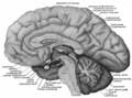 Mesal aspect of a brain sectioned in the median sagittal plane.
 -
 The hypophysis cerebri in position. Shown in sagittal section.


See also
External links
- University of Idaho
- Temple University
- http://isc.temple.edu/neuroanatomy/lab/atlas/pdhn/
![]() This article incorporates text in the public domain from page 816 of the 20th edition of Gray's Anatomy (1918)
This article incorporates text in the public domain from page 816 of the 20th edition of Gray's Anatomy (1918)
This developmental biology article is a stub. You can help Wikipedia by expanding it. |
This neuroanatomy article is a stub. You can help Wikipedia by expanding it. |