
Pelvic floor: Difference between revisions
Undid revision 1216396275 by Autisticeditor 20 (talk) reverted #diff-undo |
|||
| (44 intermediate revisions by 27 users not shown) | |||
| Line 1: | Line 1: | ||
{{short description|Anatomical structure}} |
{{short description|Anatomical structure}} |
||
{{more medical citations needed|date=August 2018}} |
{{more medical citations needed|date=August 2018}} |
||
{{cs1 config|name-list-style=vanc|display-authors=6}} |
|||
[[File:Pelvic Muscles (Female Side).png|thumb|246x246px|Female Pelvic Muscles[[File:Pelvic Muscles (Male Side).png|thumb|246x246px|Male Pelvic Muscles]] [[File:Vesicovaginal Fistula.png|thumb|246x246px|Vesicovaginal Fistula]]]]The '''pelvic floor''' or '''pelvic diaphragm''' is composed of muscle fibers of the [[levator ani]], the [[coccygeus muscle]], and associated [[connective tissue]] which span the area underneath the [[pelvis]]. The pelvic diaphragm is a muscular partition formed by the levatores ani and coccygei, with which may be included the parietal pelvic fascia on their upper and lower aspects. The pelvic floor separates the [[pelvic cavity]] above from the perineal region (including [[perineum]]) below. Both males and females have a pelvic floor. To accommodate the birth canal, a female's pelvic cavity is larger than a male's. |
|||
[[File:Pelvic Muscles (Female Side).png|thumb|246x246px|Female pelvic muscles]] |
|||
[[File:Pelvic Muscles (Male Side).png|thumb|246x246px|Male pelvic muscles]] |
|||
The '''pelvic floor''' or '''pelvic diaphragm''' is an anatomical location in the human body,<ref>{{cite book | vauthors = Bordoni B, Sugumar K, Leslie SW | chapter = Anatomy, Abdomen and Pelvis, Pelvic Floor |date=2023 | chapter-url = http://www.ncbi.nlm.nih.gov/books/NBK482200/ | title = StatPearls |access-date=2023-10-13 |place=Treasure Island (FL) |publisher=StatPearls Publishing |pmid=29489277 }}</ref> which has an important role in urinary and anal continence, sexual function and support of the pelvic organs.<ref>{{cite journal | vauthors = Fernandes AC, Palacios-Ceña D, Hay-Smith J, Pena CC, Sidou MF, de Alencar AL, Ferreira CH | title = Women report sustained benefits from attending group-based education about pelvic floor muscles: a longitudinal qualitative study | journal = Journal of Physiotherapy | volume = 67 | issue = 3 | pages = 210–216 | date = July 2021 | pmid = 34147398 | doi = 10.1016/j.jphys.2021.06.010 | s2cid = 235492234 | doi-access = free }}</ref> The pelvic floor includes muscles, both skeletal and smooth, ligaments and fascia.<ref>{{cite journal | vauthors = Roch M, Gaudreault N, Cyr MP, Venne G, Bureau NJ, Morin M | title = The Female Pelvic Floor Fascia Anatomy: A Systematic Search and Review | journal = Life | volume = 11 | issue = 9 | pages = 900 | date = August 2021 | pmid = 34575049 | pmc = 8467746 | doi = 10.3390/life11090900 | doi-access = free | bibcode = 2021Life...11..900R }}</ref> and separates between the [[pelvic cavity]] from above, and the [[perineum]] from below.{{Citation needed|date=June 2023}} It is formed by the [[levator ani|levator ani muscle]] and [[coccygeus muscle]], and associated [[connective tissue]].<ref>{{Cite web |title=Pelvic Floor Muscles: Anatomy, Function & Conditions |url=https://my.clevelandclinic.org/health/body/22729-pelvic-floor-muscles |access-date=2023-03-16 |website=Cleveland Clinic |language=en}}</ref> |
|||
| ⚫ | The pelvic floor has two [[hiatus (anatomy)|hiatuses]] (gaps): (anteriorly) the [[urogenital hiatus]] through which [[urethra]] and [[vagina]] pass, and (posteriorly) the rectal hiatus through which the [[anal canal]] passes.<ref name=":0">{{cite book | vauthors = Daftary S, Chakravarti S | chapter = Reproductive Anatomy |title=Manual of Obstetrics | publisher=Elsevier |year=2011 |isbn=978-81-312-2556-1 |edition=3rd |pages=1–16}}</ref> |
||
| ⚫ | |||
The right and left levator ani lie almost horizontally in the floor of the pelvis, separated by a narrow gap that transmits the urethra, vagina, and anal canal. The levator ani is usually considered in three parts: [[pubococcygeus]], [[coccygeus|puborectalis]], and [[iliococcygeus]]. The pubococcygeus, the main part of the levator, runs backward from the body of the pubis toward the coccyx and may be damaged during [[birth]]. Some fibers are inserted into the prostate, urethra, and vagina. The right and left puborectalis unite behind the anorectal junction to form a muscular sling. Some regard them as a part of the [[external anal sphincter]]. The [[iliococcygeus]], the most posterior part of the levator ani, is often poorly developed. |
|||
| ⚫ | |||
The coccygeus, situated behind the levator ani and frequently tendinous as much as muscular, extends from the ischial spine to the lateral margin of the sacrum and coccyx. |
|||
=== Definition === |
|||
| ⚫ | Some sources do not consider "pelvic floor" and "pelvic diaphragm" to be identical, with the "diaphragm" consisting of only the levator ani and coccygeus, while the "floor" also includes the [[perineal membrane]] and [[deep perineal pouch]].<ref>{{cite book | vauthors = Drake RL, Vogl W, Mitchell AW |title=Gray's Anatomy For Students |year=2005 |isbn=978-0-443-06612-2 |page=391|publisher=Elsevier Health Sciences TW }}</ref> However, other sources include the [[fascia]] as part of the diaphragm.<ref>{{cite journal | vauthors = Herschorn S | title = Female pelvic floor anatomy: the pelvic floor, supporting structures, and pelvic organs | journal = Reviews in Urology | volume = 6 | issue = Suppl 5 | pages = S2–S10 | date = 2004 | pmid = 16985905 | pmc = 1472875 }}</ref> In practice, the two terms are often used interchangeably.{{Citation needed|date=June 2023}} |
||
=== Relations === |
|||
The [[pelvic cavity]] of the [[true pelvis]] has the pelvic floor as its inferior |
The [[pelvic cavity]] of the [[true pelvis]] has the pelvic floor as its inferior boundary (and the [[pelvic brim]] as its superior boundary). The [[perineum]] has the pelvic floor as its superior boundary.{{Citation needed|date=June 2023}} |
||
| ⚫ | |||
| ⚫ | Some sources do not consider "pelvic floor" and "pelvic diaphragm" to be identical, with the "diaphragm" consisting of only the levator ani and coccygeus, while the "floor" also includes the [[perineal membrane]] and [[deep perineal pouch]].<ref>{{cite book | |
||
| ⚫ | |||
| ⚫ | |||
| ⚫ | It is important in providing support for pelvic [[viscera]] (organs), e.g. the [[urinary bladder|bladder]], [[intestine]]s, the [[uterus]] (in females), and in maintenance of [[wikt:continence|continence]] as part of the [[Urethral sphincter|urinary]] and [[Human anus|anal]] sphincters. It facilitates birth by resisting the descent of the presenting part, causing the fetus to rotate forwards to navigate through the pelvic girdle. It helps maintain optimal intra-abdominal pressure.<ref name=":0" /> |
||
| ⚫ | The pelvic floor has two [[hiatus (anatomy)|hiatuses]] (gaps): |
||
| ⚫ | |||
| ⚫ | It is important in providing support for pelvic [[viscera]] (organs), e.g. the [[urinary bladder|bladder]], [[intestine]]s, the [[uterus]] (in females), and in maintenance of [[wikt:continence|continence]] as part of the [[Urethral sphincter|urinary]] and [[anus|anal]] sphincters. It facilitates birth by resisting the descent of the presenting part, causing the fetus to rotate forwards to navigate through the pelvic girdle. It helps maintain optimal intra-abdominal pressure.<ref name=":0" /> |
||
==Clinical significance== |
==Clinical significance== |
||
[[File:1116 Muscle of the Female Perineum.png|thumb|179x179px|The female pelvic floor]] |
|||
[[File:1116 Muscle of the Male Perineum.png|thumb|186x186px|The male pelvic floor]] |
|||
The pelvic floor is subject to clinically relevant changes that can result in: |
The pelvic floor is subject to clinically relevant changes that can result in: |
||
* '''Anterior vaginal wall prolapse''' |
* '''Anterior vaginal wall prolapse''' |
||
| Line 32: | Line 35: | ||
** [[Vaginal vault]] prolapse (roof of vagina) - after [[hysterectomy]] |
** [[Vaginal vault]] prolapse (roof of vagina) - after [[hysterectomy]] |
||
Pelvic floor dysfunction can result after treatment for gynecological cancers.<ref>{{ |
Pelvic floor dysfunction can result after treatment for gynecological cancers.<ref>{{cite journal | vauthors = Ramaseshan AS, Felton J, Roque D, Rao G, Shipper AG, Sanses TV | title = Pelvic floor disorders in women with gynecologic malignancies: a systematic review | journal = International Urogynecology Journal | volume = 29 | issue = 4 | pages = 459–476 | date = April 2018 | pmid = 28929201 | pmc = 7329191 | doi = 10.1007/s00192-017-3467-4 }}</ref> |
||
Damage to the pelvic floor not only contributes to urinary incontinence but can lead to pelvic organ [[prolapse]]. Pelvic organ prolapse occurs in women when pelvic organs (e.g. the vagina, bladder, rectum, or uterus) protrude into or outside of the vagina. The causes of pelvic organ prolapse are not unlike those that also contribute to urinary incontinence. These include inappropriate (asymmetrical, excessive, insufficient) muscle tone and asymmetries caused by trauma to the pelvis. Age, pregnancy, family history, and hormonal status all contribute to the development of pelvic organ prolapse. The vagina is suspended by attachments to the perineum, pelvic side wall and sacrum via attachments that include collagen, elastin, and smooth muscle. Surgery can be performed to repair pelvic floor muscles. The pelvic floor muscles can be strengthened with [[Kegel exercise]]s.<ref name=pmid12806450 /> |
Damage to the pelvic floor not only contributes to urinary incontinence but can lead to pelvic organ [[prolapse]]. Pelvic organ prolapse occurs in women when pelvic organs (e.g. the vagina, bladder, rectum, or uterus) protrude into or outside of the vagina. The causes of pelvic organ prolapse are not unlike those that also contribute to urinary incontinence. These include inappropriate (asymmetrical, excessive, insufficient) muscle tone and asymmetries caused by trauma to the pelvis. Age, pregnancy, family history, and hormonal status all contribute to the development of pelvic organ prolapse. The vagina is suspended by attachments to the perineum, pelvic side wall and sacrum via attachments that include collagen, elastin, and smooth muscle. Surgery can be performed to repair pelvic floor muscles. The pelvic floor muscles can be strengthened with [[Kegel exercise]]s.<ref name=pmid12806450 /> |
||
Disorders of the posterior pelvic floor include [[rectal prolapse]], [[rectocele]], [[perineal hernia]], and a number of functional disorders including [[anismus]]. [[Constipation]] due to any of these disorders is called "functional constipation" and is identifiable by clinical diagnostic criteria.<ref name="pmid16720016">{{cite journal | |
Disorders of the posterior pelvic floor include [[rectal prolapse]], [[rectocele]], [[perineal hernia]], and a number of functional disorders including [[anismus]]. [[Constipation]] due to any of these disorders is called "functional constipation" and is identifiable by clinical diagnostic criteria.<ref name="pmid16720016">{{cite journal | vauthors = Berman L, Aversa J, Abir F, Longo WE | title = Management of disorders of the posterior pelvic floor | journal = The Yale Journal of Biology and Medicine | volume = 78 | issue = 4 | pages = 211–221 | date = July 2005 | pmid = 16720016 | pmc = 2259151 }}</ref> |
||
[[File:Bowel and bladder control postitions.png|thumb|Different positions to perform pelvic floor exercises]] |
|||
Pelvic floor exercise (PFE), also known as [[Kegel exercises]], may improve the tone and function of the pelvic floor muscles, which is of particular benefit for women (and less commonly men) who experience [[stress urinary incontinence]].<ref name="pmid15971718">{{cite journal | vauthors = Kielb SJ | title = Stress incontinence: alternatives to surgery | journal = International Journal of Fertility and Women's Medicine | volume = 50 | issue = 1 | pages = 24–29 | year = 2005 | pmid = 15971718 }}</ref><ref name="pmid12806450">{{cite journal | vauthors = Harvey MA | title = Pelvic floor exercises during and after pregnancy: a systematic review of their role in preventing pelvic floor dysfunction | journal = Journal of Obstetrics and Gynaecology Canada | volume = 25 | issue = 6 | pages = 487–498 | date = June 2003 | pmid = 12806450 | doi = 10.1016/s1701-2163(16)30310-3 }}</ref> However, compliance with PFE programs often is poor,<ref name="pmid15971718"/> PFE generally is ineffective for urinary incontinence unless performed with [[biofeedback]] and trained supervision,<ref name="pmid12806450"/> and in severe cases it may have no benefit. Pelvic floor muscle tone may be estimated using a [[perineometer]], which measures the pressure within the vagina.<ref>{{cite journal | vauthors = Barbosa PB, Franco MM, ((Souza Fd)), Antônio FI, Montezuma T, Ferreira CH | title = Comparison between measurements obtained with three different perineometers | journal = Clinics | volume = 64 | issue = 6 | pages = 527–533 | date = June 2009 | pmid = 19578656 | pmc = 2705146 | doi = 10.1590/s1807-59322009000600007 }}</ref> Medication may also be used to improve continence.<ref name="my.clevelandclinic.org">{{Cite web |title=Pelvic Floor Dysfunction: Symptoms, Causes & Treatment |url=https://my.clevelandclinic.org/health/diseases/14459-pelvic-floor-dysfunction |access-date=2023-03-16 |website=Cleveland Clinic |language=en}}</ref> In severe cases, surgery may be used to repair or even to reconstruct the pelvic floor.<ref name="my.clevelandclinic.org"/> One surgery which interrupts pelvic floor musculature in males is a [[radical prostatectomy]]. With the removal of the [[prostate]], many males experience [[urinary incontinence]] post operation; pelvic floor exercises may be used to counteract this pre and post operation. Pre-operative pelvic floor exercising significantly decreases the prevalence of urinary incontinence post radical prostatectomy. <ref>{{cite journal | vauthors = Zhou L, Chen Y, Yuan X, Zeng L, Zhu J, Zheng J | title = Preoperative pelvic floor muscle exercise for continence after radical prostatectomy: a systematic review and meta-analysis | journal = Frontiers in Public Health | volume = 11 | pages = 1186067 | date = 2023 | pmid = 37588123 | pmc = 10425962 | doi = 10.3389/fpubh.2023.1186067 | doi-access = free }}</ref> Prostatitis and prostatectomies are two contributors to [[erectile dysfunction]]; following a radical prostatectomy studies show that erectile dysfunction is improved by pelvic floor muscle training under the supervision of physical therapists certified in pelvic floor rehabilitation .<ref>{{cite journal | vauthors = Wong C, Louie DR, Beach C | title = A Systematic Review of Pelvic Floor Muscle Training for Erectile Dysfunction After Prostatectomy and Recommendations to Guide Further Research | journal = The Journal of Sexual Medicine | volume = 17 | issue = 4 | pages = 737–748 | date = April 2020 | pmid = 32029399 | doi = 10.1016/j.jsxm.2020.01.008 }}</ref> |
|||
| ⚫ | [[Perineology]] or pelviperineology is a specialty dealing with the functional troubles of the three axes (urological, gynecological and coloproctological) of the pelvic floor.<ref>{{cite journal | vauthors = Beco J, Mouchel J | title = Understanding the concept of perineology | journal = International Urogynecology Journal and Pelvic Floor Dysfunction | volume = 13 | issue = 5 | pages = 275–277 | date = 2002-10-01 | pmid = 12355284 | doi = 10.1007/s001920200060 | s2cid = 12964013 }}</ref> |
||
Pelvic floor exercise (PFE), also known as [[Kegel exercises]], may improve the tone and function of the pelvic floor muscles, which is of particular benefit for women (and less commonly men) who experience [[stress urinary incontinence]].<ref name="pmid15971718">{{cite journal |pmid=15971718 |year=2005 |last1=Kielb |first1=S. J. |title=Stress incontinence: Alternatives to surgery |journal=International Journal of Fertility and Women's Medicine |volume=50 |issue=1 |pages=24–9 }}</ref><ref name="pmid12806450">{{cite journal |pmid=12806450 |year=2003 |last1=Harvey |first1=M. A. |title=Pelvic floor exercises during and after pregnancy: A systematic review of their role in preventing pelvic floor dysfunction |journal=Journal of Obstetrics and Gynaecology Canada |volume=25 |issue=6 |pages=487–98 |doi=10.1016/s1701-2163(16)30310-3}}</ref> However, compliance with PFE programs often is poor,<ref name="pmid15971718"/> PFE generally is ineffective for urinary incontinence unless performed with [[biofeedback]] and trained supervision,<ref name="pmid12806450"/> and in severe cases it may have no benefit. Pelvic floor muscle tone may be estimated using a [[perineometer]], which measures the pressure within the vagina.<ref>{{Cite journal|last1=Barbosa|first1=Patrícia Brentegani|last2=Franco|first2=Maíra Menezes|last3=Souza|first3=Flaviane de Oliveira|last4=Antônio|first4=Flávia Ignácio|last5=Montezuma|first5=Thaís|last6=Ferreira|first6=Cristine Homsi Jorge|date=June 2009|title=Comparison between measurements obtained with three different perineometers|journal=Clinics|volume=64|issue=6|pages=527–533|doi=10.1590/s1807-59322009000600007|pmid=19578656|issn=1807-5932|pmc=2705146}}</ref> Medication may also be used to improve continence.{{Citation needed|date=January 2018}} In severe cases, surgery may be used to repair or even to reconstruct the pelvic floor.{{citation needed|date=May 2015}} |
|||
| ⚫ | |||
| ⚫ | [[Perineology]] or pelviperineology is a |
||
| ⚫ | |||
<gallery> |
<gallery> |
||
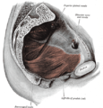
Gray404.png|The pelvic floor muscles span the bottom of the pelvis. This image shows the left [[levator ani]] from within. |
Gray404.png|The pelvic floor muscles span the bottom of the pelvis. This image shows the left [[levator ani]] from within. |
||
| Line 51: | Line 54: | ||
</gallery> |
</gallery> |
||
==See also== |
== See also == |
||
* [[Coccyx]] (tailbone) |
* [[Coccyx]] (tailbone) |
||
| ⚫ | |||
| ⚫ | |||
* [[Pelvic floor dysfunction]] |
* [[Pelvic floor dysfunction]] |
||
* [[Perineology]] |
* [[Perineology]] |
||
* [[Perineal hernia]] |
* [[Perineal hernia]] |
||
| ⚫ | |||
| ⚫ | |||
* [[Vaginal support structures]] |
* [[Vaginal support structures]] |
||
* [[Vesicovaginal fistula]] |
|||
==References== |
== References == |
||
{{Gray's}} |
{{Gray's}} |
||
{{Reflist|30em}} |
{{Reflist|30em}} |
||
==External links== |
== External links == |
||
* [https://www.nlm.nih.gov/research/visible/vhpconf98/AUTHORS/VENUTI/PELVDIAF.HTM Overview at nih.gov] |
* [https://www.nlm.nih.gov/research/visible/vhpconf98/AUTHORS/VENUTI/PELVDIAF.HTM Overview at nih.gov] |
||
Latest revision as of 16:30, 1 April 2024
This article needs more reliable medical references for verification or relies too heavily on primary sources. (August 2018) |  |
.png)
.png)
The pelvic floor or pelvic diaphragm is an anatomical location in the human body,[1] which has an important role in urinary and anal continence, sexual function and support of the pelvic organs.[2] The pelvic floor includes muscles, both skeletal and smooth, ligaments and fascia.[3] and separates between the pelvic cavity from above, and the perineum from below.[citation needed] It is formed by the levator ani muscle and coccygeus muscle, and associated connective tissue.[4]
The pelvic floor has two hiatuses (gaps): (anteriorly) the urogenital hiatus through which urethra and vagina pass, and (posteriorly) the rectal hiatus through which the anal canal passes.[5]
Structure
[edit]Definition
[edit]Some sources do not consider "pelvic floor" and "pelvic diaphragm" to be identical, with the "diaphragm" consisting of only the levator ani and coccygeus, while the "floor" also includes the perineal membrane and deep perineal pouch.[6] However, other sources include the fascia as part of the diaphragm.[7] In practice, the two terms are often used interchangeably.[citation needed]
Relations
[edit]The pelvic cavity of the true pelvis has the pelvic floor as its inferior boundary (and the pelvic brim as its superior boundary). The perineum has the pelvic floor as its superior boundary.[citation needed]
Posteriorly, the pelvic floor extends into the anal triangle.[citation needed]
Function
[edit]It is important in providing support for pelvic viscera (organs), e.g. the bladder, intestines, the uterus (in females), and in maintenance of continence as part of the urinary and anal sphincters. It facilitates birth by resisting the descent of the presenting part, causing the fetus to rotate forwards to navigate through the pelvic girdle. It helps maintain optimal intra-abdominal pressure.[5]
Clinical significance
[edit]

The pelvic floor is subject to clinically relevant changes that can result in:
- Anterior vaginal wall prolapse
- Cystocele (bladder into vagina)[8]
- Urethrocele (urethra into vagina)
- Cystourethrocele (both bladder and urethra)
- Posterior vaginal wall prolapse
- Enterocele (small intestine into vagina)
- Rectocele (rectum into vagina)
- Apical vaginal prolapse
- Uterine prolapse (uterus into vagina)
- Vaginal vault prolapse (roof of vagina) - after hysterectomy
Pelvic floor dysfunction can result after treatment for gynecological cancers.[9]
Damage to the pelvic floor not only contributes to urinary incontinence but can lead to pelvic organ prolapse. Pelvic organ prolapse occurs in women when pelvic organs (e.g. the vagina, bladder, rectum, or uterus) protrude into or outside of the vagina. The causes of pelvic organ prolapse are not unlike those that also contribute to urinary incontinence. These include inappropriate (asymmetrical, excessive, insufficient) muscle tone and asymmetries caused by trauma to the pelvis. Age, pregnancy, family history, and hormonal status all contribute to the development of pelvic organ prolapse. The vagina is suspended by attachments to the perineum, pelvic side wall and sacrum via attachments that include collagen, elastin, and smooth muscle. Surgery can be performed to repair pelvic floor muscles. The pelvic floor muscles can be strengthened with Kegel exercises.[10]
Disorders of the posterior pelvic floor include rectal prolapse, rectocele, perineal hernia, and a number of functional disorders including anismus. Constipation due to any of these disorders is called "functional constipation" and is identifiable by clinical diagnostic criteria.[11]

Pelvic floor exercise (PFE), also known as Kegel exercises, may improve the tone and function of the pelvic floor muscles, which is of particular benefit for women (and less commonly men) who experience stress urinary incontinence.[12][10] However, compliance with PFE programs often is poor,[12] PFE generally is ineffective for urinary incontinence unless performed with biofeedback and trained supervision,[10] and in severe cases it may have no benefit. Pelvic floor muscle tone may be estimated using a perineometer, which measures the pressure within the vagina.[13] Medication may also be used to improve continence.[14] In severe cases, surgery may be used to repair or even to reconstruct the pelvic floor.[14] One surgery which interrupts pelvic floor musculature in males is a radical prostatectomy. With the removal of the prostate, many males experience urinary incontinence post operation; pelvic floor exercises may be used to counteract this pre and post operation. Pre-operative pelvic floor exercising significantly decreases the prevalence of urinary incontinence post radical prostatectomy. [15] Prostatitis and prostatectomies are two contributors to erectile dysfunction; following a radical prostatectomy studies show that erectile dysfunction is improved by pelvic floor muscle training under the supervision of physical therapists certified in pelvic floor rehabilitation .[16]
Perineology or pelviperineology is a specialty dealing with the functional troubles of the three axes (urological, gynecological and coloproctological) of the pelvic floor.[17]
Additional images
[edit]-
 The pelvic floor muscles span the bottom of the pelvis. This image shows the left levator ani from within.
 -

-


See also
[edit]- Coccyx (tailbone)
- Female genital prolapse
- Pelvic floor dysfunction
- Perineology
- Perineal hernia
- Pubococcygeus muscle
- Vaginal support structures
- Vesicovaginal fistula
References
[edit]![]() This article incorporates text in the public domain from page 420 of the 20th edition of Gray's Anatomy (1918)
This article incorporates text in the public domain from page 420 of the 20th edition of Gray's Anatomy (1918)
- ^ Bordoni B, Sugumar K, Leslie SW (2023). "Anatomy, Abdomen and Pelvis, Pelvic Floor". StatPearls. Treasure Island (FL): StatPearls Publishing. PMID 29489277. Retrieved 2023-10-13.
- ^ Fernandes AC, Palacios-Ceña D, Hay-Smith J, Pena CC, Sidou MF, de Alencar AL, et al. (July 2021). "Women report sustained benefits from attending group-based education about pelvic floor muscles: a longitudinal qualitative study". Journal of Physiotherapy. 67 (3): 210–216. doi:10.1016/j.jphys.2021.06.010. PMID 34147398. S2CID 235492234.
- ^ Roch M, Gaudreault N, Cyr MP, Venne G, Bureau NJ, Morin M (August 2021). "The Female Pelvic Floor Fascia Anatomy: A Systematic Search and Review". Life. 11 (9): 900. Bibcode:2021Life...11..900R. doi:10.3390/life11090900. PMC 8467746. PMID 34575049.
- ^ "Pelvic Floor Muscles: Anatomy, Function & Conditions". Cleveland Clinic. Retrieved 2023-03-16.
- ^ a b Daftary S, Chakravarti S (2011). "Reproductive Anatomy". Manual of Obstetrics (3rd ed.). Elsevier. pp. 1–16. ISBN 978-81-312-2556-1.
- ^ Drake RL, Vogl W, Mitchell AW (2005). Gray's Anatomy For Students. Elsevier Health Sciences TW. p. 391. ISBN 978-0-443-06612-2.
- ^ Herschorn S (2004). "Female pelvic floor anatomy: the pelvic floor, supporting structures, and pelvic organs". Reviews in Urology. 6 (Suppl 5): S2–S10. PMC 1472875. PMID 16985905.
- ^ "Cystocele (Prolapsed Bladder) | NIDDK". National Institute of Diabetes and Digestive and Kidney Diseases. Retrieved 2017-12-02.
- ^ Ramaseshan AS, Felton J, Roque D, Rao G, Shipper AG, Sanses TV (April 2018). "Pelvic floor disorders in women with gynecologic malignancies: a systematic review". International Urogynecology Journal. 29 (4): 459–476. doi:10.1007/s00192-017-3467-4. PMC 7329191. PMID 28929201.
- ^ a b c Harvey MA (June 2003). "Pelvic floor exercises during and after pregnancy: a systematic review of their role in preventing pelvic floor dysfunction". Journal of Obstetrics and Gynaecology Canada. 25 (6): 487–498. doi:10.1016/s1701-2163(16)30310-3. PMID 12806450.
- ^ Berman L, Aversa J, Abir F, Longo WE (July 2005). "Management of disorders of the posterior pelvic floor". The Yale Journal of Biology and Medicine. 78 (4): 211–221. PMC 2259151. PMID 16720016.
- ^ a b Kielb SJ (2005). "Stress incontinence: alternatives to surgery". International Journal of Fertility and Women's Medicine. 50 (1): 24–29. PMID 15971718.
- ^ Barbosa PB, Franco MM, Souza Fd, Antônio FI, Montezuma T, Ferreira CH (June 2009). "Comparison between measurements obtained with three different perineometers". Clinics. 64 (6): 527–533. doi:10.1590/s1807-59322009000600007. PMC 2705146. PMID 19578656.
- ^ a b "Pelvic Floor Dysfunction: Symptoms, Causes & Treatment". Cleveland Clinic. Retrieved 2023-03-16.
- ^ Zhou L, Chen Y, Yuan X, Zeng L, Zhu J, Zheng J (2023). "Preoperative pelvic floor muscle exercise for continence after radical prostatectomy: a systematic review and meta-analysis". Frontiers in Public Health. 11: 1186067. doi:10.3389/fpubh.2023.1186067. PMC 10425962. PMID 37588123.
- ^ Wong C, Louie DR, Beach C (April 2020). "A Systematic Review of Pelvic Floor Muscle Training for Erectile Dysfunction After Prostatectomy and Recommendations to Guide Further Research". The Journal of Sexual Medicine. 17 (4): 737–748. doi:10.1016/j.jsxm.2020.01.008. PMID 32029399.
- ^ Beco J, Mouchel J (2002-10-01). "Understanding the concept of perineology". International Urogynecology Journal and Pelvic Floor Dysfunction. 13 (5): 275–277. doi:10.1007/s001920200060. PMID 12355284. S2CID 12964013.
External links
[edit]| International | |
|---|---|
| National | |
| Other | |