
Anterior superior iliac spine: Difference between revisions
m Bot: removing deprecated anatomy infobox parameters (Task 11) |
|||
| (30 intermediate revisions by 18 users not shown) | |||
| Line 1: | Line 1: | ||
{{Short description|Bony projection of the iliac bone}} |
|||
{{Infobox bone |
{{Infobox bone |
||
| Name = Anterior superior iliac spine |
| Name = Anterior superior iliac spine |
||
| Latin = |
| Latin = spina iliaca anterior superior |
||
| Image = Gray435.png |
| Image = Gray435.png |
||
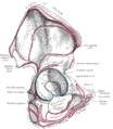
| Caption = The [[obturator membrane]] (anterior superior iliac spine visible in upper right of illustration) |
| Caption = The [[obturator membrane]] (anterior superior iliac spine visible in upper right of illustration) |
||
| Image2 = |
| Image2 = Gray abdomen front surface en.png |
||
| Caption2 = Anterior superior iliac spine labeled second to bottom, right |
| Caption2 = Anterior superior iliac spine labeled second to bottom, right |
||
}} |
}} |
||
The '''anterior superior iliac spine''' ('''ASIS''') is a bony projection of the [[iliac bone]], and an important landmark of [[surface anatomy]]. It refers to the anterior extremity of the [[iliac crest]] of the [[pelvis]]. It provides attachment for the [[inguinal ligament]], and the [[sartorius muscle]].<ref name=":0">{{Citation|last1=Chaitow|first1=Leon|title=Chapter 12 - The hip|date=2011-01-01|url=http://www.sciencedirect.com/science/article/pii/B9780443068157000127|work=Clinical Application of Neuromuscular Techniques, Volume 2 (Second Edition)|pages=391–445|editor-last=Chaitow|editor-first=Leon|place=Oxford|publisher=Churchill Livingstone|language=en|doi=10.1016/b978-0-443-06815-7.00012-7|isbn=978-0-443-06815-7|access-date=2020-12-15|last2=DeLany|first2=Judith|editor2-last=DeLany|editor2-first=Judith}}</ref> The [[tensor fasciae latae muscle]] attaches to the lateral aspect of the superior anterior iliac spine, and also about 5 [[Centimetre|cm]] away at the [[iliac tubercle]].<ref name=":4">{{Citation|last=Garten|first=Hans|title=M. tensor fasciae latae|date=2013|url=http://dx.doi.org/10.1016/b978-0-7020-3739-9.00091-2|work=The Muscle Test Handbook|pages=236–237|publisher=Elsevier|doi=10.1016/b978-0-7020-3739-9.00091-2|isbn=978-0-7020-3739-9|access-date=2020-12-15}}</ref><ref name=":5">{{Citation|last1=Chaitow|first1=Leon|title=Chapter 11 - The pelvis|date=2011-01-01|url=http://www.sciencedirect.com/science/article/pii/B9780443068157000115|work=Clinical Application of Neuromuscular Techniques, Volume 2 (Second Edition)|pages=299–389|editor-last=Chaitow|editor-first=Leon|place=Oxford|publisher=Churchill Livingstone|language=en|doi=10.1016/b978-0-443-06815-7.00011-5|isbn=978-0-443-06815-7|access-date=2020-12-15|last2=DeLany|first2=Judith|editor2-last=DeLany|editor2-first=Judith}}</ref> |
|||
The '''anterior superior iliac spine''' ([[abbreviated]]: '''ASIS''') is a bony projection of the [[iliac bone]] and an important landmark of [[surface anatomy]]. |
|||
It refers to the anterior extremity of the [[iliac crest]] of the [[pelvis]], which provides attachment for the [[inguinal ligament]], and the [[sartorius muscle]]. The [[Tensor fasciae latae muscle]] attaches about 5cm away at the [[iliac tubercle]]. |
|||
== Structure == |
|||
The anterior superior iliac spine provides a clue in identifying some other clinical landmarks, including: |
|||
The anterior superior iliac spine refers to the anterior extremity of the [[iliac crest]] of the [[pelvis]]. This is a key [[Surface anatomy|surface landmark]], and easily [[Palpation|palpated]]. It provides attachment for the [[inguinal ligament]], the [[sartorius muscle]],<ref name=":0" /><ref name=":2">{{Citation|last=Jacob|first=S.|title=Chapter 6 - Lower limb|date=2008-01-01|url=http://www.sciencedirect.com/science/article/pii/B9780443103735500099|work=Human Anatomy|pages=135–179|editor-last=Jacob|editor-first=S.|publisher=Churchill Livingstone|language=en|doi=10.1016/b978-0-443-10373-5.50009-9|isbn=978-0-443-10373-5|access-date=2020-12-15}}</ref> and the [[tensor fasciae latae muscle]].<ref name=":4" /><ref name=":5" /> |
|||
# [[McBurney's point]] |
|||
# [[Nelaton's line|Roser-Nélaton line]] |
|||
# True leg length (see [[unequal leg length]]) |
|||
A variety of structures lie close to the anterior superior iliac spine, including the [[subcostal nerve]],<ref name=":1" /> the [[femoral artery]] (which passes between it and the [[pubic symphysis]]),<ref name=":2" /> and the [[iliohypogastric nerve]].<ref name=":3">{{Citation|last=Mirjalili|first=S. Ali|title=Chapter 45 - Anatomy of the Lumbar Plexus|date=2015-01-01|url=http://www.sciencedirect.com/science/article/pii/B9780124103900000470|work=Nerves and Nerve Injuries|pages=609–617|editor-last=Tubbs|editor-first=R. Shane|place=San Diego|publisher=Academic Press|language=en|doi=10.1016/b978-0-12-410390-0.00047-0|isbn=978-0-12-410390-0|access-date=2020-12-15|editor2-last=Rizk|editor2-first=Elias|editor3-last=Shoja|editor3-first=Mohammadali M.|editor4-last=Loukas|editor4-first=Marios}}</ref> |
|||
==Etymology== |
|||
'''A''' – Anterior (front, like the [[face]])<br/> |
|||
== Clinical significance == |
|||
'''S''' – Superior (towards the [[head]], opposite to feet)<br/> |
|||
{{See also|Unequal leg length}} |
|||
'''I''' – [[Ilium (bone)|Iliac]] ([[Pelvis]] bone)<!-- double check --><br/> |
|||
The anterior superior iliac spine provides a clue in identifying some other clinical landmarks, including [[McBurney's point]], [[Nelaton's line|Roser-Nélaton line]], and true leg length. It is an important surface landmark for various [[Surgery|surgical approaches]], such as treatment of [[hernia]].<ref>{{Citation|last=Molloy|first=Robert E.|title=Chapter 75 - Truncal Blocks: Intercostal, Paravertebral, Interpleural, Suprascapular, Ilioinguinal, and Iliohypogastric Nerve Blocks|date=2005-01-01|url=http://www.sciencedirect.com/science/article/pii/B9780443066511500794|work=Essentials of Pain Medicine and Regional Anesthesia (Second Edition)|pages=636–644|editor-last=Benzon|editor-first=Honorio T.|place=Philadelphia|publisher=Churchill Livingstone|language=en|doi=10.1016/b978-0-443-06651-1.50079-4|isbn=978-0-443-06651-1|access-date=2020-12-15|editor2-last=Raja|editor2-first=Srinivasa N.|editor3-last=Molloy|editor3-first=Robert E.|editor4-last=Liu|editor4-first=Spencer S.}}</ref> The severity of symptoms of damage to the [[iliohypogastric nerve]] can show whether damage occurred above or below the anterior superior iliac spine.<ref name=":3" /> |
|||
'''S''' – [[Bone#Terminology|Spine]] |
|||
Bone may be [[Bone grafting|harvested]] from the nearby iliac crest for use elsewhere in the body.<ref name=":1">{{Citation|last=Rea|first=Paul|title=Chapter 3 - Lower Limb Nerve Supply|date=2015-01-01|url=http://www.sciencedirect.com/science/article/pii/B9780128030622000036|work=Essential Clinically Applied Anatomy of the Peripheral Nervous System in the Limbs|pages=101–177|editor-last=Rea|editor-first=Paul|publisher=Academic Press|language=en|doi=10.1016/b978-0-12-803062-2.00003-6|isbn=978-0-12-803062-2|access-date=2020-12-15}}</ref> As the [[subcostal nerve]] lies close to the anterior superior iliac spine, this is put at risk of damage.<ref name=":1" /> |
|||
The [[iliotibial tract]] may be irritated where it passes over the anterior superior iliac spine in [[iliotibial band syndrome]].<ref name=":5" /> |
|||
The line around anterior superior iliac spine is sometimes called the '''panty line''' or "bikini line".<ref>{{cite journal | last=Kim | first=Ji Hun | title=Robotic cholecystectomy with new port sites | journal=World Journal of Gastroenterology | volume=19 | issue=20 | date=2013 | issn=1007-9327 | pmid=23716987 | pmc=3662947 | doi=10.3748/wjg.v19.i20.3077 | doi-access=free | page=3077}}</ref> It is considered to be a "discreet" location for concealing cosmetic surgery scars and [[Port (medical)|ports]].<ref>{{cite journal | last=Leggett | first=P.L. | last2=Bissell | first2=C.D. | last3=Churchman-Winn | first3=R. | title=Cosmetic minilaparoscopic cholecystectomy | journal=Surgical Endoscopy | volume=15 | issue=10 | date=2001 | issn=0930-2794 | doi=10.1007/s004640041018 | pages=1229–1231}}</ref> |
|||
== Additional images == |
== Additional images == |
||
<gallery> |
<gallery> |
||
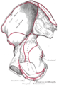
File:Gray235.png|Right hip bone. External surface. |
|||
File:Gray236.png|Right hip bone. Internal surface. |
|||
File:Gray393.png|The subcutaneous inguinal ring |
|||
File:Sobo 1909 573-574.png|ASIS visible at top left, as the origin of several muscles |
|||
File:McBurney's point.jpg|Location of [[McBurney's point]] (1), which is located two thirds the distance from the [[Navel|umbilicus]] (2) to the anterior superior iliac spine (3) |
|||
</gallery> |
</gallery> |
||
| Line 36: | Line 39: | ||
* [[Ilium (bone)]] |
* [[Ilium (bone)]] |
||
* [[Human anatomical terms#Anatomical directions|Human anatomical terms]] |
* [[Human anatomical terms#Anatomical directions|Human anatomical terms]] |
||
== References == |
|||
<references /> |
|||
==External links== |
==External links== |
||
{{Commons category|Anterior superior iliac spine}} |
{{Commons category|Anterior superior iliac spine}} |
||
| ⚫ | * {{cite web |url=http://www.tk.de/rochelexikon/pics/s03281.000-3.html |title=Anatomy diagram: 03281.000-3 |work=Roche Lexicon - illustrated navigator |publisher=Elsevier |archiveurl=https://web.archive.org/web/20120722052404/http://www.tk.de/rochelexikon/pics/s03281.000-3.html |archivedate=2012-07-22}} |
||
* {{SUNYAnatomyLabs|17|os|01|05}} – "Major Joints of the Lower Extremity: Hip bone (lateral view)" |
|||
* {{SUNYAnatomyLabs|35|os|01|03}} – "Anterior Abdominal Wall: Osteology and Surface Anatomy" |
|||
| ⚫ | * {{cite web|url=http://www.tk.de/rochelexikon/pics/s03281.000-3.html|title=Anatomy diagram: 03281.000-3|work= |
||
* [https://wayback.archive-it.org/all/20081217070227/http://academic.wsc.edu/faculty/jatodd1/351/pelvis_anterior.jpg Diagram at Wayne State] |
* [https://wayback.archive-it.org/all/20081217070227/http://academic.wsc.edu/faculty/jatodd1/351/pelvis_anterior.jpg Diagram at Wayne State] |
||
{{Pelvis}} |
{{Pelvis}} |
||
{{Authority control}} |
|||
| ⚫ | |||
[[Category:Pelvis]] |
|||
[[Category:Skeletal system]] |
|||
[[Category:Bones of the pelvis]] |
[[Category:Bones of the pelvis]] |
||
[[Category:Ilium (bone)]] |
[[Category:Ilium (bone)]] |
||
| ⚫ | |||
Latest revision as of 22:44, 11 March 2024
| Anterior superior iliac spine | |
|---|---|
 The obturator membrane (anterior superior iliac spine visible in upper right of illustration) | |
 Anterior superior iliac spine labeled second to bottom, right | |
| Details | |
| Identifiers | |
| Latin | spina iliaca anterior superior |
| TA98 | A02.5.01.111 |
| TA2 | 1327 |
| FMA | 49465 |
| Anatomical terms of bone | |
The anterior superior iliac spine (ASIS) is a bony projection of the iliac bone, and an important landmark of surface anatomy. It refers to the anterior extremity of the iliac crest of the pelvis. It provides attachment for the inguinal ligament, and the sartorius muscle.[1] The tensor fasciae latae muscle attaches to the lateral aspect of the superior anterior iliac spine, and also about 5 cm away at the iliac tubercle.[2][3]
Structure
[edit]The anterior superior iliac spine refers to the anterior extremity of the iliac crest of the pelvis. This is a key surface landmark, and easily palpated. It provides attachment for the inguinal ligament, the sartorius muscle,[1][4] and the tensor fasciae latae muscle.[2][3]
A variety of structures lie close to the anterior superior iliac spine, including the subcostal nerve,[5] the femoral artery (which passes between it and the pubic symphysis),[4] and the iliohypogastric nerve.[6]
Clinical significance
[edit]The anterior superior iliac spine provides a clue in identifying some other clinical landmarks, including McBurney's point, Roser-Nélaton line, and true leg length. It is an important surface landmark for various surgical approaches, such as treatment of hernia.[7] The severity of symptoms of damage to the iliohypogastric nerve can show whether damage occurred above or below the anterior superior iliac spine.[6]
Bone may be harvested from the nearby iliac crest for use elsewhere in the body.[5] As the subcostal nerve lies close to the anterior superior iliac spine, this is put at risk of damage.[5]
The iliotibial tract may be irritated where it passes over the anterior superior iliac spine in iliotibial band syndrome.[3]
The line around anterior superior iliac spine is sometimes called the panty line or "bikini line".[8] It is considered to be a "discreet" location for concealing cosmetic surgery scars and ports.[9]
Additional images
[edit]-
 Right hip bone. External surface.
 -
 Right hip bone. Internal surface.
 -
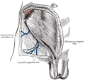 The subcutaneous inguinal ring
 -
 ASIS visible at top left, as the origin of several muscles
 -
 Location of McBurney's point (1), which is located two thirds the distance from the umbilicus (2) to the anterior superior iliac spine (3)


See also
[edit]References
[edit]- ^ a b Chaitow, Leon; DeLany, Judith (2011-01-01), Chaitow, Leon; DeLany, Judith (eds.), "Chapter 12 - The hip", Clinical Application of Neuromuscular Techniques, Volume 2 (Second Edition), Oxford: Churchill Livingstone, pp. 391–445, doi:10.1016/b978-0-443-06815-7.00012-7, ISBN 978-0-443-06815-7, retrieved 2020-12-15
- ^ a b Garten, Hans (2013), "M. tensor fasciae latae", The Muscle Test Handbook, Elsevier, pp. 236–237, doi:10.1016/b978-0-7020-3739-9.00091-2, ISBN 978-0-7020-3739-9, retrieved 2020-12-15
- ^ a b c Chaitow, Leon; DeLany, Judith (2011-01-01), Chaitow, Leon; DeLany, Judith (eds.), "Chapter 11 - The pelvis", Clinical Application of Neuromuscular Techniques, Volume 2 (Second Edition), Oxford: Churchill Livingstone, pp. 299–389, doi:10.1016/b978-0-443-06815-7.00011-5, ISBN 978-0-443-06815-7, retrieved 2020-12-15
- ^ a b Jacob, S. (2008-01-01), Jacob, S. (ed.), "Chapter 6 - Lower limb", Human Anatomy, Churchill Livingstone, pp. 135–179, doi:10.1016/b978-0-443-10373-5.50009-9, ISBN 978-0-443-10373-5, retrieved 2020-12-15
- ^ a b c Rea, Paul (2015-01-01), Rea, Paul (ed.), "Chapter 3 - Lower Limb Nerve Supply", Essential Clinically Applied Anatomy of the Peripheral Nervous System in the Limbs, Academic Press, pp. 101–177, doi:10.1016/b978-0-12-803062-2.00003-6, ISBN 978-0-12-803062-2, retrieved 2020-12-15
- ^ a b Mirjalili, S. Ali (2015-01-01), Tubbs, R. Shane; Rizk, Elias; Shoja, Mohammadali M.; Loukas, Marios (eds.), "Chapter 45 - Anatomy of the Lumbar Plexus", Nerves and Nerve Injuries, San Diego: Academic Press, pp. 609–617, doi:10.1016/b978-0-12-410390-0.00047-0, ISBN 978-0-12-410390-0, retrieved 2020-12-15
- ^ Molloy, Robert E. (2005-01-01), Benzon, Honorio T.; Raja, Srinivasa N.; Molloy, Robert E.; Liu, Spencer S. (eds.), "Chapter 75 - Truncal Blocks: Intercostal, Paravertebral, Interpleural, Suprascapular, Ilioinguinal, and Iliohypogastric Nerve Blocks", Essentials of Pain Medicine and Regional Anesthesia (Second Edition), Philadelphia: Churchill Livingstone, pp. 636–644, doi:10.1016/b978-0-443-06651-1.50079-4, ISBN 978-0-443-06651-1, retrieved 2020-12-15
- ^ Kim, Ji Hun (2013). "Robotic cholecystectomy with new port sites". World Journal of Gastroenterology. 19 (20): 3077. doi:10.3748/wjg.v19.i20.3077. ISSN 1007-9327. PMC 3662947. PMID 23716987.
- ^ Leggett, P.L.; Bissell, C.D.; Churchman-Winn, R. (2001). "Cosmetic minilaparoscopic cholecystectomy". Surgical Endoscopy. 15 (10): 1229–1231. doi:10.1007/s004640041018. ISSN 0930-2794.
External links
[edit]- "Anatomy diagram: 03281.000-3". Roche Lexicon - illustrated navigator. Elsevier. Archived from the original on 2012-07-22.
- Diagram at Wayne State
This human musculoskeletal system article is a stub. You can help Wikipedia by expanding it. |